R E V I E W
Open Access
Functional cellulose-based hydrogels as
extracellular matrices for tissue engineering
Sayan Deb Dutta
1, Dinesh K. Patel
2and Ki-Taek Lim
1*Abstract
Cellulose-based hydrogels are immensely important for tissue engineering. In this review, we attempt to document
the source, nature, and application of cellulose-based hydrogels as an extracellular matrix for tissue growth and
regeneration. Hydrogels can be prepared either from native cellulose, including both bacterial and plant sources or
from cellulose derivatives, such as methyl cellulose, carboxymethylcellulose, and hydroxypropylmethylcellulose or
even metal ions such as silver. Cellulose-polymer composite (polymers that include natural sources including
chitosan, starch, alginates, collagen, hyaluronic acid, and chitin) are an attractive, inexpensive, and advantageous
structural material that is easy to use. Cellulose-based scaffolding materials are widely used in the regeneration of
various tissues, such as bone, cartilage, heart, blood vessel, nerve, and liver, among others. In this review, we discuss
the most important applications of cellulosic hydrogels in tissue engineering based on their structural
compositions.
Keywords:
Cellulose, Hydrogels, Scaffolds, Extracellular matrices, Tissue engineering
Introduction
Cells communicate with each other either directly via
molecular interactions or through the secretion of
differ-ent hormones or mediators which systematically regulate
various cell functions. Growth factors are also secreted
during cellular crosstalk and may be pro-proliferative or
anti-proliferative in nature, being mainly involved in cell
differentiation, migration, adhesion, and gene expression.
Natural and synthetic materials may be used as bulking
agents for the binding of various growth factors by
mim-icking natural extracellular matrix (ECM) molecular
self-assembly via secondary forces, such as ionic or
hydrogen bonds, whereas chemical gels are result of
co-valent bonds [
1
–
5
].
Hydrogels have potential applications in various fields
such as agriculture, food, biomaterials, water purification,
biomedicine, and pharmaceuticals, among others. [
6
–
8
].
Hydrogels are primarily made up of natural living tissue
rather than synthetic biomaterials, as a result have a high
water content and a soft consistency similar to natural
tis-sues [
9
]. Moreover, the high water content of these
materials contributes to their biocompatibility. Thus,
hydrogels can be used as membranes for biosensors
[
10
,
11
], in artificial heart and skin [
12
,
13
], contact
lenses [
14
,
15
], and drug delivery [
3
,
6
,
16
].
Cross-linking synthetic polymer-based hydrogels have been
reported, including poly (ethylene glycol) [
17
,
18
],
poly (vinyl alcohol) [
18
,
19
], poly (amido-amine) [
20
],
poly (N-isopropylacrylamide) [
21
], polyacrylamide [
18
,
22
], and poly (acrylic acid) [
18
,
23
].
In tissue engineering, hydrogels are the most extensively
used biopolymer due to their highly swollen three-
dimen-sional (3D) environment, which is very similar to soft
tis-sues and allows for the diffusion of nutrients, growth
factors and cellular waste through the elastic network and
for the regeneration of damaged tissues [
13
,
18
,
24
,
25
]. In
regenerative medicine, hydrogel used to repair and assist
regeneration of various soft and hard tissues, such as
cartil-age, bone and vascular tissues [
26
–
28
]. Natural hydrogels
include the bioprocessing of natural polymer-based
mate-rials such as proteins, including collagen, gelatin, and fibrin,
and polysaccharides, including alginate chitosan, hyaluronic
acid, dextran, and cellulose which are used as extracellular
matrices (ECM).
Cellulose is a fibrous, tough, water-insoluble substance,
found in the protective cell walls of plants, particularly in
© The Author(s). 2019Open AccessThis article is distributed under the terms of the Creative Commons Attribution 4.0 International License (http://creativecommons.org/licenses/by/4.0/), which permits unrestricted use, distribution, and reproduction in any medium, provided you give appropriate credit to the original author(s) and the source, provide a link to the Creative Commons license, and indicate if changes were made. The Creative Commons Public Domain Dedication waiver (http://creativecommons.org/publicdomain/zero/1.0/) applies to the data made available in this article, unless otherwise stated.
* Correspondence:[email protected]
1Biorobotics Laboratory, Department of Biosystems Engineering, Kangwon
National University, Chuncheon, Republic of Korea
stalks, stems, trunks, and all woody portions. However, it
is also produced by some animals (e.g., tunicates), fungi
and a few bacteria [
29
–
31
]. Due to the presence of
abun-dant hydroxyl groups in the cellulose molecule, cellulose
can be used to prepare hydrogels with varying structures
and properties to act as a platform for advanced tissue
en-gineering and regenerative medicine. Cellulose-based
mate-rials represents a naturally occurring
‘
nanomaterial
’
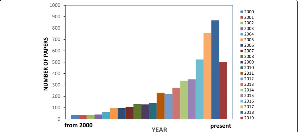
, and has
attracted the attention of researchers all over the world, as
shown by the increasing number of annual publications
appearing in
‘
Science Direct
’
with
‘
cellulose-based hydrogels
for tissue engineering
’
(Fig.
1
) as the search item. However,
furthur studies are needed for the development and
appli-cation of cellulose-based hydrogels. This review highlights
the recent development and use of various cellulose-based
hydrogels as an ECM and their structural properties for
ap-plications in advanced tissue engineering.
Structure of cellulosic biomass
Cellulose is the most abundant biopolymer and is
distrib-uted throughout nature in plants, animals, algae, fungi, and
minerals. A major source of cellulose is plant fiber.
Cellu-lose is the main structural component of plants that
pro-vides them with their mechanical as well as structural
integrity as it contributes approximately 40% to the carbon
fraction in plants (Additional file
1
: Table S1). Cellulose can
be found in its pure form in plants with hemicelluloses,
lig-nins, and other components [
32
]. Surprisingly, a large
frac-tion of cellulose is produced from trees (wood fiber) with a
global production of approximately 1,750,000 kt. Annual
plants such as bamboo, cotton linters, jute, flax, sisal, hemp,
and ramie also produces significant amount of cellulosic
biomass (Additional file
1
: Figure S1) [
33
]. In addition,
some fungi and green algae produce cellulose (e.g.
Valonia
ventricular
,
Glaucocystis
) and some marine ascidians
con-tain cellulose in their outer cell membrane. Some bacterial
genera, such as,
Gluconacetobacter
,
Agrobacterium
,
Pseudo-monas
,
Rhizobium
, and
Sarcina
are able to synthesize
bac-terial cellulose either from glucose or other carbon sources
[
34
–
36
]. Purified bacterial cellulose is highly crystalline and
possess a high degree of polymerization (DP). One of the
crucial features of cellulose is its micro-crystalline structure
and its synthesis in nature as individual molecules (linear
chain of glucosyl residues) which undergo self-assembly at
the site of biosynthesis [
37
].
Molecular structure of cellulose
Cellulose mainly consist of D-glucopyranose ring units in
a
4C
1configuration, which exhibits the lowest energy
con-formation [
38
]. Each unit is linked by
β
-1, 4-glycosidic
linkage that results in an alternate turning of the cellulose
chain axis by 180° [
39
–
41
]. Within the cellulose chain,
three reactive hydroxyl groups (
−
OH) exist in each
anhy-droglucose unit (AGU). The
–
OH groups of the AGU, the
oxygen atoms of the D-glucopyranose ring, and the
glyco-sidic linkage interacts with each other within the chain or
another cellulose chain by intermolecular and
intramo-lecular hydrogen bonds [
42
]. The presence of hydrogen
bond provides stability to the cellulose molecule and
al-lows it to be a functionally active biomolecule (Additional
file
1
: Figure S2).
X-ray diffraction studies revealed the crystalline
struc-ture of cellulose, and NMR experiments have confirmed
its dimorphic and polymorphic nature [
43
,
44
]. Different
polymorphs of cellulose are listed in Table
1
. Solid-state
13
C-NMR was used to identify different polymorphs,
de-noted as cellulose I
αand I
β. Cellulose I
βis naturally
oc-curring in plants, whereas cellulose produced by
primitive organisms crystallizes in the I
αform [
55
].
Cellulose chains are arranged in a basic fibrillary unit
or elementary fibrils with a length of 0.1 to 0.2
μ
m and
have a characteristic lateral dimension of 0.0015
μ
m to
0.0035
μ
m [
56
,
57
]. Such fibrils are known as cellulose
fi-brils. These fibrils are further assembled into microfibrils
with a width of 0.1
μ
m and a length of 0.1 to 1
μ
m (Fig.
2
a). This fibrillary architecture can be found in both
na-tive and man-made fibers [
39
].
Structure of plant cellulose (PC)
In the case of plant cell walls, a sheath of amorphous
cellu-lose surrounded by a hemicellucellu-lose layer covers the
micro-fibrils [
33
]. Fibers from different plants vary in morphology
and dimension. Additional file
1
: Figure S3 clearly shows
the variations in the fiber morphologies of cotton (S3a),
spruce wood (S3b), and ramie plant (S3c). Surprisingly, all
three plants share a common internal structure made up of
multiple cell wall layers [
58
]. During the early growth
phase, plant fibers develop a primary cell wall (P layer) that
is much thinner than the secondary wall (S layer) formed
on its inner side. Inside the S wall, a tertiary cell wall (T
layer) is present, which is typically an open, hollow area or
lumen-like structure. The cell wall thickness and length of
the plant fiber are approximately 4
–
630
μ
m and 15
–
30
μ
m,
respectively. The swelling characteristics as well as their
physical and chemical properties are strongly influenced by
the configuration, composition, and structure of the P layer,
which contains microfibrils crisscrossed onto each other to
make a net-like helical structure (S3d-e). The secondary
layer is 3
–
5
μ
m in thickness and comprises three sublayers
(S
1, S
2, and S
3) of which S
2is the thickest e (approximately
3
–
5
μ
m thickness) as shown in Additional file
1
: Figure
S3d. The S
2layer contains microfibrils arranged in parallel
[
58
–
60
].
Structure of bacterial cellulose (BC)
Bacterial cellulose (BC) can be obtained in pure form.
Compared to PC, BC contains no hemicellulose or lignin
and only a very small amount of carbonyl and carboxyl
moieties are present [
61
]. BC possesses a high degree of
crystallinity (above 80%) with a good water retention
capacity, and an extraordinary mechanical strength,
par-ticularly under wet conditions. One important advantage
of using BC is its in-situ molding ability, i.e. shaping
during biosynthesis [
62
]. The culturing and production
of BC is the most important part, although it is also
im-portant to maintain the pH of the culture medium, since
a low pH can often led to the accumulation by-products,
such as of gluconic, acetic, or lactic acids [
63
]. Figure
2
b
clearly shows the structure and formation of bacterial
cellulose in
Acetobacter xylinum
.
Role of extracellular matrix (ECM)
ECMs are used in tissue engineering and regenerative
medicine as a natural model for bioactive modifications.
Compared to other ECMs, hydrogels have provided
op-portunities for the use of a natural ECM as a model for
designing biomimetic scaffolds.
Structure and composition of ECM
The tissues of the human body contain a significant
amount of extracellular space, into which ECM molecules
are secreted by cells to form a large and complex network.
The ECM of the extracellular space provides tissue with
mechanical strength, organizes cells into specific tissues,
Table 1
Polymorphs of cellulose
Source Cellulose
polymorphs
Features References
Valonia ventricosa1
(bubble algae)
Acetobacter xylinum
(bacteria)
Microdictyon(green algae)
Halocynthia(tunicates)
Cellulose I Native cellulose, found in nature, interconvertible, stable. Crystalline forms are termed as Iαand Iβ. Iαconsidered as primitive type, while higher plants possess Iβ.
Marchessault and Sarko, 1967 [45]
[46] [47] [48]
Halicystis(green algae)2
Mutant strain ofA. xylinum
Cellulose II Obtained from cellulose I, interconvertible, also found in nature. [49] [50] [51] Kuga et al., 1993 [52]
Chemical conversion ofValonia
cellulose I and cellulose II
Cellulose III Interconvertible and not found in nature. Two crystalline forms isolated as IIIIand
IIIIIrespectively.
[49] [50]
Chemical conversion and heating of cellulose IIIIand IIIII
Cellulose IV Interconvertible and not found in nature. Two crystalline forms isolated as IVIand
IVIIrespectively.
[53] [54]
1
highly crystalline cellulose obtained fromValonia
2
and controls cell behavior and cell differentiation. Two
crucial components of the ECM are proteins and glycans,
in particular fibrous proteins (e.g., collagen, laminin, and
elastin) and glycosaminoglycans (GAGs) [
64
,
65
]. Fibrous
proteins act as a scaffold and provide adhesion to matrix
structure that are initially embedded in GAGs [
65
]. Thus,
cell-matrix adhesions mediate various physiological
re-sponses including cell growth, migration, differentiation,
survival, tissue organization and matrix remodeling [
66
].
Function of ECM
The ECM components undergo self-assembly to form a
complex 3D network [
18
]. Figure
3
shows the role of
ECMs in various cellular responses. Cell receptors bind
both soluble and tethered signaling cues from the ECM
environment. In turn, these receptor-ligand interactions
trigger complex cascades of intracellular enzymatic
reac-tions that regulate gene and protein expression and
de-fine the fate of a cell in a specific tissue [
18
,
66
]. Cell can
also transmits a signal to actively construct and degrade
their microenvironment. Thus, the ECMs acts as both a
space-filling mechanical scaffold and a bioactive and
dynamic environment to mediate cellular functions
[
64
,
65
]. However, natural ECMs also provide cellular
adhesion, proteolytic degradation and growth factor
(GF)- binding [
18
].
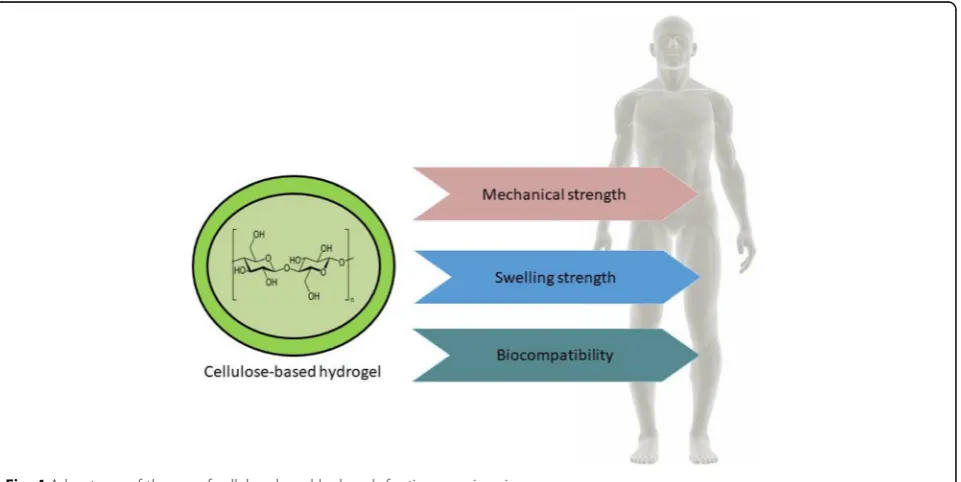
Basic properties of hydrogels
Hydrogels are a type of polymer biomaterials with
vari-ous properties. In the field of pharmaceutical and
bio-medical engineering, hydrogels are very important due
to their in-vivo swelling properties, mechanical strength
and compatibility with biological tissues, facilitating
binding (Fig.
4
) [
68
–
70
].
Mechanical properties
The mechanical properties of hydrogels are significant from
both a pharmaceutical and biomedical point of view [
68
].
The optimum mechanical strength of a hydrogel is an
es-sential requirement for its successful implementation as a
drug delivery system. The excellent mechanical properties
of hydrogels allows its physical integrity to be maintained
until the cargo molecules are released at a predetermined
rate for a predetermined time. The optimum degree of
cross-linking may lead to a hydrogel with a suitable
mech-anical strength. However, by increasing the degree of
cross-linking, a stronger form of the hydrogel can be
pre-pared, such as brittle hydrogel that exhibits a decreased
percentage of elongation [
68
,
71
].
Swelling properties
Hydrogels are polymer-based biomaterials developed by the
physical or chemical linking of polymers. When hydrogels
are exposed to water, they can absorb the water or aqueous
Fig. 3Schematic representation of the extracellular matrix (ECM). In a natural environment, cells (green) use specific markers (pink) to bind to a mechanical support matrix of polysaccharides or hydrogel (yellow) and fibrous proteins (blue). Dissolved proteins like growth factors (purple) enable communication between the cells and matrix-degrading enzymes (black), thus remodeling the matrix [67]
fluids without dissolving. This swelling continues until
there is an equilibrium between the water and the polymer
is established. On the other hand, the elasticity of this
bio-material results from the polymer-polymer interactions that
prevent the water flux inside the hydrogel resulting in a
state known as
“
equilibrium swelling
”
[
72
].
Biocompatibility
In the case of tissue engineering and regenerative medicine,
hydrogels must be compatible and non-toxic.
Biocompati-bility is a process that deals with the aBiocompati-bility of a hydrogel to
perform an appropriate host response in a specific
applica-tion. Biosafety and bio-functionality are the two keys factors
regulating
biocompatibility
[
73
].
Polysaccharide-based
hydrogels are strikingly important among the polymer
hydrogels due to the variety of chemical structures and
functional properties [
74
,
75
]. Hydrogels also act as
revers-ible gels with enlargements, such as ionic, H-bonding, or
hydrophobic forces which play a crucial role in forming the
network [
76
–
78
]. The extensive use of hydrogels in the
bio-medical field is a direct result of their capacity to hold high
amount of water, elasticity, biocompatibility, and
non-toxi-city, among others. The swelling properties of hydrogels
re-sults from the presence of hydrophilic groups, such as,
−
OH,
−
COOH,
−
CONH
2, and -SO
3H in polymer chains
[
79
]. Swelling is a crucial property of hydrogels for use in
biomedical applications, such as in wound dressings [
80
].
Cellulose-based hydrogel production
The production of cellulose and cellulose-based hydrogel
has many advantages in the biomedical and
pharmaceut-ical industries [
76
]. In addition to plant cellulose (PC)
production, microbial cellulose (MC; also known as
bac-terial cellulose or BC) production is of great importance
and is normally carried out using Gram-negative
bac-teria, such as
Acetobacter xylinum
[
81
]. Other bacteria
used to produces cellulose are listed in Table
2
. Bacterial
cellulose is produced using either static or shaking
cul-ture methods. However, the shaking culcul-ture method is
more effective than the static culture method; due to the
increased growth of bacteria and the high cellulose yield
(Fig.
5
) [
90
]. One of the essential features of bacterial
cellulose (BC) is the presence of a fine microfibrillar
structure that is entirely responsible for its high tensile
strength, high crystallinity index, and high degree of
polymerization. A previous study found that a hydrogel
obtained from BC (0.8%) had a good biocompatibility
for use in tissue remodeling[
91
]. The study also showed
the high degree of crystallinity of BC around 89% [
92
], a
high degree of polymerization [
93
], and a high specific
surface area (37 m
2/g) [
94
]. Again, BC also showed a
large surface area, high aspect ratio, and low bulk
dens-ity, as well as hydrophilicity [
76
]. For this reason, BC is
widely used in healthcare and medicinal research [
95
].
Processing of cellulose-based hydrogels
Various methods have been employed for the production
and processing of hydrogels based on cellulosic
mate-rials. Hydrogels can be obtained either directly from
na-tive cellulose or from cellulose derivana-tives [
96
]. A list of
cellulose derivatives, and their solvents, and processing
methods is presented in Table
3
.
Hydrogels obtained from native cellulose
A cellulose-based hydrogel can be obtained from a
cellu-lose solution through physical cross-linking. Due to the
presence of hydroxyl groups in cellulose, it can easily form
cross-linking through hydrogen bonding. The highly
ex-tended hydrogen-bonded structure of cellulose results in a
compact such that it is not easily dissolved in common
solvents [
113
]. Various solvents have been used to dissolve
cellulose. Nowadays, new solvents, such as
N
-methylmor-pholine-
N
-oxide (NMMO), ionic liquids (ILs), and alkali/
urea (or thiourea) aqueous systems have been developed
to dissolve cellulose, with important applications in
hydro-gel research. However, certain bacterial species are
in-volved in the processing of nearly-pure cellulose hydrogels
[
96
]. Many solvent systems are used to obtain hydrogels
Table 2
List of some bacteria producing cellulose
Type of bacteria Example Application References
Gram-negative Acetobacter xylinum Tissue repair material, human tissue substitute or artificial skins; wound dressing [81]; [82]; [83]; [84]
Gluconacetobacter hansenii Medical pads, artificial skins [85]
Acetobacter pasteurianus Medical pads, membranes [86]; [87]
Rhizobiumsp. Tissue repair material [82]; [88]
Agrobacteriumsp. Tissue repair material [82]; [88]
Aerobactersp. Tissue repair material [88]
Azotobactersp. Tissue repair material [88]
Salmonellasp. Tissue repair material [88]
Achromobactersp. Tissue repair material [88]
from native cellulose. One such systems involves the use
of LiCl/DMAc which consists of a mixture of 3 to 15%
lithium chloride/LiCl (w/w), dimethylacetamide/DMAc,
and 1-methyl-2-pyrrolidinone under specific temperature
conditions (normally less than 150 °C) [
114
]. Cellulose is
then dissolved in amide and LiCl in the absence of any
polar medium other than amide to obtain hydrogels.
However, [
99
] described the processing of cellulose
hydro-gels in bead form via the dropwise addition of cellulose
so-lution into DMAc and LiCl to azeotropic methanol or
isopropanol as a non-solvent (Fig.
6
a). The size of the
beaded hydrogels obtained from this method may varies
from 100 to 1500
μ
m [
99
]. In the LiCl/DMAc system, the
cellulose concentration has been determined to be 7 wt%.
The presence of water in the cellulose solution is a critical
factor for hydrogel production [
96
]. There have been
re-ports of the rapid dissolution of cellulose at room
temperature (around 25 °C) using solvent system with a
mixture of dimethylsulfoxide/tertrabutylaluminium
fluor-ide trihydrate (DMSO/TBAF) [
116
]. Due to its ability to
form hydrated dipoles in aqueous solution, TBAF is
con-sidered as a suitable solvents for cellulose.
The NMMO solvent system also provides a method
for the production of regenerated cellulose fibers, films,
food casings, membranes, sponges, and beads, among
others without the formation of hazardous byproducts
Fig. 5Schematic representation of strategy for BC production [73] BC: bacterial cellulose
Table 3
Summary of some cellulose derivatives and its corresponding hydrogel processing methods
Cellulose/cellulose derivatives Nature of solvents Solvent systems
Corresponding hydrogels preparation methods References
Cellulose form wood Polar solvents NMMO Solution polymerization at 85 °C [97]
Cellulose from cotton pulp Polar solvents LiCl/DMAc Solution polymerization at 75–90 °C [98]; [99]; [100]
Filter paper Ionic solvents [Amim]Cl Solution polymerization at 70 °C, 2 h ([101]; [102])
Tunicate cellulose Alkali aqueous systems
Alkali/urea Polymerization at−12 to−10 °C, 5–10 min [103]
Cotton linter Alkali aqueous systems
Alkali/ thiourea
Polymerization at−5 °C, 2–10 min [104]
Carboxymethylcellulose (CMC)
Polar solvents H2O Solution polymerization, In situ polymerization [105]; [106];
[107]
Methyl cellulose (MC) Polar solvents DCM/DMSO Solution polymerization, In situ polymerization [106]; [108]; [109]
Hydroxyethyl cellulose (HEC) Polar solvents H2O Solution polymerization, cryogenic treatment [106]; [110]
Hydroxypropyl methyl cellulose (HEMP)
Polar solvents H2O/ethanol Solution polymerization, inverse-phase suspension
polymerization
[106]; [111]
from cellulose solution [
115
]. Fiber formation occurs in
a dry jet-wet spinning process, taking into account
sev-eral physical factors (e.g. nozzle and air-gap dimensions,
drew-down ratio, take-up speed) and dopinge
character-istics (cellulose DP and concentration, temperature,
modifiers) which influence the shaping process and the
final fibers properties. Tertiary amine oxides are also
capable of dissolving up to 10% cellulose [
117
]. A novel
method has been developed which produces highly
con-centrated cellulose, up to 23%, by treating cellulose with
NMMO and water [
118
]. The cellulose fibers generated
using the NMMO system are of two types: NMMO fiber
and viscose fiber. The NMMO fiber is typically round/
oval, homogenous/dense, highly amorphous, and
crystal-line, as shown in Fig.
6
b. On the other hand, viscose
fi-bers are lobate, less homogenous, and more or less
amorphous, as indicated in Fig.
6
c [
119
].
Ionic liquids (ILs) also served as a suitable solvent for
cellulose and cellulosic materials. Hydrophilic ILs, such as
1-butyl-3-methylimidazolium chloride (BMIMCl) and
1-allyl-3-methylimidazolium chloride (AMIMCl) are
com-monly used to dissolve cellulose at room temperature
(around 25 °C) [
120
,
121
]. After treatment with AMIMCl,
regenerated cellulose exhibited excellent mechanical
prop-erties. Thus, room temperature ILs represents a new and
versatile platform for the comprehensive utilization of
cel-lulose
resources
and
the
manufacturing
of
novel
cellulose-based materials with unique properties [
121
].
Similar to ILs, a cellulose solvent with fast dissolution
was developed using a mixture of precooled (
−
12 °C) 7
wt% NaOH and 12 wt% urea aqueous solution [[
103
,
122
,
123
] in]. Native cellulose dissolved within 2 min in
NaOH/urea solution. Thus, this alkali/urea solvent
sys-tem provides a rapid and convenient method for the
rapid-rate dissolution of cellulose.
Hydrogels obtained from cellulose derivatives
Water-soluble cellulose derivatives are generally
biocom-patible, and can therefore be used as thickening agents,
binding agents, emulsifiers, film formers, suspension aids,
surfactants, lubricants, and stabilizers, and in particular as
additives in the food, pharmaceutical, and cosmetic
indus-tries. Selective cellulose derivatives, including methyl
cellulose (MC), hydroxypropyl cellulose (HPC),
hydroxy-propylmethyl cellulose (HPMC), and carboxymethyl
cellu-lose (CMC) have been used to fabricate cellucellu-lose-based
hydrogels through physical and chemical cross-linking. In
the case of physically cross-linked gels, no covalent
bond-ing formation or breakage takes place, and the
cross-linked network is formed through ionic bonding,
hydro-gen bonding, or an associative polymer- polymer
inter-action [
96
]. On the other hand, chemical cross-linked
hydrogels are prepared through cross-linking two or more
kinds of polymer chains either with a functionalized
cross-linker [
124
] or under UV irradiation [
125
].
Physic-ally cross-linked hydrogels are widely used in different
biomedical fields, including as scaffolds for cell cultures,
in cartilage models, and as implants in bone defects [
126
].
Silated-hydroxypropylmethyl
cellulose
(Si-HPMC)
hydrogels are generally developed for use as scaffold in
3D cultures of osteogenic cells, and are suitable for both
in vivo injection and in vitro culturing. However, a
previ-ous study presented the use of Si-HPMC hydrogels in
osteoblastic survival, proliferation, and differentiation
when used as a new scaffold and provided a new
treat-ment technique after bone replacetreat-ment surgery [
127
].
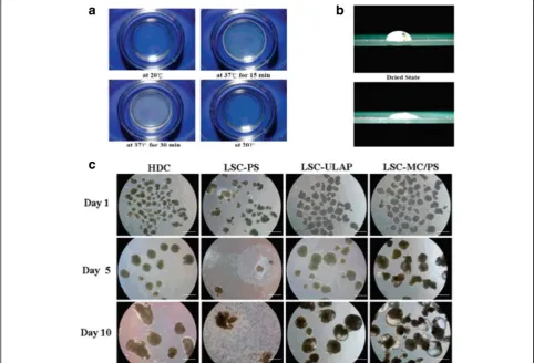
MC hydrogels are widely used to mount the surface of
polystyrene dishes and are used to cultivate human
em-bryonic stem cells (hESCs) for the formation of
embry-onic bodies (EBs) in liquid suspension cultures [
96
,
128
,
129
]. The EBs developed from the hESCs are shown to
express molecular markers specific for representative
cells from the three embryonic germ layers, indicating
the use of MC-coated dish for the large-scale production
of EBs from hESCs as shown in Fig.
7
a-c.
Mixed hydrogels
The mixing or blending of different polymers, such as, a
cellulose-polymer composite is a desirable, inexpensive
and advantageous method for obtaining novel structural
materials [
6
]. Cellulose (or its derivatives) blended with
natural biodegradable polymers, such as chitin, chitosan
[
130
], starch [
131
,
132
], alginates [
133
,
134
], and
hyalur-onic acid [
135
], has been used to created novel materials
for specific applications. Some examples include the
blending of a cellulose-polymer composite with chitosan
for the removal of heavy metals, with starch for the food
industry, and with alginates for tissue engineering
[Chang, 2011].
Cellulose-chitosan hydrogel beads areprepared by
blending cellulose powder to chitosan solution [
136
].
Chitosan is perviously blended with a highly
concen-trated carboxymethylated cellulose solution to form
physical hydrogels, which is then cross-linked by
irradi-ation [
137
]. This cellulose-chitosan duplex has been
shown to exert non-diffusible antibacterial properties
[
128
,
129
]. A novel microporous hydrogel produced by
mixing of cellulose with sodium alginate (SA) solution
and then cross linking with epichlorohydrin. The final
cellulose/SA hydrogels were characterized by
solid--state,
13C NMR, wide-angle X-ray diffraction (WXRD),
thermo-gravimetric analysis (TGA), scanning electron
microscopy (SEM), rheological measurement, dynamic
mechanical analysis (DMA), and swelling test analyses
to evaluate the structure and morphology of the
hydro-gels (Fig.
8
a-c) [
138
].
Currently,
polymeric-inorganic
hybrid
compounds
have been widely used in various fields, such as
elec-trical, optical, magnetic, and biological fields, among
others [
138
]. A novel method for the incorporation of
inorganic materials and cellulose hydrogels has been
studied in New Zealand white rabbits with
critically-sized bone defects in the distal femoral epiphyses [
139
].
In the experimental process, the researchers used an
in-jectable and self-cross-linkable bone substitute (IBS2)
composed of Si-HPMC viscous solution (3 wt%) in
alka-line medium, supplemented with biphasic calcium
phos-phate (BCP) ceramic particles. The diameter of the BCP
particles ranged from 40 to 80
μ
m. After a number of
weeks, centripetal bone formation was observed near the
defects, with a yield strength that was significantly
higher than that of the host trabecular bone tissue.
Fig. 7MC-coated hydrogel dishes for hESCs differentiation.aOriginal photograph s of the MC Hydrogel-coated in a polystyrene dish at distinct temperatures;bPhotograph of a water drop on the surface of the MC hydrogel coated in a polystyrene dish in the dried or hydrated state;c
Figure
9
a-c shows how bone regeneration occurs after
the application of Si-HPMC/BCP materials. The use of
BC from
Gluconacetobacter hansenii
along with a novel
composite material composed of calcium-deficient
hy-droxyapatite (CdHAP) for orthopedic use has been well
characterized and described by [
140
]. On the other hand,
[
141
] reported the use of heparin/cellulose/charcoal
composites to understand the mechanism and crosstalk
among cells. To study intracellular drug delivery systems
and cellular proliferation, single-walled carbon
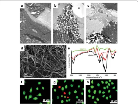
nano-tubes (SWCNTs) wrapped with cellulose have been
ob-served in HeLa cells [
101
,
102
]. Researchers developed
SWCNTs with a cellulose solution, dissolved in ionic
li-quid 1-butyl-3-methylimidazolium bromide (Fig.
9
d-e).
Another study showed that long cellulose/SWCNT
scaf-folds could promote the growth of HeLa cells, whereas
short cellulose/SWCNT were found to only have a small
effect on cell proliferation of HeLa cells (Fig.
9
f-h).
Healthy cells have a green nucleus, uniform chromatin,
and an intact cell membrane, whereas necrotic cells or
late apoptotic cells have red nuclei with damaged cell
membranes. Cells cultured on a composite scaffold and
a glass slide are healthy with a green nucleus (Fig.
9
f and
h), however, some cells culture on purified SWCNTs are
in the late apoptotic stage (Fig.
9
g). Thus,
inorganic-based cellulosic hydrogels provide a wide range of
appli-cations in the biomedical and tissue engineering field.
Application of cellulose hydrogels in tissue
engineering
Cellulose-based hydrogels are used in different fields
re-lated to tissue engineering. Patterned macroporous (PM)
with a diameter larger than 100
μ
m were introduced to
pristine 3D nanofibrous BC scaffolds using infrared (IR)
micromachining techniques to create an in vitro culture
model for breast cancer cells (BCs) [
142
]. PM-BC scaffolds
were found to be promote cellular adhesion, growth,
pro-liferation, and infiltration of BCs.
A. xylinum
BC also
pro-motes wound healing as it maintains the wound moist by
controlling the wound exudates and also heals severe
second-degree burns [
143
,
144
]. Hydroxyethylcellulose
(HEC) and carboxymethyl cellulose sodium salt (CMCNa)
cross-linked with hyaluronic acid allow for the
prolifera-tion of keratinocytes in an in vitro culture [
144
]. Bacterial
nanocellulose (BNC) has great potential for use as a
scaf-fold in tissue engineering, since BC is more effective than
PC, which accounts for BC being the first choice in
med-ical and tissue engineering applications.
BC hydrogels in biomedical applications
BC has promising features due to the similar of its
nano-structure and morphology to collagen making BC an
at-tractive choice for use in the support and immobilization
of cells. The architecture of BC materials can be
engi-neered at range of scales, ranging from the nano to
mac-roscale by controlling the biofabrication process. BC fibers
are solid and, when used in combination with other
bio-compatible materials, produce nanocomposites
particu-larly suitable for use in human and veterinary medicine
[
76
]. The applications of BC composite hydrogels in
bio-medicine and tissue engineering are listed in Table
4
. BC
composites can also be used in cornea formation after
cor-nea surgical treatment, as well as heart and vascular tissue
regeneration [
148
].
Bioactive cartilage implantation
Since, BC gels are free from the action of proteolytic
en-zymes and reactive oxygen species (ROS), they protects
the body from carcinogenesis and prevents the appearance
of inflammation. Some examples of cartilage implants
composed of BC are septum implants, ear implants, and
intervertebral discs, among others [
176
]. Now a days, the
use of bio-mimicking scaffolds has led to the exploration
of BC as a potential scaffolding material. A previous study
showed that BC did not induce the activation of pro-
in-flammatory cytokines during in vitro macrophage
screen-ing, but rather stimulated the biogenesis of collagen type
II with chondrocytes seeded on BC membranes, indicating
the suitability of BC as bio-mimicking scaffold [
177
].
An-other more recent study showed the synthesis of
modified bacterial cellulose (MBC) from metabolically
engineered
Gluconacetobacter xylinus
with a high
proliferation level of human mesenchymal stem cells
(hMSCs) compared to native cellulose. This material
was reported to be a novel in vivo degradable scaffold
for chondrogenesis [
178
,
179
].
Blood vessel prototypes
Artificial blood vessel-like structures composed of BC
are almost 5
–
25 cm long, which are stable,
mechanic-ally strong and resistant to water, aqueous liquids, ions,
and small particles, among others. Such vessel-like
structure are often used as main platforms for
neuro-transmitters. Natural BNC has promising mechanical
properties,
including
tear
resistance
and
shape-retention properties, such that it is better suited for use
as biological vessels [
176
].
Wound dressing materials
BC has been successfully used as wound dressing
ma-terial since the 1980s. BC composite mama-terials are
used in medicine due to their biocompatible, sterile,
porous, and flexible nature. The use of BC sheets
al-lows for wounds to breathe, and prevent the
forma-tion of scabs and scars. On the other hand, the use
of BC in dressing materials also reduces the amount
of pain, protects the skin from infections, and reduces
the loss of body fluids. As such, BC composite
mate-rials are an ideal candidate for the treatment of
wounds and burns [
180
]. Some examples of
commer-cially available BC composite gels are listed in Table
5
.
A novel type of BC-based wound dressing, which is
impregnated with superoxide dismutase and poviargol,
Fig. 9TEM of IBS2-filled bone defects after 8 weeks (a-c).aThe image clearly showed the mature bone tissue (B) containing the osteocytes (Os);
was found to stimulate the healing of thermal skin
burns resulting from acute radiation disease [
183
].
Surprisingly, BC/collagen type I composite was found
to promote the reduction of protease, interleukins,
and ROS activity in an in vitro culture study [
184
].
Surgical implants
BCs and BNCs can be used in the form of tracheotomy
tubes for reconstructive surgery, such as for artificial heart
valves, and as blood vessels in the form of nanotubes or
neurotubes for the regeneration of coronary blood vessel
Table 4
Uses of plant cellulose (PC), microbial cellulose (MC) and bacterial cellulose (BC) composite hydrogels in tissue engineering
Sl. No.
Hydrogel composite Applications References
1 Plant cellulose (PC) purified Tissue engineering and regenerative medicine [145]; Liu et al., 2014 [146]
2 Algal cellulose (AC) Bone tissue and cartilage engineering [147]
3 Bacterial cellulose (BC) purified Bone tissue engineering, cornea treatment, heart and vascular muscle regeneration
[148]
4 Carboxymethyl cellulose (CMC) Drug loading and controlled release of drugs, nucleus pulposus [149]; [148]
5 Polyvinylpyrrolidone (PVP) Soft-tissue replacement wound management [149]
6 Gelatin Wound dressing, tissue regeneration [80]; [150], [151]
7 Starch Reinforcement agent for bionanocomposites [152]
8 Alginate, sodium alginate High strength hydrogel preparation [153]
9 Acrylic acid Burn wound healing [154]
10 Graphene oxide (GO) Biomedicine [155]
11 Vaccarin Cell growth carrier wound dressing [156]
12 Hyaluronic acid (HA) Wound dressing, tissue engineering [157]
13 Chondroitin sulfate (CS) Dental material scaffold Opera et al., 102
14 Calcium phosphate (CP) Bone substitute [158]
15 Ca2+activated cellulose, cellulose/ lactide
Bone tissue engineering [148]
16 2-hydroxyethyl methacrylate (PHEMA)
Contact lenses and optic component for biosensors [159]
17 Polyacrylamide Cartilage replacement [160] & [161]
18 Gellan gum High strength hydrogel for synthetic connective tissue [153]
19 L-carrageenan High strength hydrogel for synthetic connective tissue [153]
20 Hydroxyapatite Bone scaffold substitute, bone tissue engineering [162]; [163]; [164]; [165]; [166]
21 Nanohydroxyapatite Bone tissue engineering
22 Polyvinyl alcohol (PVA) Cardiovascular soft tissue replacement, artificial cornea biomaterials ([167]; [168]); [169]; ([170]; [171])
23 Polylacitide and glycidyl methacrylate
Skin repair material [172]
24 Collagen Wound dressing for skin regeneration [173]; [148]
20 Silver Antimicrobial wound dressing [174]; [175]
Table 5
Commercially available hydrogel wound dressing contains cellulose or its sodium salt. Most dressings are available in two
forms, either as sheets or as amorphous gels. Products containing silver ions show antimicrobial property
The hydrogel wound dressing (producer) Composition References
IntraSite™Gel (Smith and Nephew) Carboxymethycellulose sodium (CMCNa), propylene glycol and water
GranuGel™(ConvaTec) Carboxymethycellulose sodium (CMCNa), Propylene glycol, pectin and water [181], [182]
Purilon Gel™(ColoPlast) Carboxymethycellulose (CMC), calcium alginate and water
Aquacel Ag™(ConvaTec) Carboxymethycellulose sodium (CMCNa) and silver ions (1.2%)
and nerves. Previous studies have found new epithelial cell
layers to form over these artificial BC tubes,
demonstrat-ing the successful application of BC in tissue implantation
[
185
]. The use of PVA/BC nanocomposites for the
placement of cardiovascular tissues has also been
re-ported, since these would mimic the role of natural
collagen and elastin (a connective tissue protein that helps
skin to return to its original position [
167
,
168
]
Potential drug delivery material
Transdermal systems can act as an entry gate for BCs into
the domain of drug delivery systems [
186
]. BC dry films
have been obtained after the successful immersion of
these in benzalkonium chloride (an antimicrobial agent).
Their subsequent drug loading capacity was found to be
0.116 mg/cm
2(per unit surface area), and the effect of
drug was found to last for at least 24 h against
Staphylo-coccus aureus
and
Bacillus subtilis
applied to the wounded
area [
187
]. Silver nanoparticle-coated BC fibers showed
99.99% antimicrobial activity against
Escherichia coli
and
S. aureus
[
164
]. Despite these promising results, the
appli-cation of BC hydrogels involves certain clinical and
pharmacological limitations. However, despite these
limi-tations, the complex nanofibrillar structure of BC
repre-sents a suitable macromolecular support for the inclusion
of drugs, i.e. for use as a drug carrier [
188
].
Artificial grafting of cornea
Corneal disease is a serious health problem that can lead
to partial or complete blindness. An estimated 10
mil-lion people have lost their eyesight due to corneal
infec-tion or similar diseases. With this in mind, researchers
around the world have developed biomaterials for the
treatment of defective corneas. The properties of
bacter-ial cellulose, including its nanoporous structure, and
ex-cellent mechanical properties, make it an ideal candidate
for use as an artificial cornea to help maintain the
intra-ocular pressure of the eye and re-establish intra-ocular
pellu-cidity. The BC/polyvinyl alcohol (BC/PVA) hydrogel has
a water content and light transmittance comparable to
that of natural cornea and was successfully synthesized
and described by Wang et al. for this end.
Dental implants
BC composite hydrogels were prepared from
Acetobacter
hansenii
by [
189
] for used in dental root canal treatment
(RCT) due to intracanal asepsis. Dental RCT is required
when dental caries progress to infection of the dental
pulp. From a materials point of view, BC has superior
properties compared to plant cellulose (paper points) for
the use in dental RCT. Moreover, research has
demon-strated the tissue regeneration of periodontal cells after
the application of BC hydrogels [
190
,
191
].
Other applications
Biomimetic scaffolds are of great interest to tissue
engin-eering as they supports essential cell functions. BNC
scaffolds in combination with soluble collagen-I
stimu-late estrogenic differentiation of mesenchymal stem cells
(MSCs) [Vielreicher et al., 2018]. The use of cell-derived
ECM collagen-I holds good potential, particularly for the
tissue engineering of mechanically-challenged tissues.
An optimized method for the purification of nano-
fibril-lated cellulose (NFC) and hydrogel production from
wood cellulose was described for the development of a
wound dressing material [
192
]. Inflammation, autolytic
debridement, granulated tissue formation, and re-
epi-thelialization are the processes that generally occur
dur-ing wound healdur-ing. Wound dressdur-ings are designed to
promote healing while protecting the wounds from
in-fection. This is particularly important in cases of chronic
wounds (e.g., ulcers), which fail to heal properly. Since a
moist environment encourages rapid healing, hydrogels
are optimal candidates for the development of wound
dressings, either as sheets or in an amorphous form
[
193
]. Various types of hydrogel dressings have been
pat-ented so far and are currently commercially available
(Table
5
), based on synthetic or natural polymers, or a
combination of these. Among the most recent patents, it
is worth citing those describing in situ forming gels (e.g.,
based on sprayable formulations [
194
] and on coalescing
nanoparticles [
195
]), and those exploring radiation
crosslinking as a stabilization technique, which allows to
obtain sterile and cross-linked hydrogel films in a
sin-gle-step process [
196
,
197
].
Scaffold attempts to mimic natural ECMs. The most
common method of tissue engineering includes the use of
biodegradable scaffolds to support the growth and
develop-ment of cells into tissues or by injecting the isolated single
cell suspensions [
5
]. Cellulose-based scaffolding materials
are widely used to regenerate various tissues, such as bone,
cartilage, heart, blood vessel, nerve, and liver, among others.
However, the design of scaffolds often involves issues related
to the need requirement for adequate cell-cell adhesion,
cell-cell communication, and cell-ECM communication,
which are crucial features of tissue functioning [
198
]. To
overcome these problems, biodegradable scaffolds have
been developed. Since, natural polymers are biocompatible,
their use allows us to avoid stimulating chronic
inflamma-tion or immunological reacinflamma-tions or toxicity. Therefore,
hydrogels are used extensively in tissue engineering due to
their high swelling properties and their biocompatibility. As
a result, they can be incorporated the cells of soft tissues
and bioactive molecules via gelling process [
199
].
Conclusion and future directions
materials that have widespread applications in the field
of tissue engineering and regenerative medicine. In these
areas, scaffolds played a significant role and have been
developed to form temporary, artificial ECMs to support
cell attachment and three-dimensional (3D) tissue
for-mation. Due to their high mechanical strength and
ther-mostability, bacterial cellulose derivatives are widely used
for wound dressing and healing, providing a novel method
for the treatment of epidermal burns. Most interestingly,
the work of researchers across the globe in the fields of
cellulose hydrogel development and characterization seem
to indicate that hydrogels based on cellulosic biomaterials
could be potential candidates for applications in the field
of tissue engineering. However, the research outcomes
ap-pear somewhat different from the promising predictions.
For example, while using hydrogels in bioengineering
ap-plications, researchers have encountered a number of
problems. These include difficulties in the handling,
main-tenance, storage of hydrogels, for example, for hydrogels
designed using bioprinters, which are not as much
mech-anically strong as was theoretically determined. During in
vitro experiments it was more difficult to sterilize
scaffold-ing structures than, for example, the cell culture media.
Sterilizing by means of autoclaving can cause the
func-tional properties of cellulose-based hydrogels to change.
However, their sterilization is necessary since the use of
hydrogels without proper sterilization could be a large
source of contamination during in vivo and in vitro
exper-iments in laboratory. Researchers have also often
encoun-tered difficulties while loading hydrogels with drugs or
cells for controlled drug delivery. Further research into
hydrogels will be required for the development of new
methods and protocols in order to overcome these
limita-tions. Despite these issues, the use of BC hydrogels
compared to plant-derived or manmade hydrogels is
cur-rently on the rise due to the cost-effective production of
BC hydrogels using stirred-tank or static bioreactors.
However, more needs to be done to improve plant-derived
cellulosic gel production (PC hydrogels). The use of
cellulose-based hydrogels in tissue engineering has both
advantages and disadvantages, the latter of which will
need to be resolved before cellulosic hydrogels can be
more widely applied.
Researchers are also working to improve our
under-standing of the mechanism behind the molecular
inter-action involved in cellulose ECM materials so that, in
the future, materials that mimic natural ECMs in terms
of their composition, structural characteristics, and
mechanical properties can be developed. The proper
development of 3D scaffolding materials could be used
to replace conventional tissue engineering techniques to
a great extent. Cellulose- based hydrogels have
import-ant applications in tissue engineering due to their high
biocompatibility and environment- friendly properties.
Cellulose-based hydrogels have been recently modified
using a nontoxic cross-linking agent or cross-linking
treat-ments, to improve the yield of both the final product and
the manufacturing processes. However, further research is
needed to develop more advanced cellulose-based
hydro-gels for use in healthcare and medicine.
Additional file
Additional file 1:Table S1.α-Cellulose content of some plant products [197–204].Figure S1:Source of some naturally occurring cellulose. a. hard wood (beech tree); b. cotton tree; c. bamboo; d.Gluconacetobacter xylinum; e. ascidians.Figure S2.Hydrogen bonding pattern in cellulose molecule. The hydrogen bonding within or between cellulose molecules represents its crystalline nature while studying through X-ray diffraction or NMR technique.Figure S3.Microphotograph showing variation in morphology of different fibers. a. twisted cotton fibers; b. tracheids of spruce wood; c. straight fibers of ramie. Copyright permission from [205]; simplified model of plant cell wall. d. structure of S1-S3layer; e-f. Cellulose assembly with pectin, hemicellulose, and lignin. Copyright permission from ([49]; [206–208]). (DOCX 312 kb)
Abbreviations
3D:Three-dimensional; AMIMCl: 1-allyl-3-methylimidazolium chloride; BC: Bacterial cellulose; BCP: Biphasic calcium phosphate; BMIMCl: 1-butyl-3-methylimidazolium chloride; BNC: Bacterial nanocellulose; CdHAP: Calcium-deficient hydroxyapatite; CMC: Carboxymethyl cellulose;
CMCNa: Carboxymethylcellulose sodium salt; DMA: Dynamic mechanical analysis; DMAc: Dimethylacetamide; DMSO/TBAF: Dimethysulfoxide/ tertrabutylaluminium fluoride trihydrate; DP: Dope characteristics; EBs: Embryonic bodies; ECM: Extracellular matrix; GAGs: Glycosaminglycans; HEC: Hydroxyethylcellulose; hESCs: Human embryonic stem cells; HPC: Hydroxypropyl cellulose; HPMC: Hydroxypropylmethyl cellulose; ILs: Ionic liquids; IR: Infrared; LiCl: Lithium chloride; MBC: Modified bacterial cellulose; MC: Methyl cellulose; MSCs: Mesenchymal stem cells;
NaOH: Sodium hydroxide; NFC: Nano-fibrillated cellulose; NMMO:N -methylmorpholine-N-oxide; NMR: Nuclear magnetic resonance; PC: Plant cellulose; PM: Patterned macroporous; ROS: Reactive oxygen species; SA: Sodium alginate; SEM: Scanning electron microscopy; Si-HPMC: Silated-hydroxypropylmethyl cellulose; SWCNTs: Single-walled carbon nanotubes; TGA: Thermo-gravimetric analysis; UV: Ultra-violet; WXRD: Wide-angle X-ray diffraction
Acknowledgements
The authors would like to thank Prof. Lim for his continuous support to write the manuscript.
Funding
This research was supported by‘Co-operative Research Program for Agriculture Science and Technology Development (No. PJ012854012017)’, Rural Development Administration, Republic of Korea and‘Basic Science Research Program’through the‘National Research Foundation of Korea’ funded by the Ministry of Education (No. 2018R1A6A1A03025582) and the
‘National Research Foundation of Korea’(NRF-2016R1D1 A3B03932921).
Availability of data and materials
Not applicable.
Authors’contributions
SDD wrote the manuscript. DKP and KTL reviewed the manuscript, edited, and provided feedback. KTL read and approved the final manuscript.
Ethics approval and consent to participate
Not applicable.
Consent for publication
Competing interests
The authors declare no competing interests.
Publisher
’
s Note
Springer Nature remains neutral with regard to jurisdictional claims in published maps and institutional affiliations.
Author details
1
Biorobotics Laboratory, Department of Biosystems Engineering, Kangwon National University, Chuncheon, Republic of Korea.2The Institute of Forest
Science, Kangwon National University, Chuncheon 24341, Republic of Korea.
Received: 24 February 2019 Accepted: 10 May 2019
References
1. Silva AKA, Richard C, Bessodes M, Scherman D, Merten OW. Growth factor delivery approaches in hydrogels. Biomacromolecules. 2009.https://doi.org/ 10.1021/bm801103c.
2. Peppas NA, Bures P, Leobandung W, Ichikawa H. Hydrogels in
pharmaceutical formulations. Eur J Pharm Biopharm. 2000.https://doi.org/ 10.1016/S0939-6411(00)00090-4.
3. Peppas NA, Mikos AG. Preparation methods and structure of hydrogels. In: Peppas NA, editor. Hydrogels in medicine and pharmacy. Florida: CRC Press; 1986. p. 1–27.
4. Stauffer SR, Peppas NA. Poly (vinyl alcohol) hydrogels prepared by freezing-thawing cyclic processing. Polymer. 1992. https://doi.org/10.1016/0032-3861(92)90385-A.
5. Drury JL, Mooney DJ. Hydrogels for tissue engineering: scaffold design variables and applications. Biomaterials. 2003.https://doi.org/10.1016/j. biomaterials.2010.02.044.
6. Bajpai AK, Shukla SK, Bhanu S, Kankane S. Responsive polymers in controlled drug delivery. Prog Polym Sci. 2008.https://doi.org/10.1016/j.progpolymsci. 2008.07.005.
7. Vinogradov SV, Bronch TK, Kabanov AV. Nanosized cationic hydrogels for drug delivery preparation, properties and interactions with cells. Adv Drug Deliv Rev. 2002.https://doi.org/10.1016/S0169-409X(01)00245-9.
8. Ostrovidova GU, Makeev AV, Shamtsian MM. Polyfunctional film coatings for medical use. J Mater Sci Eng. 2003. https://doi.org/10.1016/S0928-4931(03)00031-6.
9. Ratner, BD, Hoffman AS. Process of radiation grafting hydrogels onto organic polymeric substrates. 1976; US Patent US3939049A.
10. Lee YJ, Braun PV. Tunable inverse opal hydrogel pH sensors. Adv Mater. 2003.https://doi.org/10.1002/adma.200304588.
11. Sorber J, Steiner G, Schulz V, Guenther M, Gerlach G, Salzer R, et al. Hydrogel-based piezoresistive pH sensors: Investigations using FT-IR attenuated total reflection spectroscopic imaging. Anal Chem. 2008.https:// doi.org/10.1021/ac702598n.
12. Khan F, Tare R, Richard O, Oreffo R, Bradley M. Versatile biocompatible polymer hydrogels: scaffolds for cell growth. Angewandate Chemie Int Edn. 2009.https://doi.org/10.1002/anie.200804096.
13. Lee KY, Mooney DJ. Hydrogel for tissue engineering. Chem Rev. 2001.
https://doi.org/10.1021/cr000108x.
14. Katsoulos C, Karageorgiadis L, Vasileiou N, Mousafeiropoulos T, Asimellis G. Customized hydrogel contact lenses for keratoconus incorporating correction for vertical coma aberration. Ophthalmic Physiol Opt. 2009.
https://doi.org/10.1111/j.1475-1313.2009.00645.x.
15. Yasuda H. Biocompatibility of nanofilm-encapsulated silicone and silicone-hydrogel contact lenses. Macromol Biosci. 2006.https://doi.org/10.1002/ mabi.200500153.
16. Wu D, Wang T, Lu B, Xu X, Cheng S, Jiang X, et al. Fabrication of supramolecular hydrogels for drug delivery and stem cell encapsulation. Langmuir. 2008.https://doi.org/10.1021/la8006876.
17. Nagahama K, Ouchi T, Ohya Y. Temperature-induced hydrogels through self-assembly of cholesterol-substituted star PEG-b-PLLA copolymers: an injectable scaffold for tissue engineering. Adv Funct Mat. 2008.https://doi. org/10.1002/adfm.200700587.
18. Zhu J. Bioactive modification of poly (ethylene glycol) hydrogels for tissue engineering. Biomaterials. 2010.https://doi.org/10.1016/j.biomaterials.2010. 02.044.
19. Martens PJ, Bryant SJ, Anseth KS. Tailoring the degradation of hydrogels formed from multivinyl poly (ethylene glycol) and poly (vinyl alcohol) macromers for cartilage tissue engineering. Biomacromolecules. 2003.
https://doi.org/10.1021/bm025666v.
20. Ferruti P, Bianchi S, Ranucci E, Chiellini F, Piras AM. Novel agmatine-containing poly (amidoamine) hydrogel as scaffolds for tissue engineering. Biomacromolecules. 2005.https://doi.org/10.1021/bm050210+.
21. Nayak S, Lee H, Chmielewski J, Lyon LA. Folate-mediated cell targeting and cytotoxicity using thermoresponsive microgels. J Am Chem Soc. 2004.
https://doi.org/10.1021/ja0474143.
22. Gao D, Xu H, Philbert MA, Kopelman R. Ultrafine hydrogel nanoparticles: synthetic approach and therapeutic application in living cells. Angewandate Chemie Int Edn. 2007.https://doi.org/10.1002/ange.200603927.
23. Tomatsu I, Hashidzume A, Harada A. Contrast viscosity changes upon photoirradiation for mixtures of poly(acrylic acid)-basedα-cyclodextrin and azobenzene polymers. J Am Chem Soc. 2006.https://doi.org/10.1021/ ja058345a.
24. Kim DG, Seo SW, Cho BK, Lohumi S, Hong SJ, Lee WH. Review of current approaches for implementing metabolic reconstruction. J Biosyst Eng. 2018.
https://doi.org/10.5307/JBE.2018.43.1.045.
25. Hoffman AS. Hydrogels for biomedical applications. Adv Drug Deliv Rev. 2002;43:3–12.
26. Temenoff JS, Mikos AG. Injectable biodegradable materials for orthopedic tissue engineering. Biomaterials. 2002. https://doi.org/10.1016/S0142-9612(00)00108-3.
27. Buxton AN, Zhu J, Marchant RE, West JL, Yoo JU, Johnstone B. Design and characterization of poly(ethylene glycol) photopolymerizable semi-interpenetrating networks for chondrogenesis of human mesenchymal stem cells. Tissue Eng. 2007.https://doi.org/10.1089/ten.2007.0075. 28. Hahn MS, McHale MK, Wang E, Schmedlen RH, West JL. Physiologic pulsatile
flow bioreactor conditioning of poly (ethylene glycol)-based tissue engineered vascular grafts. Ann Biomed Eng. 2007.https://doi.org/10.1007/ s10439-006-9099-3.
29. O’Sullivan AC. Cellulose: the structure slowly unravels. Cellulose. 1997;4:173– 207.
30. Eichhorn SJ, Young RJ, Davies GR. Modeling crystal and molecular deformation in regenerated cellulose fibers. Biomacromolecules. 2005.
https://doi.org/10.1021/bm049409x.
31. Schurz J.“Trend in polymer science”a bright future for cellulose. Prog Polym Sci. 1999;24:481.
32. Hon DNS. Cellulose and its derivatives: structures, reactions, and medical uses. In: Dumitriu S, editor. Polysaccharides in medical applications. New York: Marcel Dekker; 1996. p. 87–105.
33. Eichhorn SJ, Baillie CA, Zafeiropoulos N, Mwaikambo LY, Ansell MP, Dufresne A, et al. Current international research into cellulosic fibers and composites. J Mater Sci. 2001.https://doi.org/10.1023/A:1017512029696.
34. Vandamme EJ, De Baets S, Vanbaelen A. Improved production of bacterial cellulose and its application potential. Polym Degrad Stab. 1998;59:93–9.
35. Jonas R, Farah LF. Production and application of microbial cellulose. Polym Degrad Stab. 1998.https://doi.org/10.1016/S0141-3910(97)00197-3. 36. Dutta SD, Tarafder M, Islam R, Datta BS. Characterization of cellulolytic
enzymes of fusarium soli isolates. Biocatal Agri Biotechnol. 2018.https://doi. org/10.1016/j.bcab.2018.03.011.
37. Brown RM Jr, Saxena IM. Cellulose biosynthesis: a model for understanding the assembly of biopolymers. Plant Physiol Biochem. 2000.https://doi.org/ 10.1016/S0981-9428(00)00168-6.
38. Rao VSR, Sundararajan PR, Ramakrishnan C, Ramachandran GN. Conformational studies of amylose. In: Ramachandran GN, editor. Conformation of biopolymers (Vol II). London: Academic; 1967. p. 721–37. 39. Krassig HA. Cellulose: structure, accessibility and reactivity. Gordon and
Breach Science: Yverdon; 1993.
40. Sorieul M, Dickson A, Hill SJ, Pearson H. Plant fibre: molecular structure and biomechanical properties, of a complex living material, influencing its deconstruction towards a biobased composite. Materials. 2016.https://doi. org/10.3390/ma9080618.
41. Seo YR, Kim JW, Hoon S, Kim J, Chung JH, Lim KT. Cellulose-based nanocrystals: sources and applications via agricultural by-products. J Biosyst Eng. 2018.https://doi.org/10.5307/JBE.2018.43.1.059.
![Fig. 2 Structure of cellulose and bacterial cellulose.xylinum a structure of cellulose fibrils (0.2 μm) and microfibrils (1 μm); b SEM images of Acetobacter and formation of bacterial cellulose [53] SEM: Scanning electron micrograph](https://thumb-us.123doks.com/thumbv2/123dok_us/9109668.1903633/4.595.58.538.87.498/structure-microfibrils-acetobacter-formation-bacterial-cellulose-scanning-micrograph.webp)
![Fig. 5 Schematic representation of strategy for BC production [73] BC: bacterial cellulose](https://thumb-us.123doks.com/thumbv2/123dok_us/9109668.1903633/7.595.62.539.506.715/fig-schematic-representation-strategy-bc-production-bacterial-cellulose.webp)
![Fig. 6 a Cellulose hydrogel beads with an average size of 467 μm [99], b NMMO fibers, c Viscose fibers [115]NMMO: N-methylmorpholine-N-oxide](https://thumb-us.123doks.com/thumbv2/123dok_us/9109668.1903633/8.595.59.538.88.198/cellulose-hydrogel-average-fibers-viscose-fibers-nmmo-methylmorpholine.webp)
![Fig. 8 Original photograph (a), SEM image (b), and compressive stress-strain curve (c) of cellulose/SA hydrogel [139] SEM: scanning electronmicroscopy; SA: sodium alginate](https://thumb-us.123doks.com/thumbv2/123dok_us/9109668.1903633/10.595.59.539.87.201/original-photograph-compressive-cellulose-hydrogel-scanning-electronmicroscopy-alginate.webp)