Iranian Biomedical Journal 2 (1): 21-25 (January 1998)
*Corresponding Author.
PCR-mediated Expression of the Human GM-CSF Gene in
Escherichia coli
Mana Oloomi
l*, Saeid Bouzari and Veladimir O. Rechinsky
Molecular Biology unit, Pasteur Institute of Iran, Pasteur Ave, Tehran 13164, I.R., Iran
ABSTRACT
Four exons of the human genomic GM-CSF gene were assembled together using gene splicing by overlap extension (SOE) method. The resulting nucleotide sequence was cloned in the pET23a(+) ex-pression vector under the control of strong bacteriophage T7 transcription and translation signals. The construct obtained was Transferred into the E. coli strain, BL21(DE3) pLysS and IPTG was used for induction of GM-CSF gene and production of the target protein. one mg of protein per liter of cell culture, was obtained as revealed by ELISA.Iran. Biomed. J. 2: 21-25, 1998.
Keywords: SOE method, Polymerase Chain Reaction, human GM-CSF, Gene expression, E. coli
INTRODUCTION
Colony-stimulating factors are a group of molecules that can stimulate hemopoiesis in vitro. They were first described during the 1960s but were available only in small quantities as crude extracts for about a quarter of century. Human granulocyte-macrophage colony stimulating factor (hGM-CSF) was originally identified and characterized by its ability to support the proliferation and maturation of bone marrow-derived myeloid progenitor cells[1, 2]. The colonies formed in response to this factor consist primarily of neutrophils, monocytes and granulocytes, hence the name GM-CSF was given [2]. With the advent of recombinant DNA technology, sufficient amounts of pure substances could be produced to allow research and development for clinical use [1].
The gene for hGM-CSF has been cloned and ex-pressed in mammalian [3, 4], yeast [5, 6] and bacterial cells [7, 8]. Protein synthesized by bacteria differs from the native one in that it has an additional methionine residue at the N-terminus and is not glycosylated but was found to exhibit the same biological activities expect the erythroid burst-promoting activity [8]. The recombinant hGM-CSF has considerable therapeutic potential [9], being used for the stimulation and/or reconstitution of the immune system, particularly in chemotherapy induced leukopenia[10].
In this study the Polymerase Chain Reaction technique was used for processing of the human
genomic GM-CSF gene and the resulting PCR product was cloned in a pET vector (plasmid for expression by 77 RNA polymerase) and expressed in Escherichia coli [11].
MATERIALS AND METHODS
Strains and plasmids.pUC18 vector (Pharmacia), together with E. coli strain XL-1 blue (recAl, endAl, gyrA96, thi-1, hsdR17, supE44, relAl, Lac [F 'proAB, LacIqZΔM15, Tn10(tetr)]), were used for initial cloning and maintaining DNA fragments. For protein production, an expression vector pET23a(+) (Novagen) carrying strong bacteriophage T7 transcription and translation signals, coupled with the E. coli strain, BL21(DE3)pLysS (F- ompT hsdSB
(rB
-mB
-) dcm gal (DE3-) pLysS, Cmr)as host strain was used. It contains a chromosomal copy of the T7 RNA polymerase gene under the control of the inducible LacUV5 promoter. Addition of IPTG to a growing culture induces the enzyme, which in turn transcribes the target DNA in the plasmid. A plasmid containing the human genomic GM-CSF gene was kindly provided by Dr. V.G. Krobko (Institute for Bioorganic chemistry of the Russian Academy Science). This DNA was used as a template for PCR. Enzymes were pur chased from GIBCO-BRL and used under conditions recommended by supplier. Bacterial transformation and plasmid DNA purification were
performed as described by Sambrook et al. [12]. DNA sequencing was performed by the dideoxy chain termination technique of Sanger et al. [13].
Polymerase Chain Reaction (PCR). PCR was carried out in a Pharmacia LKB Gene ATAQ con-troller and oligonucleotide primers prepared on an applied Biosystems DNA synthesizer model 360B and Vent DNA polymerase from New England Biolabs. The amplification was conducted according to the following program. Denaturation at 94°C for 1 min, annealing at 47°C and 60°C for 30 sec, extension at 70°c for 1 min. To amplify each exon 30 cycles of PCR were conducted to recover the exon junction products at every stage of assembly, 20 cycles of PCR were used. Amplification products were analyzed by electrophoresis in 1.2-2.0% agarose gels and purified from the low melting point temperature agarose (Sigma, Type VII).
Cloning and expression of the rhGM-CSF gene. The resulting amplification product was gel purified, phosphorylated with the T4 polynucleotide Kinase, and blunt-end ligated into the pUC18 vector that had been cleaved with HindII. Recombinant plasmids were transformed into E.coli strain, XL1-blue and selected on LB plates containing ampicillin (100 µg/m1) and Xgal (40 µg/m1). Correct constructs were initially identified by restriction endonuclease analysis of miniprep plasmid pu-rification and ultimately confirmed by direct DNA sequencing. Then the GM-CSF gene was sub-cloned into the pET23a expression vector (pET-GMCSF). E. coli BL21(DE3)pLYSs transformed with pET-GMCSF was cultured in LB supple-mented with ampicillin (100 µg/ml) at 37°C with good aeration to early exponential phase (A 600 approximately 0.2-0.5 ). IPTG was added to a final concentration of 0.5 mM and the culture was al-lowed to continue growing for 1-3 h. Bacteria were harvested by centrifugation and GM-CSF extracted essentially as described by pET System manual [14].
SDS-polyacrylamide gel electrophoresis (SDS-PAGE) and Western Blot. Analyses were carried out as described by Maniatis [12]. Nitrocellulose paper, 0.45 µm pore size (Pharmacia-LKB) was used for blotting of the protein. Rabbit Polyclonal hGMCSF antibody (R&D Systems) and Anti-Rabbit Antibody/HRP (Sigma) was used for detec-tion of the protein.
Enzyme-Linked immunosorbent assay
(ELISA). ELISA was performed by using a kit From R&D Systems for detection of GM-CSF. This assay employed the quantitative sandwich enzyme immunoassay technique in order to measure the amount of recombinant product using standard curve prepared for diluted hGM-CSF.
RESULTS AND DISCUSSION
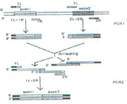
Construction of an expression plasmid. The hGM-CSF gene requires deletion of three introns before it can be expressed in bacteria. For this pur-pose, a technique based on splicing by overlap ex-tension (SOE) PCR was employed [15], which is in common use now. The procedure outlined in Fig.1 is as follows. To generate a continuous sequence, the two exons to be joined are firstly amplified in separate PCR reactions, using primers containing complementary sequences at ends. When these PCR products are mixed, denatured and annealed, the complementary ends hybridize thus allowing the second strand to be synthesized. The result is that the two exons are spliced together and can be further amplified as a unit by a second PCR with the "external" primers only.
In the first stage of the "spliced" hGM-CSF gene construction, each exon was amplified separately using a corresponding pair of primers (see Table 1) designed on the basis of the known nucleotide sequence of the gene [16]. Then exon 1 was coupled
Fig. 1. Outline of the SOE strategy.
performed as described by Sambrook et al. [12]. DNA sequencing was performed by the dideoxy chain termination technique of Sanger et al. [13].
Polymerase Chain Reaction (PCR). PCR was carried out in a Pharmacia LKB Gene ATAQ con-troller and oligonucleotide primers prepared on an applied Biosystems DNA synthesizer model 360B and Vent DNA polymerase from New England Biolabs. The amplification was conducted according to the following program. Denaturation at 94°C for 1 min, annealing at 47°C and 60°C for 30 sec, extension at 70°c for 1 min. To amplify each exon 30 cycles of PCR were conducted to recover the exon junction products at every stage of assembly, 20 cycles of PCR were used. Amplification products were analyzed by electrophoresis in 1.2-2.0% agarose gels and purified from the low melting point temperature agarose (Sigma, Type VII).
Cloning and expression of the rhGM-CSF gene. The resulting amplification product was gel purified, phosphorylated with the T4 polynucleotide Kinase, and blunt-end ligated into the pUC18 vector that had been cleaved with HindII. Recombinant plasmids were transformed into E.coli strain, XL1-blue and selected on LB plates containing ampicillin (100 µg/m1) and Xgal (40 µg/m1). Correct constructs were initially identified by restriction endonuclease analysis of miniprep plasmid pu-rification and ultimately confirmed by direct DNA sequencing. Then the GM-CSF gene was sub-cloned into the pET23a expression vector (pET-GMCSF). E. coli BL21(DE3)pLYSs transformed with pET-GMCSF was cultured in LB supple-mented with ampicillin (100 µg/ml) at 37°C with good aeration to early exponential phase (A 600 approximately 0.2-0.5 ). IPTG was added to a final concentration of 0.5 mM and the culture was al-lowed to continue growing for 1-3 h. Bacteria were harvested by centrifugation and GM-CSF extracted essentially as described by pET System manual [14].
SDS-polyacrylamide gel electrophoresis (SDS-PAGE) and Western Blot. Analyses were carried out as described by Maniatis [12]. Nitrocellulose paper, 0.45 µm pore size (Pharmacia-LKB) was used for blotting of the protein. Rabbit Polyclonal hGMCSF antibody (R&D Systems) and Anti-Rabbit Antibody/HRP (Sigma) was used for detec-tion of the protein.
Enzyme-Linked immunosorbent assay
(ELISA). ELISA was performed by using a kit From R&D Systems for detection of GM-CSF. This assay employed the quantitative sandwich enzyme immunoassay technique in order to measure the amount of recombinant product using standard curve prepared for diluted hGM-CSF.
RESULTS AND DISCUSSION
Construction of an expression plasmid. The hGM-CSF gene requires deletion of three introns before it can be expressed in bacteria. For this pur-pose, a technique based on splicing by overlap ex-tension (SOE) PCR was employed [15], which is in common use now. The procedure outlined in Fig.1 is as follows. To generate a continuous sequence, the two exons to be joined are firstly amplified in separate PCR reactions, using primers containing complementary sequences at ends. When these PCR products are mixed, denatured and annealed, the complementary ends hybridize thus allowing the second strand to be synthesized. The result is that the two exons are spliced together and can be further amplified as a unit by a second PCR with the "external" primers only.
In the first stage of the "spliced" hGM-CSF gene construction, each exon was amplified separately using a corresponding pair of primers (see Table 1) designed on the basis of the known nucleotide sequence of the gene [16]. Then exon 1 was coupled
Fig. 1. Outline of the SOE strategy.
Iranian Biomedical Journal 2 (1): 21-25 (January 1998)
23
Table 1. Oligonucleotide Primers used in splicing of the hGM-CSF gene. The ends of exons are
marked off with points. The NdeI and HindIII restriction exonuclease sites are underlined.
Oligonucleotide Sequence
1L 5'-CACCATATG.GCACCCGCCCGCTGCCCCAG-3'
1R 5'-TACTGTTTCATT.CATCTCAGCAGCAGTGTC-3'
2L 5'-GCTGCTGAGATG.AATGAAACAGTAGAAGTC-3'
2R 5'-GCAGGTCGGCTC.CTGGAGGTCAAACATTTC-3'
3L 5'-TTTGACCTCCAG.GAGCCGACCTGCCTACAG-3'
3R 5'-ACAGGAACTTTC.CGGGGTTGGAGGGCAGTG-3'
4L 5'-CCTCCAACCCCG.GAAACTTCCTGTGCAACC-3'
4R 5'-CACAAGCTTATCA.CTCCTGGACTGGCTCCCA-3'
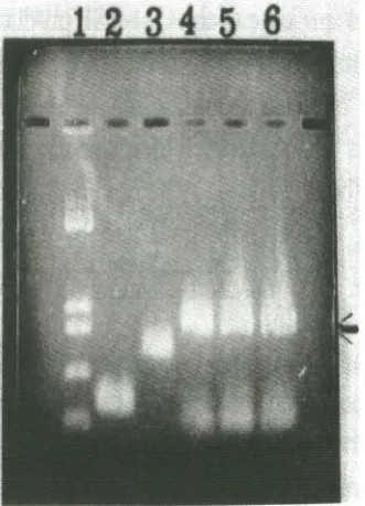
to exon2, and exon 3-to exon 4 in the same manner (Fig. 2 lane 2,3,4). Finally, the entire GM-CSF-encoding sequence was assembled by PCR with the terminal primers 1L and 4R containing NdeI and HindIII restriction endonuclease sites for subse-quent insertion of the target gene into the expres-sion vector. The above technique relies on the use of a DNA polymerase that has a proofreading (i.e. 3'5' exonuclease)activity, such as Vent DNA polymerase. This derives higher fidelity than that observed for Taq DNA polymerase. In the PCR Vent polymerase produces fragments with blunt ends that match exactly the primer sequence, unlike Taq DNA polymerase, which adds a single A over-hang [17].
Fig. 2. GM-CSF gene amplification. Lane 1, lengths of
marker fragments (pUC18 digested with Hinf I); lane 2, amplified exon 1; lane 3, amplified exon 2, 3, and 4; lanes 4-6 (arrow), GM-CSF gene constructed of exons 14 with different concentrations of Mg2+(lane 4, 1 mM; lane 5, 3 mM and lane 6, 5 mM).
In study conducted by Slobodkina et. al [18] the same technique was employed and they found dif-ferent concentration of Mg 2+ is needed to amplify different exons. However in our experiment the concentration of Mg2
+
was not very critical (Fig. 2 lane 4-6).
Sequencing of the GM-CSF gene. The resulting sequence was cloned in pUC18 by blunt end liga-tion (Hinc II) and it's sequence was confirmed by Sanger method [14]. NdeI restriction site was used at the 5' end of the insert coding sequence, to add ATG initiation codon for translation and Hind III restriction site was used for cloning of the gene into the pET vector. The pET system is developed for the cloning and expression of recombinant proteins in E. coli. For expression of the protein we used pET system because of it's strong and specific promoter. Moreover RNA polymerase of bacterio-phage T7 is very selective for specific promoters that are rarely encountered in DNA unrelated to T7 DNA. A very active enzyme, T7 RNA polymerase elongates chains about five times faster than E. coli RNA polymerase. These properties, make T7 RNA polymerase attractive as the basis for expression systems [19]. The NdeI-HindIII fragment coding for GM-CSF was subcloned into the pET expression vector under the control of strong bacteriophage T7 transcription and translation signals, yielding a plasmid named pET-GMCSF. The resulting plasmid was transformed into E. coli host strain, B121(DE3)pLYSs and IPTG was used for induc-tion.
Preparation and analysis of rGM-CSF. Ex-pression of the GM-CSF gene was analyzed by SDS-PAGE and the resulting protein was run on 18% SDS-PAGE along with standard molecular weight marker. The gel was stained with Coomassie blue and the band with corresponding molecular
Fig. 3. (Panel A) SDS-PAGE analyses of rGM-CSF. Lane 1, inclusion bodies from IPTG- induced cells resuspended in 6M urea (arrow shows
GM-CSF); lane 2, proteins from uninduced cells; lane 3, proteins from negative control induced cells; lane 4, proteins from negative control uninduced cells; lane M, Amersham rainbow Low molecular weight markers (46, 30, 21.5, 14.3 in kDa). (Panel B) Western blot analyses of rGM-CSF. Lane 1, negative control; lane 2, rGM-CSF; lane 3, positive control; lane 4 Molecular weight markers (24, 18, 14.3 in kDa.)
weight marker of 14 kDa was identified. As is seen in Figure 3A. Also in western blot analysis (Lane 2 in Fig. 3B) the corresponding band could be detected. The supernatant of induced cell extract was assayed by ELISA for the presence of GM-CSF protein. The results were noted at 450 nm and it was found that the yield of the protein to be 1 mg per liter of bacterial culture (i.e. 1-2 g of bacterial cell). The purification of GM-CSF protein from natural sources is not easy because of its low content. Therefore different approaches have been under taken to purify this protein using various methods. hGM-CSF has been partially purified from placental conditioned medium [20], and medium conditioned by the hairy T-cell leukemia cell line Mo [21]. Although it has been possible to purify it, detailed analysis of many aspects of their biology and biochemistry has been hampered by the limited quantities available. This problem can be largely circumvented by molecular cloning of the corre-sponding gene sequences [22]. Another way for obtaining hGM-CSF is purification of the mRNA which is complicated. The procedure of selecting this gene from a human cDNA library by hybridi-zation with oligonucleotide probes is rather expen-sive and needs more procedure.
Considering the above mentioned difficulties, the use of PCR technique, used in this study accelerates significantly the process of detection, cloning and purification as compared to classical methods.
Moreover it is more convenient when the nucleo-tide sequence of the gene is known, and can be synthesized by the amplification technique directly on the basis of genomic DNA. On the other hand in bacteria no glycosylation takes place and the influence of carbohydrate on the bioactivities is different among various proteins [23]. Nevertheless it was indicated that removal of N-linked oligosaccharides in case of the GM-CSF produced in yeast and mammalian cells, increased their immunoreactivities by 4 to 8-fold. Removal of these oligosaccharides also increased their specific biological activity about 20-fold. The E. coli produced-protein lacking any carbohydrate had by far the highest specific activity observed for the recombinant hGM-CSFs [23].
Considering these above mentioned facts, the PCR technique employed was quite successful in order to process the gene suitable for expression in prokaryotic system and more over, the resulting construct in E.coli produced active protein. The activity of the produced protein was determined by the stimulation action on proliferation of KG1 cell line, using (3H TdR method) incorporation (data not shown).
In conclusion, the results of this study revealed that, PCR can be good method for construction of an eukaryotic gene and its subsequent expression in E. coli. It is also confirmed the ability of E. coli to produce such proteins in active form.
Iranian Biomedical Journal 2 (1): 21-25 (January 1998)
25 REFERENCES
1. Steward, W.P. (1993) Granulocyte Macrophage Colony Stimulating Factor. The Lancet 342: 153- 15 7.
2. Mire-Sluis, A.R., Gaires Das, R., Thorpe, R. (1995) The international standard for Granulocyte-macrophage colony stimulating factor (GM-CSF). I Immunol. Methods 179: 127-135.
3. Gasson, J.C., Baldwin, G.C., Sakamoto, K.M. and Di persio, J.F. (1990) The biology of human granulo-cyte-macrophage colony stimulating factor (GM-CSF). In: Progress in clinical and biological Re-search. (Dainiak , N., eds), Wiely liss New York. 4. Wong, G.G., Wirek, J.S., Temple, P.A., Wilkens,
K.M., Leary, A.c., Luxenberg, D.P., Shoemaker, S.S., Brown, E.L., Kay, R.M., Orr, E.C., Hewick, R.M., Wang, E.A. and Clark, S.C. (1985) Human GM-CSF: molecular cloning of the complementary DNA and purification of the natural and recombinant proteins. Science 228: 810-815.
5. Lee, F., Yokota, T., Otsuka, T., Gemmell, L., Larson, N., Luh, J., Arai, K-I., and Rennick, D. (1985) Isolation of cDNA for a human granulocyte-macrophage colony-stimulating factor by functional expression in mammalian cells. Proc. Natl. Acad. Sci. 82: 4360-4364.
6. Cantrell, M.A., Anderson, D., Cerretti, D.P., Price, V., Mckereghan, K., Ttshinski, R.J., Mochizuki, D.Y., Larsen, A., Grabstein, K., Gillis, S., and Cos-man, D. (1985) Cloning, sequence, and expression of a human granulocyte/macrophage colony-stimulating factor. Proc. Natl. Acad. Sci. 82: 6250-6254.
7. Miyajima, A., Otsu, K., Schreurs, J., Bond, M. W., Abrams, J.S., and Aria, K. (1986) Expression of murine and human granulocyte-macrophage colony stimulating factors in S. cerevisiae: mutagenesis of the potential glycosylation sites. EMBO J. 5: 1193-1197.
8. Burgess, A.W., Begley, C.G., Johnson, G.R., Lopez, A.F., Williamson, D.J., Mermod, J.J., Simpson, R.J., Schmitz, A. and Delamarter, J.F. (1987) Purification and properties of bacterially synthesized human granulocyte-macrophage colony stimulating factor. Blood 69: 43-51.
9. Weiss, M. and Belohradsky, B.H. (1992) Granulo-cyte-macrophage colony stimulating factor (GM-CSF). A variety of possible applications in clinical medicine. Infection 20: S81-S83.
10. Metcalf, D. (1990) The colony-stimulating factors. Discovery, Development and clinical Applications. Cancer 65: 2185-2195.
11. Studier, F.W. and Moffatt, B.A. (1986) Use of
bac-teriophage T7 RNA polymerase to direct selective high-level expression of cloned genes. J. Mol. Biol. 18.9: 113-130.
12. Sambrook, J., Fritsch, E.F. and Maniatis, T. (1989) Molecular Cloning.: A Laboratory manual, 2nd Edi-tion. Cold Spring Harbor Press, Cold Spring Harbor, NY. pp. 368 and 458.
13. Sanger, F., Nicklen, S. and Coulson, A.R. (1977) DNA sequencing with chain-terminating inhibitors. Proc. Natl. Acad. Sci. 74: 5463-5467.
14. Novagen Inc. (1992) pET System Manual. 5th Edi-tion. Madison, WI, USA.
15. Lefebvre, B., Formstecher, P. and Lefebvre P. (1995) Improvement of the gene Splicing Overlap (SOE) method. Biotechniques 19: 186-188.
16. Kaushansky, K., O'Hara, P., Berkner, K., Segal, G., Hagen, F., and Adamson, J.W. (1986) Genomic cloning, characterization, and multilineage growth-promoting activity of human granulocyte-macrophage colony-stimulating factor. Proc. Natl. Acad. Sci. 83: 3101-3105.
17. Clark, J.M. (1988) Novel non-template nucleotide addition reactions catalyzed by prokaryotic and eu-karyotic DNA polymerases. Nucleic acids Res. 16: 9677-9688.
18. Slobodkina, G.V., Kotova, E.Yu., Braga, E.A., Chi-syakov, D., Marchenko, G.N., Tsygankov, Yu. D., and Noikov, V.V. (1994) Construction of Human GM-CSF Gene Using Polymerase Chain Reaction and it's Expression in Pseudomonas putida Cells. Molecular biology 28: 553-558.
19. Goeddel, D.V. (1991) Gene expression technology. Academic press, Inc. Pp. 60-89.
20. Nicola, N.A., Metcalf, D., Johnson, G.R. and Bur-gess, A.W. (1979) Separation of functionally distinct human granulocyte-macrophage colony-stimulating factors. Blood 54: 614-627.
21. Gasson, J.C., Weisbart, R.H., Kaufman,S.E., Clark, S. C., Hewick, R. M., Wong, G.G. and Golde, D. W. (1984) Purified human granulocyte-macrophage col-ony-stimulating factor: Direct action on neutrophils. Science 226: 1339-1342.
22. Gough, N.M., Gough, J., Metcalf, D., Kelso, A., Grail, D., Nicola, N.A., Burgess, A.W. and Dunn, A.R. (1984) Molecular cloning of cDNA encoding a murine hematopoietic growth regulator, granulocyte-macrophage colony stimulating factor. Nature 309: 763-767.
23. Moonen, P., Mermod, J-J., Ernst, J.F., Hirschi, M. and Delamarter, J.F. (1987) Increased biological ac-tivity of deglycosylated recombinant human granulo-cyte/macrophage colony-stimulating factor produced by yeast or animal cells. Proc. Natl. Acad. Sci. 84: 4428-4431.