Short Report
Gene Expression Profile of CatSper3 and CatSper4 during
Postnatal Development of Mouse Testis
Malek Hossein Asadi, Seyed Javad Mowla
*and Parvaneh Nikpoor
Dept. of Genetics, Faculty of Basic Science, Tarbiat Modarres University, Tehran, 14115-175, Iran
Received 23 July 2005; revised 12 October 2005; accepted 12 October 2005
ABSTRACT
Channel activities, particularly those of calcium channels, have vital roles in the process of sperm maturation, motility and sperm-egg interaction. A group of the recently discovered ion channels associated with these processes is four novel channel-like proteins known as CatSper (cation channel sperm) gene family. CatSper1 and CatSper2 show sperm specific expression patterns. However, neither CatSper1 nor CatSper2 have been shown to function as cation channels when transfected into cells, singly or together. It has been suggested that the known CatSper proteins require additional subunits and/or interaction partners to function. Using in silico gene identification and prediction
techniques, two further members of the CatSper family, CatSper3 and CatSper4, have been identified. It has been hypothesized that the CatSper proteins may form a functional tetrameric channel. Based on these findings, we evaluated the temporal pattern of CatSper3 and CatSper4 genes during mouse testis development to assess any potential co-incidence of their expression with CatSper1 and CatSper2. In the present study, a total of 55 male BALB/c mice were categorized into 11 age groups: 1 day, 1 week, 2 weeks, 16, 18, 20, 22, 24 and 26 days, 1 month as well as adult mice. Testes were removed from each mouse, total RNA was extracted and RT-PCR was performed with specific primers for CatSper3, CatSper4 and beta-2-microglobin (as an internal control). Our results revealed that CatSper3 and CatSper4 genes are expressed predominantly at 3 week of age. The obtained data demonstrate that: 1) The expression of CatSper3 and CatSper4 in mouse is developmentally regulated. 2) There is a direct correlation between the temporary expressions of these genes and the other two members of the family, CatSper1 and CatSper2. Iran. Biomed. J. 10 (2): 111-115, 2006
Keywords:CatSper, Fertility, Sperm motility, Testis, Gene expression
INTRODUCTION
t is well known that Ca2+ ions and cyclic
nucleotides play vital roles during various stages of fertilization, especially in facilitating spermatozoal motility [1]. Accordingly, any increase in the concentration of intracellular Ca2+
ions leads to changes in spermatozoal motility [2, 3]. However, the mechanisms controlling swimming behavior, the entrance site of Ca2+ into
spermatozoa and the molecular identity of the Ca2+
channels remain poorly understood [4].
Recently, two novel channel-like proteins: CatSper1 (Cation channel Sperm 1) and CatSper2 were identified to be exclusively expressed in
spermatozoa and are linked to sperm mobility [5, 6]. The human and mouse CatSper channels, CatSper1 and CatSper2, both carry a single six-transmembrane spanning unit analogous to one of the four repeats found in voltage-dependent Ca2+
channels. Analysis of the pore forming region within the repeat suggested that CatSper1 and CatSper2 are Ca2+ selective. Further evidence
supporting channel activity has been provided for CatSper1 by gene-targeting experiments in the mouse. Notably in sperm from the mice carrying two null alleles for the CatSper1 gene, cAMP and cGMP induced Ca2+ influx are lost [5]. Moreover,
CatSper1 has been shown to be required for normal sperm motility and egg penetration. However,
I
functional channel. The fact that CatSper1 and CatSper2 share features of a single repeat of a four repeat channel suggests that two additional members might exist [5, 6]. In another attempt, two additional channels in the human and mouse genomes, CatSper3 and CatSper4 were predicted by means of bioinformatics search tools [7]. Both channels contain a single six-transmembrane repeat domain which contain the T× D×W pore-lining consensus sequence present in CatSper1 and CatSper2. Based on accompanying EST, cDNA library source information and Taqman data, both genes are expressed in testis. Furthermore, they noted that each CatSper channel is predicted to contain a coiled-coil motif, and a protein-protein interaction interface in its intracellular C-terminal tail. Based on EST data and the fact that each CatSper protein is predicted to contain a coiled-coil domain, they hypothesized that different members of CatSper come together to form functional tetrameric channels either by direct interaction of their coiled-coil motif or through interaction with additional factors [7].
In the present study, we have determined the expression pattern of CatSper3 and CatSper4 during mouse testis development and compared the data with our previous observation about CatSper [8]. The similar temporal patterns of gene expression for members of CatSper gene family support a potential formation of functional tetrameric channel.
MATERIALS AND METHODS
Animals. A total of 55 male BALB/c mice were
obtained from Razi institute for serums and vaccines (Tehran, Iran) and categorized into 11 age groups: 1 day, 1 week, 2 weeks, 16, 18, 20, 22, 24 and 26 days, 1 month as well as adult mice. Animals were housed in small groups under standard lightening conditions with free access to water and food. They were rapidly sacrificed by cervical dislocation and both testes were removed.
RNA extraction and reverse transcription. Total
RNA was isolated from frozen tissues using RNX plus solution (CinnaGen, Tehran, Iran) according to the manufacturer’s instructions. Purity and integrity of the extracted RNA was measured by optical density (260/280-nm ratio) as well as visual
was synthesized at 42C for 60 min in presence of 1 µg RNA and 200 U/μl MMLV reverse transcriptase (Gibco BRL, Germany), 20 U RNase inhibitor, dNTP mix (final concentration of 1 mM) with oligo(dT) 18 priming in a 20 µl
reaction [9].
PCR. PCR primers were designed using
Genrunner software (version 3.02; Hastings software Inc.) according to previously described mouse CatSper3, CatSper4 and 2m sequences. (GenBank accession numbers: AK014942, AK077145 and NM-009735, respectively). As following:
2m-Mouse-forward:
5`TGACCGGCTTGTATGCTATC 3`;
2m-Mouse-reverse:
5`CACATGTCTCGATCCCAGTAG3` CatSper3-Mouse-forward:
5`GCTCTTCCTCCTCATGTTTG3`; CatSper3-Mouse-reverse:
5`TCTTCCAACATCAGGCTCAG 3`; CatSper4-Mouse-forward:
5`AAGGGGACACAGCAAAGATG3`; CatSper4-Mouse-reverse:
5`TATTCCAGCCATCCTTCCAG3`;
The primers amplified a 597 and 417-bp segment from CatSper3 and 4 cDNA, respectively and a 316-bp segment from 2m cDNA. The PCR amplification of cDNA was carried out by using PCR mix (2 μl the first cDNA strand, 2 units Taq DNA polymerase, 5 μl 10x PCR buffer, 5 μl 2 mM dNTPs, 1 μl 20 pmol forward primer, 1 μl 20 pmol reverse primer and water to 50 μl) and under the following condition: initial denaturation at 94C for 2 min, a 30 cycle amplification (94C for 30 s, 58.5C for 30 s, 72C for 1 min), and a final extension at 72C for 5 min.
DNA sequencing and enzymatic digestion. To
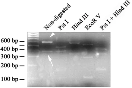
confirm the specificity of CatSper3 bands, PCR product of CatSper3 was digested with PstI, HindIII and EcoRV restriction enzymes (Fermentaz Company, Lithuania). To verify the identity of an unexpected shorter band of amplified CatSper3, the band was extracted from 1% agarose gel by Fermentaz kit. The extracted band was sequenced by MWG Company (Germany).
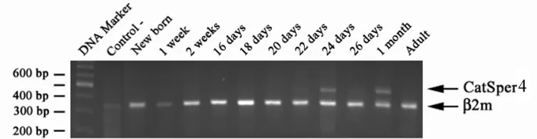
Fig. 1. Postnatal time-course analysis of mouse CatSper3 and ß2m gene expression in mouse testis. Total mRNA from mouse testis at different ages was reversed-transcribed and amplified using gene-specific primers for mouse CatSper3 and ß2m genes. A 316 bp RT-PCR product corresponding to the amplified ß2m fragment was observed in all age groups examined. In contrast, the 597 bp RT-PCR product corresponding to the amplified CatSper3 fragment was only observed in mouse testis starting at 3 weeks of age, with no detectable expression at earlier ages. Note the existence of a lower band at approximately bp, which is highlighted with a question mark.
RESULTS AND DISCUSSION
Detection of CatSper3 and 4 in testis. In order to
determine the temporal profile of CatSper3 and 4 genes expression during postnatal development of mouse testis, different age groups of mice (premature and mature) were examined. Our data revealed that the expression of the genes is developmentally controlled; when it is predominantly started at the age of 22 days (Figs. 1 and 2). The expression of CatSper3 begins postnatally at the age of 16 days, but the expression was weak until 22ndday, when it reached the peak
of its expression. The expression of CatSper4 begins at the age of 22 days and there were no expression prior to this time (Fig. 1).
In mammals, spermatozoa must swim a long distance to reach the site of egg to begin the process of fertilization. The female reproductive tract represents several natural barriers in the way of millions of released spermatozoa, and sperm motility is vital for the success of pregnancy [10].
For a long time, scientists who work on
contraceptive medicine and infertility treatment have searched to find molecules that control sperm motility [1]. Recently, a report from Clapham lab described cloning and characterization of a novel gene, CatSper1, which has a key role in controlling sperm motility. The gene codes for a unique Ca2+
channel, which is exclusively expressed in the testis. The channel controls the influx of calcium ions into the spermatozoa and the influx of calcium ions in turn regulates the sperm motility. Knocking out the gene in mice diminished sperm motility and its penetration ability to enter the egg’s outer layer. Except for being sterile, the appearance and sexual behavior of CatSper -/- mice are indistinguishable from the wild type males [5].
Comparing the CatSper1 and latterly discovered CatSper2 [6] with other 4 units Ca2+ channels, two
additional CatSper channels in the human and mouse genomes, CatSper3 and CatSper4 were predicted [7]. Both channels contain a single six-transmembrane repeat domain, which contain the T D W pore-lining consensus sequence present
Fig. 2. Postnatal time-course analysis of mouse CatSper4 and ß2m gene expression in mouse testis. Total mRNA from mouse testis at different ages was reversed-transcribed and amplified using gene-specific primers for mouse CatSper3 and ß2m genes. A 316 bp RT-PCR product corresponding to the amplified ß2m fragment was observed in all age groups examined. In contrast, the 417 bp RT-PCR product corresponding to the amplified CatSper4 fragment was only observed in mouse testis starting at 3 weeks of age, with no detectable expression at earlier ages. Reduced signal intensity was observed for adult mouse testis in all 3 repeats of the experiment.
the fact that each CatSper protein is predicted to contain a coiled-coil domain, they hypothesized that the CatSper come together to form a functional tetrameric channels either by direct interaction of their coiled-coil motif or through interaction with additional factors [7].
According to our previous work [8], the expression of CatSper1 is developmentally regulated, where its expression begins postnatally at the age of three weeks. Since the current knowledge about CatSper3 and 4 is based on bioinformatics search, rather than laboratory research, we decided to empirically determine the expression of the mentioned members of the family. Our results revealed that CatSper3 and 4 are expressed predominantly at three weeks of age. However, the expression profile of CatSper3 is slightly different from others, as it is expressed from birth to adulthood. Nevertheless, the intensity of CatSper3 band is very low before the age of 3 weeks. During the evaluation of CatSper3 expression, an unexpected shorter band (around 400 bp) was seen repeatedly in all age groups suggesting the existence of a potential alternatively spliced variant CatSper3.
By changing the condition of PCR reaction, the shorter band didn’t disappear. To test its relatedness to CatSper3, we performed restriction mapping on the PCR product of CatSper3. We selected various enzymes that are capable of cutting the different exons of the gene. By digesting the PCR product with pstI, HindIII, EcoRV (Fermentaz, Company, Lithuania); singly or in combination, the shorter band confirmed to be related to CatSper3 (Fig. 3). To further determine the exact identity of the shorter band, we extracted and sequenced the band. The sequencing data revealed that the shorter band is indeed a truncated form of CatSper3, however not a spliced variant. According to the sequencing data, the forward primer had been bound with two sites of cDNA template. Therefore, the shorter band is amplified by internally hybridization of forward primer to the cDNA.
The timing of expression of CatSper3 and 4 coincides with other members of the family, CatSper1 and 2. A finding that supports the potential tetrameric assembly model was proposed by Lobley et al. [7]. The timing is also well
matched with some newly reported transcription factors expressed primarily in testis. The expression
Fig. 3. Restriction enzymatic digestion of PCR products of CatSper3 and its corresponding shorter form by different restriction enzymes. The calculation of the sizes of the bands generated with different enzyme digestion revealed the correct identity of both CatSper3 and its shorter form.
of Tex1, a novel transforming growth factor beta-induced factor (TIGF) subclass homeobax gene [11] and Trif (testis-specific ring factor), a novel member of the RBCC family [12], begins in testis at around day 20 of age. The potential involvement of these and other genes in controlling the expression of CatSper3 and 4 should be investigated in future.
In conclusion, the obtained data demonstrate that: 1) The expression of CatSper3 and 4 in mouse is developmentally regulated. 2) There is a direct correlation between the temporary expressions of these genes.
ACKNOWLEDGEMENTS
We would like to thank Esmaeil Babaei, Mahmood Faraz and Mojdtaba Emadi at Genetics Laboratory of Tarbiat Modares University for their technical advice. We also received valuable help from graduate students at Virology and Biochemistry Labs of Tarbiat Modares University.
REFERENCES
1. Darszon, A., Labarca, P., Nishigaki, T. and Espinosa, F. (1999) Ion channels in sperm physiology. Physiol. Rev. 79: 481-510.
2. Ward, G.E., Brokaw, C.J., Garbeos, D.L. and Vacquier, V.D., (1985) Chemotaxis of Arbacia punctulata spermatozoa to react a peptide from the egg jelly layer. J. Cell Biol. 101: 2324-2329. 3. Cook, S.P., Brokaw, C.J., Muller, C.H. and Babcock,
D.F. (1994) Sperm chemotaxis: egg peptides control cytosolic calcium to regulate flagellar responses. Dev. Biol. 165: 10-19.
4. Wiesner, B., Weiner, J., Middendorff, R., Hagen, V., Kaupp, U.B. and Weyand, I. (1998) Cyclic nucleotide-gated channels on the flagellum control. Ca2+entry into Sperm. J. Cell Biol. 142: 473-484.
5. Ren, D., Navarro, B., Perez, G., Jackson, A.C., Hsu, S., Shi, Q., Tilly, J.L. and Clapham, D.E. (2001) A sperm ion channel required for sperm motility and male fertility. Nature 413: 603-609.
6. Quill, T.A., Ren, D., Clapham, D.E. and Garbers, D.L. (2001) A voltage-gated ion channel expressed specifically in spermatozoa. Proc. Natl. Acad. Sci. USA 98: 12527-12531.
7. Lobley, A., Pierron, V., Reynolds, L., Allen, L. and Michalovich, D. (2003) Identification of human and
mouse CatSper3 and CatSper4 genes:
Characterization of a common interaction domain and evidence for expression in testis. Reprod. Biol. Endocr. 1: 53
8. Nikpoor, P., Mowla, S.J., Movahedin, M., Ziaee, A.M. and Tiraihi, T. (2004) CatSper gene expression in postnatal development of mouse testis and in subfertile men with deficient sperm motility. Hum. Reprod. 19: 124-128.
9. Sambrook, J. and Russel, D.W. (2001) Molecular cloning: a laboratory manual. 3rd ed., Cold Spring
Harbor Laboratory Press, New York, USA. pp. 8.21-8.53.
10. Olds-Clarke, P. (1996) How does poor motility alter sperm fertilizing ability? J. Androl. 17: 183-186. 11. Lai, Y.L., Li, H., Chiang, H.S., Hsieh-Li, H.M.
(2002) Expression of a novel TGIF subclass homeobox gene, Tex1, in the spermatids of mouse testis during spermatogenesis. Mech. Dev. 113: 185-187.
12. Shyu, H.W., Hsu, S.H., Hsieh-Li, H.M. and Li, H. (2001) A novel member of the RBCC family, Trif, expressed specifically in the spermatids of mouse testis. Mech. Dev. 108: 213-216.