DOI
10.17219/acem/68902
Copyright
© 2018 by Wroclaw Medical University This is an article distributed under the terms of the Creative Commons Attribution Non-Commercial License (http://creativecommons.org/licenses/by-nc-nd/4.0/)
Address for correspondence
Agnieszka Rusak E-mail: [email protected]
Funding sources
Financial support of the National Science Centre (Poland) in the course of realization of the project "Preparation and characterization of nanoapatites doped with rare earth ions and their biocompos-ites" (No. UMO-2012/05/E/ST5/03904).
Conflict of interest
None declared
Received on September 15, 2016 Reviewed on December 14, 2016 Accepted on February 9, 2017
Abstract
Background. Venous insufficiency is still a serious clinical problem. The exact cause and molecular mecha-nisms of this disease are still unknown. In this study, we try to identify whether there is a difference in the level of trace elements between healthy and pathological veins. Our results show that insufficient veins have different levels of some trace elements: magnesium, calcium, manganese, and silicon compared to control samples. This study could lead to a better understanding of the molecular causes of venous insufficiency and may help to develop better methods of treatment.
Objectives. Nowadays, venous diseases are a very common clinical phenomenon. Venous insufficiency is thought to be one of the most common vein diseases. The exact mechanisms of its etiology are still unknown, although from a clinical point of view some risk factors include gender, age, changing hormone levels, heredity, and standing or sitting for long periods. An imbalance in trace elements could also play a crucial role in the development and/or progression of venous insufficiency.
Material and methods. The trace element content in varicose vein walls and in normal vein walls was measured using an inductively coupled plasma-optical emission spectrometer (ICP-OES) after sample miner-alization. Statistical analysis (the Mann-Whitney U test and the Friedman ANOVA) was performed to compare insufficient veins to controls (healthy veins).
Results. This study found statistically significant higher magnesium (Mg) ion levels in varicose veins com-pared to controls (p = 0.0067) and differences close to statistical significance in calcium (Ca), manganese (Mn), and silicon (Si) ion levels.
Conclusions. The results obtained could indicate oxidative stress occurring in chronic venous insufficiency as well as free radical neutralization pathways due to superoxide dismutase (SOD) activity with Mg, Mn and copper (Cu) ion involvement. Our results are consistent with literature data and are preliminary in nature.
Key words: trace elements, venous insufficiency, venous pathology
Venous insufficiency: Differences in the content of trace elements.
A preliminary report
Agnieszka Rusak
1,B–D, Ewa Karuga-Kuźniewska
2,A,E, Benita Wiatrak
3,D,E, Maria Szymonowicz
4,C,D,
Mateusz Stolarski
5,B, Małgorzata Radwan-Oczko
6,B, Rafał J. Wiglusz
7,C–F, Paweł Pohl
8,A,C, Zbigniew Rybak
4,D–F1 Division of Histology and Embryology, Department of Human Morphology and Embryology, Wroclaw Medical University, Poland
2 Division of Infectious Diseases of Animals and Veterinary Administration, Department of Epizootiology and Clinic of Bird and Exotic Animals, Faculty of Veterinary Medicine,
Wroclaw University of Environmental and Life Sciences, Poland
3 Department of Basic Medical Sciences, Wroclaw Medical University, Poland
4 Department of Experimental Surgery and Biomaterials Research, Wroclaw Medical University, Poland
5 Department of Trauma Surgery, Knappschaftskrankenhaus Bochum-Langendreer, University Hospital Bochum, Germany 6 Department of Periodontology, Wroclaw Medical University, Poland
7 Institute of Low Temperature and Structure Research, Polish Academy of Sciences, Wrocław, Poland
8 Division of Analytical Chemistry and Chemical Metallurgy, Faculty of Chemistry, Wroclaw University of Science and Technology, Poland
A – research concept and design; B – collection and/or assembly of data; C – data analysis and interpretation; D – writing the article; E – critical revision of the article; F – final approval of the article
Introduction
Venous disease with concomitant varicose veins is pres-ently a very common clinical phenomenon consisting of the destruction of vein valves, backward blood flow (venous reflux) and dilated veins.1 This in turn, may lead
to serious complications, such as thrombosis, skin changes and leg ulcers. There are many genetic, hormonal and en-vironmental factors affecting the development of varicose veins. The exact mechanism of their formation has not been identified. The main destructive factor is venous hypertension, which may lead to the remodeling of vein walls and vein valves.2,3 It is thought that long-term high
pressure in veins may activate matrix metalloproteinases (MMPs), initiate coagulation and complement cascades, and activate platelets, leukocytes and macrophages as well.1,2,4 Matrix metalloproteinases destroy the
extracel-lular matrix, affect smooth muscle cells, and change the properties of the endothelium layer. These processes tend to reduce the flexibility of vein walls. Activated leukocytes escape via damaged endothelium outside the capillaries, starting inflammatory processes. Then, proteins like fi-brinogen build a cuff around the capillaries, interfering with the gas exchange between the blood and the extra-cellular environment, which then leads to further damage of vein walls.1,2
The role of trace elements in vein pathology is not yet fully understood, but it is known that they play a significant role in tissue metabolism. Many studies suggest that iron (Fe) could be important in the development of venous diseases, but the cause of these iron deposits in the legs of patients with venous diseases is not yet known.5 It has been
suggest-ed that this process is connectsuggest-ed with activatsuggest-ed MMPs or free radicals.5–7 There is also a hypothesis that iron is not
a direct cause of venous abnormalities, but only a factor which intensifies the autoimmune response.8 Zinc (Zn),
manganese (Mn) and copper (Cu) ions are associated with an active center of superoxide dismutase (SOD) and par-ticipation in free radical neutralization.9–11 It is thought
that reactive oxygen species (ROS) play a significant role in the development of the endothelial dysfunctions which are connected with venous diseases, such as varicose veins and venous ulcers.12–14 Zinc, Cu and Fe ions also
partici-pate in free oxygen radical formation and may be involved in damage leading to chronic venous insufficiency.7,15 Zinc
is an important trace element for the immune system.16,17
In in vitro tests, high levels of Zn led to cell apoptosis. Most of the zinc (90%) in the human body is contained in the muscles and bones.16 Copper is also an important
trace element for human health – the body needs Cu in the appropriate amount, but excessive amounts are confirmed to have toxic properties.18,19 Copper deficiency causes many
abnormalities, e.g., problems with the absorption of iron leading to cellular iron deficiency, disorders of the immune system and weakness of the walls of the blood vessels.7,20,21
Copper is needed as a structural component or catalyst by many proteins.7 One of the important
copper-contain-ing proteins relevant to the construction of the blood ves-sels is lysyl oxidase. It is responsible for stiffness and elas-ticity as a result of the fact that it allows bonding between collagen and elastin.7,22 Manganese is needed for the proper
functioning and metabolism of cells.23 However, in vivo
and in vitro studies showed the toxic properties of Mn in high doses, especially for the central nervous system and PC12 cells (used as an in vitro model of neuronal cells).23–25
Age-related changes were previously reported in the arteries and veins.26 The magnesium (Mg) content in the
veins increases with aging, which may play a role in the development of vein wall diseases.27 Magnesium Mg2+
ions are known as an enzyme cofactor.28 Signaling
path-ways, ATPase activity, and the channel and metabolic regulation of the cell cycle are all dependent on Mg2+
content. Magnesium deficiency is connected with the occurrence of many disorders, such as osteoporosis, hy-pertension, heart arrhythmia, impaired glucose tolerance, and serum cholesterol.29–31 A reduced level of magnesium
also increases oxidative stress and can reduce erythrocyte SOD concentrations.30
Calcium (Ca) is involved in many metabolic processes. It occurs in the human body mainly in the bones and teeth in the form of calcium hydroxyapatite. Accumulated cal-cium in the bones constitutes a reserve for the function-ing of the body.32 Calcium ions are used to carry cellular
signals and as transporters across cell membranes.33,34
They also regulate intracellular mechanisms by binding to proteins. The cellular level of Ca2+ is precisely
regu-lated – a significant increase in the calcium level can lead to apoptotic changes.34 Metabolic processes involving Ca
ions are critical for the functioning of the vessels, muscles, nerves, and the endocrine system.32
Silicon (Si) is an important element in the biology of all living organisms – small amounts of this element are es-sential for their proper functioning.35,36 It plays
an impor-tant role in the connective tissue (bone, cartilage, skin, and blood vessels).37,38 It is believed that Si is associated with
the content of collagen and glycosaminoglycans in the con-nective tissue matrix.38,39 Changes in the Si level
of patho-logical tissues have been observed (e.g., in atherosclerotic tissue).40 In vitrostudies suggest that Si could have
neu-roprotective or neurotoxic properties (depending on the concentration).41
In our study, we determined the content of trace ele-ments – Cu, Fe, Mn, Mg, Zn, Ca, and Si – found in hu-man veins affected by chronic venous insufficiency and compared this data with the levels of the same trace el-ements found in healthy, sufficient human veins. It was confirmed that the concentrations of elements in tissue are often correlated with each other.42 For this reason, the
Material and methods
This study was conducted over a span of 2 years, between 2013 and 2014, at the Wroclaw Medical University and the Institute of Low Temperature and Structure Research, Polish Academy of Sciences. It was performed in compli-ance with the ethical principles of the Declaration of Hel-sinki and Good Clinical Practice. Legal representatives read, signed and dated the form before taking part in any study activity. Consent from the Bioethical Commission of Wroclaw Medical University was granted for the study (No. KB-87/2013).
Reagents and solutions
EMSURE® ACS grade reagents, i.e., concentrated HNO
3
(65%), HCl (36%) and H2O2 (30%) solutions were purchased
from Merck Millipore (Merck KGaA, Darmstadt, Ger-many). Aqua regia was freshly prepared by mixing con-centrated HCl and HNO3 solutions at a 3:1 volume ratio.
Deionized water from an EASYpure™ water purification
system (Barnstead Thermolyne Corp., Dubuque, USA) was used throughout. A Certipur® multi-element stock
(1000 μg/mL) ICP standard solution IV (Merck KGaA) was used for preparing simple and matrix-matching standard solutions for the calibration of the inductively coupled plas-ma-optical emission spectrometry (ICP-OES) instrument.
Samples and their preparation
An inorganic solution, made directly from human veins, was fabricated using microwave-assisted wet digestion, whereby the disinfected human veins were combined with HNO3 and H2O as well as NH4OH, for pH control. First
of all, the received disinfected human veins were subjected to thermal treatment using an electric furnace at a tem-perature of 550°C for 1 h. Following typical preparation pro-cedures for a final product containing all microelements, the inorganic powder was first dissolved in an excess of HNO3
in order to create an inorganic solution. This inorganic so-lution was then transferred into a teflon vessel and placed in a microwave reactor. After 60 min of microwave-stimu-lated hydrothermal processing at 200°C, and under an au-togenous pressure of 45 atm, the pH of the dispersion was then adjusted to approx.7 with the addition of NH4OH.
The resulting composition of the prepared solutions was then determined by inductively coupled plasma-optical emission spectrometry (ICP-OES).
Apparatus
A bench-top optical emission spectrometer, model 720 (Agilent, Santa Clara, USA), with an axially viewed Ar-ICP and a 5-channel peristaltic pump, was used to mea-sure the concentrations of Cu, Mn, Fe, Mg, Zn, Ca, and Si (trace elements). The instrument was equipped with
a high-resolution echelle-type polychromator and a Vis-taChip II CCD detector (Agilent)cooled down to –35°C on a triple-stage Peltier device. The plasma was sustained in a standard 1-piece, low-flow, extended quartz torch with a 2.4 mm inside diameter injector tube. A single-pass glass cyclonic spray chamber and a OneNeb pneumatic concen-tric nebulizer made of a high-tech PFA and PEEK poly-mers were used to introduce the sample solutions by pneu-matic nebulization. Operating conditions recommended by the manufacturer for solutions containing high levels of dissolved solids were applied: an RF power of 1200 W, a plasma gas flow rate of 15.0 L min−1, an auxiliary gas flow
rate of 1.5 L min−1, a nebulizer gas flow rate of 0.75 L min−1,
a sample flow rate of 0.75 mL min−1, a stabilization delay
of 15 s, a sample uptake delay of 30 s, a rinse time of 10 s, a replicate read time of 1 s, and 3 replicates. A fitted back-ground mode with 7 points per line profile was applied for the background correction. Background-corrected intensi-ties of analytical lines were used for calibration graphs.
Characteristic of groups
In this study, 2 different groups of patients were enrolled. One group of 11 patients (study group) was referred for surgery due to symptomatic vein insufficiency in 1 or both legs ac-cording to Clinical, Etiological, Anatomical, and Pathophysi-ological (CEAP) classification C2S, Ep, As, or Pr. The other group, consisting of 14 patients (control group), underwent cardiac coronary bypass grafting due to symptomatic arterial coronary insufficiency. The proximal and distal segments of the great saphenous vein were obtained from patients via a striping procedure (study group), while the patients with healthy veins (control group) underwent elective coronary artery bypass grafting, whereby excess vein was obtained from the proximal and distal vein segments. Patients were matched for age, sex and major risk factors (data not shown). Vessel segments were harvested using a non-touch technique before surgical distension or rapid removal. The segments were immediately transferred to physiological saline solution and stored in a refrigerator at a temperature of –20°C.
Statistical analysis
The results were statistically analyzed using the STATIS-TICA v. 12 software (StatSoft, Tulsa, USA). The Shapiro-Wilk test showed no normal distribution in the samples. The Mann-Whitney U test, Spearman’s rank order cor-relation (R), Friedman ANOVA, and Kendall’s coefficient of concordance were applied.
Results
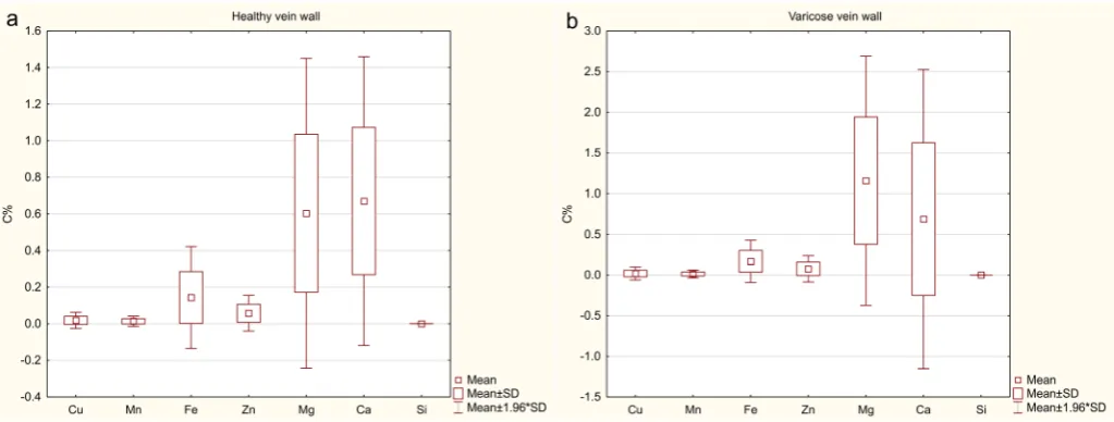
Statistically significant differences (the Mann-Whitney U test) compared to controls were observed (Fig. 2). A sta-tistically significant difference (p = 0.007) in the Mg ion level in varicose veins compared to controls was noted (Fig. 2e). Differences in Mn (p = 0.067), Ca (p = 0.085) and Si (p = 0.075) levels were close to the margin of statistical significance (Fig. 2b, 2f, 2g). The concentrations of mi-croelements in the studied human veins are presented as mean % mass concentration (C%) of trace elements in the mineral part of tissue samples.
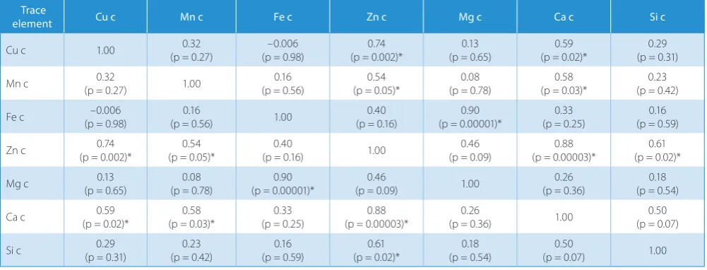
A statistically significant (p > 0.05), positive and strong correlation (Spearman’s rank order correlation) between the levels of some trace elements were observed for the study group (Table 1) and control group (Table 2). In the investigated group, correlations between the Ca and Mn ion levels (R = 0.71; p = 0.01), and the Ca and Zn levels (R = 0.93; p = 0.00004) were observed. In the control group, a correlation between these pairs was also observed
(R = 0.58; p = 0.03 and R = 0.88; p = 0.00003, respectively), with a common correlation for both groups being noted.
Discussion
Similar research has already been done, but there is very little literature available for comparison. The pioneer study of ferrous content in veins was performed by Krzyściak et al.6
One of the mechanisms involved in chronic venous dis-ease is the destructive effect of free oxygen and free ni-trogen radicals. The authors showed that Fe ions were in-volved in the oxidative damage mechanism which caused tissue altering via the Fenton reaction. Oxidative stress correlated with insufficiency in the venous tissue, and increased SOD activity was observed compared to nor-mal venous tissue.15 An increased level of iron in the skin
of patients with chronic venous disease was described
Table 1. Spearman’s rank order correlation (R) in the study group of insufficient veins
Trace
element Cu Mn Fe Zn Mg Ca Si
Cu 1.00 0.13
(p = 0.73)
–0.01 (p = 0.99)
0.05 (p = 0.88)
0.16
(p = 0.65) 0.10 –0.10
Mn (p = 0.73)0.13 1.00 (p = 0.13)0.48 (p = 0.15)0.46 (p = 0.03)*0.65 (p = 0.01)*0.71 (p = 0.008)*0.75
Fe (p = 0.99)–0.01 (p = 0.13)0.48 1.00 (p = 0.27)0.36 (p = 0.32)0.33 (p = 0.26)0.37 (p = 0.67)0.15
Zn (p = 0.88)0.05 (p = 0.15)0.46 (p = 0.27)0.36 1.00 (p = 0.00004)*0.93 (p = 0.00004)*0.93 (p = 0.17)0.45
Mg (p = 0.65)0.16 (p = 0.03)*0.65 (p = 0.32)0.33 (p = 0.00004)*0.93 1.00 (p = 8.4E–08)*0.98 (p = 0.05)0.59
Ca 0.10
(p = 0.78)
0.71 (p = 0.01)*
0.37 (p = 0.26)
0.93 (p = 0.00004)*
0.98
(p = 8.4E–08)* 1.00
0.61 (p = 0.04)*
Si (p = 0.78)–0.10 (p = 0.008)*0.75 (p = 0.67)0.15 (p = 0.17)0.45 (p = 0.05)0.59 (p = 0.04)*0.61 1.00
* statistically significant values (p < 0.05).
Fig. 2. Levels of ions, presented as mean % mass concentration (C%) of trace elements in the mineral part of tissue samples in insufficient veins compared to healthy veins (Mann-Whitney U test): a) Cu ions (p = 0.723); b) Mn ions (p = 0.067); c) Fe ions (p = 0.529); d) Zn ions (p = 0.848); e) Mg ions (p = 0.007); f) Ca ions (p = 0.085); and g) Si ions (p = 0.075)
by Myers in 1966.43 Degradation of hemoglobin, as well
as Fe release, was associated with erythrocyte dysfunc-tion. Moosavi et al. showed increased concentrations of iron and copper in the walls of varicose veins com-pared to controls, using a proton-induced X-ray emission (PIXE) analysis.44
Determining the content of trace elements in tissues is still a subject of research, though environmental con-tamination due to metal ions and the bioaccumulation ratio needs to be assessed as well.45,46 The level of trace
elements in tissue is a reflection of the general health and metabolic status of the individual.47 The level of metal ions
significantly depends on the type and location of the tissue being measured. Determining the level of trace elements may have a diagnostic application in such tissues as brain and blood serum.47,48
The level of SOD, oxidative DNA damage, and increased levels of iron, copper and zinc ions were also observed.27,30
SOD functions as a free radical scavenger. In oxidative stress conditions, increased antioxidant activity must be correlated with increased levels of enzymatic cofactors like Zn, Mn and Cu ions.15
The analysis of our results shows the differences between normal and insufficient veins. The Cu, Zn, Mn, and Ca ion levels had higher maximum values and slightly higher mean values compared to normal. Increased levels of these ions were observed in chronic venous insufficiency (CVI). Moreover, the Mn and Ca ion levels were close to being statistically significantly different from the control. These results are consistent with the literature and suggest a role of an SOD-, Cu-, Mn-, and Zn-dependent enzyme in CVI. Even though only a few articles dealing with a similar sub-ject were found, the results obtained in this study were consistent with the data in the existing literature.6,15
The iron ion levels were the highest detected, but the difference between altered and normal veins was not sta-tistically significant.
The reduction of vein wall elasticity is one of the symp-toms of venous insufficiency, which, in our opinion, could be associated with a decreased level of silicon, considering the role of this trace element in fiber synthesis. Our study shows decreased levels of Si in insufficient veins. These levels were considerably lower, as predicted, and the results were close to statistically significant. To our knowledge, at present, there are no other reports in the literature about the Si levels in similar research.
We found that the magnesium ion levels were higher in varicose veins compared to the control (p = 0.0067). This result may suggest that magnesium ions play an important role in the mechanism which occurs during venous insuf-ficiency, and very probably in vein wall structures as well. Differences in the correlation patterns of trace elements between normal and varicose veins may suggest homeo-static destabilization associated with vein disease. In ad-dition, they could also suggest a significant role of trace elements in chronic venous disease.
The current study is only a preliminary one. There is a need to perform further research, with an increased size of the study and control groups. Future studies are planned to assess the impact of gender on the levels of trace elements in a larger group.
Conclusions
This paper shows the levels of certain ions in insufficient and normal veins, particularly the differences between the concentrations of Mg, Mn, Ca, and Si. This study is pre-liminary in nature, but the data obtained suggests that the oxidative damage mechanism is involved in the devel-opment of varicose veins and chronic venous insufficiency.
Table 2. Spearman’s rank order correlation (R) in the control group (c) of normal veins
Trace
element Cu c Mn c Fe c Zn c Mg c Ca c Si c
Cu c 1.00 0.32
(p = 0.27)
–0.006 (p = 0.98)
0.74 (p = 0.002)*
0.13 (p = 0.65)
0.59 (p = 0.02)*
0.29 (p = 0.31)
Mn c (p = 0.27)0.32 1.00 (p = 0.56)0.16 (p = 0.05)*0.54 (p = 0.78)0.08 (p = 0.03)*0.58 (p = 0.42)0.23
Fe c (p = 0.98)–0.006 (p = 0.56)0.16 1.00 (p = 0.16)0.40 (p = 0.00001)*0.90 (p = 0.25)0.33 (p = 0.59)0.16
Zn c (p = 0.002)*0.74 (p = 0.05)*0.54 (p = 0.16)0.40 1.00 (p = 0.09)0.46 (p = 0.00003)*0.88 (p = 0.02)*0.61
Mg c (p = 0.65)0.13 (p = 0.78)0.08 (p = 0.00001)*0.90 (p = 0.09)0.46 1.00 (p = 0.36)0.26 (p = 0.54)0.18
Ca c 0.59
(p = 0.02)*
0.58 (p = 0.03)*
0.33 (p = 0.25)
0.88 (p = 0.00003)*
0.26
(p = 0.36) 1.00
0.50 (p = 0.07)
Si c (p = 0.31)0.29 (p = 0.42)0.23 (p = 0.59)0.16 (p = 0.02)*0.61 (p = 0.54)0.18 (p = 0.07)0.50 1.00
References
1. Raffetto JD, Khalil RA. Mechanisms of varicose vein formation: Valve dysfunction and wall dilation. Phlebology. 2008;23:85–98. 2. Kucukguven A, Khalil RA. Matrix metalloproteinases as potential
tar-gets in the venous dilation associated with varicose veins. Curr Drug Targets. 2013;14:287–324.
3. Simka M. Cellular and molecular mechanisms of venous leg ulcers development: The “puzzle” theory. Int Angiol. 2010;29:1–19. 4. Raffetto JD, Qiao X, Koledova VV, Khalil RA. Prolonged increases
in vein wall tension increase matrix metalloproteinases and decrease constriction in rat vena cava: Potential implications in varicose veins.
J Vasc Surg. 2008;48:447–456.
5. Zamboni P, Izzo M, Tognazzo S, et al. The overlapping of local iron overload and HFE mutation in venous leg ulcer pathogenesis. Free Radic Biol Med. 2006;40:1869–1873.
6. Krzyściak W, Kowalska J, Kózka M, Papież MA, Kwiatek WM. Iron con-tent (PIXE) in compecon-tent and incompecon-tent veins is related to the vein wall morphology and tissue antioxidant enzymes. Bioelectrochemistry. 2012;87:114–123.
7. Arredondo M, Núñez MT. Iron and copper metabolism. Mol Aspects Med. 2005;26:313–327.
8. Simka M, Rybak Z. Hypothetical molecular mechanisms by which local iron overload facilitates the development of venous leg ulcers and multiple sclerosis lesions. Med Hypotheses. 2008;71:293–297. 9. Skrzycki M, Czeczot H. The role of superoxide dismutase in the
aris-ing of tumors. Postępy Nauk Med. 2005;4:7–15.
10. Xu B, Wu SW, Lu CW, et al. Oxidative stress involvement in manga-nese-induced alpha-synuclein oligomerization in organotypic brain slice cultures. Toxicology. 2013;305:71–78.
11. Prasad AS, Beck FW, Bao B, et al. Zinc supplementation decreases incidence of infections in the elderly: Effect of zinc on generation of cytokines and oxidative stress. Am J Clin Nutr. 2007;85:837–844. 12. Ojeda R, Aljama PA. Chronic microinflammation and endothelial
damage in uremia. Nefrologia. 2008;28:583–586.
13. Chiu JJ, Chien S. Effects of disturbed flow on vascular endothelium: Pathophysiological basis and clinical perspectives. Physiol Rev. 2011; 91:327–387.
14. Karatepe O, Unal O, Ugurlucan M, et al. The impact of valvular tive stress on the development of venous stasis ulcer valvular oxida-tive stress and venous ulcers. Angiology. 2010;61:283–288. 15. Krzyściak W, Kózka M, Kowalska J, Kwiatek WM. Role of Zn, Cu-trace
elements and superoxide dismutase (SOD) in oxidative stress pro-gression in chronic venous insufficiency (CVI). Przegląd Lek. 2010;67: 446–449.
16. Plum LM, Rink L, Haase H. The essential toxin: Impact of zinc on human health. Int J Environ Res Public Health. 2010;7:1342–1365. 17. Chasapis CT, Loutsidou AC, Spiliopoulou CA, Stefanidou ME. Zinc
and human health: An update. Arch Toxicol. 2012;86:521–534. 18. Leone N, Courbon D, Ducimetiere P, Zureik M. Zinc, copper, and
mag-nesium and risks for all-cause, cancer, and cardiovascular mortality.
Epidemiology. 2006;17:308–314.
19. Tisato F, Marzano C, Porchia M, Pellei M, Santini C. Copper in diseas-es and treatments, and copper-based anticancer strategiin diseas-es. Med Res Rev. 2010;30:708–749.
20. Saari JT, Schuschke DA. Cardiovascular effects of dietary copper defi-ciency. Biofactors. 1999;10:359–375.
21. Gupta A, Lutsenko S. Human copper transporters: Mechanism, role in human diseases and therapeutic potential. Future Med Chem. 2009; 1:1125–1142.
22. Cromwell GL. Copper as a nutrient for animals. In: Richardson HW, ed.
Handbook of Copper Compounds and Applications. Boca Raton, FL: CRC Press; 1997.
23. Tuschl K, Mills PB, Clayton PT. Manganese and the brain. Int Rev Neu-robiol. 2013;110:277–312.
24. Hirata Y. Manganese-induced apoptosis in PC12 cells. Neurotoxicol Teratol. 2002;24:639–653.
25. Erikson KM, Aschner M. Manganese neurotoxicity and glutamate-GABA interaction. Neurochem Int. 2003,43:475–480.
26. Tohno S, Tohno Y, Minami T, et al. A high accumulation of minerals in human internal jugular vein. Biol Trace Elem Res. 1998;62:17–23. 27. Tohno S, Tohno Y, Masuda M, et al. A possible balance
of magne-sium accumulations among bone, cartilage, artery, and vein in sin-gle human individuals. Biol Trace Elem Res. 1999;70:233–241. 28. Pilotelle-Bunner A, Cornelius F, Sebban P, Kuchel PW, Clarke RJ.
Mecha-nism of Mg2+ binding in the Na+, K+-ATPase. Biophys J. 2009;96: 3753–3761. 29. Rude RK, Singer FR, Gruber HE. Skeletal and hormonal effects
of mag-nesium deficiency. J Am Coll Nutr. 2009;28:131–141.
30. Nielsen FH, Milne DB, Klevay LM, Gallagher S, Johnson L. Dietary magnesium deficiency induces heart rhythm changes, impairs glu-cose tolerance, and decreases serum cholesterol in post menopaus-al women. J Am Coll Nutr. 2007;26:121–132.
31. Kolte D, Vijayaraghavan K, Khera S, Sica DA, Frishman WH. Role of magnesium in cardiovascular diseases. Cardiol Rev. 2014;22:182–192. 32. Ross AC, Taylor CL, Yaktine AL, Del Valle HB. Dietary Reference Intakes for Calcium and Vitamin D. Washington, DC: National Academies Press; 2011.
33. Yáñez M, Gil-Longo J, Campos-Toimil M. Calcium binding proteins.
Adv Exp Med Biol. 2012;740:461–482.
34. Brini M, Ottolini D, Calì T, Carafoli E. Calcium in health and disease.
Met Ions Life Sci. 2013;13:81–137.
35. Pruksa S, Siripinyanond A, Powell JJ, Jugdaohsingh R. Silicon bal-ance in human volunteers: A pilot study to establish the varibal-ance in silicon excretion versus intake. Nutr Metab (Lond). 2014;11(1):4. doi: 10.1186/1743-7075-11-4
36. Martin KR. Silicon: The health benefits of a metalloid. Met Ions Life Sci. 2013;13:451–473.
37. Jugdaohsingh R, Watson AIE, Pedro LD, Powell JJ. The decrease in sili-con in sili-concentration of the in sili-connective tissues with age in rats is a mark-er of connective tissue turnovis a mark-er. Bone. 2015;75:40–48.
38. Gropper S, Smith J. Nonessential Trace and Ultratrace Elements. Advanced Nutrition and Human Metabolism. 6th ed. Belmont, CA: Wad-sworth; 2013.
39. Mertz W, ed. Trace Elements in Human and Animal Nutrition. Volume 2. 5th ed. Orlando, FL: Academic Press; 2012.
40. Nakashima Y, Kuroiwa A, Nakamura M. Silicon contents in normal, fatty streaks and atheroma of human aortic intima: Its relationship with glycosaminoglycans. Br J Exp Pathol. 1985;66:123–127. 41. Garcimartín A, Merino JJ, Santos-López JA, et al. Silicon as
neuropro-tector or neurotoxic in the human neuroblastoma SH-SY5Y cell line.
Chemosphere. 2015;135:217–224.
42. Tohno S, Tohno Y, Moriwake Y, Azuma C, Ohnishi Y, Minami T. Quan-titative changes of calcium, phosphorus, and magnesium in com-mon iliac arteries with aging. Biol Trace Elem Res. 2001;84:57–66. 43. Myers HL. Topical chelation therapy for varicose pigmentation.
Angi-ology. 1966;17:66–68.
44. Moosavi K, Vatankhah S, Salimi J, Moradi M. A proton induced X-ray emission (PIXE) analysis of concentration of trace elements in vari-cose veins. Int J Radiat Res. 2010;8:117–121.
45. Aubail A, Méndez-Fernandez P, Bustamante P, et al. Use of skin and blubber tissues of small cetaceans to assess the trace element con-tent of internal organs. Mar Pollut Bull. 2013;76:158–169.
46. Heidari B, Riyahi Bakhtiari A, Shirneshan G. Concentrations of Cd, Cu, Pb and Zn in soft tissue of oyster (Saccostrea cucullata) collected from the Lengeh Port coast, Persian Gulf, Iran: A comparison with the permissible limits for public health. Food Chem. 2013;141:3014–3019. 47. Döker S, Hazar M, Uslu M, Okan İ, Kafkas E, Boşgelmez İİ. Influence
of training frequency on serum concentrations of some essential trace elements and electrolytes in male swimmers. Biol Trace Elem Res. 2014;158:15–21.