Mr. Sayantan Sadukhan Department of Pharmaceutics
Gupta College of Technological Sciences Asansol-713301,
West Bengal, India
E-mail: [email protected] Address for correspondence
Access this article online www.japer.in
Tailored Bio-Polymeric Nanomicellar Carriers: A Promising
approach for the delivery of poorly water soluble Drugs
INTRODUCTION
The oral bioavailability of drugs is strongly influenced by two important parameters, solubility and permeability. Based on the intestinal permeability and solubility of drugs, the Biopharmaceutical Classification System (BCS) defines four categories of drugs. Many existing and new therapeutic entities are categorized as BCS Class II (low solubility and high permeability) or BCS Class IV (low solubility & low permeability). However, their low solubility limits the drug dissolution rate, consequently results in low oral bioavailability.
Several strategies have been proposed including the use of auxiliary solvents, alteration in pH of the drug microenvironment, soluble salt formation, micronization, complexation with β-cyclodextrin,
microemulsification, and surfactant micelles. Each of the methods has its own limitations; e.g., the use of surfactant micelles to solubilize hydrophobic drugs pose problems as most of the surfactants are relatively toxic and that precipitation of hydrophobic drugs occurs when subjected to dilution in vivo [1]. The use of organic solvents in the cosolvency approach limits its extensive use. The solubilization of poorly soluble drug candidates sometimes requires extremes of pH which is physiologically incompatible. Due to the relatively smaller cavity of β-cyclodextrin, it can solubilize relatively small amount of drug in aqueous media. The major disadvantages of solid dispersion are related to their instability with aging and lack of suitable manufacturing techniques that could be scaled up to commercial production. Among these approaches, polymeric micelles have gained considerable attention in the last two decades. Due to their nanoscopic size, ability to solubilize hydrophobic drugs in large amounts & achieve site specific delivery, polymeric micelles hold promise to obtain desirable biopharmaceutical and pharmacokinetic properties of drugs and enhance their bioavailability.
Review ReviewReview
Review ArticleArticleArticle Article
About 40% of the drugs currently being discovered suffered from their poor water solubility and thus exhibited dissolution rate limited absorption following oral administration. Because of this, their formulation development has been halted for several years and presents a challenge to the formulation scientists. Various strategies have been adopted in order to improve their solubility and bioavailability. However, each of them has its own drawbacks and none was found appropriate for all the drug candidates in surmounting the oral bioavailability problems. In continuation with the search for an effective solubilization technique, surfactant micellar system was found useful. However, the surfactants have high critical association concentration and thus can cause premature release of drugs. On contrary, copolymer micellar systems are found stable and useful for solubilization and delivery of poorly water soluble drugs. Although, synthetic copolymers are investigated to a greater extent, the investigation on biopolymer-based micellar systems are limited. The purpose of this review is to highlight the basic understanding of the copolymer micellar systems, biopolymer studied so far in the design of micelles, drug loading strategies, recent developments and their future prospects in drug delivery.
Keywords: Copolymers, Micelles, Critical association concentration (CAC),
Bio-polymers, Drug delivery ABSTRACT
ABSTRACT ABSTRACT ABSTRACT Sayantan Sadukhan*,
Paromita Bakshi, Sabyasachi Maiti
Department of Pharmaceutics, Gupta College of Technological Sciences, Ashram More, G.T. Road, Asnasol-713301, West Bengal, India
Micelles are self-assembled, nanosized colloidal particles with a hydrophobic core and hydrophilic shell when amphiphiles are placed in water [2]. The micelles are formed when the concentration of the amphiphiles in aqueous solution increases above a certain concentration named the critical micelle concentration (CMC). At the CMC, hydrophobic segments of amphiphiles start to associate to minimize the contact with water molecules, leading to the formation of a vesicular or core-shell micellar structure. Theoretically, the formation of micelles is driven by decrease of free energy. The removal of hydrophobic fragments from the aqueous environment and the reestablishing of hydrogen bond network in water decrease free energy of the system and finally form the micelles [3, 4].Micelles withinthe inner core of assembled hydrophobic segments can solubilize lipophilic substances and the outer hydrophilic corona serves as a stabilizing interface between the hydrophobic core and the external aqueous environment [5]. Since several new drug candidates under development suffer from poor aqueous solubility, micelles are found to be valuable in enhancing solubility and hence bioavailability of these drugs. These features along with their easily controlled release property make micellar carriers a promising avenue for drug delivery research since 1960’s [6].
However, the micelles made of nonionic surfactants, such as Tween 80 are widely used as adjuvants and drug carrier systems in many areas of pharmaceutical technology (viz. emulsion), the CMC of these surfactants is much higher (2.2 mM) [1].A higher CMC of the nonionic surfactants means that a higher concentration of the surfactant is required to keep them in the micelle form. At high concentrations, the surfactants may cause toxicity problems. Cationic surfactants such as cetyl trimethyl ammonium bromide have a CMC of 0.92 mM [7]. However, quaternary ammonium compounds can potentially be toxic due to their ability to interact with the negatively charged cell membranes, thereby causing cell damage
[2]. Again due to high CMC values, surfactant micelles rapidly break apart upon dilution in the bloodstream or other biological fluids following administration and lead to precipitation of the drugs in situ. These limitations of surfactant micelles as drug delivery carriers triggered the search for micelles with high stability and solubilizing power [8].
In recent years, the polymer-based micelles draw considerable attention. Like surfactants, amphiphilic polymers associate in water to form polymeric micelles [9]. Ideally, the core compartment of the pharmaceutical polymeric micelles should demonstrate a high loading capacity, a controlled release profile for the incorporated drug, and good compatibility between the core-forming block and the incorporated drug. Polymeric micelles are self-assembled core-shell nanostructures formed from amphiphilic copolymers in aqueous solution [10, 11]. Micellar carriers have many advantages, such as: (a) they physically entrap sparingly soluble pharmaceuticals and deliver them to the desired site of action at concentrations that can exceed their intrinsic water solubility and thus, increase their bioavailability; (b) the stability of the drug is also increased through micelle incorporation. Furthermore, undesirable side effects are lessened, as contact of the drug with inactivating species, such as enzymes present in biological fluids, are minimized, in comparison with free drug [12]. They can be prepared in large quantities easily and reproducibly having small size (~10 to 30 nm) and the narrow size distribution [13].
as the carriers of drugs having poor aqueous solubility for parenteral as well as oral route.
Even if they are biocompatible, polymers have some degree of toxicity, in particular for the synthetic materials. Some concerns, including material toxicity, immunogenicity, low cellular uptake, short half-life, and tissue accumulation, have arisen [15]. Therefore, there is a need to synthesize materials that are more biocompatible, for the preparation of micelles and incorporation of drugs. Ideally, micelles developed for drug delivery should be biodegradable and should have high stability, high biocompatibility, and low immunogenicity. Bio-polymers like, natural polysaccharides meet the latter requirements and can be used to develop micelles instead of synthetic polymers. In addition, they can be readily modified and exist in positive, negative, or neutral charged states. Despite these advantages, biopolymer-based micellar systems are still under development and the outcomes have not met the clinical need. The objective
of this review article is to discuss critically some interesting reports and advancements in the arena of biopolymer-based micellar drug delivery systems, various drug loading techniques and stabilization aspects thereof.
Updates in Polymeric Micellar Carriers
In the field of drug delivery, polymeric micelles have been extensively studied as injectable carriers for poorly water soluble drugs such as paclitaxel, indomethacin, amphotericin B, adriamycin, and dihydrotestosterone and overall, they proved to be highly effective drug delivery vehicles [16-18]. To date, most contributions in the area of polymeric micelles for oral formulations involves commercially available Pluronic® triblock copolymers (Poloxamer) micelles. A wide range of hydrophilic and hydrophobic blocks have been explored, resulting in different micellar systems with distinct physicochemical properties.
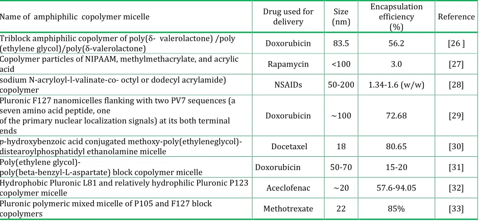
Table 1: The various synthetic copolymers used in the design of drug-loaded micelles and their important properties
Name of amphiphilic copolymer micelle Drug used for delivery
Size (nm)
Encapsulation efficiency
(%)
Reference
Triblock amphiphilic copolymer of poly(δ- valerolactone) /poly
(ethylene glycol)/poly(δ-valerolactone) Doxorubicin 83.5 56.2 [26 ] Copolymer particles of NIPAAM, methylmethacrylate, and acrylic
acid Rapamycin <100 3.0 [27]
sodium N-acryloyl-l-valinate-co- octyl or dodecyl acrylamide)
copolymer NSAIDs 50-200 1.34-1.6 (w/w) [28]
Pluronic F127 nanomicelles flanking with two PV7 sequences (a seven amino acid peptide, one
of the primary nuclear localization signals) at its both terminal ends
Doxorubicin ~100 72.68 [29]
p-hydroxybenzoic acid conjugated
methoxy-poly(ethyleneglycol)-distearoylphosphatidyl ethanolamine micelle Docetaxel 18 80.65 [30] Poly(ethylene glycol)-
poly(beta-benzyl-L-aspartate) block copolymer micelle Doxorubicin 50-70 15-20 [31] Hydrophobic Pluronic L81 and relatively hydrophilic Pluronic P123
copolymer micelle Aceclofenac ∼20 57.6-94.05 [32]
Pluronic polymeric mixed micelle of P105 and F127 block
copolymers Methotrexate 22 85% [33]
A variety of polymers have been used to build hydrophobic core-forming blocks: propylene oxide [19]; poly(L-lysine) [20]; aspartic acid [21]; poly(caprolactone) [22] and D,L-lactic acid [23]. It is interesting to note that in most of the cases, the hydrophilic outer shell consists of poly (ethylene
(PVP) is frequently considered as a primary alternative to PEG [2]. Another hydrophilic candidate is poly(vinyl alcohol). Polyvinyl alcohol substituted with oleic acid was also used for carrying lipophilic drugs [25]. The followings are the notable examples in the field of polymeric micelles and are summarized in Table 1.
Bio-Polymer-Based Micellar Drug Delivery System
Recent findings on polymeric micelles are limited
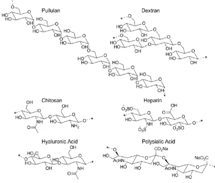
compared to synthetic polymer-based systems. The hydrophilic natural polysaccharides are known to be nontoxic, biodegradable, and biocompatible. These are amenable to easy chemical modification due to plenty of hydroxyl, amine, carboxyl, sulfate functional groups and others. The more commonly used biopolymers, focused so far in the design of nanomicellar drug delivery systems were given in Fig. 1.
Figure 1: Structures of polysaccharides that are used in the development of micelle drug delivery systems.
Chitosan-based micellar system
Chitosan is a linear heteropolymer of N-acetyl-D-glucosamine and D-N-acetyl-D-glucosamine linked by β-(1→4) glycosidic bonds. Chitosan and its derivatives have been the most widely investigated material for drug delivery as it is biocompatible and is biodegraded by enzymes such as lysozymes, some lipases and
PC was approximately 10%. The drug release strongly depended on pH and temperature: low pH and high temperature accelerated the drug release rate markedly. Another example of chitosan-based system is methotrexate (MTX)-encapsulated polymeric nanocarriers using methoxy poly(ethylene glycol) (MPEG)-grafted-chitosan copolymer [35]. These were spherical in shape and had nanometer size range (50-300nm). MTX-chitosan complexes constituted the inner core; whereas MPEG formed outer shell of the nanocarriers. Poly(ethylene glycol)-modified stearic acid-grafted chitosan (PEG-CS-SA) micelles coupled with RGD peptide was targeted with doxorubicin (DOX) to the integrin-over expressing tumor cells [36]. The CAC values of PEG-CS-SA and RGD-PEG-CS-SA were determined to be 25.9±1.2 μg/ml and 23.5±0.5 μg/ml in deionized water, respectively. The particle size of doxorubicin-loaded PEG-CS-SA and RGD-PEG-CS-SA carriers was 21.5±1.0nm and 24.8±0.6nm, respectively. The drug encapsulation efficiency reached to 90%. In vitro drug-release study suggested that the micelles could be used as a controlled drug release carrier, which was found to prolong the release of doxorubicin for 9 days. RGD-modified micelles significantly increased the DOX concentration in integrin-over expressing human hepatocellular carcinoma cell line (BEL-7402). RGD-modified PEG-CS-SA micelles were promising drug carriers for integrin-over expressing tumor active targeting therapy.
Chitosan-based micellar carriers were also evaluated for oral delivery applications. Stearic acid-g-chitosan (CS-SA) was found to form micelles by self-aggregation in aqueous medium [37]. The CAC ranged from about 0.16 to 0.25 mg/ml. The CS-SA micelles were in the size range of 33.4-130.9 nm and had positive zeta potential values (22.9-48.4 mV). The permeability and possible transport route of CS-SA micelles across the gastrointestinal tract was investigated by in vitro model Caco-2 cells. The results exhibited that the CS-SA micelles had good permeability. Energy, pH and concentration
dependent transcytosis processes were involved in transportation of the micelles. The reversible decrease in trans-epithelial electrical resistance by treatment of micelles suggested that paracellular transport pathway was alternate pathway of transport across the gastrointestinal tract. DOX transport by the CS-SA micelles could avoid efflux via P-glycoprotein. In vivo study demonstrated that the micelles were able to significantly improve the bioavailability of encapsulated drug.
Chitosan has the characteristic mucoadhesive property exerted through the interaction between the positive charges carried by the amine and the negative charges carried by membrane proteins. Hence, chitosan-based micelles have been used extensively to improve the oral drug delivery. Chitosan-based micelles inhibited the activity of P-glycoprotein 1 (P-gp) ATPase, and consequently, inhibited drug efflux and enhanced drug permeation [38,39]. Moreover, the chitosan opened the tight junctions between cells and further enhanced drug absorption. The chitosan-based micelles were characterized by their low CAC values, indicative of higher in vivo physical stability [40] and resistance to harsh conditions of the gastrointestinal tract. In summary, chitosan-based micellar carriers was reported to be a relatively safe for oral drug delivery applications [41].
Dextran-Based Micellar System
Dextran is another polysaccharide that has long history of use in pharmaceutical formulation and is reported to have no toxicity problems. For this reason, a series of studies on dextran-based micellar carriers was conducted with the aim to have an effective oral delivery system for Cyclosporin A (CPA) [42, 43]. Francis and his colleagues demonstrated that dextran-PEG-C16 had a higher loading capacity for CPA relative
to dextran-PEG-C18 (0.048 vs. 0.03 mg CPA/mg
micelle) copolymer. The size of dextran-PEG-C16 was
very small (9±0.3nm), and the drug loading did not significantly affect the size of micelles (10±0.3nm). Further, dextran-PEG-C16 showed no toxicity to Caco-2
inhibit cell growth [42]. Additional in vitro studies demonstrated that dextran-PEO-C16 could significantly
improve CPA permeability across Caco-2 cells, although the improvement was lower than that achieved by CPA loaded hydroxypropyl cellulose (HPC)-PEO-C16 and, unlike HPC-PEO-C16,
dextran-PEO-C16 showed no affinity to mucus. To improve the
relatively low transport efficiency, vitamin B12 was
conjugated to the micelle and the vitamin B12
-dextran-PEO-C16 showed increased transportation and
internalization of CPA across the Caco-2 monolayer [43].
Another highly investigated system is dextran-cholic acid. Cholic acid is one of the major bile acids that help to deliver and digest hydrophobic fats in the human small intestine via bile acid self-aggregates. Early dextran-cholic acid systems had low stability, as indicated by a high CMC value (0.02-0.2g/ml) [44]. A high CMC was suggestive of low thermodynamic stability [45]. Later on, some workers [46, 47] used periodate-oxidized dextran to enhance the stability of micellar system. The free aldehyde groups generated after partial oxidation and the remaining hydroxyl groups on dextran moiety could form hydrogen bonds and thus afforded stability to the systems. Moreover, the micelles sustained the drug release up to 14 days at acidic and neutral condition and thus served as good depots for the hydrophobic drug indomethacin (~0.299mg drug/mg micelles). More recently, dextran sulfate-cholic acid was investigated to deliver superoxide dismutase (SOD) orally [48]. They reported that SOD-loaded dextran sulfate-cholic acid had a high stability against the acidic environment of the stomach and the release of SOD in the small intestine was controlled up to 100h. Furthermore, dextran sulfate-cholic acid facilitated SOD cellular uptake, suggesting that cholic acid enhanced the interaction of micelles with the intestinal membrane. Other micellar systems were also developed by grafting polycaprolactone [49], poly (L-lactide) [50], polystyrene [51], lauryl group [52], and methyl
methacrylate-ethylene glycol dimethacrylate [45] onto the dextran polysaccharide backbone.
Pullulan-based micellar system
Pullulan is a water-soluble, neutral, non-toxic bacterial exopolysaccharides [53]. Since the first report of a stable, self-aggregated colloidal system of cholesterol-bearing pullulan (CHP) [54]; numerous studies related to or based on CHP have been carried out [55]. Originally, CHP was synthesized by grafting 1.6 cholesterol groups to every 100 glucose units on pullulan (55 kDa) in a random manner. The average hydrodynamic radius of the uniform, spherical CHP aggregates was found to be 13.3nm. CHP self-aggregates revealed very high colloidal stability [56]. Studies have shown that CHP self-aggregates can be loaded with insulin, thereby protecting the entrapped protein from thermal denaturation and enzymatic degradation [57,58].Till now, not much emphasis was given to investigate its potential as oral delivery carriers for poorly soluble drugs. Recently, a report on CHP to deliver anti-tumor agents [59,60] with improved solubility and stability has been cited in the literature.
Other pullulan based-micelle systems include pullulan acetate [61], poly (DL-lactide-co-glycolide)-graft-pullulan [62], (DL-lactide-co-glycolide)-graft-pullulan-g-poly(L-lactide) [63, 64]. These systems were mainly investigated with regards to chemical synthesis, physicochemical characterization, and drug release characteristics. However, to elucidate their potential in drug delivery, additional studies, more importantly the interaction of the micelles with cells, tissues, and living systems are warranted.
Heparin-based micellar system
inhibits angiogenesis and tumor development. [65,66]. Therefore, several studies have focused on heparin-based micellar systems for improved cancer treatment. To develop an oral anti-tumor formulation, heparin-pluronic micelles were developed to improve drug absorption. The diameter of the carriers were suitable for tumor accumulation and high loading of paclitaxel [67] and RNase A, an anti-tumor protein [68]. The therapeutic-loaded heparin-pluronic micelles exhibited 5-6 folds higher permeability through rat intestines relative to Taxol [67]. Oral availability was also improved by grafting deoxycholic acid to low molecular weight heparin. Deoxycholic acid-heparin micelles (100-200 nm) absorbed from the small intestine via a bile acid transporter, as shown with a nude mouse model [69].
Hyaluronan-based micellar system
Hyaluronic acid is bioactive in that it can bind to the CD44 receptor that is over expressed in tumor and inflammatory tissues. Thus, HA has been investigated as an active targeting agent in drug delivery for enhanced efficacy. Most recently, HA-based micellar systems aimed at cancer treatment with doxorubicin [70, 71], paclitaxel [72,73], siRNA [74], and curcumin [75]. The nano-systems (100-200nm) were physiological stable and could facilitate their passive accumulation in the tumor. Cho et al., 2012 [70]reported that, polyethylene glycol-conjugated hyaluronic acid-ceramide self-assembled micelles had significantly higher cellular uptake by a CD44 over expressed cancer cell line compared to a CD44 negative cell line, NIH3T3. The blocking of CD44 receptor with free HA molecules caused a significant decrease in their cellular uptake [72]. Therefore, HA-based micellar systems had potential in improving drug efficacy via CD44-mediated endocytosis. To further improve targeting, folic acid was conjugated to HA and higher cellular uptake was observed with folic acid-HA-octadecyl group compare to HA-octadecyl group [73]. However, due to the high affinity of HA to liver sinusodidal endothelial cells that have another HA receptors (HARE), HA-based micelles had a high
propensity for accumulation in the liver after systemic administration. In order to circumvent this, PEG was conjugated to HA-5 beta-cholanic acid and liver accumulation of micelles was significantly suppressed, while the tumor accumulation was increased to 1.6 folds. Intravital tumor imaging confirmed that PEG-HA-5 beta-cholanic acid had rapid extravasation into tumor tissues [76].
Other bio-polymer-based micellar systems
Advances of polysaccharide research have provided more candidates as potentially functional biomaterials. In addition to the most investigated polysaccharides described above, several other polysaccharides have also been used to develop micellar systems. Polysialic acid (PSA), in particular, is a non-toxic polysaccharide that can be used to protect and increase body circulation of therapeutics in the form of micelles to inflamed tissue. Using CPA as a model drug, a high loading capacity was achieved with micelles prepared from polycaprolactone (PCL) modified PSA [77]. Mannan based-micellar systems with high stability were developed by grafting cholesterol [78] or hexadecanethiol (C16) [79,80]to mannan. All the polysaccharide micelles are expected to exhibit better properties and functions.
Drug Loading Techniques into Micelles
In order to incorporate or solubilize hydrophobic drugs into the amphiphilic copolymer micelle, various methods were described [81].
Stirring: The drug was added to an aqueous solution of a copolymer and stirred for 2 to 24h to obtain micelles containing the physically entrapped drug. Heating: The drug and copolymer were dissolved in an organic solvent and the solvent was evaporated off at an elevated temperature (40 to 80°C under nitrogen
atmosphere or by rotary evaporator under vacuum). The resulting mixture was kept at a temperature of 20 to 80°C, preferably at 40 to 70°C, for 2h. Then, warm
water (40 to 70°C) was added thereto, and the mixture
Ultrasonic treatment: A mixture of drug and an aqueous solution of copolymer were subjected to ultrasonic treatment for a period ranging from about 1sec to 1h and then stirred at room temperature to obtain micelles containing the drug.
Solvent evaporation: The drug was dissolved in
water-immiscible organic solvent, for example, dichloromethane, chloroform and the like, and then added to an aqueous solution of a copolymer. Subsequently, the organic solvent was solely evaporated off at 25 to 40°C, while stirring and then
filtered to remove un-dissolved drug.
Dialysis:The drug and copolymer were dissolved in a
water-miscible organic solvent. The solution was dialyzed against a buffer solution and then against water. In the dialysis method, suitable water-miscible organic solvents for dissolving drugs were selected from the group consisting of acetonitrile, dimethylformamide, dimethylsulfoxide, dioxane, dimethylacetamide and the like.
Stability Aspects of Micellar Systems
The stabilization of micellar systems was an important consideration since reduced degradation or dissociation of the micelles might circulate for a longer period, leading to a more sustained release of the drug. Some useful strategies were described below.
Cross-linking of Hydrophilic Shell
This strategy involved the introduction of cross-linkable groups within the hydrophilic portion of the copolymer and then using polymer chemistry to cross-link the hydrophilic shell portion after the micellization of the copolymer. Such cross-linking often led to stabilization of the micellar system and delayed the degradation of the micelles. A shell cross-linked micellar system can be prepared for drug delivery. For example, the surfaces of the chitosan micelles were cross-linked with glutaraldehyde or sodium tripolyphosphate to obtain shell cross-linked nanoparticles for drug delivery. In a study, the surface of the paclitaxel-loaded stearic acid grafted chitosan micelle was cross-linked by glutaraldehyde and the
drug release behaviors were correlated with the degree of cross-linking [82]. However, the chemistry used here was not simple to perform. The shell cross-linked micelles needed to be prepared under highly dilute conditions. Moreover, the chemical groups added to the polymer may contribute to its toxicity, change its properties and may render the micelle useless for drug delivery.
Cross-linking of Hydrophobic Core
This strategy involved cross-linking of the core and the formation of a matrix that trapped the drug into its deeper confines, thereby controlling diffusion of the drug from the core. Many approaches was attempted to stabilize the core by cross-linking with different functional groups. A completely biodegradable system was prepared by Hu et al., [83] using the polymer PEG-b-PLA with 5-methyl-5-allyloxycarbonyl-1,3-dioxane-2-1 group as a polymerizable group for cross-linking of the core. This was achieved after micellization by reaction with 2, 2-azoisobutyronitrile. The resultant micelles (130nm) survived water dilution and temperature better than non-crosslinked micelles. Core cross-linked micelles were utilized for the preparation of drug-loaded micelles that offered a longer sustained release than non-modified regular micelles. The chemistry involved in such cross-linking was comparatively simpler than that was used for shell cross-linking. Moreover, the core was the part that encapsulated the drug and a stabilized core could hold the drug for a longer period of time. Strategies like this usually make the system complicated while allowing formulation of a drug in a controlled delivery system.
Use of LCST hydrogel
locked interpenetrating network in the core prevented the breakdown of the core upon dilution. This was meant that a drug loaded in the core would remain in the micelles for prolonged release. Such a system with pluronic micelles and an LCST gel was reported by Rapoport [84]. He suggested three ways to stabilize pluronic micelles, namely, core cross-linking, introducing vegetable oil in the hydrophobic portion to stabilize the micelles and polymerizing an LCST gel with the hydrophobic portion of the micelle to stabilize the core. The core cross-linking strategy decreased the drug loading capacity of the micelle. Addition of vegetable oil to the core increased the hydrophobicity of the core. However, the release was not as sustained as was seen with an LCST gel core. LCST gel in the core allows incorporation of hydrophilic as well as lipophilic drugs. One major disadvantage of using an LCST gel in the core of the micelle was that it increases the micellar size by several folds. Rapoport reported an increase in micelle size from 12-15 nm to 30-400 nm [84]. Although this strategy was employed for the stabilization of commercially available triblock copolymer (Pluronics) micelles, it was not tested yet in imparting stability to the biopolymer based micellar systems.
Usually, one important and vital reason behind the use of biopolymers was that the hydrophilic polymers remained outstretched towards the aqueous solution and thus contributed to the stability of the aqueous micellar preparation. The stability aspects were described herein for providing information to the readers and new research workers so that one of the methods can be used if situation demands, more specifically if zeta potential values were found frustrating for the systems.
CONCLUSION
A significant contribution in the field of polymeric micellar systems is dependent on synthetic polymers. However, the use of biopolymers in this regard is relatively scarce. Naturally occurring hydrophilic polymers are nontoxic, biodegradable, biocompatible
and amenable to easy chemical modification and thus these could constitute the hydrophilic segment of a copolymer. Because of the availability of large number of functional groups these polymers can easily be modified to have ideal carrier for active drug targeting. In recent years, some polymeric micellear systems have entered into various phases of clinical trials. Till date, no such biomaterial-based micellar systems are known to enter this stage in drug formulation research. With chemical approaches, it is possible to design novel amphiphilic copolymers which can entrap a reasonable amount of drug in their core and retard the drug release for a longer duration, thereby improving the therapeutic effectiveness of poorly water soluble drugs. There is a lot of scope in this area of research and in near future, such type of carriers is going to secure a vital place in the field of pharmaceutical industry.
ACKNOWLEDGEMENT
The authors wish to thank all the management members of Trinity Trust, Gupta College of Technological Sciences, Asansol, West Bengal, India for their kind support and encouragement for writing this critical review.
REFERENCES
1. Feng J., Zeng Y., Ma C. The surfactant tween 80
enhances biodesulfurization. Appl. Environ.
Microbiol. 2006; 72:7390–7393.
2. Torchilin, V.P. Micellar nanocarriers: Pharmaceutical
perspectives. Pharma. Res. 2007; 24: 1–16.
3. Xu W., Ling P., Zhang T. Polymeric Micelles, a
Promising Drug Delivery System to Enhance
Bioavailability of Poorly Water-Soluble Drugs.
Journal of Drug Delivery. 2013; 2013: 1-15.
4. Kwon G.S., Okano T. Soluble self assembled block
copolymers for drug delivery. Pharm. Res. 1999; 16:
597-600.
5. Kataoka K., Kwon G.S., Yokoyama M., Okano T.,
Sakurai Y. Block copolymer micelles as vehicles for
drug delivery. J.Controlled Release.1993; 24: 119–
6. Yokoyama M., Kwon G.S., Okano T., Sakurai Y., Seto
T., Kataoka K. Preparation of micelle-forming
polymer-drug conjugates. Bioconjugate Chem. 1992;
3: 295-301.
7. Gao H., Zhu R., Yang X. Properties of polyethylene
glycol lauryl ether with cetyltrimethylammonium
bromide in mixed aqueous solutions studied by
self-diffusion coefficient NMR. J. Colloid. Interface Sci.
2004; 273:626–631.
8. Francis M.F, Cristea M., Winnik FM. Polymeric
micelles for oral drug delivery: Why and how. Pure
Appl. Chem. 2004; 76: 1321-1335.
9. Kwon G.S., Okano T. Polymeric micelles as new drug
carriers .Adv. Drug Deliv. Rev. 1996; 21: 107-116.
10. Jones M.C. Leroux J.C. Polymeric micelles—a new
generation of colloidal drug carriers. European
Journal of Pharmaceutics and Biopharmaceutics.
1999; 48: 101–111.
11. Riess G. Micellization of block copolymers. Progress
in Polymer Science. 2003; 28: 1107–1170.
12. Kwon G.S. Block copolymer micelles as drug delivery
systems. Adv Drug Deliv. Rev. 2002; 54: 167.
13. Yokoyama M. Novel Passive Targetable Drug
Delivery with polymeric Micelles. Academic
Press:San Diego.1998: 193-229.
14. Gao Z. Eisenberg A. A model of micellization for block
copolymers in solutions. Macromolecules. 1993; 26:
7353-7360.
15. Jian F., Zhang Y., Wang J., Ba K., Mao R.Y.’ Lai W.L., Lin
Y.F. Toxicity of Biodegradable Nanoscale
Preparations. Curr. Drug Metabol. 2012; 13: 440–
446.
16. Zhang X., Burt H.M., Mangold G., Dexter, D., Von Hoff
D., Mayer L., Hunter W. L. Anti-tumor efficacy and
biodistribution of intravenous polymeric micellar
paclitaxel. Anticancer Drugs. 1997; 4: 381-388.
17. Jeong Y-I.I., Nah J.W., Lee H.C., Kim S.H., Cho C.S.
Adriamycin release from flower type polymeric
micelle based on star block copolymer composed of
poly(γ-benzyl-L-glutamate) as the hydrophobic part
and poly (ethylene oxide) as the hydrophilic part. Int.
J. pharm. 1999; 188:49-58.
18. Allen C., Han J., Yu Y., Maysinger D., Eisenberg A.
Polycaprolactone–b-poly(ethylene oxide) copolymer
micelles as a delivery vehicle for
dihydrotestosterone. J. Controlled Rel. 2000;
63:275-286.
19. Miller D.W., Batrakova E.V., Waltner T.O., Yu A.V.,
Kabanov A.V. Interactions of pluronic block
copolymers with brain microvessel endothelial cells:
evidence of two potential pathways for drug
absorption. Bioconj. Chem. 1997; 8:649-657.
20. Katayose S., Kataoka K. Remarkable increase in
nuclease resistance of plasmid DNA through
supramolecular assembly with poly (ethylene
glycol)-poly(L-lysine) block copolymer. J. Pharm. Sci.
1998; 87:160-163.
21. Harada A., Kataoka K. Novelpolyion comples micelles
entrapping enzyme molecules in the core:
Preparation of narrowly distributed micelles from
lysozyme and poly (ethylene glycol)-poly(aspartic
acid) block copolymer in aqueous medium.
Macromolecules. 1996; 31:288-294.
22. Kim S.Y., Shin I.G., Lee Y.M., Cho C.G, Sung Y.K.
Methoxy poly(ethylene glycol) and ε-caprolactone
amphiphilic block coplymeric micelles containing
indomethacin.II. Micelle formation and drug release
behaviors. J. Controlled. Rel. 1998; 51:13-22.
23. Ramaswamy M., Zhang X., Burt H.M., Wasan K.M.
Human plasma distribution of free paclitaxel and
paclitaxel associated with diblock copolymers. J.
Pharm. Sci. 1997; 86:460-464.
24. Otsuka H., Nagasaki Y., Kataoka K. Self assembly of
poly (ethylene glycol)-based block copolymers for
biomedical applications. Curr. Opin. Colloid Interface
Sci. 2001; 6:3-10.
25. Luppi B., Bigucci F., Cerchiara T., Andrisano V., Pucci
V., Mandrioli R.,Zecchi V. Micelles based on polyvinyl
alcohol substituted with oleic acid for targeting of
lipophilic drugs. Drug Deliv. 2005; 12: 21-26.
26. Nair L.K., Jagadeeshan S., Nair S.A. and Vinod Kumar
G.S. Evaluation of triblock copolymeric micelles of
δ-valerolactone and poly (ethylene glycol) as a
competent vector for doxorubicin delivery against
cancer. Journal of Nanobiotechnology. 2011; 25:
9-42.
27. Bisht S., Feldmann G., Koorstra J.B.M., Mullendore M.,
Alvarez H., Karikari C., Rudek M.A., Lee C.K., Maitra
A., Maitra A. In vivo characterization of a polymeric
nanoparticle platform with potential oral drug
delivery capabilities. Mol. Cancer. Ther. 2008; 7:
3878-3888.
28. Dutta P., Dey J., Perumal V., Mandal M. Amino acid
non-steroidal anti-inflammatory drugs:
Solubilization, in vitro release and biological
evaluation. Int J Pharm. 2011; 407: 207-216.
29. Li Y.Y., Li L., Dong H.Q., Cai X.J., Ren T.B. Pluronic
F127 nanomicelles engineered with nuclear localized
functionality for targeted drug delivery. Mater Sci.
Eng. C. Mater Biol. Appl. 2013; Mater Sci Eng C Mater
Biol Appl. 2013; 33:2698-2707.
30. Zhang Z.X., Wei X.L., Zhang X.Y., Lu W.Y.
p-Hydroxybenzoic acid (p-HA) modified polymeric
micelles for brain-targeted docetaxel delivery. Chin.
Sci. Bull. 2013; 58: 2651-2656.
31. Kataoka K., Matsumoto T., Yokoyama M., Okano T.,
Sakurai Y., Fukushima S., Okamoto K., Kwon G.S.
Doxorubicin-loaded poly(ethylene
glycol)-poly(beta-benzyl-L-aspartate) copolymer micelles: their
pharmaceutical characteristics and biological
significance. J. Control Release. 2000; 64:143-153.
32. Kulthe S.S., Inamdar N.N., Choudhari Y.M., Shirolikar
S.M., Borde L.C., Mourya V.K. Mixed micelle
formation with hydrophobic and hydrophilic
Pluronic block copolymers: implications for
controlled and targeted drug delivery. Colloids Surf B
Biointerfaces. 2011; 88: 691-6.
33. Chen Y., Sha X., Zhang W., Zhong W., Fan Z., Ren Q.,
Chen L., Fang X. Pluronic mixed micelles overcoming
methotrexate multidrug resistance: in vitro and in
vivo evaluation. International Journal of
Nanomedicine. 2013; 8: 1463 – 1476.
34. Jiang G.B., Quan D., Liao K., Wang H. Novel Polymer
Micelles Prepared from Chitosan Grafted
Hydrophobic Palmitoyl Groups for Drug Delivery.
Mol. Pharm. 2006; 3: 152–160.
35. Seo D.H., Jeong Y.I., Kim D.G., Jang M.J., Jang M.K., Nah
J.W.; Methotrexate- incorporated polymeric
nanoparticles of methoxy poly(ethylene
glycol)-grafted chitosan. Colloids and Surfaces B:
Biointerfaces. 2009; 69: 157–163.
36. Cai L.L., Liu P., Li X., Huang X., Ye Y.Q., Chen F.Y., Yuan
H., Hu F.Q., Du Y.Z. RGD peptide-mediated
chitosan-based polymeric micelles targeting delivery for
integrin-overexpressing tumor cells. International
Journal of Nanomedicine. 2011; 6: 3499–3508.
37. Yuan H., Lu L.J., Du Y.Z., Hu F.Q. Stearic
Acid-g-chitosan Polymeric Micelle for Oral Drug Delivery: In
Vitro Transport and in Vivo Absorption. Mol.
Pharmaceutics. 2011; 8: 225–238.
38. Du Y.Z., Wang L., Yuan H., Hu F.Q. Linoleic
acid-grafted chitosan oligosaccharide micelles for
intracellular drug delivery and reverse drug
resistance of tumor cells. Int. J. Biol. Macromol. 2011;
48: 215–222.
39. Du Y.Z., Cai L.L., Liu P., You J., Yuan H., Hu F.Q. Tumor
cells-specific targeting delivery achieved by A54
peptide functionalized polymeric micelles.
Biomaterials. 2012; 33: 8858–8867.
40. Jiang G.B., Lin Z.T., Xu X.J., Zhang H., Song K. Stable
nanomicelles based on chitosan derivative: In vitro
antiplatelet aggregation and adhesion properties.
Carbohydr. Polym. 2012; 88: 232–238.
41. Sonaje K., Lin K.J., Tseng M.T., Wey S.P., Su F.Y.,
Chuang E.Y., Hsu C.W., Chen C.T., Sung H.W. Effects of
chitosan-nanoparticle-mediated tight junction
opening on the oral absorption of endotoxins.
Biomaterials. 2011; 32: 8712–8721.
42. Francis M.F., Lavoie L., Winnik F.M., Leroux J.C.
Solubilization of cyclosporin A in
dextran-g-polyethyleneglycolalkyl ether polymeric micelles.
Eur. J. Pharm. Biopharm. 2003; 56: 337–346.
43. Francis M.F., Cristea M., Winnik F.M. Exploiting the
vitamin B-12 pathway to enhance oral drug delivery
via polymeric micelles. Biomacromolecules. 2005; 6:
2462–2467.
44. Nichifor M., Lopes A., Carpov A., Melo E. Aggregation
in water of dextran hydrophobically modified with
bile acids. Macromolecules. 1999; 32: 7078–7085.
45. Krasznai D.J., McKenna T.F.L., Cunningham M.F.,
Champagne P., Smeets N.M.B.
Polysaccharide-stabilized core cross-linked polymer micelle
analogues. Polym. Chem. 2012; 3: 992–1001.
46. Xu Q.G., Yuan X.B., Chang J. Self-aggregates of cholic
acid hydrazide-dextran conjugates as drug carriers. J.
Appl. Polym. Sci. 2005; 95: 487–493.
47. Yuan X.B., Li H., Zhu X.X., Woo H.G. Self-aggregated
nanoparticles composed of periodate-oxidized
dextran and cholic acid: Preparation, stabilization
and in-vitro drug release. J. Chem. Technol.
Biotechnol. 2006; 81: 746–754.
48. Xiong Y., Qi J., Yao P. Amphiphilic
cholic-acid-modified dextran sulfate and its application for the
controlled delivery of superoxide dismutase.
Macromol. Biosci. 2012; 12: 515–524.
49. Liu J.Y., Zhang L.M. Preparation of a
characteristics. Carbohydr. Polym. 2007; 69: 196–
201.
50. Nagahama K., Ouchi T., Ohya Y. Biodegradable
nanogels prepared by self-assembly of
poly(L-lactide)-grafted dextran: Entrapment and release of
proteins. Macromol. Biosci. 2008; 8: 1044–1052.
51. Houga C., Giermanska J., Lecommandoux S., Borsali
R., Taton D., Gnanou Y., Le Meins J.F. Micelles and
polymersomes obtained by self-assembly of dextran
and polystyrene based block copolymers.
Biomacromolecules. 2009; 10: 32–40.
52. Daoud-Mahammed S., Couvreur P., Bouchemal K.,
Cheron M., Lebas G., Amiel C., Gref R. Cyclodextrin
and polysaccharide-based nanogels: Entrapment of
two hydrophobic molecules, benzophenone and
tamoxifen. Biomacromolecules. 2009; 10: 547–554.
53. Cheng K.C., Demirci A., Catchmark J.M. Pullulan:
Biosynthesis, production, and applications. Appl.
Microbiol. Biotechnol. 2011; 92: 29–44.
54. Akiyoshi K., Yamaguchi S., Sunamoto J.
Self-aggregates of hydrophobic polysaccharide
derivatives. Chem. Lett. 1991; 1263–1266.
55. Morimoto N., Nomura S.I.M., Miyazawa N., Akiyoshi
K. Nanogel engineered designs for polymeric drug
delivery. ACS Sym. Ser. 2006; 924: 88–101.
56. Akiyoshi K., Deguchi S., Moriguchi N., Yamaguchi S.,
Sunamoto J. Self-aggregates of hydrophobized
polysaccharides in water. Formation and
characteristics of nanoparticles. Macromolecules.
1993; 26: 3062–3068.
57. Akiyoshi K., Nishikawa T., Shichibe S., Sunamoto J.
Stabilization of insulin upon supramolecular
complexation with hydrophobized polysaccharide
nanoparticle. Chem. Lett. 1995; 24: 707–708.
58. Akiyoshi K., Kobayashi S., Shichibe S., Mix D., Baudys
M., Kim S.W. Sunamoto J. Self-assembled hydrogel
nanoparticle of cholesterol-bearing pullulan as a
carrier of protein drugs: Complexation and
stabilization of insulin. J. Contr. Release. 1998; 54:
313–320.
59. Satoh K., Chen F., Aoyama A., Date H., Akiyoshi K.
Nanoparticle of cholesterol-bearing pullulan as a
carrier of anticancer drugs. EJC Suppl. 2008; 6: 139–
139.
60. Shimizu T., Kishida T., Hasegawa U., Ueda Y.,
Imanishi J., Yamagishi H., Akiyoshi K., Otsuji E.,
Mazda O. Nanogel DDS enables sustained release of
IL-12 for tumor immunotherapy. Biochem. Biophys.
Res. Commun. 2008; 367: 330–335.
61. Jung S.W., Jeong Y.I., Kim S.H. Characterization of
hydrophobized pullulan with various
hydrophobicities. Int. J. Pharm. 2003; 254: 109–121.
62. Jeong Y.I., Na H.S., Oh J.S., Choi K.C., Song C.E., Lee H.C.
Adriamycin release from self-assembling
nanospheres of poly(DL-lactide-co-glycolide)-grafted
pullulan. Int. J. Pharm. 2006; 322: 154–160.
63. Ouchi T., Minari T., Ohya Y. Synthesis of
poly(L-lactide)-grafted pullulan through coupling reaction
between amino end-capped poly(L-lactide) and
carboxymethyl pullulan and its aggregation behavior
in water. J. Polym. Sci. A Polym. Chem. 2004; 42:
5482–5487.
64. Seo S., Lee C.S., Jung Y.S., Na K. Thermo-sensitivity
and triggered drug release of polysaccharide
nanogels derived from pullulan-g-poly(L-lactide)
copolymers. Carbohydr. Polym. 2012; 87: 1105–
1111.
65. Karti S.S., Ovali E., Ozgur O., Yilmaz M., Sonmez M.,
Ratip S., Ozdemir F. Induction of apoptosis and
inhibition of growth of human hepatoma HepG2 cells
by heparin. HepatoGastroenterology. 2003; 50:
1864–1866.
66. Niers T.M.H., Klerk C.P.W., DiNisio M., van Noorden
C.J.F., Buller H.R., Reitsma P.H., Richel D.J.
Mechanisms of heparin induced anti-cancer activity
in experimental cancer models. Crit. Rev. Oncol.
Hematol. 2007; 61: 195–207.
67. Dahmani, F.Z., Yang H., Zhou J.P., Yao J., Zhang T.,
Zhang Q. Enhanced oral bioavailability of paclitaxel
in pluronic/LHR mixed polymeric micelles:
Preparation, in vitro and in vivo evaluation. Eur. J.
Pharm. Sci. 2012; 47: 179–189.
68. Choi J.H., Jang J.Y., Joung Y.K., Kwon M.H., Park K.D.
Intracellular delivery and anti-cancer effect of
self-assembled heparin-Pluronic nanogels with RNase A.
J. Contr. Release. 2010; 147: 420–427.
69. Park K., Kim K., Kwon I.C., Kim S.K., Lee S., Lee D.Y.,
Byun Y. Preparation and characterization of
self-assembled nanoparticles of heparin-deoxycholic acid
conjugates. Langmuir. 2004; 20: 11726- 11731.
70. Cho H.J., Yoon I.S., Yoon H.Y., Koo H., Jin Y.J., Ko S.H.,
Shim J.S., Kim K., Kwon I.C., Kim D.D. Polyethylene
self-assembled nanoparticles for targeted delivery of
doxorubicin. Biomaterials. 2012; 33: 1190–1200.
71. Wu J.L., Liu C.G., Wang X.L., Huang Z.H. Preparation
and characterization of nanoparticles based on
histidine-hyaluronic acid conjugates as doxorubicin
carriers. J. Mater. Sci. Mater. Med. 2012; 23: 1921–
1929.
72. Li J., Huo M., Wang J., Zhou J., Mohammad J.M., Zhang
Y., Zhu Q., Waddad A.Y., Zhang Q. Redox-sensitive
micelles self-assembled from amphiphilic hyaluronic
acid-deoxycholic acid conjugates for targeted
intracellular delivery of paclitaxel. Biomaterials.
2012; 33: 2310–2320.
73. Liu Y.H., Sun J., Cao W., Yang J.H., Lian H., Li X., Sun
Y.H., Wang Y.J., Wang S.L., He Z.G. Dual targeting
folate-conjugated hyaluronic acid polymeric micelles
for paclitaxel delivery. Int. J. Pharm. 2011; 421: 160–
169.
74. Shen Y., Wang B.H., Lu Y., Ouahab A., Li Q., Tu J.S. A
novel tumor-targeted delivery system with
hydrophobized hyaluronic acid-spermine conjugates
(HHSCs) for efficient receptor-mediated siRNA
delivery. Int. J. Pharm. 2011; 414: 233–243.
75. Manju S., Sreenivasan K., Conjugation of curcumin
onto hyaluronic acid enhances its aqueous solubility
and stability. J. Colloid Interface Sci. 2011; 359: 318–
325.
76. Choi K.Y., Min K.H., Yoon H.Y., Kim K., Park J.H., Kwon
I.C., Choi K., Jeong S.Y. PEGylation of hyaluronic acid
nanoparticles improves tumor targetability in vivo.
Biomaterials. 2011; 32: 1880–1889.
77. Bader R.A., Silvers A.L., Zhang N. Polysialic
acid-based micelles for encapsulation of hydrophobic
drugs. Biomacromolecules. 2011; 12: 314–320.
78. Ha W., Wu H., Wang X.L., Peng S.L., Ding L.S., Zhang S.,
Li B.J. Self-aggregates of cholesterol-modified
carboxymethyl konjac glucomannan conjugate:
Preparation, characterization, and preliminary
assessment as a carrier of etoposide. Carbohydr.
Polym. 2011; 86: 513–519.
79. Ferreira S.A., Pereira P., Sampaio P., Coutinho P.J.G.,
Gama F.M. Supramolecular assembled nanogel made
of mannan. J. Colloid Interface Sci. 2011; 361: 97–
108.
80. Ferreira S.A., Coutinho P.J.G., Gama F.M.
Self-assembled nanogel made of mannan: Synthesis and
characterization. Langmuir. 2010; 26: 11413–11420.
81. Kim S.-C., Chang E.-O., Pai C.-M. Biodegradable
polymeric micelle-type drug composition and
method for the preparation thereof. US Patent
6322805B1, 2001.
82. Hu F.Q., Ren G.F., Yuan H., Du Y.Z., Zeng S. Shell
cross-linked stearic acid grafted chitosan oligosaccharide
selfaggregated micelles for controlled release of
paclitaxel. Colloids Surf B Biointerfaces. 2006; 50:
97-103.
83. Hu X., Chen X., Wei J., Liu S., Jing X. Core crosslinking
of biodegradable block copolymer micelles based on
poly(ester carbonate). Macromol. Biosci. 2009; 9:
456–463.
84. Rapoport N. Stabilization and activation of Pluronic
micelles for tumor-targeted drug delivery. Colloids
Surf. B Biointerfaces. 1999; 16: 93–111.
How to cite this article: Sayantan Sadukhan*, Paromita Bakshi, Sabyasachi Maiti; Tailored Bio-Polymeric Nanomicellar Carriers: A Promising approach for the delivery of poorly water soluble Drugs; J. Adv. Pharm. Edu. & Res. 2014: 4(1): 41-53.