A
LICJAK
ĘDZIA1, J
OWITAW
OŹNIAK1, K
RZYSZTOFD
UDEK1, 2Relationship of the Radial Nerve
and Lateral Intermuscular Septum
of the Arm During the Fetal Period
Relacje nerwu promieniowego względem przegrody
międzymięśniowej bocznej ramienia w okresie prenatalnym
1Department of Normal Anatomy, Wroclaw Medical University, Poland
2Institute of Machines Design and Operation, Technical University of Wrocław, Poland Adv Clin Exp Med 2009, 18, 3, 235–242
ISSN 1230−025X
ORIGINAL PAPERS
© Copyright by Wroclaw Medical University
Abstract
Objectives.The aim of the study was to analyze the location of radial nerve transit through the lateral intermus− cular septum before its division into its deep and superficial branches during the human fetal period.
Material and Methods. The analysis included 140 preparations of the arms of 70 fetuses (33 female) aged 15–28 weeks (CRL: 90–225 mm). The methods comprised morphological and preparative analysis, digital image acquisition with a digital camera, the Scion Image computer measurement system for Windows v. Alpha 4.0.3.2, and statistical analysis. The point of radial nerve division (point W), the distance between the nerve’s septum tran− sition and the lateral epicondyle of the humerus (lWB), and the humeral length (lAB) were measured in each bone. Correlation between the two lengths and age was determined, symmetry and sexual dimorphism were examined, and the clinical significance of the problem discussed.
Results. In the examined material the lWB/lAB ratio was in the range of 0.16–0.42 (mean ± SD: 0.28 ± 0.05). The arms were divided into three groups with lWB/LABas the division criterion. The first subgroup consisted of arms with ratios smaller than the typical range of variability (–x± s), the second of arms within the range, and the third of arms with ratios greater than typical.
Conclusions. Of the 140 arms, the level of the nerve’s lateral division was higher than 1/3 of the bone length in 23 cases (16%), with similar percentages in the left (16%) and right (17%) arms. The difference was not statisti− cally significant. Sex also did not influence the prevalence frequency of arms with a lWB/lABratio > 1/3; the per− centages for female and male arms were 17% and 16%, respectively. No statistically significant relationship between lWB/lAB and fetal age was observed (Adv Clin Exp Med 2009, 18, 2, 231–238).
Key words:radial nerve, lateral intermuscular brachial septum, arm, fetal period.
Streszczenie
Cel pracy.Analiza umiejscowienia (wysokości) przejścia nerwu promieniowego przez przegrodę międzymięśnio− wą boczną ramienia przed oddaniem gałęzi głębokich i powierzchownych w okresie prenatalnym człowieka.
Materiał i metody. Analizie poddano 140 preparatów ramion 70 płodów w wieku 15–28 tygodni, CRL: 90–255 mm. W grupie tej były 33 płody żeńskie. Wykorzystano analizę morfologiczną i preparatykę, cyfrową akwizycję obrazu za pomocą kamery cyfrowej, komputerowe pomiary w systemie Scion Image for Windows w. Alpha 4.0.3.2 , statystyczną analizę (testy: χ2Pearsona, Kołmogorowa−Smirnowa, logarytmiczne modele regresyjne). Dla każdej kości wyznaczono punkt wyjścia nerwu promieniowego (W) – jako odległość między wyjściem przez przegrodę a nadkłykciem bocznym kości ramiennej (lWB) oraz zmierzono długość kości ramiennej (lAB). Wyznaczono korela− cje między tymi wymiarami a wiekiem, zbadano symetrię i dymorfizm płciowy oraz omówiono znaczenie klinicz− ne badanego problemu.
Wyniki. W badanym materiale stosunek wymiarów lWB/lAB mieścił się w przedziale 0,16–0,42 mm (średnia
x
–= 0,28, odchylenie standardowe SD= 0,05). Badane ramiona podzielono na trzy podgrupy, przyjmując jako kry−
The radial nerve penetrates the lateral inter− muscular septum before its division into its deep and superficial branches. According to Fleming et al. [5], such a nerve position may cause prob− lems when operating on the humerus. The authors analyzed selective material composed of 140 upper extremities and determined the “predicted point” as being between the proximal two−thirds and the distal one−third of the distance between the lateral border of the acromion process of the scapula and the lateral epicondyle of the humerus. Humeral lengths and the distances between the lat− eral intermuscular septum penetration site and lat− eral epicondyle (point W) were measured. The mean distance between the predicted point and the nerve’s actual division site was 2.6 mm.
Shah et al. [11] reported that in 2–18% of patients, humeral fractures are always accompa− nied by radial nerve damage. They analyzed the results and radiograms of 62 patients with humer− al fracture and radial nerve damage. Fifteen of them (24%) had penetrating fracture, 73% had pri− mary nerve paralysis, and the others were qualified for secondary nerve analysis. After surgery, 95% of the patients regained normal or almost normal nerve function. Conservative treatment is recom− mended in patients with radial nerve damage accompanying humeral fractures. Sonnevald et al. analyzed clinical cases of radial nerve damage coex− isting with humeral fracture in a similar way, [13]. In their material of 111 patients with humeral frac− ture, nerve damage occurred in 17 (15%) cases. In early intraoperative examinations the radial nerve remained undamaged in 13 of 14 cases. In the authors’ opinion, nerve damage concurrent with humeral fracture is not an indication for early sur− gical treatment.
Bono et al. [2] examined radial nerve paralysis in 50 adult corpses. They measured the length of the humerus as well as the distance between the site of radial nerve transit through the lateral intermus− cular septum (point W) and the distal part of the humerus (point B). The average distance (lWB) was
16 ± 0.4 cm. This value is of a special importance in the risk rating of implantation of the distal part of the humerus as well as in post−fracture bone fix− ation. Guse and Ostrum [6] carried out postmortem examinations of 24 arms to define the radial
nerve’s position at the posterior arm surface in rela− tion to the epicondyles of the humerus, the tip of the acromion, as well as the brachial triceps heads and aponeurotic membranes. The nerve length measured on the arm amounted to 124 mm and the mean distance between the lateral epicondyle and the division site on the bone posterior surface was 126 mm; this value was never below 100 mm. Carlan et al. [3] analyzed the course of the radial nerve in 27 cadavers. They examined the correla− tion between the nerve’s location and the risk of intraoperative damage during the reconstruction of humeral fractures. Blackburn et al. [1] analyzed the radial nerve’s course between the brachial plexus division site and the lateral epicondyle of the humerus in 40 adult cadavers. They described the distance between the level of the division of the radial nerve’s superficial branch running along the arm’s lateral surface and the lateral humeral epi− condyle and ulnar olecranon. The aim of their stud− ies was to implement the results in brachial surgery. Shinohara et al. [12] presented one of the few papers in the literature which includes a prenatal analysis describing upper−extremity nerve develop− ment in the early embryonic period based on post− mortem examinations of eight human embryos (CRL: 5.1–21.1 cm). Between the 38th and 40th
weeks of human life (CRL: 9.2–11.5 cm), the radi− al nerve reaches the hand plate. In the prenatal peri− od, acromial delivery constitutes a danger of brachial plexus injury as well as shoulder girdle fractures. This kind of delivery frequently results from the mother’s obesity, diabetes, or fetal macro− somy. Shoulder dystocia is a prolonged head−body delivery time of more than 60 seconds and/or the necessity to use ancillary obstetric maneuvers [9].
The available literature does not include stud− ies analyzing the site of radial nerve transmission through the lateral intermuscular septum in the prenatal period. The objectives of this survey were to make up for this lack and to compare results with those observed in adults.
Material and Methods
The material consisted of 140 preparations of fetal arms aged 15–28 weeks (70 fetuses, 37 male
Wnioski.Wśród 140 ramion poziom wysokości odejścia nerwu na stronę boczną ramienia wyższy niż w 1/3 dłu− gości kości zaobserwowano w 23 przypadkach (16%). Odsetek ramion z taką cechą był podobny po stronie lewej (16%) i prawej (17%). Różnica nie była istotna statystycznie, co zweryfikowano testem niezależności χ2Pearso− na. Płeć również nie miała wpływu na częstość występowania ramion o stosunku lWB/lAB> 1/3. Odpowiednie od− setki dla płodów żeńskich i męskich to 17% oraz 16%. Nie zaobserwowano również statystycznie istotnego związ− ku między lWB/lABa wiekiem płodów (Adv Clin Exp Med 2009, 18, 2, 231–238).
and 33 female) and with crown−rump lengths (CRL) of 90–255 mm. The material was stored in formaldehyde solution and at present it is impossi− ble to conciliate any new one. The fetuses used in the survey were preserved for similar times, so the influence of the agent was similar in all the exam− ined structures. All the material originated from the collection of the Department of Normal Anatomy, Wroclaw Medical University. Their morphological ages were determined according to the dependence described by Scammon and Cal− kins [4]. The methods applied comprised anatom− ical and preparative procedures as well as image acquisition with a digital camera and statistical analysis (Pearson’s chi−squared test, Kolmogorov− −Smirnov test). The measurements were carried out on the Scion Image 4.0.3.2 Alpha System [8]. The photos were prepared using the Gimp 2.2 [7] and Bimp 1.43 programs.
Results
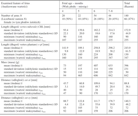
Measurements of the level of radial nerve tran− sit through the lateral intermuscular septum in the arms of the 70 fetuses were statistically analyzed. Table 1 presents the basic statistics of the somatic features characteristic of the examined fetuses. The lengths analyzed in this paper are shown in Fig. 1 and Table 2, including their basic statistics. In the examined material, the lWB/lAB ratio was
within the range of 0.16–0.42 (mean ± SD: 28 ± ± 0.05). The arms were divided into three sub− groups according to this ratio. The first subgroup (“less”) consisted of arms for which the ratio was smaller than the expected range of variability (–x± s), the second (“typical”) contained arms with ratios within the range, and the third (“more”) arms with ratios larger than typical. Table 2 also presents the sizes of these subgroups. In the first group were 10 female (15%) and 10 male (14%) arms: 12 (17%) left and 8 (11%) right. In the second (most numerous) group were 52 (70%) male and 45 (68%) female arms: 47 (67%) left and 50 (71%) right. In the third group were 11 (17%) male and 12 (16%) female arms: 11 (17%) left and 12 (16%) right.
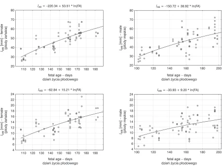
The increases in humeral length (lAB) and the
distance between the radial nerve’s site of division and the lateral epicondyle (lAB) in both the male and
female fetuses are exponential (Fig. 2). However, the ratio of these lengths changed with fetal age (Fig. 3). Of the 140 arms, the level of the nerve’s division towards the lateral side (lWB) was higher
than 1/3 of the bone length in 23 (16%). The per− centage of arms with this feature was similar on the left (11 arms, 16%) and right side (12 arms, 17%,
Table 2). The difference was not statistically signif− icant (Pearson’s chi−squared test, Fig. 4). Sex did not influence the prevalence of the lWB/labratio. No
correlation between the prevalence of the lWB/lAB
ratio and fetal age was found (Fig. 5). The lWB/lAB
ratio did not depend on sex or on left or right side and showed a normal distribution with a mean value of 0.28 and a standard deviation of 0.05 (Fig. 6).
Together with the developmental process, the average distance between the nerve division site (point W) and the “predicted point” (lWP) increased
towards point A, which is visible in a comparative diagram of developmental age (Fig. 7). A compari− son of the authors’ material of fetal arms and arms in adults [5] in subgroups with different lWB/lAB ratios
(Fig. 8) showed a statistically significant difference.
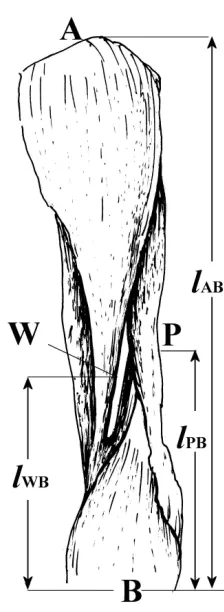
Fig. 1.Left arm: A – humerus proximal point (caput humeri); B – humerus distal point (humerus lateral epicondyle), W – point of nerve divergence towards arm lateral part, P – point at 1/3 of humeral length (“predicted point”), lAB– humeral length, lWB– dis− tance between nerve transit through the lateral inter− muscular septum and the bone’s distal point
Fig. 4.Numbers of fetuses in the subgroups according to sex and lWB/lABratio and to side and lWB/lABratio
Ryc. 4.
Liczebności płodów w pod− grupach różniących się płcią i sto− sunkiem lWB/lAB oraz stroną i stosunkiem lWB/lAB
lAB= –220.34 + 53.51 * ln(FA)
110 120 130 140 150 160 170 180 190
fetal age – days dzieñ ¿ycia p³odowego 20 30 40 50 60 70 80 lWB – female (p³ody ¿eñskie) [mm]
lAB= –150.72 + 38.92 * ln(FA)
100 120 140 160 180 200
20 30 40 50 60 70 80
fetal age – days dzieñ ¿ycia p³odowego
lWB
– male
(p³ody mêskie)
[mm]
Fig. 2.Correlation diagrams of humeral length (lAB) and nerve divergence height (lWB) in male and female fetuses with morphological age (FA) together with regressive logarithmic models
Ryc. 2. Diagramy korelacyjne długości kości ramiennej lAB; wysokości odejścia nerwu na boczną stronę ramienia lWB płodów żeńskich i męskich z wiekiem morfologicznym oraz logarytmiczne modele regresyjne
lWB= –62.84 + 15.21 * ln(FA)
110 120 130 140 150 160 170 180 190
4 6 8 10 12 14 16 18 20 22 24
fetal age – days dzieñ ¿ycia p³odowego
lWB
– female
(p³ody ¿eñskie)
[mm]
lWB= –33.93 + 9.20 * ln(FA)
100 120 140 160 180 200
4 6 8 10 12 14 16 18 20 22 24
fetal age – days dzieñ ¿ycia p³odowego
lWB
– male
(p³ody mêskie)
[mm]
Fig. 3.Correlation diagrams of nerve divergence height and humeral length ratio (lWB/lAB) with morpho− logical age
Ryc. 3.Diagramy korelacyjne stosunku wysokości odejścia nerwu na stronę boczną ramienia do długości kości ramiennej lWB/lABz wiekiem morfologicznym
100 120 140 160 180 200
0,10 0,15 0,20 0,25 0,30 0,35 0,40 0,45 typical typowy less mniejszy more wiêkszy
fetal age – days dzieñ ¿ycia p³odowego
lWB
/
Fig. 5.Numbers of fetuses in the subgroups accord− ing to age and lWB/lABratio and chi−squared test results
Ryc. 5.Liczebno− ści płodów w pod− grupach różnią− cych się wiekiem i stosunkiem lWB/lABoraz wynik testu χ2
Fig. 6.lWB/lABratio histogram with the normal distribution N(0.28, 0.05) and normality test results
Ryc. 6.Histogram stosunku wymiarów lWB/lAB badanych płodów na tle rozkładu normalnego N(0,28; 0,05) i wynik testu normalności Kołmogorowa− −Smirnowa
K-S test:d= 0,049,p> 0,20
0,10 0,15 0,20 0,25 0,30 0,35 0,40 0,45
0 10 20 30 40 50 60 70
number of observations
liczba obserwacji
lWB/lAB
Fig. 7.Comparison of distance points P and W in the four fetal age groups and in adults [5]
Ryc. 7.Porównanie odległości między punktami P i W w czterech grupach wiekowych płodów i u osób dorosłych [5]
IV V VI VII adult
doroœli fetal age – months
miesi¹c ¿ycia p³odowego -10
-8 -6 -4 -2 0 2 4 6
median 25%–75% min–max
lWB
–
lPB
[mm]
Fig. 8.Comparison of fetal (own material) and adult [5] arm numbers in subgroups according to lWB/lAB ratio
Ryc. 8.Porównanie liczby ramion płodów (materiał własny) i dorosłych [5] w podgrupach różniących się stosunkiem lWB/lAB
fetuses adults
(płody) (dorośli)
less lWB/lAB < 0.23 lWB/lAB < 0.327 (mniejszy)
typical 0.23 ≤lWB/lAB≤0.33 0.327 ≤lWB/lAB≤0.345 (typowy)
more lWB/lAB > 0.33 lWB/lAB > 0.345 (większy)
Discussion
Fig. 9. Comparison of lWB/lABratios in fetuses and adults (difference statistically significant)
Ryc. 9.Porównanie stosunku lWB/lAB płodów i osób dorosłych (różnica statystycznie istotna)
Z = 5.356; p< 0.0001
foetus N = 140 p³ody
adult N = 20 doroœli 0,15
0,20 0,25 0,30 0,35 0,40 0,45
median 25%–75% min–max
llWB
AB
/
Table 1.Characteristics of the analyzed group of fetuses
Tabela 1.Charakterystyka analizowanej grupy płodów
Examined feature of fetus Fetal age – months Total
(Analizowane wartości) (Wiek płodu – miesiąc) (Razem)
4 5 6 7–8
Number of arms N 20 22 54 44 140
(Liczebność ramion N) 10 (50%) 10 (45%) 26 (48%) 20 (45%) 66 (47%)
female (w tym płodów żeńskich) Length (długość vertex−tuberale) CRL[mm]
mean (średnia) –x 93.4 139.6 182.2 214.3 172.9
standard deviation (odchylenie standardowe) SD 22.1 20.0 18.6 17.6 44.9
minimum (wartość minimalna) xmin 90 116 160 160 90
maximum (wartość maksymalna) xmax 107 187 255 235 255
Length (długość vertex−plantare) v−pl[mm]
mean (średnia) –x 141.9 189.1 258.8 298.2 243.0
standard deviation (odchylenie standardowe) SD 9.8 22.9 18.9 38.2 61.5
minimum (wartość minimalna) xmin 130 160 224 208 130
maximum (wartość maksymalna) xmax 160 234 295 347 347
Mass (masa) [g]
mean (średnia) –x 73 197 407 633 396
standard deviation (odchylenie standardowe) SD 15 115 114 150 231
minimum (wartość minimalna) xmin 50 110 240 225 50
maximum (wartość maksymalna) xmax 94 465 606 842 842
Distance (odległość) sst−xi[mm]
mean (średnia) –x 43.7 60.8 109.6 94.1 88.8
standard deviation (odchylenie standardowe) SD 3.1 14.0 49.7 20.4 38.1
minimum (wartość minimalna) xmin 40 50 28 25 25
maximum (wartość maksymalna) xmax 47 90 170 113 170
Distance (odległość) sst−sy[mm]
mean (średnia) –x 88.7 122.8 111.7 178.7 140.3
standard deviation (odchylenie standardowe) SD 4.4 22.4 35.6 39.9 48.2
minimum (wartość minimalna) xmin 83 105 75 72 72
maximum (wartość maksymalna) xmax 92 169 159 206 206
0.16–0.42 (mean: 0.28 ± 0.05). Sixteen percent of the examined arms had a ratio less than the typical variability range (– x ± s), 69% were within this
range, and 16% had a ratio greater than normal. Comparing the present results with Flemings’ reports it was observed that the W point (point of radial nerve divergence) was shifted upwards in adult patients, together with age (Fig. 9).
Blackburn et al. [1], Bono et al. [2], Carlan et al. [3], and Guse and Ostrum [6] examined arm geometry, the height of radial nerve transit on the arm’s lateral surface, as well as the clinical impli− cations connected with the division patterns, applied mainly in brachial surgery. Their papers comprised adult postmortem analyses. The possi− bility of radial nerve paralysis with humeral shaft fracture at a high level of the division was recog− nized as an important clinical aspect. It is worth mentioning that such incidents may happen during improper intradermal injection or perinatal dam− age by brachial dystocia. Long−term observations indicate that such clinical complications may pre− vail in 13% of the examined population, indepen− dent of sex and body side.
Table 2.Basic statistics of the arms’ geometry
Tabela 2.Podstawowe statystyki geometrii badanych ramion
Quantitative feature Gender Body side Total
(Cecha ilościowa) (Płeć) (Strona ciała) (Razem)
N = 140
F M left right
N= 66 N= 74 N = 70 N= 70 lAB[mm] – length of humerus:
(Długość kości ramiennej):
mean –x(średnia) 47.3 44.9 46.2 45.8 46.0
standard deviation (odchylenie standardowe) SD 11.2 11.5 11.6 11.2 11.4
median Me (mediana) 49.4 45.7 47.7 45.8 46.5
minimum (wartość minimalna) xmin 25.6 24.0 24.7 24.0 24.0
maximum (wartość maksymalna) xmax 67.5 72.2 72.2 68.6 72.2
lWB[mm] – distance between lateral epicondyle and the nerve divergence site
(Odległość między nadkłykciem bocznym a miejs− cem wyjścia nerwu na część boczną ramienia):
mean –x(średnia) 13.3 12.3 12.7 12.8 12.8
standard deviation (odchylenie standardowe) SD 3.5 3.4 3.5 3.6 3.5
median Me (mediana) 13.4 12.0 12.3 12.8 12.5
minimum (wartość minimalna) xmin) 6.5 5.1 6.5 5.1 5.1
maximum (wartość maksymalna) xmax 23.1 19.9 23.1 19.9 23.1
lWB/lAB– lWB to lABratio (Stosunek wymiarów lWBi lAB):
mean –x(średnia) 0.28 0.28 0.28 0.28 0.28
standard deviation (odchylenie standardowe) SD 0.05 0.05 0.05 0.04 0.05
median Me(mediana) 0.28 0.27 0.28 0.28 0.27
minimum (wartość minimalna) xmin 0.18 0.16 0.16 0.18 0.16
maximum (wartość maksymalna) xmax 0.40 0.42 0.42 0.39 0.42
LWB – lPB [mm] – distance from ”predicted point” (Odległość od „predicted point”):
mean –x(średnia) –2.5 –2.7 –2.7 –2.5 –2.6
standard deviation (odchylenie standardowe) SD 2.5 2.4 2.7 2.1 2.4
median Me(mediana) –2.3 –2.8 –2.4 –2.7 –2.5
minimum (wartość minimalna) xmin –8.9 –9.1 –8.9 –9.1 –9.1
maximum (wartość maksymalna) xmax 2.4 3.3 3.3 2.4 3.3
Number of arms (%) with ratios
(Liczba ramion (odsetek) o stosunku wymiarów):
lWB/lAB < 0.23 10 (15%) 10 (14%) 12 (17%) 8 (11%) 20 (14%)
0.23 ≤lWB/lAB≤0.33 45 (68%) 52 (70%) 47 (67%) 50 (71%) 97 (69%)
0.33 < lWB/lAB 11 (17%) 12 (16%) 11 (16%) 12 (17%) 23 (16%)
lar septum before its division into its deep and super− ficial branches in the prenatal period proved to be an individual feature which was symmetric and did not
show sexual dimorphism. In fetuses this site is locat− ed at 5/7 of the humeral length and in adults this point shifts upwards to 2/3 of the humeral length.
References
[1] Blackburn SC, Wood CPJ, Evans DJR, Watt DJ:Radial nerve contribution to brachialis in the UK Caucasian population: position is predictable based on surface landmarks. Clin Anat 2007, 1, 20, 64–67.
[2] Bono CM, Grossman MG, Hochwald N, Tornetta PII:Radial and axillary nerves. Anatomic considerations for humeral fixation. Clin Orthop 2000, 373, 259–264.
[3] Carlan D, Pratt J, Patterson MM, Weiland AJ, Boyer MI, Gelberman RH:The radial nerve in the brachium: an anatomic study in human cadavers. J Hand Surg [Am] 2007, 32, 8, 1177–1182.
[4] Carlson BM:Human embryology and developmental. Mosby, St. Louis 1999, 450.
[5] Fleming F, Lenehan B, Sankar R, Folan−Curran J, Curtin W:One−third, two−thirds: Relationship of the radi− al nerve to the lateral intermuscular septum in the arm. Clin Anat 2004, 17, 26–29.
[7] http://www.gimp.org/
[8] http://www.scioncorp.com/
[9] Jevitt CM:Shoulder Dystocia: Etiology, common risk factors, and management. J Midwifery Womens Health 2005, 50, 6, 458–497.
[10] Schocke M, Bodner G, Buchberger W, Bale R, Huber B, Harpf C, Gassner E, Jaschke W:Radial nerve palsy associated with humeral shaft fracture: evaluation with US–initial experience. Radiology 2001, 219, 811–816.
[11] Shah JJ, Bhatti NA:Radial nerve paralysis associated with fractures of the humerus. A review of 62 cases. Clin Orthop 1983, 172, 171–176.
[12] Shinohara H, Naora H, Hashimoto R, Hatta T, Tanaka O: Development of the upper limb of staged human embryos. Acta Anat 1990, 138, 3, 265–269.
[13] Sonneveld GJ, Patka P, von Mourik JC, Broere G:Treatment of fractures of the shaft of the humerus accom− panied by paralysis of the radial nerve. Injury 1987, 18, 404–406.
Address for correspondence:
Alicja Kędzia
Department of Normal Anatomy Wroclaw Medical University Chałubińskiego 6a
50−368 Wrocław Poland
Tel.: +48 71 784 00 80
E−mail: [email protected]
Conflict of interest: None declared
![Fig. 8. Comparison of fetal (own material) and adult[5] arm numbers in subgroups according to lWB/lABratio](https://thumb-us.123doks.com/thumbv2/123dok_us/8771981.1757506/5.651.102.313.91.462/comparison-fetal-material-adult-numbers-subgroups-according-labratio.webp)