R E S E A R C H
Open Access
Cryptic amyloidogenic elements in mutant
NEFH causing Charcot-Marie-Tooth 2
trigger aggresome formation and neuronal
death
Arnaud Jacquier
1,2†, Cécile Delorme
3†, Edwige Belotti
1, Raoul Juntas-Morales
4, Guilhem Solé
5, Odile Dubourg
6,
Marianne Giroux
7, Claude-Alain Maurage
8, Valérie Castellani
1, Adriana Rebelo
9, Alexander Abrams
9,
Stephan Züchner
9, Tanya Stojkovic
10*†, Laurent Schaeffer
1,11*†and Philippe Latour
2†Abstract:Neurofilament heavy chain (NEFH) gene was recently identified to cause autosomal dominant axonal Charcot-Marie-Tooth disease (CMT2cc). However, the clinical spectrum of this condition and the physio-pathological pathway remain to be delineated. We report 12 patients from two French families with axonal dominantly inherited form of CMT caused by two new mutations in theNEFHgene. A remarkable feature was the early involvement of proximal muscles of the lower limbs associated with pyramidal signs in some patients. Nerve conduction velocity studies indicated a predominantly motor axonal neuropathy. Unique deletions of two nucleotides causing frameshifts near the end of theNEFHcoding sequence were identified: in family 1, c.3008_3009del (p.Lys1003Argfs*59), and in family 2 c.3043_3044del (p.Lys1015Glyfs*47). Both frameshifts lead to 40 additional amino acids translation encoding a cryptic amyloidogenic element. Consistently, we show that these mutations cause protein aggregation which are recognised by the autophagic pathway in motoneurons and triggered caspase 3 activation leading to apoptosis in neuroblastoma cells. Using electroporation of chick embryo spinal cord, we confirm that NEFH mutants form aggregates in vivo and trigger apoptosis of spinal cord neurons. Thus, our results provide a physiological explanation for the overlap between CMT and amyotrophic lateral sclerosis (ALS) clinical features in affected patients.
Introduction
Charcot-Marie-Tooth disease (CMT) refers to a hetero-geneous group of chronic inherited motor and sensory disorders of the peripheral nervous system. CMT are classified according to their axonal or demyelinating fea-ture on nerve conduction studies, and their mode of in-heritance [10]. The autosomal dominant axonal forms are termed CMT2. The number of genes associated with CMT is progressively expanding, particularly since the development of next-generation sequencing. Several of these genes are expressed in both the central and per-ipheral nervous system, such as neurofilaments, which
have been implicated in several neurodegenerative dis-eases, including ALS [24]. Neurofilaments are intermedi-ate filaments exclusively expressed in neurons in the central and peripheral nervous system. They have im-portant cytoskeletal functions such as the regulation of axonal growth and diameter [15]. Neurofilaments are composed of three subunits defined by their molecular weight: NEFL (light), NEFM (medium), and NEFH (heavy) [23, 24], encoded by NEFL, NEFM and NEFH genes, respectively. Mutations in NEFL are known to cause both axonal and demyelinating forms of CMT and manifest with various clinical phenotypes, sometimes with additional pyramidal signs [2, 4, 13, 19, 25]. Muta-tions in the NEFH gene have been suggested to play a role in the pathogenesis of sporadic amyotrophic lateral sclerosis (ALS), but with conflicting results [28].
Recently, NEFH mutations have been identified as a rare cause of autosomal dominant CMT, with two * Correspondence:[email protected];[email protected]
†Equal contributors 10
Institut de Myologie, Hôpital Pitié-Salpêtrière, 47-83 boulevard de l’Hôpital, 75013 Paris, France
1Institut NeuroMyoGène, Université Lyon1 - CNRS UMR 5310 - INSERM
U1217, Lyon, France
Full list of author information is available at the end of the article
families reported to date [27]. The clinical and electro-physiological phenotype in these two families was char-acterized by a severe, predominantly motor, axonal neuropathy, with significant walking difficulties in early adulthood. Similar to our families, the two mutations (c.3010_3011delGA and c.3017_3020dup) cause the loss of the stop codon and the translation of 40 additional amino acids which encode a cryptic amyloidogenic elem-ent (CAE) and cause protein aggregation [27].
Here, we report two French families presenting with an axonal, dominantly inherited form of CMT characterized by prominent motor deficit affecting both the distal and proximal muscles, and signs of central nervous system in-volvement, caused by two previously unreported mutations in the NEFH gene. We show that those new mutations cause protein aggregation, not only in neuroblastoma cells as similar mutations previously reported, but also in pri-mary mouse motoneurons. We further show that this type of mutations also induces neuronal apoptosis, both in neuroblastoma cells and in vivo in spinal cord neurons
using in ovo chick spinal cord electroporation. Our results thus provide a physiological basis to the pathogenicity of
NEFHmutations that interfere with neurofilament assem-bly via protein sequestration and cause neurotoxicity, which explains the overlapping clinical features of NEFH mutations with those of motor neuron disease.
Materials and methods
Patients
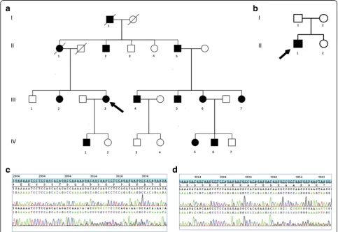
The patients were identified as part of our on-going gen-etic studies in CMT. Patients were all of French ascen-dance. Patients were recruited, enrolled and sampled according to the protocols of the institutional review board at the Pitié-Salpêtrière Hospital. Written informed consent was obtained for participation in the study. Pa-tients displayed a clinical and electrical phenotype of axonal motor and sensory neuropathy, with no muta-tions in known CMT2 genes at that time. Twelve pa-tients belonging to two different families (Fig. 1) were included in the study.
Fig. 1Pedigree of the two families.aFamily 1.bFamily 2.Arrowindicates the proband.Slash linesindicate dead individuals.Squaresare males
andcirclesare females.Filled symbolsrepresent affected subjects andempty symbolsunaffected subjects.cFamily 1 -NEFHC-terminal sequence
Clinical assessment
Patients were seen in neuromuscular centres and assessed by senior neurologists specialized in neuromus-cular disorders in Paris (TS and OD), Lille (TS), Bor-deaux (GS), Valenciennes (MG) and Montpellier (RJM). Clinical assessment included medical history and neuro-logical examination.
Neurophysiological study
Electrodiagnostic studies included nerve conduction studies in the upper and lower limbs, and electromyog-raphy (EMG) using concentric needle electrodes in at least three muscles. Electrodiagnostic studies were per-formed using conventional equipment and standard methods. Skin temperature was maintained in the range of 32° to 34 °C. Patients were classified as having axonal neuropathy if they had a nerve conduction velocity in the median nerve above 38 m/s [12].
Histological study
A nerve biopsy (superficial peroneal sensory nerve) was performed in patients II-1, III-4 and III-7 of family 1. A muscle biopsy (tibialis anterior) was performed in pa-tients III-3, III-4 and III-7 of family 1 and patient II-1 of family 2. Muscle and nerve biopsies were processed and assessed according to standard techniques [33].
Molecular analysis
Mutations in genes frequently associated with axonal CMT (MFN2, GJB1; MPZ, TRPV4, NEFL, GDAP) and amyotrophic lateral sclerosis (ALS), such as SOD1gene, were first excluded by Sanger sequencing. In family 1, four patients were then studied by sequencing a panel of 4813 genes associated with known clinical phenotypes (Illumina® TruSight One Sequencing Panel) on Illumina® NextSeq500 sequencer. In family 2, the index case and his two parents were sequenced by the same method. Confirmation of the putative deleterious variants in
NEFH and familial studies were done by Sanger
sequencing.
Plasmid construct and mutagenesis
The construct encoding for the human NEFH was a gift from Dr Sidransky [21] and it was subcloned in a pCA-GEN eGFP C1 plasmid allowing optimal expression in neurons. NEFH N-terminus end was fused with mono-meric eGFP (derived from peGFP-C1, clonetech) using Gibson assembly kit (E5510, NEB) following the manu-facturer recommendation to obtain pCAGEN eGFP NEFH referred to as WT NEFH thereafter. The mutant
forms of NEFH, pCAGEN eGFP-NEFH plasmid were
obtained by site-directed mutagenesis using QuickChange II XL Mutagenesis Kit (from Agilent technology #200521). Mutant NEFH plasmids harboring c.3008_3009delAA,
c.3043_3044delAA and c.3010_3011delGA are referred re-spectively as c3008, c3043 and c3010 in the text. Untagged NEFH plasmids (WT and CAE, c.3010_3011delGA muta-tion) and eGFP-NEFL plasmids were provided by Pr Zuchner (University of Miami, USA). Plasmids prepared with the EndoFree Maxiprep Kit (Macherey-Nagel, Düren, Germany) were routinely diluted at 2μg/μl.
Cell line culture and transfection
Human neuroblastoma cell line SH-EP [3, 6] were grown on 12 mm coverslips in Dulbecco’s Modified Eagle Medium (DMEM, gibco) media supplemented with 10% fetal bovine serum (FBS, Gibco) and 1% penicillin/ streptomycin (Gibco) and transfected at 60% of conflu-ence with jetPRIME (Polyplus-transfection) according to the manufacturer’s protocol. 1, 2 or 3 days after transfec-tion, cells were fixed with 4% paraformaldehyde for 20 min, and washed with PBS.
Primary motoneuron culture and transfection
Spinal cord motoneurons were prepared from E12.5 OF1 mice embryos as described by Henderson et al. [14] with minor modifications. Briefly, anterior horn of the embryo were dissected in HBSS supplemented with 4.5 g/l glucose and 7 mM HEPES (invitrogen). Motoneu-rons were purified by using a 6% OptiPrep density gradi-ent medium (D1556, Sigma). Then, motoneurons were resuspended in supplemented Neurobasal medium (Invi-trogen) containing 1 ng/ml brain-derived neurotrophic factor (Peprotech), 1 ng/ml glial cell line–derived trophic factor (Peprotech), and 10 ng/ml ciliary
neuro-trophic factor (Peprotech) and were seeded on
polyornithin/laminin-coated glass coverslips (P8638 and L2020, Sigma). After two days in vitro, plasmid transfec-tions were done by Magnetofection following the manu-facturer recommendations (OZBiosciences). Two or four days later, motoneurons were fixed using 4% paraformal-dehyde for 20 min, and washed with PBS.
Embryonic chick spinal cord electroporation
Immunocytochemistry
Fixed cells or frozen tissue sections were blocked and permeabilized with PBS containing 4% bovine serum al-bumin, 2% goat serum, 100 mM glycine and 0.3% Triton X-100. Primary antibody was applied overnight at 4 °C diluted in blocking solution, washed 3 times in PBS, in-cubated for 2 h in the secondary antibody at room temperature, with DAPI, then washed four times in PBS, mounted in Vectashield® and imaged with confocal microscope Zeiss LSM800 or imaged with conventional microscope Zeiss Axiovert 135 M equipped with the camera Leica DC 350 FX CCD monochrome. The fol-lowing antibodies were used: anti-acetylated tubulin (1/ 400, clone 6-11B-1, Sigma), anti p62 (1/200, GP62-C, Progen, Germany), anti-Ubiquitin conjugated protein1 (1/100, BML-PW8810, Enzo), anti - Lc3b (1/100, #2775, Cell Signaling Technology, Danvers), anti-beta3 tubulin
(1/500, TUJ1, Biolengend), anti-NEFM (1/500,
Poly28410, Biolegend), anti-NEFH (1/2000, Smi32, Bio-legend), anti-cleaved caspase 3 (1/100; 5A1E, Cell Signal-ing Technology, Danvers).
Western blot
Triton X100-soluble and -insoluble protein fraction-ation were performed following cell lysis in 20 mM NaCl, 20 mM Tris-HCl, pH 7.4, 5 mM MgCl2, 0.1 mM EDTA, 0.1% Triton X-100, and protease cocktail, for 30 min at 4 °C. Then, cell extracts were centrifuged for 15 min at 15000 g to separate soluble (supernatant) and insoluble (pellet) fractions. Pellet fraction was sus-pended in RIPA buffer. Soluble (S) and insoluble (I) fractions were resolved by SDS-PAGE and transferred on Nitrocellulose transfer membrane. Western blot was performed with anti-GFP (A11122, Thermo) and anti-GAPDH (2118 CST), followed by chemilumines-cent detection using horseradish peroxidase-conjugated antibodies and the SuperSignal West Pico (Thermo Scientific) reagent.
Statistical analysis
Each experiment was repeated at least twice. Data were analysed with Excel (Microsoft) or SigmaStat 3.5 (Systat Software Inc). Data from more than two groups each showing normality and equal variance were analyse with one way ANOVA followed by Dunnett’s test to compare several treatment group to a control group. Otherwise, data that do not showed a Gaussian distribution were analyse with one way Kruskal-Wallis test followed by Dunn posthoc test.
Results
Clinical phenotype
The clinical features of family 1 and 2 are summarized in Table 1.
Family 1
The affected siblings were born from non-consanguineous parents (Fig. 1a). The family displayed an autosomal dominant inheritance pattern. The propositus (III-3) was a 49-year-old woman. She had normal developmental milestones and no walking or running difficulties in child-hood. At age 27, she developed difficulties climbing stairs. Muscle weakness worsened gradually and involved both distal and proximal muscles in the lower limbs, with diffi-culties arising from squat position. At 30 years of age, she developed proximal weakness in the lower limbs, involv-ing particularly the iliopsoas muscle (MRC score 3/5), in addition to the distal weakness. At age 49, she was able to walk around 50 m with a walker, and used a wheelchair for longer distances. She was still able to climb stairs. Romberg’s test showed mild postural instability. Heel or tiptoe walking was impossible. Squatting was impossible. Gower’s manoeuvre was positive. Muscle strength exam-ination showed a severe motor deficit in both proximal and distal muscles of the lower limbs (tibialis anterior 1/5, peroneus longus and tibialis posterior muscles MRC 2/5 both sides; quadriceps 3/5 right side, 1/5 left side; ham-string muscles 2/5 both sides; gluteus medius 3/5 both sides; iliopsoas 2/5 both sides). There was distal motor deficit in the upper limbs (abductor digiti minimi 3/5 right side, 3+/5 left side; normal strength in the other muscles). There were no fasciculations. There was distal and prox-imal muscle wasting in the lower limbs, and distal muscle wasting in the upper limbs. She had pes cavus. Deep ten-don reflexes were absent and plantar reflex was flexor. She had distal hypoesthesia in the lower limbs (pin, touch and vibration). She reported frequent episodes of hypophonia, suggestive of associated vocal cord involvement. Cranial nerve examination was otherwise normal.
decade of the disease, and caused waddling gait. Motor deficit was clearly prominent in the iliopsoas whereas the quadriceps and hamstring muscles were better pre-served during the third decade. However, the weakness spread over years to all proximal muscles in the lower and to distal muscles of the upper limbs. Indeed, the propositus's mother (patient II-1) presented at the age of 70 with diffuse motor weakness involving the distal and proximal muscles of the four limbs, associated with brisk reflexes. Most patients had cramps. Pes cavus was a con-sistent feature. Deep tendon reflexes absent in most pa-tients, but three of them (II-1, III-7, IV-1) had brisk patellar reflexes. There was no Babinski or Hoffman sign. There were mild sensory abnormalities in all pa-tients, with progressively ascending superficial and deep sensory alterations. The disease evolved progressively and most patients used a wheelchair around 50 years. There were no associated features such as visual loss, deafness, cranial nerve abnormalities, cerebellar syn-drome, seizures or cognitive disturbances. One of the patients (III-4 of family 1) experienced a malignant hyperthermia following a surgical procedure.
Family 2
The propositus (II-1) was a 23-year-old man born from non-consanguineous asymptomatic parents (Fig. 1b). There was no familial history of neuropathy. He was born at term of an uncomplicated pregnancy. In early infancy, he presented with walking clumsiness with frequent falls and difficulties jumping. A pectum excava-tum was noted. He had waddling gait with difficulties climbing stairs. Progressively, he developed drop foot gait and could not walk on heels. He had several epi-sodes of ankle sprains. Since age 16, he developed pro-gressive atrophy in the lower limbs muscles and
complained of frequent cramps. At age 23, he could walk approximately one kilometre without help. He had waddling gait and bilateral foot drop. Heel or tiptoe walking and squatting were impossible. Gower’s manoeuvre was positive. He had mild postural instability at Romberg’s test. Muscle strength examination revealed a symmetric motor deficit in distal and proximal lower limbs (MRC score: tibialis anterior 1+/5; soleus 1/5, per-oneus longus 0/5, hamstring and gluteus medius 4/5, quadriceps and psoas 3/5). Upper limbs examination showed symmetric proximal and distal deficit (distal hand muscles, biceps brachii and triceps brachii 4/5 and deltoid 3/5). There was no axial deficit. There was severe muscle wasting in the lower limbs, distally and proxim-ally, and Achilles tendons contractures. There were no fasciculations. He had bilateral scapular winging and pes cavus. Deep tendon reflexes were weak, plantar re-sponses were flexor. He had distal hypoesthesia in the lower limbs (pin, touch and vibration). Neurological examination was otherwise normal. He had no associ-ated features except for gastroparesia, with episodes of morning vomiting.
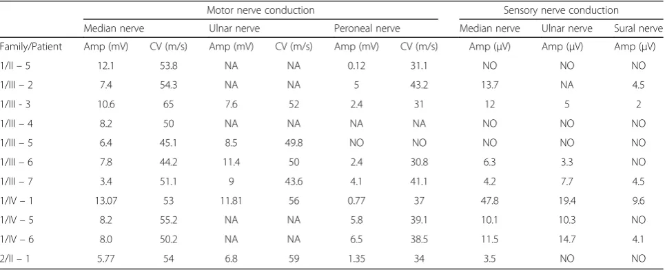
Electrophysiological findings
Nerve-conduction velocity studies are shown in Table 2 for families 1 and 2. There was evidence of a motor and sensory axonal neuropathy predominantly affecting the lower limbs. EMG showed neurogenic changes in distal muscles in all patients and in proximal muscles in the most severely affected patients). Two patients (IV - 5 and IV - 6) in family 1 underwent an electrodiagnostic study before the genetic investigation at ages 23 and 17 years, respectively, which displayed a sensorimotor axonal neuropathy.
Table 2Electrophysiological findings
Motor nerve conduction Sensory nerve conduction
Median nerve Ulnar nerve Peroneal nerve Median nerve Ulnar nerve Sural nerve
Family/Patient Amp (mV) CV (m/s) Amp (mV) CV (m/s) Amp (mV) CV (m/s) Amp (μV) Amp (μV) Amp (μV)
1/II–5 12.1 53.8 NA NA 0.12 31.1 NO NO NO
1/III–2 7.4 54.3 NA NA 5 43.2 13.7 NA 4.5
1/III - 3 10.6 65 7.6 52 2.4 31 12 5 2
1/III–4 8.2 50 NA NA NA NA NO NO NO
1/III–5 6.4 45.1 8.5 49.8 NO NO NO NO NO
1/III–6 7.8 44.2 11.4 50 2.4 30.8 6.3 3.3 NO
1/III–7 3.4 51.1 9 43.6 4.1 41.1 4.2 7.7 4.5
1/IV–1 13.07 53 11.81 56 0.77 37 47.8 19.4 9.6
1/IV–5 8.2 55.2 NA NA 5.8 39.1 10.1 10.3 NO
1/IV–6 8.0 50.2 NA NA 6.5 38.5 11.5 14.7 4.1
2/II–1 5.77 54 6.8 59 1.35 34 3.5 NO NO
Histological findings
Nerve biopsy of patient II-1 of family 1 showed signs of chronic denervation with no inflammatory infiltrates or vascular abnormalities. There was evidence of meta-chromatic staining of the Schwann cells. Muscle biopsy of patient III-3 of family 1 showed signs of chronic denervation associated with reinnervation. Some mito-chondrial abnormalities were observed with mitochon-drial loading in some fibers and three muscle fibers were Cox negative. Muscle biopsy of patient III-4 of family 1 showed muscle fiber atrophy with signs of chronic de-nervation and reinde-nervation. Cox staining was normal. Nerve biopsy of the same patient showed the rarefaction of large myelinated fibers and some fibers with thin myelin sheath (Additional file 1: Figure S1). There were no signs of inflammatory deposits or Congo red staining. Muscle biopsy of patient III-7 of family 1 showed muscle atrophy with signs of denervation following a fascicular distribution. Muscle biopsy of patient II-1 of family 2 showed atrophy and grouping of muscle fibers suggest-ive of neurogenic pattern. There were no mitochondrial abnormalities. Cox staining was normal.
Molecular analysis
In family 1, only oneNEFHvariant was shared by the four patients, and absent in ExAC database. In family 2, 30 var-iants absent in ExAC database were found for the index case. The variant inNEFHwas the only one absent in his two parents. Both variants were novel deletions of 2 nucle-otides in the extreme C-terminus ofNEFHgene: in family 1, c.3008_3009del (p.Lys1003Argfs*59), and in family 2 c.3043_3044del (p.Lys1015Glyfs*47). The segregation of the mutation in family 1 was confirmed in 13 at-risk sub-jects (11 affected, 2 no affected). The mutation occurred de novo in family 2.
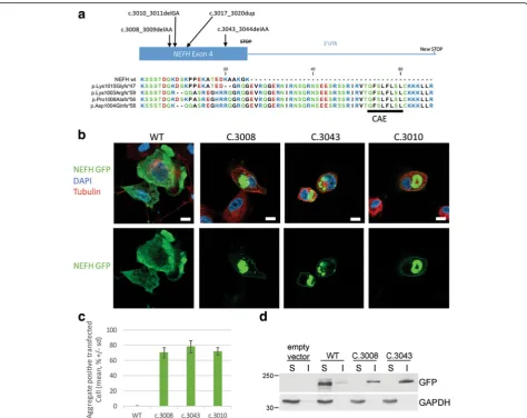
Mutant NEFH forms aggresomes in human neuroblastoma cells line
Both mutations cause the loss of the termination codon, leading to the translation of 40 additional amino acids until the next stop codon at the 3’UTR, as described in Rebelo et al. (c.3010; Fig. 2a). We thus investigated whether our mutations could cause protein aggregation. Expression vectors for eGFP tagged NEFH were trans-fected in the human neuroblastoma cell line SH-EP. Twenty-four hours after transfection all mutant forms (c.3008, c.3043 and c.3010) formed aggregates visible at 10× lens objective in more than 70% of the transfected cells, without significant difference between c.3008 (70.6% +/− 6.7), c.3043 (78.3 +/− 8.0) or c.3010 (72.3% +/−4.9) (Fig. 2c). Conversely, WT NEFH formed aggre-gates in less than 1% of the transfected cells (0.3% +/− 0.6). This result was consistent with the one obtained by Rebelo et al.
Next we investigate the subcellular distribution of mu-tant NEFH proteins, forty-eight hours after transfection. At low expression levels, WT NEFH incorporated into a filamentous network in the cytoplasm (Additional file 2: Figure S2). At higher plasmid concentration, overex-pressed WT NEFH was distributed homogenously in the cytoplasm as revealed by the co-staining with acetylated tubulin and DAPI (Fig. 2b). Conversely, mutant NEFH proteins was always found under the plasma membrane and was also either localized in small aggregates scattered over the cytoplasm or accumulated in a single prominent perinuclear aggregate next to the microtubule organization center (MTOC). Such prominent perinuclear inclusion bodies formed next to the MTOC are commonly named aggresomes [18]. Accumulation of insoluble proteins is characteristic of aggresomes. Solubility in Triton X-100 was used to evaluate the solubility of NEFH proteins expressed in SH-EP cells. As shown in Fig. 2d, WT NEFH was solubi-lized by 0.1% Triton, whereas a significant proportion of mutant NEFH proteins were insoluble. Aggresomes are also characterized by accumulation of ubiquitin conjugates. As expected, aggregates of mutant NEFH proteins colocalized with mono- and poly-ubiquitinated conjugates as evidenced by staining of ubiquitin conjugated (Fig. 3a).
Mutant NEFH proteins are addressed to the autophagic pathway
Aggresome formation has been proposed to allow isola-tion of toxic misfolded proteins and to act as key step for the disposal of protein aggregates by autophagy. In addition, the autophagy role in motoneuron and muscle disorders associated with protein aggregates is well known [5, 7, 29]. We thus investigated the distribution of the pro-tein p62/SQSTM that recruits poly ubiquitinated sub-strates to autophagy. Immunofluorescence experiments revealed that p62 colocalized with aggresomes containing mutant NEFH (Fig. 3b). Finally, we examined the distribu-tion of LC3b, an autophagosome marker. Consistently with p62 localization, we found that LC3b accumulated with aggresomes containing mutant NEFH proteins (Fig. 3c). Altogether, these observations suggest that the cells sequester mutant NEFH proteins in aggresomes from where they direct them to autophagy for degradation.
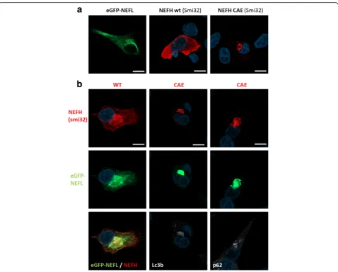
NEFH mutants perturb the neurofilament network in vitro
NEFH homogenously distributed in the cytoplasm whereas NEFH CAE mutants formed perinuclear aggre-gates, as revealed by the Smi32 staining (Fig. 4a). As ex-pected, eGFP-NEFL and WT NEFH colocalized in a dense filamentous network in SH-EP cells (Fig. 4b). Interestingly, eGFP-NEFL and mutant NEFH-CAE proteins colocalized restrictedly in an aggresome containing p62/SQSTM1 and LC3b (Fig. 4b). Therefore, NEFH mutants not only form aggresomes, but also interact with NEFL and destabilizes the neurofilaments network.
Mutant NEFH proteins activate caspase 3 dependent cell death in vitro
Aggresome formation usually indicates a cellular stress response. To determine if mutant NEFH could be
cytotoxic, we investigated for signs of cellular stress. NEFH is a key component of intermediate filaments that contribute to the formation of the cytoskeleton. We thus first examined the morphology of transfected cells 24 h after transfection. Metamorph analysis of the morph-ology of more than 1000 cells chosen stochastically dem-onstrated that cells expressing mutant NEFH presented a significantly reduced average radius and a significantly increased average shape factor compared to cells ex-pressing WT NEFH (Additional file 3: Figure S3). To-gether, this indicated that cells expressing mutant NEFH are smaller and rounder.
We next examined whether mutant NEFH proteins could alter cell viability. Caspase 3 is activated by proteo-lytic cleavage by both extrinsic (death ligand) and intrinsic
Fig. 2NEFH mutations cause protein aggregation in SH-EP.aAmino acid alignment of normal and mutantNEFHC-terminal parts reported in the CMT2cc
presenting the Cryptic Amyloidogenic Element translation and their differences. Amino acid color correlates with polarity: hydrophobic inblack, hydrophilic in
green, acidic inredand basic inblue.bConfocal images of SH-EP transfected with eGFP tagged NEFH vectors and counterstained for tubulin inredand nucleus
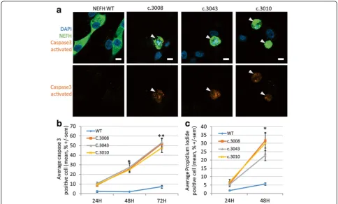
(mitochondrial) apoptotic pathways. Immunofluorescence experiments detected caspase 3 activation in numerous cells 48 h after transfection with one of the mutant NEFH expression vectors. Caspase 3 activation was often associ-ated with a pyknotic nuclei, indicating ongoing apoptosis (Fig. 5a). Quantification of the percentage of cells contain-ing activated caspase 3 at different times points after transfection revealed a progressive increase of the number
of cells triggering the apoptotic pathway. Approximately 10% of SH-EP cells expressing a mutant NEFH had acti-vated caspase 3 twenty four hours after transfection, this number increasing to 25% and 50% 48 h and 72 h after transfection, respectively (Fig. 5b).
To confirm that mutant NEFH triggered cell death over-time, we used propidium iodide (PI), a fluorescent inter-calating agent that requires broken membranes to reach
Fig. 3NEFH aggregates form aggresomes. eGFP tagged NEFH proteins form perinuclear aggregates called aggresome containing ubiquitin, p62/
nuclear DNA and thus selectively labels dying cells. Twenty-four hours after transfection with a mutant NEFH vector, 6% of cells were stained positive for PI, and 25% of cell were stained 1 day later, consistently with caspase 3 activation results (Fig. 5c). Altogether, these results show that expression of mutant NEFH strongly triggered cas-pase 3 activation and cell death.
Mutant NEFH form aggresomes in primary motoneurons in vitro
To confirm our observation in motoneurons, mouse primary motoneuron cultures were used [14, 16, 17] (Additional file 4: Figure S4). Two days after magnetofec-tion, WT and mutant eGFP-NEFH formed a filamentous network with endogenous NEFL (Fig. 6a). Interestingly, after 2 days of expression mutant eGFP-NEFH formed
aggregates along the filamentous structure, which evolved in a prominent perinuclear aggresome containing LC3b as observed 4 days after magentofection (Fig. 6a and b).
NEFH mutations cause protein aggregation and apoptosis in spinal cord neurons
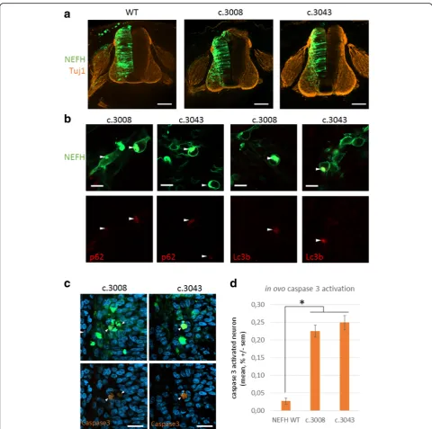
In order to evaluate the effect of NEFH mutations in vivo on spinal cord neurons, we decided to express NEFH in the spinal cord of chick embryos by in ovo electropor-ation. Electroporation of embryonic hemi neural tube al-lows transfer of expression vectors in both motor neurons and dorsal root ganglion (DRG) sensitive neurons. Con-focal images of the entire spinal cord showed that trans-fection was efficient in the hemi spinal cord neurons (Fig. 7a) and less efficient in the DRG but still sufficient to visualize some electroporated neurons (data not show). At
Fig. 4NEFH mutation destabilised NEFL filamentous network in vitro.aConfocal images of transfected SH-EP with eGFP-NEFL or untagged NEFH
higher magnification the anterior horn of these sections holding motor neurons revealed that WT NEFH distrib-uted in a filamentous network (Additional file 2: Figure S2C). Conversely, mutant NEFH proteins accumulated at intense foci in the soma of the neurons forming aggre-somes which colocalized with p62/SQSTM1 and LC3b (Fig. 6b). Caspase 3 activation was clearly detected in 0.22% +/−0.02 and 0.25% +/−0.03 neurons electroporated with mutant NEFH constructs c.3008 and c.3043 respect-ively (Fig. 7c). In these neurons, the occurrence of Caspase 3 activation 48 h after electroporation was more than 8 fold higher than in neurons expressing the WT NEFH ex-pression construct (Fig. 7d). Progressive apoptosis activa-tion is consistent with the neurodegenerative features observed in the patients.
Discussion and conclusions
The families presented here displayed a severe, predom-inantly motor, axonal autosomal dominant form of CMT with proximal motor involvement in the lower limb as-sociated with two previously unreported mutations in theNEFHgene, including a de novo mutation. The clin-ical phenotype varied in term of age at onset, ranging from 5 to 40 years, with a median around 30 years. A
remarkable and consistent feature in all patients was the early involvement of proximal muscles of the lower limbs, occurring approximately 10 to 15 years after the onset of motor deficit. Proximal deficit involved pre-dominantly the iliopsoas muscle, whereas quadriceps and hamstring muscles were relatively preserved. Prox-imal muscles weakness is rare in any form of CMT. Proximal lower-limb weakness with waddling gait is also described in recessive forms of CMT due toNEFL muta-tions [2]. There was muscle wasting predominant in the distal lower limbs muscles progressively ascending to proximal limbs muscles and distal upper limbs muscles. Muscle weakness and muscle wasting were quickly evolving, with most of the patients needing walking as-sistance after 20 years of disease evolution. Three pa-tients in family 1 had brisk reflexes associated with distal and proximal weakness. Pyramidal signs have also been shown in CMT caused by NEFLmutations [2, 13]. The phenotype was mainly motor but there were also sensory alterations, with prominent involvement of large sensory nerves. There were no associated features except for gas-troparesia in one patient and vocal cord involvement in another patient. Muscle pathology and electrophysio-logical studies were consistent with a symmetrical,
Fig. 5NEFH mutations trigger caspase 3 dependent death in vitro.aConfocal images of transfected SH-EP counterstained with DAPI inblueand activated
progressive distal and proximal sensorimotor axonal neuropathy. These findings are in accordance with the previously published paper [27]. It is worth noticing that electrophysiological studies of two asymptomatic patients of family 1 (IV5, IV6) showed a sensorimotor neuropathy respectively at the age of 23 and 17 years, indicating that the neuropathy may exist in a latent state. Patients of these IV generation have been diag-nosed early in their life since they were aware of symptoms related to the neuropathy in the family. Al-though we could not exclude the hypothesis of an
an-ticipation, it must be stressed that even mild
neurological symptoms led their parents to seek for a
neurological and neurophysiological exam that would confirm the neuropathy.
The pathogenicity of the NEFH mutations was con-firmed by the easy segregation of the mutation in both families. The clinical phenotype was also very similar in the two families. Analysis of family 1 suggests an antici-pation pattern given that the proband had an earlier age of onset and more severe manifestations than did the previous generations. This anticipation pattern has already been reported in the previously reported patients [27]. Nevertheless, this anticipation phenotype is prob-ably rather due to a diagnostic bias than to biological phenomenon. Indeed, in families with known familial CMT, clinical signs of the disease are searched earlier and thus detected sooner in the offsprings. The pene-trance of the disease appeared to be complete. Muscle weakness was more severe in patient II-1 of family 2, who carries a de novo mutation in NEFH gene, than in patients belonging to family 1 at the same age. The two previously reported families had frameshift variants in the extreme C-terminus of NEFH [27]. Our two families present with original deletions of two nucleotides close to the normal stop codon. Mutation in family 1 (c.3008_3009delAA; p.Lys1003Argfs*59) leads to the same defective protein as in family UK1 at the exception of one amino acid. Mutation in family 2 is located just three amino acids before the normal stop codon, and is the most subterminal variant in the four families re-ported thus far. Both mutations translate into the same alternative Open Reading Frame (ORF), which is also identical to the previously reported families [27].
The predominance of motor involvement in both proximal and distal muscles associated with brisk re-flexes in some members of family 1 initially prompted us to study the SOD1 gene implicated in genetic ALS, but no mutation was found. In ALS, there is accumulation of neurofilaments in motor neurons. Mouse models with overexpression of neurofilaments subunits have a motor neuropathy resembling ALS [20]. Rare mutations in the peripherin and in the KSP repeats motifs have been re-ported [1, 8], and a latter study showed that a short al-lele of the NEFH tail was associated with ALS [30]. Other studies found no mutations ofNEFHin ALS [9, 28]. The pathogenicity of NEFH mutations in ALS thus re-mains unclear. Nevertheless, the proposed association of
NEFH mutations with ALS and the finding that some CMT patients withNEFHmutations have unusual clin-ical signs related to ALS is intriguing and consistent with the notion that mutations causing the addition of a cryptic amyloidogenic element to NEFH proteins cause a particular neuropathy with overlapping clinical fea-tures of both CMT and ALS.
Our in vitro and in vivo experiments provide a rational basis to these observations by demonstrating thatNEFH
Fig. 6NEFH mutations form aggresome in primary motoneuron in
mutations causing the addition of a CAE induce aggre-some formation and neuronal apoptosis. Loss of motor neurons is a hallmark of ALS, which is also increasingly associated with the presence of protein aggregates in motoneurons. This is especially true for ALS caused by mutations in SOD1, TDP43 and C9ORF72 [32]. The
presence of intracellular protein inclusions is a common hallmark of a wide variety of human disorders. These in-clude neurofibrillary tangles in Alzheimer’s disease, Lewy bodies in Parkinson’s disease, polyglutamine enriched in-clusions in Huntington’s disease, as well as intermediate filament inclusions in specific myopathies. Once formed,
Fig. 7mutant NEFH aggregate in ovo and trigger apoptosis.aconfocal images of chick embryo spinal cord cryosection after in ovo electroporation.
protein aggregates tend to be insoluble, refractory to pro-teolysis and to accumulate in inclusion bodies which are usually present in low copy number, most often only one per cell [18]. This is consistent with the features of the NEFH protein aggregates we observed, which mostly ac-cumulated in one spot and contained insoluble proteins.
Autophagy is the major cellular process by which large cytoplasmic components, including ribosomes, organ-elles and protein aggregates, are degraded. Autophagy plays a key role in the maintenance of homeostasis and the quality of the cellular component for the survival of the neuron in their functional context. The finding that the autophagy proteins p62 and LC3 are present in NEFH aggregates indicates that the presence of the CAE triggers autophagy. Although the precise role of autoph-agy in motoneuron disease is unclear, emerging evidence supports the notion that defects in autophagic pathway may contribute to pathogenic mechanism and could constitute valuable therapeutic targets [5, 7, 22, 31].
Mutations inNEFH cause a sensorimotor axonal neur-opathy, characterized by distal lower limbs motor deficit with early and prominent involvement proximal of the iliopsoas muscle, associated in some patients with pyram-idal signs. Clinical features of NEFH mutations clearly overlap with those of motor neuron disease. At the cellular level, mutant NEFH is sequestered in a prominent peri-nuclear inclusion body, the aggresome which is addressed to the autophagic pathway. Mutant NEFH proteins are nevertheless toxic and progressively trigger caspase 3 de-pendant apoptosis. Interestingly, motoneuron death and intracellular protein inclusions are common hallmarks in ALS. Characterization of cellular effects of theNEFH mu-tations provides a rational basis to the clinical continuum between CMT and ALS in affected patients.
Additional files
Additional file 1: Figure S1.Superficial peroneal sensory nerve biopsy
(case III4): Semi-thin section. A. Note the rarefaction of large myelinated fibers. B. Several fibers have a thin myelin sheath (arrowhead) and some of them present myelin sweling (arrow). (Original magnification ×100). (PNG 947 kb)
Additional file 2: Figure S2.eGFP-NEFH WT form filamentous network
in vitro and in ovo. A. monomeric eGFP tag NEFH WT expression can form visible filamentous network in SH-EP under lower expression condi-tion when transfected at low concentracondi-tion (optimal recommended con-centration diluted four time). eGFP-NEFH WT form filamentous network in spinal motoneuron in vitro (B) and in vivo (C). Scale bar 10μm. (TIFF 156 kb)
Additional file 3: Figure S3.NEFH mutations modify cell morphology
in vitro.A Mutant NEFH expression induces morphological changes as seen on 10× microscopic images. Scale bar represent 100μm. B-C. Quantification of the average shape factor and radius of transfected SH-EP cells. Values represent means in percent +/−standard deviation of at least 15 fields (Cells analyzed >1000 per condition) and analyzed by Kruskal-Wallis one way ANOVA on ranks test followed by Dunn’s methods (*P< 0.001). Shape factor equal to one usually represents an ideal circle. (PNG 334 kb)
Additional file 4: Figure S4.Representative primary motoneuron in its
entirety in vitro. A Non transfected motoneuron revealed by SMI-32 stain-ing. B. Magnetofected motoneuron with eGFP tag NEFH WT or mutated form, without counterstaining. (PNG 156 kb)
Acknowledgements
We thank Aurora Pignata for her technical help on chick embryo experiment.
Funding
AJ was funded by ADN“Association pour le Développement de la Neurogénétique”. SZ is the recipient of NIH R01NS072248 and NIH U54NS092091 (CReATe).
Authors’contributions
TS, CD, OD, GS, MG and RJ did the clinical assessment. TS and CD performed clinical meta-analysis. PL was in charge of the molecular analysis of the patients. AJ and LS designed experimental studies in vitro and in ovo. AJ and EB performed the in vitro studies. AJ and VC performed the in ovo studies. AJ, LS, TS, PL, SZ, AR and AA wrote the paper. All authors read and approved the final manuscript.
Ethics approval and consent to participate
All procedures performed in studies involving human participants were in accordance with the ethical standards of the institutional and/or national research committee and with the 1964 Helsinki declaration and its later amendments or comparable ethical standards. Informed consent was obtained from all individual participants included in the study. All procedures performed in studies involving animals were in accordance with the ethical standards of the institution or practice at which the studies were conducted.
Competing interests
The authors declare that they have no competing interests.
Publisher’s Note
Springer Nature remains neutral with regard to jurisdictional claims in published maps and institutional affiliations.
Author details
1Institut NeuroMyoGène, Université Lyon1 - CNRS UMR 5310 - INSERM
U1217, Lyon, France.2Unité fonctionnelle de neurogénétique moléculaire, CHU de Lyon - HCL groupement Est, Bron, France.3Département de Neurologie, Hôpital Pitié-Salpêtrière, Paris, France.4Clinique du motoneurone et pathologies neuromusculaires, CHRU de Montpellier, Montpellier, France. 5Centre de références des maladies neuromusculaires, CHU de Bordeaux,
Bordeaux, France.6Centre de référence des maladies neuromusculaires, Hôpital Pitié-Salpêtrière, Paris, France.7Service de Neurologie, CH de Valenciennes, Valenciennes, France.8Institut de pathologie, CHU de Lille, Lille, France.9Dr John T. MacDonald Foundation Department of Human Genetics, Institute of Human Genomics, University of Miami, Miller School of Medicine, Miami, USA.10Institut de Myologie, Hôpital Pitié-Salpêtrière, 47-83 boulevard de l’Hôpital, 75013 Paris, France.11Centre de Biotechnologie Cellulaire, CBC Biotec, CHU de Lyon - HCL groupement Est, Bron, France.
Received: 27 June 2017 Accepted: 28 June 2017
References
1. Al-Chalabi A, Andersen PM, Nilsson P, Chioza B, Andersson JL, Russ C, Shaw CE, Powell JF, Leigh PN (1999) Deletions of the heavy neurofilament subunit tail in amyotrophic lateral sclerosis. Hum Mol Genet 8:157–164
2. Berciano J, García A, Peeters K, Gallardo E, Vriendt ED, Pelayo-Negro AL, Infante J, Jordanova A (2015) NEFL E396K mutation is associated with a novel dominant intermediate Charcot–Marie–Tooth disease phenotype. J Neurol 262:1289–1300. doi:10.1007/s00415-015-7709-4
3. Biedler JL, Helson L, Spengler BA (1973) Morphology and growth, tumorigenicity, and cytogenetics of human neuroblastoma cells in continuous culture. Cancer Res 33:2643–2652
and rodent NF-L subunits. J Biol Chem 273:5101–5108. doi:10.1074/jbc. 273.9.5101
5. Chen S, Zhang X, Song L, Le W (2012) Autophagy dysregulation in amyotrophic lateral sclerosis. Brain Pathol Zurich Switz 22:110–116. doi:10. 1111/j.1750-3639.2011.00546.x
6. Ciccarone V, Spengler BA, Meyers MB, Biedler JL, Ross RA (1989) Phenotypic diversification in human neuroblastoma cells: expression of distinct neural crest lineages. Cancer Res 49:219–225
7. Cipolat Mis MS, Brajkovic S, Frattini E, Di Fonzo A, Corti S (2016) Autophagy in motor neuron disease: key pathogenetic mechanisms and therapeutic targets. Mol Cell Neurosci 72:84–90. doi:10.1016/j.mcn.2016.01.012 8. Figlewicz DA, Krizus A, Martinoli MG, Meininger V, Dib M, Rouleau GA,
Julien J-P (1994) Variants of the heavy neurofilament subunit are associated with the development of amyotrophic lateral sclerosis. Hum Mol Genet 3:1757–1761
9. Garcia ML, Singleton AB, Hernandez D, Ward CM, Evey C, Sapp PA, Hardy J, Brown RH, Cleveland DW (2006) Mutations in neurofilament genes are not a significant primary cause of non-SOD1-mediated amyotrophic lateral sclerosis. Neurobiol Dis 21:102–109. doi:10.1016/j. nbd.2005.06.016
10. Gutmann L, Shy M (2015) Update on Charcot-Marie-Tooth disease. Curr Opin Neurol 28:462–467. doi:10.1097/WCO.0000000000000237 11. Hamburger V, Hamilton HL (1992) A series of normal stages in the
development of the chick embryo. 1951. Dev Dyn Off Publ Am Assoc Anat 195:231–272. doi:10.1002/aja.1001950404.
12. Harding AE, Thomas PK (1980) The clinical features of hereditary motor and sensory neuropathy types I and II. Brain J Neurol 103:259–280
13. Hashiguchi A, Higuchi Y, Nomura M, Nakamura T, Arata H, Yuan J, Yoshimura A, Okamoto Y, Matsuura E, Takashima H (2014) Neurofilament light mutation causes hereditary motor and sensory neuropathy with pyramidal signs. J Peripher Nerv Syst 19:311–316
14. Henderson CE, Bloch-Gallego E, Camu W Purified embryonic motoneurons. In: Cohen J, Wilkin G (eds) Nerve cell cult. Pract. Approach, Oxford Univ. Press, London, pp 69–81
15. Hoffman PN, Cleveland DW, Griffin JW, Landes PW, Cowan NJ, Price DL (1987) Neurofilament gene expression: a major determinant of axonal caliber. Proc Natl Acad Sci U S A 84:3472–3476
16. Jacquier A, Bellouze S, Blanchard S, Bohl D, Haase G (2009) Astrocytic protection of spinal motor neurons but not cortical neurons against loss of Als2/alsin function. Hum Mol Genet 18:2127–2139. doi:10.1093/hmg/ddp136 17. Jacquier A, Buhler E, Schäfer MK, Bohl D, Blanchard S, Beclin C, Haase G
(2006) Alsin/Rac1 signaling controls survival and growth of spinal motoneurons. Ann Neurol 60:105–117. doi:10.1002/ana.20886.
18. Johnston JA, Ward CL, Kopito RR (1998) Aggresomes: a cellular response to misfolded proteins. J Cell Biol 143:1883–1898
19. Jordanova A, Jonghe PD, Boerkoel CF, Takashima H, Vriendt ED, Ceuterick C, Martin J-J, Butler IJ, Mancias P, Papasozomenos SC, Terespolsky D, Potocki L, Brown CW, Shy M, Rita DA, Tournev I, Kremensky I, Lupski JR, Timmerman V (2003) Mutations in the neurofilament light chain gene (NEFL) cause early onset severe Charcot–Marie–Tooth disease. Brain 126:590–597. doi:10.1093/ brain/awg059
20. Julien JP, Côté F, Collard JF (1995) Mice overexpressing the human neurofilament heavy gene as a model of ALS. Neurobiol Aging 16:487–490 discussion 490-492
21. Kim MS, Chang X, LeBron C, Nagpal JK, Lee J, Huang Y, Yamashita K, Trink B, Ratovitski EA, Sidransky D (2010) Neurofilament Heavy Polypeptide Regulates the Akt-β-Catenin Pathway in Human Esophageal Squamous Cell Carcinoma. PLoS ONE 5:e9003. doi:10.1371/journal.pone.0009003. 22. Lee JK, Shin JH, Lee JE, Choi E-J (2015) Role of autophagy in the
pathogenesis of amyotrophic lateral sclerosis. Biochim Biophys Acta (BBA) -Mol Basis Dis 1852:2517–2524. doi:10.1016/j.bbadis.2015.08.005
23. Lee MK, Xu Z, Wong PC, Cleveland DW (1993) Neurofilaments are obligate heteropolymers in vivo. J Cell Biol 122:1337–1350
24. Liu Q, Xie F, Siedlak SL, Nunomura A, Honda K, Moreira PI, Zhua X, Smith MA, Perry G (2004) Neurofilament proteins in neurodegenerative diseases. Cell Mol Life Sci CMLS 61:3057–3075. doi:10.1007/s00018-004-4268-8 25. Mersiyanova IV, Perepelov AV, Polyakov AV, Sitnikov VF, Dadali EL, Oparin
RB, Petrin AN, Evgrafov OV (2000) A new variant of Charcot-Marie-Tooth disease type 2 is probably the result of a mutation in the neurofilament-light gene. Am J Hum Genet 67:37–46. doi:10.1086/302962
26. Moret F, Renaudot C, Bozon M, Castellani V (2007) Semaphorin and neuropilin co-expression in motoneurons sets axon sensitivity to environmental semaphorin sources during motor axon pathfinding. Dev Camb Engl 134:4491–4501. doi:10.1242/dev.011452.
27. Rebelo AP, Abrams AJ, Cottenie E, Horga A, Gonzalez M, Bis DM, Sanchez-Mejias A, Pinto M, Buglo E, Markel K, Prince J, Laura M, Houlden H, Blake J, Woodward C, Sweeney MG, Holton JL, Hanna M, Dallman JE, Auer-Grumbach M, Reilly MM, Zuchner S (2016) Cryptic Amyloidogenic elements in the 3′UTRs of Neurofilament genes trigger axonal neuropathy. Am J Hum Genet 98:597–614. doi:10.1016/j.ajhg.2016.02.022
28. Rooke K, Figlewicz DA, Han FY, Rouleau GA (1996) Analysis of the KSP repeat of the neurofilament heavy subunit in familiar amyotrophic lateral sclerosis. Neurology 46:789–790
29. Sandri M (2010) Autophagy in skeletal muscle. FEBS Lett 584:1411–1416. doi: 10.1016/j.febslet.2010.01.056
30. Skvortsova V, Shadrina M, Slominsky P, Levitsky G, Kondratieva E, Zherebtsova A, Levitskaya N, Alekhin A, Serdyuk A, Limborska S (2004) Analysis of heavy neurofilament subunit gene polymorphism in Russian patients with sporadic motor neuron disease (MND). Eur J Hum Genet 12: 241–244. doi:10.1038/sj.ejhg.5201144
31. Song C, Guo J, Liu Y, Tang B (2012) Autophagy and its comprehensive impact on ALS. Int J Neurosci 122:695–703. doi:10.3109/00207454.2012. 714430
32. Taylor JP, Brown RH, Cleveland DW (2016) Decoding ALS: from genes to mechanism. Nature 539:197–206. doi:10.1038/nature20413
33. Uro-Soste E, Fernandez C, Authier F-J, Bassez G, Butori C, Chapon F, Delisle M-B, Dubourg O, Feasson L, Gherardi R, Lacroix C, Laquerriere A, Letournel F, Magy L, Maisonobe T, Marcorelles P, Maurage C-A, Mezin P, Mussini J-M, Penisson-Besnier I, Romero N-B, Streichenberger N, Vallat J-M, Viennet G, Vital A, Voit T, Boucharef W, Figarella-Branger D, Société française de myologie, Association française contre les myopathies (2010, 166) Management of muscle and nerve biopsies: expert guidelines from two French professional societies, Société française de myologie et de l’Association française contre les myopathies. Rev Neurol (Paris) 477:–485
• We accept pre-submission inquiries
• Our selector tool helps you to find the most relevant journal
• We provide round the clock customer support
• Convenient online submission
• Thorough peer review
• Inclusion in PubMed and all major indexing services
• Maximum visibility for your research
Submit your manuscript at www.biomedcentral.com/submit