R E S E A R C H A R T I C L E
Open Access
Biocidal effects of stem bark extract of
Chrysophyllum albidium
G. Don on
vancomycin-resistant
Staphylococcus aureus
David A. Akinpelu
1,2,3, Joseph O. Odewade
1, Olayinka A. Aiyegoro
2,3*, Anofi O. T. Ashafa
3, Oluseun F. Akinpelu
4and Mayowa O. Agunbiade
3Abstract
Background:Staphylococcus aureuscauses variety of infections in humans and animals worldwide and predominates in surgical wound infections. This study assessed the antimicrobial potential of the stem bark extract ofChrysophyllum albidumagainst an array of vancomycin resistantStaphylococcus aureus(VRSA) isolated from clinical samples.
Methods:The methanolic crude extract of the plant was preliminary screened for the presence of phytochemicals; after then, the extract was partitioned into n-hexane, chloroform, ethyl acetate and butanol fractions. A range of concentrations of the plant extract fractions was prepared to assess its antimicrobial potency; the minimum inhibitory concentrations (MICs); the minimum bactericidal concentrations (MBCs); the rate of killing; the potassium ion leakage potential and nucleotides leakage ability against the VRSAs.
Results:The phytochemical screening revealed the presence of tannins, alkaloids, flavonoids, saponins, steroids, reducing sugars and terpenoids as major phytoconstituents resident in the crude plant extract. The two active fractions (n-hexane and butanol) at a concentration of 10 mg/ml exhibited antibacterial activities with the MIC and MBC values for the fractions ranged between 0.63–10 mg/ml and 1.25–10 mg/ml respectively. The time kill assay revealed that the antibacterial action of the two fractions are time and concentration dependent; the n-hexane and butanol fractions achieved 100 % kill on the test isolates at a concentration of 3 × MIC and 2 × MIC respectively after 120 min of reaction time. Varying amount of potassium ions as well as nucleotides were leaked from the test cells by n-hexane and butanol fractions.
Conclusions:This study has established the possibility of developing antimicrobial agents of natural origin to manage possible infection from vancomycin resistantStaphylococcus aureusthat are now developing multi-resistance against many antibiotics.
Keyword:Chrysophyllum albidum, Vancomycin resistantStaphylococcus aureus, Clinical samples, Methanolic extract, Phytochemicals, Minimum inhibitory concentrations, Minimum bactericidal concentrations, Time kill, Potassium ion, Nucleotide
* Correspondence:[email protected] 2
GI Microbiology and Biotechnology Unit, Agricultural Research Council, Animal Production Institute, Irene, Pretoria 0062, South Africa
3Plant Science Department, University of the Free State, Qwaqwa Campus, South Africa
Full list of author information is available at the end of the article
Background
Staphylococcus aureus causes variety of infections in humans and animals worldwide and predominates in surgical wound infections [1]. This organism causes super-ficial skin infection and are also responsible for some life-threatening diseases such as sepsis, respiratory tract and bloodstream infections [2, 3]. The emergence of vanco-mycin resistant Staphylococcus aureus is a big concern that is threatening human’s health globally and has led to an increase in morbidity and mortality. The abrupt and continued increase in the use of vancomycin worldwide resulted in the increasing frequency of VRSA infections in hospitals [4]. The diseases caused by these multiple-drug resistant microorganisms are associated with prolonged hospitalization [5].
Methicillin a derivative of penicillin was used to treat infection caused by S. aureus after its development of resistant to penicillin but resistant to methicillin finally emerged in 1962 [6, 7]. Vancomycin was then considered the drug of choice for the treatment of infections caused by S. aureus for many years before the emergence of VRSA [8]. Emergence of clinical infection due toS. aureus
in a patient in USA with high resistance to vancomycin was reported in 2002 [9]. Thus, there is an urgent need to source for antimicrobial agents especially of natural origin to combat the activities of these pathogens. Emergence of vancomycin resistance in S. aureus is a global issue, and thus, this study focused on the potency of stem bark ex-tract ofChrysophyllum albidiumon vancomycin resistant
S. aureusisolated from clinical samples.
Chrysophyllum albidium belongs to the family sapota-ceae. It is commonly calledAgbalumoin Yoruba,Udalain Igbo andAgbalubain Hausa languages. This plant is also referred to as“The white star apple”[10].Chrysophyllum albidiumis a popular tropical fruit tree and widely distrib-uted in the low land rain forest zones [11].
In Nigeria this plant is distributed within the south-western part of the country [12].Chrysophyllum albidium
is rich in natural antioxidants which promote health [13]. This plant is highly rich in flavonoids, steroids, glyco-sides and saponins and thus serves as a source of anti-inflammatory, anti-spasmodic, as well as possesses diuretic properties [14]. Extracts from seeds and roots ofC. albidium effectively arrested bleeding from fresh wounds and also promote wound healing [15].Chrysophyllum albi-diumleaves are used as emollients and for the treatment of skin eruptions, diarrhoea and stomach ache [16]. The stem bark of this plant is used in preparation of decoction for the treatment of fever and black coated tongues caused by increase population of bacteria and yeast in the mouth [17]. Leaves extract from C. albidium exhibited antibacterial activities against S. aureus, Escherichia coli, Salmonella typhiandShigellaspecies [18]. Seeds and root extracts ob-tained fromC. albidiumexhibited inflammatory,
anti-diarrhoeal and anti-haemorrhoidal [14]. Onyeka [19] dem-onstrated hypoglycaemic, antioxidant and hepatoprotective activities of root bark extract of C. albidium in alloxan-induced diabetic rats.
Methods
Cultures of clinical strains of vancomycin-resistant Staphylococcus aureusand standard cultures of Staphylococcus aureus
Various clinical strains of VRSA used for this study were collected from stock collection of Microbiology Laboratory of both Obafemi Awolowo University Teaching Hospital, Ile Ife, Osun State, Nigeria and University College Hospital, Ibadan, Oyo State, Nigeria. These isolates were first sub cultured on mannitol salt agar medium (Biolab) to confirm their purity and strains before use. The isolates were con-firmed to be true VRSA by subjecting them to susceptibil-ity testing against vancomycin (Duchefa). The standard strains ofStaphylococcus aureus of National Collection of Industrial Bacteria (NCIB) and American Typed Culture Collection (ATCC) were obtained from culture collection of Prof David Akinpelu, Department of Microbiology, Obafemi Awolowo University, Ile Ife, Osun State, Nigeria.
The inoculum of the test isolates were prepared using the colony suspension method as described by European Committee for Antimicrobial Susceptibility Testing (EUCAST) (2000). The inoculum were standardized using 0.5 McFarland standards.
Preparation of plant materials and extraction
Fresh stem bark of Chrysophyllum albidium used for this study was collected from Opa Village, Ile Ife (7.4667° N, 4.5667° E), Osun State, Nigeria in April, 2014. The plant sample was identified in Herbarium of Department of Botany, Obafemi Awolowo University, Ile Ife, Osun State, Nigeria. Voucher specimen of the plant sample was prepared (Voucher Number IFE-17419) and deposited for reference purposes. The plant sample was air-dried until a constant weight was obtained. The air-dried stem bark was ground into powder using a mill (CHRISTY LABMILL, Christy and Norris Ltd; Process Engineers, Chelmsford, England), the powdered material was stored in an air-tight container for further use.
Exactly 1500 g of powdered sample was soaked in solution of methanol and sterile distilled water (3:2) (v/v) for 96 h. The mixture was regularly agitated throughout the period. The mixture was filtered into a clean flask and the filtrate collected was concentrated
Test for phytochemical compounds present inC. albidum extract
The phytochemical analysis of the extract was done using Trease and Evans [20] and Harborne [21] methods. The test include determination of the presence of tannins, alkaloids, flavonoids, saponins, steroids, reducing sugars and terpenoids in the extract.
Fractionation of the crude extract fromC. albidiumstem bark
The crude extract was fractionalized using different organic solvents in order of their polarity. The solvent used are n-hexane, chloroform, ethyl acetate and n-butanol in that order. The yield obtained for each fraction was noted and kept in an air-tight container for further use.
The antibiograms of the fractions obtained from the crude extract against the test isolates
The antibiogram of the fractions was determined using Akinpelu and Kolawole method [22]. The test isolates were re-activated in nutrient broth for 18 h before use. Exactly 0.1 mL of the standardized test isolates was transferred into molten Mueller-Hinton agar medium at 40 °C, thoroughly mixed and then poured into a sterile Petri dish. The plates were allowed to set and wells were bored into the medium with the aid of a sterile 6 mm cork borer. These wells were then filled up with the sus-pension of the fractions at a concentration of 10 mg/mL. Streptomycin and ampicillin each at a concentration of 1 mg/mL were used as positive control, while sterile dis-tilled water in separate wells were used as negative con-trol. The plates were allowed to stand on the laboratory bench for 1 h to allow proper in-flow of the extract into the medium. The plates were later incubated at 37 °C for 24 h, after which they were observed for zones of in-hibition which indicates susceptibility of the test isolates to the extract.
The minimum inhibitory concentrations (MIC) of the fractions against the test isolates
The MIC of the fractions against the test isolates was de-termined using EUCAST [23] method. Dilution of the fractions ranging between 0.313 and 5.00 mg/mL were prepared and incorporated into molten nutrient agar at 45 °C and poured into sterile plates. The plates were allowed to set and left on the laboratory bench overnight to ascertain of non-contaminant in the prepared plates. The plates were then inoculated with the standardized inoculum of the test isolates by streaking across the plate surface. These plates were incubated aerobically at 37 °C for 48 h and observed for any growth. The MIC was taken as the lowest concentration of the extract that inhibited the growth of the test isolates.
The minimum bactericidal concentration (MBC) of the fractions against the test isolates
The MBC of the fractions against the test isolates was de-termined using Spencer [24] and Okore [25] methods with some modifications. Samples were taken from plates with no visible growth in MIC assay and sub-cultured onto freshly prepared nutrient agar plates and incubated at 37 °C for 72 h. The MBC was taken as the concentration of the extract that did not show any bacterial growth on fresh nutrient agar plates.
Determination of rate of kill of the test isolates by the fractions
The assay of the rate of kill of the test isolates was deter-mined using Odenholt method [26] with some modifica-tion. Culture of the test isolates was first standardized to approximately 106cfu/mL before use. Exactly 0.5 mL of the standardized suspension of the culture was added to 4.5 mL of different concentrations of the fraction relative to MIC. These were held at room temperature over a period of 2 h to determine the killing rate. Exactly 0.5 mL of the standardized suspension of the culture was added to 4.5 mL of different concentrations of the fraction rela-tive to MIC. These were held at room temperature over a period of 2 h to determine the killing rate. A volume of 0.5 mL of each suspension was withdrawn at time inter-vals and transferred to 4.5 mL of recovery medium con-taining 3 % “Tween 80” to shake off the effect of the extract carry-overs from the test isolates. The suspension was then serially diluted and plated for viable counts. The plates were incubated at 37 °C for 48 h before reading. Control plates containing the test cells without the extract were set up along with the experimental. The emergent bacterial colonies were counted and compared with the counts of the culture control.
Determination of potassium ion leakage from the test isolates by the fractions
from the cells in the suspension was measured at time intervals over 2 h as a potential difference in mV. These values were converted to concentrations of K+ions (M) by reference to a conversion graph, which had been con-structed earlier using KCl standard solutions. The concen-tration of K+ions released was plotted against time.
Determination of nucleotides leakage from the test isolates by the fractions
The leakage of nucleotides from the test cells was deter-mined using Heipieper method [27]. Washed cells of 18 h old test isolates was standardized (approximately 106cfu/mL) and treated with different concentrations of the fractions relative to the MIC at various contact time intervals over 2 h. Each suspension was centrifuged at 10,000 rpm and decanted. Wavelength of the super-natant collected was determined at 260nm to quantify the amount of nucleotide leaked by comparing with the standard curve already plotted (A range of concentration from 0.5 to 5 μM of dNTP mix (dA, dC, dG, dT) (INQABA© Biotech, Pretoria, South Africa) was pre-pared in TE buffer and used to generate the standard curve). The blank constitute sterile distilled water inocu-lated with the test isolates.
Statistics and data processing
All experiments were done in triplicates. Data was analysed for a 4 × 4 Latin square design with the statistical program using the GLM model (Statistical Analysis Systems, SAS Institute, Cary, NC, USA, 2001). Results were contrasted with negative and a positive control. The means of the values was compared using independent t test of signifi-cance (p< 0.05).
Results
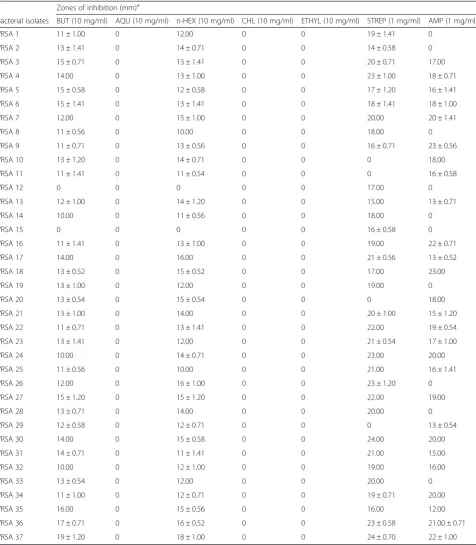
Five fractions were obtained from the crude extract ofC. albidium and they are n-butanol, n-hexane, chloroform, ethyl acetate and aqueous fractions. Two of the fractions (n-butanol and n-hexane) exhibited antimicrobial activities against the test isolates while other fractions were not ac-tive against the test isolates of VRSA. The zones of inhib-ition exhibited by both n-butanol and n-hexane fractions against the test isolates ranged between 10 and 16 mm. The zone of inhibition exhibited by n-butanol against S. aureus (NCIB 8588) was 17 mm while that exhibited againstS. aureus(ATCC 6538) was 19 mm. On the other hand, zones of inhibition observed for n-hexane fraction against S. aureus (NCIB 8588) was 16 mm and for S. aureus (ATCC 6538) was 18 mm (Table 1). Strepto-mycin and ampicillin inhibited 33 out of 37 and 27 out of 37 test isolate respectively. The zones of inhibition exhibited by streptomycin against the test isolates ranged between 14 and 24 mm. On the other hand, ampicillin ex-hibited between 12 and 23 mm zones of inhibition against
the test isolates. Overall, the two active fractions com-pared favourably with the two standard antibiotics used as positive control for this study.
The MIC and MBC exhibited by both n-butanol and n-hexane fractions against the test isolates were also determined. The MIC exhibited by n-butanol fraction ranged between 0.63 and 5.00 mg/mL while that of n-hexane was between the same range with that of n-butanol. The minimum bactericidal effects showed against the test isolates by both fractions ranged between 1.25 and 10.00 mg/mL. From all indications both n-butanol and n-hexane fractions exhibited equal antimicrobial activ-ities against the test isolates (Table 2).
The phytochemical compounds present in the extract were also investigated. The extract revealed the presence of tannins, alkaloids, flavonoids, saponins, steroids, redu-cing sugars and terpenoids (Table 3).
The bactericidal potentials of the two fractions were determined on the test isolates by assaying for the killing rate, potassium ion leakage and leakage of nucleotides. Figure 1a shows the extent and killing rate of the test isolates by n-hexane fraction. At a concentration of 1 × MIC after 15 min of contact time of the isolates with the fraction, 44.5 % of the test isolates was killed and this rose to 55.5 % at 30 min of contact time. At 60 min con-tact time the population of the test isolates killed was 61.3 % while the population rose to 73.0 % at 90 min contact time. Finally, at 120 min contact time 87.7 % of the test isolates were killed. This monophasic effects were also observed when the n-hexane fraction concen-trations were increased to 2 × MIC and 3 × MIC. Finally 100 % cells killing was achieved at 3 × MIC concentra-tion after 120 min of contact time of the test cells with the fraction (Fig. 1a). The same trend of reactions were observed when the test isolates were subjected to n-butanol fraction (Fig. 1b). The rate at which the test cells were killed increases with the concentrations as well as increase in contact time. This is also an indication of monophasic effects. Thus, the bactericidal effects of both n-hexane and n-butanol were through cell membrane disruption as indicated in the experiment.
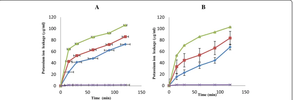
at 1 × MIC and 120 min contact time the quantity of po-tassium ion leaked out of the test cells was 67.83μg/mL. At concentrations of 2 × MIC and 3 × MIC and contact time of 120 min, the quantities of potassium ion leaked
out of the test cells were 83.59 and 103.0μg/mL respect-ively (Fig. 2b).
[image:5.595.60.537.98.644.2]The extent and damaged done to the test cells mem-brane was also observed through nucleotide leakage
Table 1Sensitivity patterns exhibited by fractions obtained fromChrysophyllum albidumagainst the bacterial isolates
Zones of inhibition (mm)a
Bacterial isolates BUT (10 mg/ml) AQU (10 mg/ml) n-HEX (10 mg/ml) CHL (10 mg/ml) ETHYL (10 mg/ml) STREP (1 mg/ml) AMP (1 mg/ml)
VRSA 1 11 ± 1.00 0 12.00 0 0 19 ± 1.41 0
VRSA 2 13 ± 1.41 0 14 ± 0.71 0 0 14 ± 0.58 0
VRSA 3 15 ± 0.71 0 13 ± 1.41 0 0 20 ± 0.71 17.00
VRSA 4 14.00 0 13 ± 1.00 0 0 23 ± 1.00 18 ± 0.71
VRSA 5 15 ± 0.58 0 12 ± 0.58 0 0 17 ± 1.20 16 ± 1.41
VRSA 6 15 ± 1.41 0 13 ± 1.41 0 0 18 ± 1.41 18 ± 1.00
VRSA 7 12.00 0 15 ± 1.00 0 0 20.00 20 ± 1.41
VRSA 8 11 ± 0.56 0 10.00 0 0 18.00 0
VRSA 9 11 ± 0.71 0 13 ± 0.56 0 0 16 ± 0.71 23 ± 0.56
VRSA 10 13 ± 1.20 0 14 ± 0.71 0 0 0 18.00
VRSA 11 11 ± 1.41 0 11 ± 0.54 0 0 0 16 ± 0.58
VRSA 12 0 0 0 0 0 17.00 0
VRSA 13 12 ± 1.00 0 14 ± 1.20 0 0 15.00 13 ± 0.71
VRSA 14 10.00 0 11 ± 0.56 0 0 18.00 0
VRSA 15 0 0 0 0 0 16 ± 0.58 0
VRSA 16 11 ± 1.41 0 13 ± 1.00 0 0 19.00 22 ± 0.71
VRSA 17 14.00 0 16.00 0 0 21 ± 0.56 13 ± 0.52
VRSA 18 13 ± 0.52 0 15 ± 0.52 0 0 17.00 23.00
VRSA 19 13 ± 1.00 0 12.00 0 0 19.00 0
VRSA 20 13 ± 0.54 0 15 ± 0.54 0 0 0 18.00
VRSA 21 13 ± 1.00 0 14.00 0 0 20 ± 1.00 15 ± 1.20
VRSA 22 11 ± 0.71 0 13 ± 1.41 0 0 22.00 19 ± 0.54
VRSA 23 13 ± 1.41 0 12.00 0 0 21 ± 0.54 17 ± 1.00
VRSA 24 10.00 0 14 ± 0.71 0 0 23.00 20.00
VRSA 25 11 ± 0.56 0 10.00 0 0 21.00 16 ± 1.41
VRSA 26 12.00 0 16 ± 1.00 0 0 23 ± 1.20 0
VRSA 27 15 ± 1.20 0 15 ± 1.20 0 0 22.00 19.00
VRSA 28 13 ± 0.71 0 14.00 0 0 20.00 0
VRSA 29 12 ± 0.58 0 12 ± 0.71 0 0 0 13 ± 0.54
VRSA 30 14.00 0 15 ± 0.58 0 0 24.00 20.00
VRSA 31 14 ± 0.71 0 11 ± 1.41 0 0 21.00 15.00
VRSA 32 10.00 0 12 ± 1.00 0 0 19.00 16.00
VRSA 33 13 ± 0.54 0 12.00 0 0 20.00 0
VRSA 34 11 ± 1.00 0 12 ± 0.71 0 0 19 ± 0.71 20.00
VRSA 35 16.00 0 15 ± 0.56 0 0 16.00 12.00
VRSA 36 17 ± 0.71 0 16 ± 0.52 0 0 23 ± 0.58 21.00 ± 0.71
VRSA 37 19 ± 1.20 0 18 ± 1.00 0 0 24 ± 0.70 22 ± 1.00
Key: BUT = Butanol fraction, AQU = Aqueous fraction, n-HEX = n-Hexane Fraction, CHL = Chloroform fraction, ETHYL = Ethy1 acetate fraction, STREP = Streptomycin and AMP = Ampicillin,a
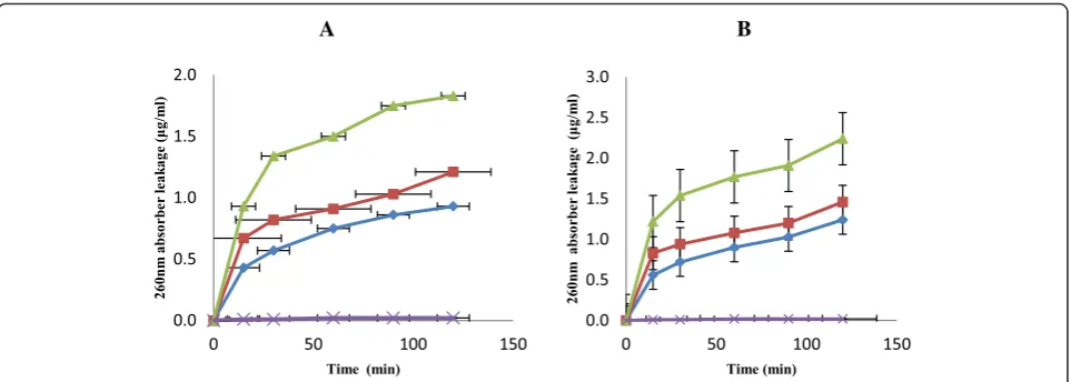
(Fig. 3). The two fractions exhibited appreciable nucleo-tide leakage as observed in the results. When the test cells were subjected the effects of n-hexane fraction at a concentration of 1 × MIC and at 15 min contact time the quantity of nucleotides leaked from the cells was 0.43μg/mL. At 30 min contact time, the quantity leaked increased to 0.57 μg/mL and got to 0.75 μg/mL after 60 min of contact time. There was an increase in the quan-tity of nucleotides leaked out of the cells to 0.93 μg/mL when the contact time was increased to 120 min. The same trend of nucleotide leakage from the test cells was observed when the concentrations of n-hexane fraction were icreased by 2 × MIC and 3 × MIC (Fig. 3a). The effects of n-butanol fraction against the test isolates was also observed as shown in Fig. 3b. There was an increase in the quantity of nucleotides leaked from the cells as the concentrations and contact time increased. For example, at 1 × MIC, 2 × MIC and 3 × MIC concentrations after 120 min of contact time, the quantities of nucleotides leaked from the cells were 1.03, 1.46 and 2.24μg/mL respectively (Fig. 3b). Discussion
The antimicrobial potentials of stem bark extract of
[image:6.595.306.539.112.222.2]Chrysophyllum albidium were investigated on various isolates of vancomycin resistant S. aureus isolated from clinical samples along with two typed cultures, one of NCIB and the other of ATCC. Staphylococcus aureusis a pathogen and is responsible for various ailments in humans and animals. This organism has developed re-sistant to many antibiotics used as therapy against infec-tions caused by it. Methicillin was later chosen as a drug of choice for the treatment of infections caused by this pathogen but later developed resistant to it [6, 7]. Later, vancomycin was adopted to combat the activities of S. aureus and this drug is no longer showing potency towards the treatment of infection caused by this patho-gen [8]. The results obtained from our studies on frac-tions obtained from C. albidium stem bark extract showed that the extract exhibited high antibacterial ac-tivities towards various isolates of vancomycin resistant
Table 2The minimum inhibitory and bactericidal concentrations exhibited by the butanol and n-hexane fractions against susceptible bacterial isolates
Butanol fraction n-Hexane fraction Bacterial
isolates
MIC (mg/ml) MBC (mg/ml) MIC (mg/ml) MBC (mg/ml)
VRSA 1 5.00 10.00 2.50 5.00
VRSA 2 ND ND 5.00 10.00
VRSA 3 1.25 2.50 1.25 2.50
VRSA 4 1.25 2.50 1.25 2.50
VRSA 5 2.50 5.00 5.00 10.00
VRSA 6 1.25 2.50 0.63 1.25
VRSA 7 2.50 5.00 1.25 2.50
VRSA 8 ND ND 5.00 10.00
VRSA 9 5.00 10.00 ND ND
VRSA 10 5.00 10.00 1.25 2.50
VRSA 11 1.25 2.50 2.50 5.00
VRSA 12 ND ND ND ND
VRSA 13 ND ND ND ND
VRSA 14 5.00 10.00 ND ND
VRSA 15 ND ND ND ND
VRSA 16 1.25 2.50 1.25 2.50
VRSA 17 1.25 2.50 0.63 1.25
VRSA 18 ND ND 1.25 2.50
VRSA 19 5.00 10.00 ND ND
VRSA 20 5.00 10.00 2.50 5.00
VRSA 21 2.50 5.00 ND ND
VRSA 22 ND ND ND ND
VRSA 23 1.25 2.50 1.25 2.50
VRSA 24 1.25 2.50 0.63 1.25
VRSA 25 2.50 5.00 2.50 5.00
VRSA 26 1.25 2.50 1.25 2.50
VRSA 27 2.50 5.00 2.50 5.00
VRSA 28 1.25 2.50 0.63 1.25
VRSA 29 1.25 2.50 5.00 10.00
VRSA 30 2.50 5.00 5.00 10.00
VRSA 31 0.63 1.25 0.63 1.25
VRSA 32 1.25 2.50 2.50 5.00
VRSA 33 2.50 5.00 0.63 1.25
VRSA 34 1.25 2.50 1.25 2.50
VRSA 35 ND ND ND ND
VRSA 36 0.63 1.25 1.25 2.50
VRSA 37 1.25 2.50 2.50 5.00
[image:6.595.57.289.122.675.2]KEY: VRSA = Vancomycin resistantStaphylococcus aureus(Clinical strains), MIC = Minimum Inhibitory Concentration, MBC = Minimum Bactericidal Concentration, VRSA 36 =Staphylococcus aureus(NCIB 8588), VRSA 37 =Staphylococcus aureus(ATCC 6538), ND = Not Done
Table 3Preliminary phytochemical compounds obtained from the stem bark extract ofChrysophyllum albidum
Chemical test Result
Tannins Positive
Alkaloids Positive
Flavonoids Positive
Saponins Positive
Steroids Positive
Reducing sugars Positive
S. aureus used for this studies. Fractions obtained from this extract compared favourably with the two standard antibiotics–ampicillin and streptomycin used as positive controls. This is an indication that C. albidium stem bark extract could serve as a remedy to combat the in-fections caused by VRSA along with other pathogens that have now developed resistance to various antibiotics now in circulation used as therapy against infections caused by microorganisms. Chrysophyllum albidium is used among many tribes in West Africa for the treat-ment of various infections caused by microorganisms. Extracts obtained fromC. albidiumexhibited the presence of phytochemicals which include flavonoids, tannins, sa-ponins, alkaloids, steroids, reducing sugars and terpenoids. These phytochemicals contributed to the bioactive poten-tials of this plant. Phytochemicals have been reported to be biologically active [19] and thus contributed to the
antimicrobial activities of this plant. These facts supported the usefulness of C. albidium in folklore remedies. The natural products and phytochemical compounds present in this plant can be exploited for the development of novel bioactive compounds for the treatment of infections caused by VRSA and other pathogens that are now be-coming resistant to antibiotics.
Mode of action of n-hexane and n-butanol fractions from crude extract ofC. albidiumwere also investigated through rate of killing, leakages of potassium ions and nucleotides from the test cells.Chrysophyllum albidium
extract exerted a biocidal effects on the test cells and this might have been contributed by the phytochemicals present in this plant’s extract. The killing rate exhibited by both n-hexane and n-butanol showed a 100 % kill of the test cells at a concentration of 3 × MIC within the shortest time of 120 min of the contact time (Fig. 1).
Fig. 1The extent and the rate of killing of test cells by n- hexane (a) and butanol (b) fractions at 1 × MIC ( ), 2 × MIC ( ), 3 × MIC ( ) and control ( ). Each point represents the log10of mean survival of bacterial cells at a particular time interval in the presence of the fraction
A B
[image:7.595.55.540.87.237.2] [image:7.595.59.540.529.693.2]The ability of plant extract to eliminate or kill organisms at the shortest period of time is generally accepted defin-ition of bactericidal activity in antibiotics [28]. These fractions also exhibited appreciable potassium ions and nucleotides leakage from the test cells which also led to the death of the organisms (Figs. 2 and 3). Leakage of potassium ions from bacterial cell could lead to deactiva-tion of important enzymes required for cell metabolism and this effects could be lethal on the cells [29]. Potas-sium is the major monovalent intracellular cations in cells, and its uptake is essential for all living organisms [30]. Potassium has many key functions within bacterial cells and this include activation of intracellular enzymes. It also act as an intracellular second messenger and in-volved in the maintenance of a constant internal pH and membrane potential [31]. In addition, potassium plays an important function as an osmotic solute. Potassium ions transport is a critical determinant of growth and survival through its role in regulating cytoplasmic pH and cell turgor [32, 33]. Thus leakage of potassium ions from bacterial cells will have a serious effect on such cells as this leads to their death. Leakage of intracellular materials from the cells is an indication of damage to the cell membrane. Thus permeability of the cytoplasmic membrane will lead to the loss of cellular matters and consequently results in cell death [32]. From our results in this study, C. albidium extract might have caused disruption in cell membrane of VRSA used for this study and thus led to their death. This observation was also noted by [33]) in their work on Candida albicans
andC. krusei.
Conclusion
The bactericidal activities exhibited byC. albidiumextract against VRSA used in this study revealed a significant
therapeutic potential of this plant and supported its useful-ness in folklore remedies for the management of infections caused by pathogens. The ability of the plant extract frac-tions obtained fromC. albidiumto kill VRSA in this study at low concentration and minimal contact time has estab-lished the potential of the plant as a template for future drugs that could be formulated to combat infections caused by VRSA; such drug would be useful in combating the menace of VRSAs in human and animal health.
Competing interests
All the authors declares that there is no competing interest.
Authors’contributions
DAA: Design, supervise the research and write section of the article. JOO: Bench work and writing of the article. OAA: Analyse the data and write section of the article. AOTA: Write and edited section of the article. OFA: Bench work and writing of the article. MOA: Bench work and writing of the article. All authors read and approved the final manuscript.
Acknowledgment
The authors acknowledges Obafemi Awolowo University, Ile-Ife, Nigeria, Agricultural Research Council, South Africa and the University of the Free State, South Africa for the provisions of research space and financial grants.
Author details
1Department of Microbiology, Obafemi Awolowo University, Ile Ife, Osun State, Nigeria.2GI Microbiology and Biotechnology Unit, Agricultural Research Council, Animal Production Institute, Irene, Pretoria 0062, South Africa.3Plant Science Department, University of the Free State, Qwaqwa Campus, South Africa.4Department of Biological Science, Faculty of Agriculture and Biotechnology, North West University, Mahikeng Campus, Mmabatho 2735, South Africa.
Received: 14 October 2015 Accepted: 9 March 2016
References
1. Pelczar MJ, Chan EC, Kruz NR. Microbiology. 5th ed. New Delhi, India: McGraw-Hill Publishing Company Limited; 2006.
2. Jarvis WR, Jarvis AA, Chinn RY. National prevalence of methicillin-resistant Staphylococcus aureusin inpatients at United States health care facilities. Am J Infect Control. 2012;40:194–200.
A B
[image:8.595.58.540.87.259.2]3. Coltman K. Urinary tract infections. New thoughts on an old subject. The Practitioner. 1981;223:351–5.
4. Gardete S, Tomasz A. Mechanisms of vancomycin resistance inStaphylococcus aureus. J Clin Invest. 2014;24(7):2836–40.
5. Cunha BA. Antibiotic side effects. Med Clin. 2001;85:149–85. 6. Johnson AP, Aucken HM, Cavendish S, Ganner M, Wale MC, Warner M,
Livermore DM, Cookson BD. Dominance of EMRSA-15 and -16 among MRSA causing nosocomial bacteraemia in the UK: analysis of isolates from the European Antimicrobial Resistance Surveillance System (EARSS). J Antimicrob Chemother. 2001;48(1):143–4.
7. Lowy FD.Staphylococcus aureusinfections. N Engl J Med. 1998;339(8):520–32. 8. Cosgrove SE, Carroll KC, Perl TM.Staphylococcus aureuswith reduced
susceptibility to vancomycin. Clin Infect Dis. 2004;39:539–45.
9. Sievert DM, Rudrik JT, Patel JB, McDonald LC, Wilkins MJ, Hageman JC. Vancomycin-resistantStaphylococcus aureusin the United States, 2002–2006. Clin Infect Dis. 2008;46(5):668–74.
10. Idowu TO, Iwelewa EO, Aderogba MA, Akinpelu BA, Ogundaini AO. Biochemical and behavioural effects of eleagine fromC. albidum. J Biol Sc. 2006;6:1029–34. 11. Madubuike FN, Ogbonnaya O. The potential use of white star apple seed
(C. albidum) and physic nut (Jatropha curcas) as feed ingredients for rat. J Agric Vet Med. 2003;1:97–105.
12. Oyebade BA, Ekeke BA, Adeyemo FC. Fruits categorization and diagnostic analysis ofChrysophyllum albidumG. Don in Nigeria. Adv Appl Sc Res. 2011; 2(1):7–15.
13. Burits M, Bucar F. Antioxidant activity ofChrysophyllum albidumessential oil. Phyt Res. 2002;14:323–8.
14. Savithramma N, Linga RAOM, Beena P. Phytochemical studies ofDysophylla myosuroides(Roth.) Benth. In. wall andTalinum cuneifolium(Vahl.) wild. Res J Phyt. 2011;5:163–9.
15. Okoli BJ, Okere OS. Antimicrobial activity of the phytochemical constituents ofChrysophyllum albidumG.Don. (African Star apple) Plant J Res Nat Dev. 2010;8(1):35.
16. Adisa SA. Vitamin C, protein and mineral contents of african apple (Chrysophyllum albidum). In: Proceedings of the 18th Annual Conference of NIST (eds). 2000. 17. Olapade EO. Medicinal importance ofChrysophyllum albidum. In: Proceedings
of a National Workshop on the Potentials of African Star Apple. 1997. p. 36–8. 18. Duyilemi OP, Lawal IO. Antibacterial activity and phytochemical screening
ofChrysophyllum albidumleaves. Asian J Fd Agro Ind. 2009;2:S75–9. 19. Onyeka CA, Aligwekwe AU, Olawuyi TS, Nwakama EA, Kalu EC, Oyeyemi AW.
Antifertility effects of ethanolic root bark extract ofChrysophyllum albidum in male albino rats. Int J Appl Res Nat Pr. 2012;5(1):12–7.
20. Trease GE, Evans WC. Textbook of Pharmacognosy. London, UK: Balliere, Tindall; 2002. p. 57–9. 343–383.
21. Harborne JB. Phytochemical methods: a guide to modern techniques of plant analysis. London, UK: Chapman and Hall; 1998. p. 60–6.
22. Akinpelu DA, Kolawole DO. Phytochemical and antimicrobial activity of leaf extract ofPiliostigma thonningii(Schum.). Sc Focus. 2004;7:64–70. 23. European Committee for Antimicrobial Susceptibility Testing (EUCAST)
of the European Society for Clinical Microbiology and Infectious Diseases (ESCMID). Determination of minimum inhibitory concentrations (MICs) of antibacterial agents by agar dilution. Cl Microbiol Inf. 2000;6: 509–15.
24. Spencer RC. Infections in continuous ambulatory peritoneal dialysis. J Med Microbiol. 1998;27:1–9.
25. Okore VC. Evaluation of chemical antimicrobial agents. Bacterial resistance to antimicrobial agents. Pharm Microbiol. 2005;55–120.
26. Odenholt I, Lowdinand E, Cars O. Pharmacodynamics of telithromycin in vitroagainst respiratory tract pathogens. Antimicrob Ag Chemoth. 2001; 45:23–9.
27. Heipieper HJ, Diefenbach R, Kewelol H. Conversion of cis unsaturated fatty acids to Trans, a possible mechanism for the protection of phenol degradingPseudomonas putidaP8 from substrate toxicity. Appl Env Microbiol. 1992;58:1847–51.
28. Pankey GA, Sabath LD. Clinical relevance of bacteriostatic against bactericidal mechanisms of action in the treatment of Gram-positive bacterial infections. Clin Infect Dis. 2004;38:864–70.
29. Cox SD, Mann CM, Marhan JL, Gustafson JE, Warmington JR, Wyllie SG. Determining the antimicrobial action of tea tree oil. Molecules. 2001;6:87–91. 30. Gründling A. Potassium uptake systems inStaphylococcus aureus: new
stories about ancient systems. Mol Biol. 2013;4(5):784-13.
31. Booth IR. Regulation of cytoplasmic pH in bacteria. Microbiol Rev. 1985;49: 359–78.
32. Epstein W. Osmoregulation by potassium transport inEscherichia coli. FEMS Microbiol Lett. 1986;39:73–80.
33. Cao M, Wang T, Ye R, Helmann JD. Antibiotics that inhibit cell wall biosynthesis induce expression of theBacillus subtilissigma(W) and sigma(M) regulons. Mol Microbiol. 2002;45(5):1267–76.
• We accept pre-submission inquiries
• Our selector tool helps you to find the most relevant journal • We provide round the clock customer support
• Convenient online submission • Thorough peer review
• Inclusion in PubMed and all major indexing services • Maximum visibility for your research
Submit your manuscript at www.biomedcentral.com/submit