0095-1137/06/$08.00
⫹
0
doi:10.1128/JCM.01389-06
Copyright © 2006, American Society for Microbiology. All Rights Reserved.
Development of a Serotype-Specific DNA Microarray for Identification
of Some
Shigella
and Pathogenic
Escherichia coli
Strains
䌤
Yayue Li,
1,2,3† Dan Liu,
4† Boyang Cao,
1,2,3Weiqing Han,
1,2,3Yanqun Liu,
1,2,3Fenxia Liu,
1,2,3Xi Guo,
1,2,3David A. Bastin,
4Lu Feng,
1,2,3and Lei Wang
1,2,3,4*
TEDA School of Biological Sciences and Biotechnology, Nankai University, 23 Hong Da Street, TEDA, Tianjin 300457, China
1;
Tianjin Research Center for Functional Genomics and Biochips, TEDA College, Nankai University, Tianjin 300457, China
2;
Tianjin Key Laboratory of Microbial Functional Genomics, TEDA College, Nankai University, 23 Hong Da Street, TEDA,
Tianjin 300457, China
3; and Tianjin Biochip Corporation, 23 HongDa Street, TEDA, Tianjin 300457, China
4Received 6 July 2006/Returned for modification 10 August 2006/Accepted 26 September 2006
Shigella
and pathogenic
Escherichia coli
are major causes of human infectious diseases and are responsible
for millions of cases of diarrhea worldwide every year. A convenient and rapid method to identify highly
pathogenic serotypes of
Shigella
and
E. coli
is needed for large-scale epidemiologic study, timely clinical
diagnosis, and reliable quarantine of the pathogens. In this study, a DNA microarray targeting
O-serotype-specific genes was developed to detect 15 serotypes of
Shigella
and
E. coli
, including
Shigella sonnei
;
Shigella
flexneri
type 2a;
Shigella boydii
types 7, 9, 13, 16, and 18;
Shigella dysenteriae
types 4, 8, and 10; and
E. coli
O55,
O111, O114, O128, and O157. The microarray was tested against 186 representative strains of all
Shigella
and
E. coli
O serotypes, 38 clinical isolates, and 9 strains of other bacterial species that are commonly present in
stool samples and was shown to be specific and reproducible. The detection sensitivity was 50 ng genomic DNA
or 10
4CFU per ml in mock stool specimens. This is the first report of a microarray for serotyping
Shigella
and
pathogenic
E. coli
. The method has a number of advantages over traditional bacterial culture and antiserum
agglutination methods and is promising for applications in basic microbiological research, clinical diagnosis,
food safety, and epidemiological surveillance.
Shigella
and pathogenic
Escherichia coli
are the major
caus-ative agents of diarrhea.
E. coli
clones are normally classified
by a combination of oligosaccharide (O), flagellar (H), and
capsular (K) antigens.
Shigella
clones are classified by the O
antigen only, as they lack H and K antigens (14). There are
four named species of
Shigella
classified on the basis of
bio-chemical and O-antigen serological differences:
Shigella
dysen-teriae
(consisting of 13 serotypes),
Shigella flexneri
(consisting
of 14 serotypes [including subtypes]),
Shigella boydii
(consisting
of 18 serotypes), and
Shigella sonnei
(27). Of the estimated 165
million cases of
Shigella
diarrhea per year, 69% of the episodes
occurred in children under 5 years of age, and 1.1 million
deaths were attributed to
Shigella
infections (24). In the United
States, most infections are caused by
S. sonnei
;
S. flexneri
is the
second most common serotype (4, 24).
The
E. coli
species consist of various serotypes, ranging from
highly pathogenic to nonpathogenic strains, which are from
normal intestinal flora and are often used as safe laboratory
strains (32). There are five major pathotypes of
E. coli
strains
that cause diarrhea in humans: enteropathogenic,
enterotoxi-genic, enteroinvasive, enteroaggregative, and
enterohemor-rhagic (28). These pathotypes consist of genetic clones that
often correspond to distinct O:H serotypes (15). Serotypes
O55, O111, O114, and O128 belong to the group of
entero-pathogenic
E. coli
-associated O serotypes (23).
E. coli
O157:H7 is one of the enterohemorrhagic
E. coli
strains, and it
causes significant illness and represents a serious public health
threat worldwide (5).
The O antigen, which consists of repeats of the O unit, is
part of the lipopolysaccharide in the outer membrane of
gram-negative bacteria and contributes major antigenic variability to
the cell surface. In
Shigella
and
E. coli
, genes involved in the
biosynthesis of the O antigen are normally clustered in the
chromosome between two housekeeping genes,
galF
and
gnd
,
and are classified into three main classes: the nucleotide sugar
biosynthesis pathway genes, glycosyltransferase genes, and
O-unit processing genes encoding flippase and polymerase (
wzx
and
wzy
) (14). Glycosyltransferase genes encode enzymes for
the transfer of sugars to build the O unit (12). The role of Wzx
is to translocate, or flip, the O units formed at the cytoplasmic
face of the inner membrane to the periplasmic face. The O
units are then polymerized by Wzy to form a long-chain O
antigen at the periplasmic face of the membrane (19). The
diverse forms of O antigen are almost entirely due to genetic
variation in the O-antigen gene cluster (29). Due to the
rela-tively low similarity of glycosyltransferase,
wzx
, and
wzy
genes
among different serotypes, their sequences are normally highly
specific to individual O antigens (7, 8, 11, 14, 30).
The highly variable nature of the O antigen provides the
basis for serotyping, and more than 160 different serotypes (1,
8, 28, 33, 35) have been recognized in
E. coli
. Traditional
serotyping requires the use of a large panel of antisera;
moreover, it is subjective and cross-reactive (16). In recent
years, PCR assays based on O-serotype-specific genes have
been proposed by us and others for molecular typing of
many
Shigella
and
E. coli
O serotypes (2, 3, 7, 8, 10, 11, 12,
* Corresponding author. Mailing address: TEDA School of
Biolog-ical Sciences and Biotechnology, Nankai University, 23 Hong Da
Street, TEDA, Tianjin 300457, China. Phone: 86-22-66229588. Fax:
86-22-66229596. E-mail: [email protected].
† Y. Li and D. Liu contributed equally to this report.
䌤Published ahead of print on 4 October 2006.
4376
on May 16, 2020 by guest
http://jcm.asm.org/
15, 18, 31, 34, 36). Molecular typing has many advantages
over the traditional method. However, it is difficult to
quan-tify PCR products and to differentiate bands of similar size
in a multiplex PCR mixture.
The oligonucleotide-based microarray assay is an efficient
approach for parallel analyses of a large number of specific
sequences (37). In this study, we developed a DNA microarray
based on the target genes
wzx
,
wzy
, and
wfaU
(encoding
gly-cosyltransferase) of the 15
Shigella
and
E. coli
serotypes:
S.
sonnei
;
S. flexneri
type 2a;
S. boydii
types 7, 9, 13, 16, and 18;
S.
dysenteriae
types 4, 8, and 10; and
E. coli
O55, O111, O114,
O128, and O157. The DNA microarray method described in
this communication is specific, sensitive, and reliable and
serves as a prototype for an array of all serotypes of
Shigella
and
E. coli
in our future work.
MATERIALS AND METHODS
Bacterial strains.The strains used in this study include 186 representative strains of allShigellaandE. coliO serotypes (15), 38 clinical isolates, and 9 strains ofSalmonella enterica,Staphylococcus aureus,Bacillus cereus, andVibrio cholerae(Table 1). Serotypes of the 38 clinical isolates were identified using commercial antisera from the Chengdu Institute of Biological Products, China (data not shown).
Genomic-DNA extraction.Genomic DNA was extracted from 1.5 ml of over-night broth culture (approximately 109CFU) using a DNA extraction kit
(Tian-gen, Beijing, China). The mock stool specimens were prepared as follows. A serial dilution of bacterial culture of each of the 15 serotype strains in the range of 101
to 106
[image:2.585.43.283.87.337.2]CFU per ml was prepared and mixed with 0.3 g of stool specimens from adult volunteers. DNA was extracted with a QIAamp Mini Stool Kit
TABLE 1.
Shigella
and
E. coli
clinical isolates and strains of other
bacterial species
Bacterium Serotype No. of strains of each source
Total no.
Shigella
and
E. coli
clinical isolates used for blind testing
of the microarray (
n
⫽
38)
S. sonnei
1
a, 2
b3
S. flexneri
Type 2a
1
a, 1
b2
S. boydii
Type 7
1
a, 1
b2
Type 9
1
a, 1
b2
Type 13
1
a, 1
b2
Type 16
1
a, 1
b2
Type 18
1
a, 1
b2
S. dysenteriae
Type 4
1
a, 1
b2
Type 8
1
a, 1
b2
Type 10
1
a, 1
b, 1
c3
E. coli
O55
1
a, 1
c2
O111
1
a1
O114
1
a, 7
d8
O128
1
a1
O157
1
a, 3
d4
Other bacterial species used to test the specificity
of the probes (
n
⫽
9)
Salmonella enterica
3
a3
Staphylococcus aureus
1
c, 1
e2
Bacillus cereus
2
e2
Vibrio cholerae
2
a2
a
Institute of Medical and Veterinary Science, Adelaide, Australia. b
Institute of Epidemiology and Microbiology, Chinese Academy of Preventive Medicine, Beijing, China.
c
National Center for Medical Culture Collection, China. d
Robert Koch-Institut, Germany. e
Institute of Microbiology, Chinese Academy of Sciences, China.
TABLE 2. PCR specificity test for
E. coli
O128,
S. sonnei
,
S. flexneri
type 2a ,
S. boydii
types 7 and 9, and
S. dysenteriae
types 4, 8, and 10
Serotype Specific gene
GenBank
accession no. Forward primer sequences (5⬘–3⬘) Reverse primer sequences (5⬘–3⬘)
Product size (bp)
E. coli
O128
wzx
AY217096
TCTTGCTTATAGCCAGAATT
AATAAACCGACACCGAAA
1,353
GTGAATCGCAACACTTAT
GCAAACGATAAAGGAGGC
1,035
wzy
ATGATTTCTTACGGAGTGC
CTCTAACCTAATCCCTCCC
782
ATTCTGGTATGCGGTGTT
ATAATTGCTGGGTACATC
460
S. sonnei
wzx
AF285971
ATTTCATTAACTCTGCTTGT
ACAACCGCTGCTGACCATT
967
TTGGTCGGTTTAGATGTG
TCCCTACGAAATAGATGC
516
wzy
TGAGGTTTCACGTTTCTC
AATAATCCCTAACTGAGCC
817
CCACCGCAATTATAGTAGT
TACGTATAAAAACCACCGA
687
S. flexneri
type 2a
wzx
AY900451
CACTTGTTGGGTATGCTGG
CCGGCAAACAGATTAGAAA
782
CTGAAGGTTTCGGTGTTTA
ATTACTTACTGTCATCCAACC
584
wzy
GTGGTGGAAGATTACTGGA
GCTCCAGAAGTGAGGTTAT
1,084
GTGTCGGTGCGATTATCAG
AAGATAAGCAACATAGGAACT
418
S. boydii
type 7
wzx
Laboratory stock
ATTGCTTCCCTATCTTAC
GAGAACTGAGGCTATTTG
685
TGTGACGACATCCCTATTG
AGTGCTTTATTCAACGCC
528
wzy
TCCTCGTAGGTCAACTCA
AATACAATCCTGCAACAG
384
GGCTCTGCATTATCTGTAAC
ATAAACTTACGACCTGAC
365
S. boydii
type 9
wzx
AF402315
TTTGTTGGAGGAATGTTGT
TAAACCTTCAGCGACTACA
804
CTGTTTCTTCCATTATCTGC
TTGTAGATATTTGAGGGT
1,069
wzy
GCGTTGGTTGGTGAAAGAG
TTCCCACAAATCAAACCA
877
TAAGCCACGCTATGTTGA
CTCTTAATTGATTTCCCACA
736
S. dysenteriae
type 4
wzx
Laboratory stock
AACGATTAGTTGGTTGACA
CAATAAATAGACACGCACC
1,058
CGACTTTGGGAAATGTGGA
GAAGGGTGGAAAACTGGCC
505
wzy
TGTATGCTGGTGGAGGACC
GAACCGTATAGCGGAAAAA
263
TTATGTGGGATATTGCTTC
CATTTTAACCTTCCTTCAT
719
S. dysenteriae
type 8
wzx
Laboratory stock
CTGGTATTTCAGTTGTCAC
CAGAAGCAGCGCCAACCG
1,096
TGGGTAGTTGGGCAACG
AACCATTAATACTTGCGCC
869
wzy
ATTGGCAACATTCTTTTTCC
CATTGATATAGTTAACACC
1,139
TACCATGAGTTAAATTAT
GTTATTCCCTAAAGACAC
870
S. dysenteriae
type 10
wzx
Laboratory stock
GGAGCATTGGTGGTGT
AGAACGGAAAGTTGGG
718
TGGCTTGTTATCTGCAGTAT
CTTTTACCAAAACTGACGTG
728
wzy
GACACTGAAAGACTGGCGTT
AAGAAGGTGTTCCAAGCGTA
623
CGCTGTTTCTATATTAATTG
AATTGAAGTGACCAGATAAC
707
on May 16, 2020 by guest
http://jcm.asm.org/
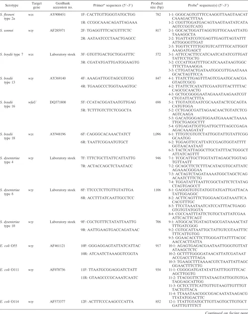
[image:2.585.56.539.407.726.2]TABLE 3. Multiplex PCR primers and oligonucleotide probes used in the study
Serotype Target gene
GenBank
accession no. Primer
asequences (5⬘–3⬘) Product
size (bp) Probe
bsequence(s) (5⬘–3⬘)
S. flexneri type 2a
wzx AY900451 1F: CACTTGTTGGGTATGCTGG 782 1-1: GGGCAGTGTTTCCAAGGTTAAGTAACAT
CAAAGACTTTAA
1R: CCGGCAAACAGATTAGAAA 1-2: CGGTTGGATGACAGTAAGTAATATCATA
AGTCCGGTCATG
S. sonnei wzy AF285971 2F: TGAGGTTTCACGTTTCTC 817 2-1: GGCACTGGATTAGGTGTTGCAAATTATG TAAAGGCTA
2R: AATAATCCCTAACTGAGCC 2-2: TGATTATCGTCGAGTTGAGTTAGTATTT
ATTGGGGTTGAT
2-3: TGGTTCTTTTGGTGTCATTTTGCATTGGT AAAGATGAGCT
S. boydiitype 7 wzx Laboratory stock 3F: GTGTTGACTGCTGGATTTC 572 3-1: ATTCCACTTCCATCAATCATATCGTTGAT TATTCCTGCTG
3R: CGATATGATTGATGGAAGTG 3-2: CCCATTGATTTTGCATCAAATAAGTGGC
TTTCTTAAAGGA
3-3: CTTGATACTGATAATGGCGTTGAATAAA GCACTAGTTCCA
S. boydii type 13
wzy AY369140 4F: AAAGATTGGTAGCGTCGG 847 4-1: TTATCTTGAGTTTAGTCGAATGCAACGA
GTAGTCGCG
4R: TGAAGCCCTGGTAAAGTGC 4-2: TTATTCTCATATTCGAATGTTACTTTTAC
CAGCGCAACTG
4-3: GCTGCGGGGAATAGAAATAAGAATCGT CTGTATTACTTTA
S. boydii type 16
wfaU DQ371800 5F: CCATACGGATAATGTTGAG 698 5-1: TTGTATGTAATCGCAAATACTCGCAGTA
CATGTGGA
5R: TCTTTGTCTTCTCGGCTA 5-2: CCTGAGCGATTAGAACAACTGTATCTCG
AGTCAAGA
5-3: GACATGGGAGTGGAATGAAAACTAAAA TTGCTGAGGCTTT
5-4: GTGAGATTGTTGATTGCTTTAGCCGAGA AGACAAAGATAT
S. boydii type 18
wzx AY948196 6F: CAGGGCACAAACTATCT 1,096 6-1: TTTGTCGTGTCTATTGGTATTGTATTCGG
GCAATGG
6R: TAATTCGGAATGTGCT 6-2: TGGAGTTCCATTATCCGAGTGGTATTTT
GGTAACAATAAT
6-3: TACTCATTAGGCTTGCTATTTACTGGGCT ATTATCAGTTT
S. dysenteriae type 4
wzy Laboratory stock 7F: TTTCTGCTTATTCATTATTG 946 7-1: TCGCATTGCTTGGTATTAGAGCTGGTAG
TGTTAATT
7R: ACTACCAGCTCTAATACC 7-2: GCAGCTTCTCTTTACATACGTGCATTATC
AGAAACGGGAA
7-3: ACTAGTCTAGATAAAATGGCTAGCTCAG ACAATCTTTCTG
7-4: TGGATATTTAATTCGGCTATTCTCTATAG CTAGTGAGCCT
S. dysenteriae type 8
wzy Laboratory stock 8F: TTCCCTCTTGTTGTATTGA 1106 8-1: GAGGGTGTGTATGGTATGATTGATTACA
TATTGGAGGC
8R: ACCTTTATCAATTGCCTCC 8-2: ACTTCAGTTTCTGGGAACGATAAATTCA
CACGTTTGC
8-3: TTCCTAAATAATCATCCATTTACTGAGG GTGTGTATGGTA
8-4: CGCCAATTTATTCTGTGCTATTATCGAA ATTCACTTCAGT
S. dysenteriae type 10
wzy Laboratory stock 9F: CGCTGTTTCTATATTAATTG 706 9-1: ATGGCACTGATAGTAGCGATAAAACTAT
TTTGATCGGG
9R: AATTGAAGTGACCAGATAAC 9-2: CGTGCATTAATTGCTATTGTCGTAATTTC
TTTCATTGTGG
9-3: GGAACACCTTCTTGGGATTATTTTACGC AACCACTTATTA
E. coliO55 wzy AF461121 10F: GGGAGGAGTATTATCATTAC 917 10-1: AGAGTGAGACGAATAATTGGGTGTTAT ATAAGCTCTC
10R: ATCAATCTAAAGGTCGGTA 10-2: GCTTTTGGGGATAACATTATCGATAAT
ACCGACCTTTAGA
10-3: TGAAGCTTTAAAACGTCTAATTATTAGC GGAACTTTCTTG
E. coliO111 wzy AF078736 11F: TTAATGCGGAGGATCTATT 934 11-1: CGGGGATGATATATTATTTGGTTTCAC AGCTTGGTG
11R: GTAAGCCCGCAAATCAATC 11-2: TTACGGTTCTTTATAAGTATTGGTGTGA
TAGGAGCATTGG
11-3: GCTCCTTTCATTGTTGTAAGTTGTTTGT TACTGTTACA
11-4: TTAAATAACGGCGGACAATATAAGACG TTATATGGACTTC
E. coliO114 wzy AF573377 12F: ACTTTCCCAAGCCCATTA 852 12-1: TTATTGTATGCTTGTTAGTGCTTGTGCT GATTTGTTTTCT
Continued on facing page
on May 16, 2020 by guest
http://jcm.asm.org/
(QIAGEN GmbH, Hilden, Germany). For each strain, at least 10 extractions were repeated to verify the reproducibility of the DNA extraction method.
Identification of O-serotype-specific genes by PCR. Genomic DNAs were prepared from 186 representative strains of allShigellaandE. coliO serotypes and examined for quality by PCR amplification of themdhgene coding for malate dehydrogenase, as described previously (30). A total of 13 pools of DNA were made, each containing DNAs from 12 to 19 strains (15). The pools were screened using primers based on thewzxandwzygenes ofE. coliO128;S. sonnei; S. flexneritype 2a;S. boydiitypes 7 and 9; andS. dysenteriaetypes 4, 8, and 10 (Table 2). The PCR cycles used were as follows: 30 cycles of denaturation at 94°C for 2 min, annealing at 45°C for 30 s, and extension at 72°C for 1 min, with a final extension at 72°C for 5 min. An aliquot of 10l of the PCR product was examined on an agarose gel.
Primer design.The sources of the O-antigen gene cluster sequences of the 15 serotypes are listed in Table 3. Based on the O-serotype-specific genes of the 15 serotypes, the 15 compatible primer pairs in a multiplex PCR were designed: 10 primer pairs for the 10 serotypes ofShigellaand 5 primer pairs for the 5 serotypes of pathogenicE. coli(Table 3). There was also one primer pair for amplifying the 16S rRNA genes ofShigellaandE. coli. All 16 primer pairs were contained in one multiplex PCR.
Multiplex PCR and labeling of the target genes.Each multiplex PCR ampli-fication was performed with 30l of reaction mixture consisting of 50 to 100 ng of DNA; 1⫻PCR buffer (50 mM KCl, 10 mM Tris-HCl [pH 8.3]), 0.5 mM MgCl2, 100M concentration of deoxynucleoside triphosphate, 1.5 UTaqDNA
polymerase, 0.047M each of two primers based on the 16S rRNA gene se-quence, 0.14M each of primers based on each target gene, and 0.15 nM cyanine dye Cy3-dUTP (Amersham Biosciences UK Ltd., Little Chalfont, England). The reaction parameters were 94°C for 5 min; 35 cycles of 94°C for 30 s, 50°C for 1 min, and 72°C for 1 min; and a final extension at 72°C for 5 min. An aliquot of 2l of PCR product was run on an agarose gel to check the amplified DNA, and the rest was stored at⫺20°C in the dark.
Oligonucleotide probe design.For each serotype, two to four probes were designed with OligoArray 2.0 based on GenBank and an in-house database of all 33 of the O-antigen gene clusters ofShigellaand 175 O-antigen gene clusters of E. coli. Two probes based on the 16S rRNA genes of all knownShigellaandE. colistrains were designed as positive controls. A probe containing 40 poly(T) oligonucleotides was used as the negative control. A probe labeled with 3⬘Cy3
was used as the positional reference and printing control. Each probe was 5⬘
amino modified. All of the oligonucleotide probes are listed in Table 3.
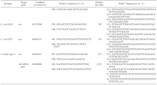
DNA array preparation.The probes were dissolved in 50% dimethyl sulfoxide at a final concentration of 1g/l and printed onto aldehyde group-modified glass slides (CEL Corporation) using SpotArray72 (Perkin-Elmer Corporation). Each probe was spotted in triplicate. The printed slides were dried for 24 h at room temperature, cross-linked by UV cross-linker (UVP Corporation), and stored at room temperature in the dark. Each slide consisted of four microarrays framed with a 12-l Geneframe (Beijing Capital Biochip Corporation, Beijing, China), which constituted individual reaction chambers. One of the four microarrays was tested with the positive control standard, 100 ng/lS. dys-enteriaetype 8 genomic DNA, to ensure that the reagents were effective. Another was tested with the negative control standard, sterile deionized water, to show that the reagents were uncontaminated. The other two were used to detect samples. A schematic diagram of the probe positions on the microarray is shown in Fig. 1.
Hybridization process.An aliquot of 15l of labeled PCR product was baked for about 1.5 h at 65°C until it was dry and diluted in 13l of hybridization buffer (25% formamide, 0.1% sodium dodecyl sulfate, 6⫻SSPE [1⫻SSPE is 0.18 M NaCl, 10 mM NaH2PO4, and 1 mM EDTA {pH 7.7}]). After denaturation at
98°C for 5 min, an aliquot of 12l of labeled target DNA was hybridized with the probes at 40°C for 16 h. After hybridization, the Geneframe was removed and the slide was washed with solution A (1⫻SSC [1⫻SSC is 0.15 M NaCl plus 0.015 M sodium citrate], 0.1% sodium dodecyl sulfate) for 3 min, followed by solution B (0.05⫻SSC) for 3 min, and finally by solution C (95% ethanol) for 1.5 min. The slide was dried under a gentle air stream before it was scanned. For each DNA, at least three hybridization reactions were replicated to verify the reproducibility of the microarray method.
[image:4.585.41.543.80.353.2]Data acquisition and automated analysis.The slide was scanned with a laser beam at 532 nm using the GenePix personal 4100A (Axon Instruments), and two files were generated, one for the images, saved as TIF, and the other for the signal intensity, saved as GPR. The signal-to-noise ratio was calculated for each spot using the Bactarray Analyzer 1.0, developed in-house, with the threshold set at 3.0. For each serotype, two to four probes were used, and each probe was printed in triplicate to eliminate any possible physical defects in the glass slide. A serotype was confirmed and reported when the following conditions existed: (i) the positive standard, the negative standard, the two positive control probes, the
TABLE 3—
Continued
Serotype Target gene
GenBank
accession no. Primer
asequences (5⬘–3⬘) Product
size (bp) Probe
bsequence(s) (5⬘–3⬘)
12R: CAGCACAAGCACTAACAAG 12-2: TGAGATGCTTAAATTAGGTGGATGGAA
TGTTAATGGG
12-3: GTTGTTCATATGCTCAGGGGAAAATCA GAAGAGTATCTAT
12-4: TGGATGGAATGTTAATGGGTTATTTAT TTCAGAAGCATG
E. coliO128 wzy AY217096 13F: ATGATTTCTTACGGAGTGC 782 13-1: TCTGATCTTGGATTAAGTAAGATGTAC CCAGCAA
13R: CTCTAACCTAATCCCTCCC 13-2: CGGTGTTTTGCAAGAGATATAAAAGAG
TTAGCTTTAGCAT
13-3: GCTAGGTATTTAGCAAATTCAACAGAT TTGGCTGACTTTG
E. coliO157 wzy AF061251 14F: TTGCTGCTGTAGTTTTATTTCTT 555 14-1: CGATTTCTTTCCGACACCAGAGTTAGA AAAGGAATT
14R: TGATGCTTTATTCCCTGTA TTCT
14-2: TAGAGCAAGTTGAAAGTGTTCCATATG TTGTTTCTGAATC
14-3: GTATGCTCGTTGTTTTATCTAAGTTTAG GACAAGACGGAG
S. boydiitype 9 wzy AF402315 15F: GCGTTGGTTGGTGAAAGAG 877 15-1: AACTGAGTTCACTTATGGTTCGAGAAC CTTTACTCCATTT
15R: TTCCCACAAATCAAACCA 15-2: GGATTTTAATACAACTGAGTTCACTTAT
GGTTCGAGAACC 16S rRNA
gene
AE000406 16F: GACGGGTGAGTAATGTCTGG 1,251 16-1: CGGGAACTCAAAGGAGACTGCCAGTG
ATAA
16R: ATCCACGATTACTAGCGATTCC 16-2: CGGGAACTCAAAGGAGACTGCCAGTG
ATAAACTGGAG
17: TTTTTTTTTTTTTTTTTTTTTTTTTTTTTTTT TTTTTTTT
18: TTTTTTTTTTTTTTTTTTTTTTTTTTTTTTTT TTTTTTT_Cy3
aF, forward primer; R, reverse primer.
b16-1 and 16-2, positive control probes; 17, negative control probe; 18, positional reference and printing control probe.
on May 16, 2020 by guest
http://jcm.asm.org/
negative control probe, and the printing control probe all provided the expected signals and (ii) more than half of all the probes of the given serotype generated positive signals above the signal-to-noise ratio threshold.
RESULTS
Identification of specific genes.
The O-serotype-specific
genes of each of the
S. boydii
types 13, 16, and 18 and
E. coli
O55, O111, O114, and O157 have been reported previously (7,
12, 13, 15, 22, 33, 36). In this study, we identified the specific
genes of the other eight serotypes:
S. sonnei
;
S. flexneri
type 2a;
S. boydii
types 7 and 9;
S. dysenteriae
types 4, 8, and 10; and
E.
coli
O128. For each of them, two primer pairs based on each of
the
wzx
and
wzy
genes were designed (Table 2) and used to
screen DNA pools containing 186 representative strains of all
Shigella
and
E. coli
O serotypes. With primer pairs of
S. flexneri
type 2a, only the pools containing
E. coli
O13, O129, and O135,
which shared the same O antigen with
S. flexneri
type 2a (9), in
addition to
S. flexneri
2a, gave PCR products of the expected
size. With the other primer pairs, only the bands of correct size
were observed in the pool containing the target strains.
There-fore, both the
wzx
and
wzy
genes were specific to the eight
serotypes. For each serotype, one of the O-serotype-specific
genes was chosen as a target gene to identify
Shigella
and
E.
coli
(Table 3).
Multiplex PCR to amplify the target genes.
Multiplex PCR
was used to streamline the overall test while maintaining
spec-ificity for individual amplicons. In the presence of one target
strain, only the 16S rRNA gene primer pair and one of the
serotype-specific primer pairs performed, while none of the
other 14 specific primer pairs annealed with the template
DNA. Therefore, for each of the 15 serotype strains, two bands
were generated from the PCRs: one was the 16S rRNA gene,
and the other was the specific gene (Fig. 2). The amplicon size
ranged from 555 bp to about 1.25 kb in length (Table 3). The
Shigella
and
E. coli
representative strains belonging to other
serotypes (except
E. coli
O13, O129, and O135 [see below]),
S.
enterica
, and
V
.
cholerae
generated only the 16S rRNA gene
products. Strains of
B. cereus
and
S. aureus
failed to generate
any amplicon. The results showed that the 15 primer pairs were
specific and compatible in one multiplex PCR.
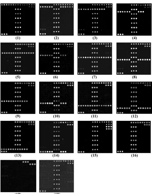
Probe specificity.
The DNA microarray was tested using 186
representative strains of all
Shigella
and
E. coli
O serotypes, 9
strains of other bacterial species (Table 1), and 40 stool
spec-imens. From 141 oligonucleotide probes initially screened, 52
FIG. 1. Probe positions on the slide. Numbers 1-1 to 15-2 are the specific probes for the target strains. Numbers 16-1 and 16-2 are the positive
control probes based on the 16S rRNA genes of all
Shigella
and
E. coli
strains. Number 17 is the negative control probe. Number 18 is the positional
reference and printing control probe.
FIG. 2. Agrose gel electrophoresis of multiplex PCR products. Lanes: Mr, molecular weight standards (lambda DNA/EcoRI plus HindIII
marker); A,
S. boydii
type 7; B,
S. boydii
type 9; C,
S. boydii
type 13; D,
S. boydii
type 16; E,
S. boydii
type 18; F,
S. dysenteriae
type 4; G,
S. dysenteriae
type 8; H,
S. dysenteriae
type 10; I,
S. flexneri
type 2a; J,
S. sonnei
; K,
E. coli
O55; L,
E. coli
O111; M,
E. coli
O114; N,
E. coli
O128; O,
E. coli
O157.
Two bands were generated from the PCR products: one was the 16S rRNA gene (1.2 kb), and the other was the gene specific to the individual
serotype.
on May 16, 2020 by guest
http://jcm.asm.org/
FIG. 3. Microarray differentiation of the pathogens. (1)
S. flexneri
type 2a and
E. coli
O13, O129, and O135. (2)
S. sonnei
. (3)
S. boydii
type 7.
(4)
S. boydii
type 13. (5)
S. boydii
type 16. (6)
S. boydii
type 18. (7)
S. dysenteriae
type 4. (8)
S. dysenteriae
type 8. (9)
S. dysenteriae
type 10. (10)
E. coli
O55. (11)
E. coli
O111. (12)
E. coli
O114. (13)
E. coli
O128. (14)
E. coli
O157. (15)
S. boydii
type 9. (16) Other serotype strains of
E. coli
or
Shigella
,
Salmonella
, and
V
.
cholerae
. (17)
B
.
cereus
and
S
.
aureus
. (18) DNAs from healthy-adult stool specimens.
4381
on May 16, 2020 by guest
probes were selected for the microarray: 48 probes for specific
genes, 2 probes for positive control, 1 for negative control, and
1 for positional reference and printing control (Table 3). All of
the representative strains belonging to the 15 serotypes
con-sistently hybridized to their corresponding probes with 100%
specificity. The hybridization results are shown in Fig. 3, arrays
1 to 15. Strains of
E. coli
O13, O129, and O135 gave the same
results as
S. flexneri
type 2a (Fig. 3, array 1), because they had
same O antigen (9). None of the 23 representative strains of
Shigella
and the 148 representative strains of
E. coli
belonging
to other serotypes, or strains of other bacterial species which
are likely present in stool samples, hybridized to the
serotype-specific probes on the microarray (Fig. 3, arrays 16 and 17).
The 40 stool specimens obtained from 20 adult volunteers
reacted only to the 16S rRNA gene probes, not the
serotype-specific probes (Fig. 3, array 18), a result that was consistent
with the fact that large numbers of nonpathogenic
E. coli
organisms exist in the stools of healthy people.
Double-blind test.
A double-blind test was performed in
order to verify the stability and specificity of the microarray. A
total of 38 clinical isolates of
Shigella
and
E. coli
(Table 1) and
70 mock stool samples were selected to hybridize to the
mi-croarray without disclosure of their identities during testing.
All the detection results were consistent with those of the
conventional methods.
Sensitivity of detection with genomic DNA.
Serial dilutions
of the genomic DNAs of 15 serotype representative strains in
the range of 1
g, 100 ng, 50 ng, 10 ng, and 1 ng were used as
the templates for multiplex PCR to test the sensitivity of the
microarray method. The positive signals could be obtained
from the dilutions of 1
g to 10 ng, while the results were
negative or the fluorescence signals were very weak with less
than 10 ng. We chose 50 ng as the most suitable DNA quantity
for this microarray, and all of the 38 clinical isolates belonging
to the 15 serotypes could be detected successfully at this level.
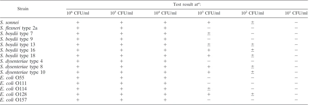
Sensitivity of detection with mock stool specimens.
Pure
cultures of each of the 15 serotype representative strains were
diluted to 10
1to 10
6CFU per ml, mixed with approximately
0.3 g of fresh stool specimens from healthy people, and tested
with the microarray. All of the targets were detected at levels
as low as 10
4CFU per ml. Some strains, such as
E. coli
O128,
S. sonnei
,
S. boydii
types 16 and 18, and
S. dysenteriae
types 8
and 10, could be detected successfully at 10
3CFU per ml
(Table 4).
DISCUSSION
Systematic O serotyping of
E. coli
began in the early 1930s
(23), and many studies showed that the O serotypes of
E. coli
are generally associated with pathogenesis (6, 17, 26, 33, 36). O
serotyping became an important tool to classify
E. coli
in
clin-ical settings. In recent years, some microarray and PCR assays
have used toxin genes as targets (20, 23, 25) to identify
patho-genic
E. coli
. However, since mutations, instability, and loss of
toxin genes among Shiga-like toxin-producing
E. coli
are quite
common (20, 25), it is not reliable to use the toxin genes, even
if a large number of primer pairs are incorporated into the test.
wzx
,
wzy
, and glycosyltransferase genes are generally specific to
individual O-antigen gene clusters. We previously suggested
the application of O-serotype-specific genes for detection and
identification of
E. coli
(14, 30). Here, we demonstrated the
feasibility of using a microarray based on
wzx
,
wzy
, or
trans-ferase genes to identify pathogens from pure culture and more
clinically relevant mock stool samples. The DNA microarray
described in this communication has paved the way for the
establishment of an array of all serotypes of
Shigella
and
E. coli
that we are currently working on.
In comparison to the traditional serotyping method, the
microarray method is high throughput, specific, and sensitive
and also avoids most cross-reactions. Nevertheless, as with
traditional serotyping, the microarray has its limits in
distin-guishing
S. sonnei
and
S. flexneri
type 2a from other strains that
share the same O-antigen structure and the corresponding
O-antigen gene cluster. Among the 46
Shigella
serotypes
rec-ognized, there are only 33 distinct O antigens. Of the 33
O-antigen forms, 12 are identical to some
E. coli
O antigens (35).
When
E. coli
O13, O129, or O135 existed in stool samples, the
[image:7.585.43.543.81.252.2]probes for
S. flexneri
type 2a would have been expected to give
positive signals. Similarly, the
S. sonnei
O-antigen gene cluster
is identical in sequence to that of
Plesiomonas shigelloides
O17
TABLE 4. Sensitivity test results for mock stool specimens
Strain
Test result ata:
106CFU/ml 105CFU/ml 104CFU/ml 103CFU/ml 102CFU/ml 101CFU/ml
S. sonnei
⫹
⫹
⫹
⫹
⫾
⫺
S. flexneri
type 2a
⫹
⫹
⫹
⫺
⫺
⫺
S. boydii
type 7
⫹
⫹
⫹
⫾
⫺
⫺
S. boydii
type 9
⫹
⫹
⫹
⫺
⫺
⫺
S. boydii
type 13
⫹
⫹
⫹
⫾
⫾
⫺
S. boydii
type 16
⫹
⫹
⫹
⫹
⫾
⫺
S. boydii
type 18
⫹
⫹
⫹
⫹
⫾
⫺
S. dysenteriae
type 4
⫹
⫹
⫹
⫺
⫺
⫺
S. dysenteriae
type 8
⫹
⫹
⫹
⫹
⫾
⫺
S. dysenteriae
type 10
⫹
⫹
⫹
⫹
⫾
⫺
E. coli
O55
⫹
⫹
⫹
⫺
⫺
⫺
E. coli
O111
⫹
⫹
⫹
⫺
⫺
⫺
E. coli
O114
⫹
⫹
⫹
⫾
⫺
⫺
E. coli
O128
⫹
⫹
⫹
⫹
⫾
⫺
E. coli
O157
⫹
⫹
⫹
⫺
⫺
⫺
a⫹, positive signal;⫺, negative signal;⫾, weak or ambiguous signal.
on May 16, 2020 by guest
http://jcm.asm.org/
(21). We therefore assumed that when
P. shigelloides
existed in
stool, the probes for
S. sonnei
would also give false-positive
results. Nevertheless, we will try to use other methods or other
genes, such as
lacZ
or
cadA
(31), which are commonly present
in
E. coli
but absent in
Shigella
, to differentiate these potential
false positives.
The microarray has many advantages over traditional
bac-terial culture and serotyping methods and is applicable to
many fields. First, it facilitates medical detection of
Shigella
and
E. coli
from stool samples in a timely fashion. Second, it
allows efficient inspection for food contamination. Third, it will
be an invaluable tool to sensitively monitor and accurately
pinpoint causative bacterial strains to prevent the occurrence
or spread of epidemics of bacterial infection.
ACKNOWLEDGMENTS
This work was supported by the NSFC Programs (30370023,
30370339, and 30530010), the Tianjin Municipal Special Fund for
Science and Technology Innovation (05FZZDSH00800), and funds
from the Tianjin Municipal Science and Technology Committee
(06YFJZJC02200) to L.W. and L.F.
REFERENCES
1.Bastin, D. A., and P. R. Reeves.1995. Sequence and analysis of the O antigen gene (rfb) cluster ofEscherichia coliO111. Gene164:17–23.
2.Beutin, L., Q. Kong, L. Feng, Q. Wang, G. Krause, L. Leomil, Q. Jin, and L. Wang.2005. Development of PCR assays targeting the genes involved in synthesis and assembly of the newEscherichia coliO174 and O177 O anti-gens. J. Clin. Microbiol.43:5143–5149.
3.Beutin, L., J. Tao, L. Feng, G. Krause, S. Zimmermann, K. Gleier, Q. Xia, and L. Wang.2005. Sequence analysis of theEscherichia coliO15 antigen gene cluster and development of a PCR assay for rapid detection of intes-tinal and extraintesintes-tinal pathogenicE. coliO15 strains. J. Clin. Microbiol.
43:703–710.
4.Bopp, C. A., F. W. Brenner, P. I. Fields, J. G. Wells, and N. A. Strockbine.
2003.Escherichia,Shigella, andSalmonella, p. 654–671.InP. R. Murray, E. J. Baron, J. H. Jorgensen, M. A. Pfaller, and R. H. Yolken (ed.), Manual of clinical microbiology, 8th ed. American Society for Microbiology, Washing-ton, D.C.
5.Call, D. R., F. J. Brockman, and D. P. Chandler. 2001. Detecting and genotypingEscherichia coliO157:H7 using multiplexed PCR and nucleic acid microarrays. Int. J. Food. Microbiol.67:71–80.
6.Coimbra, R. S., F. Grimont, P. Lenormand, P. Burguiere, L. Beutin, and P. A. Grimont.2000. Identification ofEscherichia coliO-serogroups by re-striction of the amplified O-antigen gene cluster (rfb-RFLP). Res. Microbiol.
151:639–654.
7.DebRoy, C., P. M. Fratamico, E. Roberts, M. A. Davis, and Y. Liu.2005. Development of PCR assays targeting genes in O-antigen gene clusters for detection and identification ofEscherichia coliO45 and O55 serogroups. Appl. Environ. Microbiol.71:4919–4924.
8.DebRoy, C., E. Roberts, J. Kundrat, M. A. Davis, C. E. Briggs, and P. M. Fratamico.2004. Detection ofEscherichia coliserogroups O26 and O113 by PCR amplification of thewzxandwzy genes. Appl. Environ. Microbiol.
70:1830–1832.
9.Ewing, W. H.1986. Edwards and Ewing’s identification of the Enterobacte-riaceae, 4th ed. Elsevier Science Publishers, Amsterdam, The Netherlands. 10.Feng, L., W. Han, Q. Wang, D. A. Bastin, and L. Wang.2005. Characteriza-tion ofEscherichia coliO86 O-antigen gene cluster and identification of O86-specific genes. Vet. Microbiol.106:241–248.
11.Feng, L., S. N. Senchenkova, J. Tao, A. S. Shashkov, B. Liu, S. D. Shevelev, P. R. Reeves, J. Xu, Y. A. Knirel, and L. Wang.2005. Structural and genetic characterization of enterohemorrhagicEscherichia coliO145 O antigen and development of an O145 serogroup-specific PCR assay. J. Bacteriol.187:
758–764.
12.Feng, L., S. N. Senchenkova, W. Wang, A. S. Shashkov, B. Liu, S. D. Shevelev, D. Liu, Y. A. Knirel, and L. Wang.2005. Structural and genetic characterization of theShigella boydiitype 18 O antigen. Gene355:79–86. 13.Feng, L., S. N. Senchenkova, J. Yang, A. S. Shashkov, J. Tao, H. Guo, G.
Zhao, Y. A. Knirel, P. Reeves, and L. Wang.2004. Structural and genetic characterization of theShigella boydiitype 13 O antigen. J. Bacteriol.186:
383–392.
14.Feng, L., J. Tao, H. Guo, J. Xu, Y. Li, F. Rezwan, P. Reeves, and L. Wang.
2004. Structure of theShigella dysenteriae7 O antigen gene cluster and identification of its antigen specific genes. Microb. Pathog.36:109–115. 15.Feng, L., W. Wang, J. Tao, H. Guo, G. Krause, L. Beutin, and L. Wang.2004.
Identification ofEscherichia coliO114 O-antigen gene cluster and develop-ment of an O114 serogroup-specific PCR assay. J. Clin. Microbiol.42:3799– 3804.
16.Fratamico, P. M., C. E. Briggs, D. Needle, C. Y. Chen, and C. DebRoy.2003. Sequence of theEscherichia coliO121 O-antigen gene cluster and detection of enterohemorrhagicE. coliO121 by PCR amplification of thewzxandwzy genes. J. Clin. Microbiol.41:3379–3383.
17.Gemski, P. J., D. G. Sheahan, O. Washington, and S. B. Formal.1972. Virulence ofShigella flexneri hybrids expressingEscherichia colisomatic antigens. Infect. Immun.6:104–111.
18.Guo, H., L. Feng, J. Tao, C. Zhang, and L. Wang.2004. Identification of Escherichia coliO172 O-antigen gene cluster and development of a sero-group-specific PCR assay. J. Appl. Microbiol.97:181–190.
19.Guo, H., Q. Kong, J. Cheng, L. Wang, and L. Feng.2005. Characterization of theEscherichia coliO59 and O155 O-antigen gene clusters: the atypical wzxgenes are evolutionary related. FEMS Microbiol. Lett.248:153–161. 20.Karch, H., T. Meyer, H. Russmann, and J. Heesemann.1992. Frequent loss
of Shiga-like toxin genes in clinical isolates ofEscherichia coliupon subcul-tivation. Infect. Immun.60:3464–3467.
21.Lai, V., L. Wang, and P. R. Reeves.1998.Escherichia coliclone Sonnei (Shigella sonnei) had a chromosomal O-antigen gene cluster prior to gaining its current plasmid-borne O-antigen genes. J. Bacteriol.180:2983–2986. 22.Liu, B., S. N. Senchenkova, L. Feng, A. V. Perepelov, T. Xu, S. D. Shevelev,
Y. Zhu, A. S. Shashkov, M. Zou, Y. A. Knirel, and L. Wang.Structural and molecular characterization ofShigella boydiitype 16 O antigen. Gene380:
46–53.
23.Nataro, J. P., and J. B. Kaper.1998. DiarrheagenicEscherichia coli. Clin. Microbiol. Rev.11:142–201.
24.Niyogi, S. K.2005. Shigellosis. J. Microbiol.43:133–143.
25.Parreira, V. R., and C. L. Gyles.2002. Shiga toxin genes in avianEscherichia coli. Vet. Microbiol.87:341–352.
26.Pluschke, G., J. Mayden, M. Achtman, and R. P. Levine.1983. Role of the capsule and the O-antigen in resistance of O18:K1Escherichia colito com-plement-mediated killing. J. Bacteriol.42:907–913.
27.Pupo, G. M., R. Lan, and P. R. Reeves.2000. Multiple independent origins ofShigellaclones ofEscherichia coliand convergent evolution of many of their characteristics. Proc. Natl. Acad. Sci. USA97:10567–10572. 28.Scheutz, F., T. Cheasty, D. Woodward, and H. R. Smith.2004. Designation
of O174 and O175 to temporary O groups OX3 and OX7, and six newE. coli O groups that include verocytotoxin-producingE. coli(VTEC): O176, O177, O178, O179, O180 and O181. APMIS112:569–584.
29.Senchenkova, S. N., L. Feng, J. Yang, A. S. Shashkov, J. Cheng, D. Liu, Y. A. Knirel, P. R. Reeves, Q. Jin, Q. Ye, and L. Wang.2005. Structural and genetic characterization of theShigella boydiitype 10 and type 6 O antigens. J. Bacteriol.187:2551–2554.
30.Tao, J., L. Feng, H. Guo, Y. Li, and L. Wang.2004. The O-antigen gene cluster ofShigella boydiiO11 and functional identification of itswzygene. FEMS. Microbiol. Lett.234:125–132.
31.Tao, J., L. Wang, D. Liu, Y. Li, D. A. Bastin, Y. Geng, and L. Feng.2005. Molecular analysis ofShigella boydiiO1 O-antigen gene cluster and its PCR typing. Can. J. Microbiol.51:387–392.
32.van Ijperen, C., P. Kuhnert, J. Frey, and J. P. Clewley.2002. Virulence typing ofEscherichia coliusing microarrays. Mol. Cell Probes16:371–378. 33.Wang, L., H. Curd, W. Qu, and P. R. Reeves.1998. Sequencing ofEscherichia
coliO111 O-antigen gene cluster and identification of O111-specific genes. J. Clin. Microbiol.36:3182–3187.
34.Wang, L., B. Liu, Q. Kong, H. Steinruck, G. Krause, L. Beutin, and L. Feng.
2005. Molecular markers for detection of pathogenicEscherichia colistrains belonging to serogroups O138 and O139. Vet. Microbiol.111:181–190. 35.Wang, L., W. Qu, and P. R. Reeves.2001. Sequence analysis of fourShigella
boydiiO-antigen loci: implication forEscherichia coliandShigella relation-ships. Infect. Immun.69:6923–6930.
36.Wang, L., and P. R. Reeves.1998. Organization ofEscherichia coliO157 O antigen gene cluster and identification of its specific genes. Infect. Immun.
66:3545–3551.
37.Yue, J., W. Shi, J. Xie, Y. Li, E. Zeng, L. Liang, and H. Wang.2004. Detection of rifampin-resistant Mycobacterium tuberculosis strains by using a specialized oligonucleotide microarray. Diagn. Microbiol. Infect. Dis.48:47–54.