INTRODUCTION
The mortality rate of women with cessation of ovarian hor-mones, such as in menopause and with bilateral oophorecto-my, increases due to cardiovascular diseases, such as coronary heart disease and stroke.1,2 As weight gain, a cardiovascular
risk factor, is commonly followed by menopausal transition or
bilateral oophorectomy, minimizing weight gain through diet and exercise is emphasized for reducing mortality.3,4
Ascorbic acid (vitamin C) is a cofactor in several enzymatic processes related to collagen, carnitine, and neurotransmitter synthesis, and its deficit results in scurvy.5 Collagen synthesis
by ascorbic acid affects adipogenesis. Ascorbic acid increases the synthesis of type I and IV collagen and differential expres-sion of type VI collagen in 3T3-L1 preadipocytes by enhancing differentiation of 3T3-L1 cells.6,7 Another well-known action
of ascorbic acid is its antioxidant effect as an electron donor protecting against oxidative stress.8 Ascorbic acid has been
re-ported to prevent osteoporosis and hip fracture through an antioxidant effect in postmenopausal women and ovariecto-mized mice.9-11
Although the effect of ascorbic acid on weight gain in post-menopausal women and bilateral oophorectomized patients has not been reported yet, there are several papers suggesting Received: April 4, 2017 Revised: August 21, 2017
Accepted: September 14, 2017
Corresponding author: Dr. Min-Goo Lee, Department of Physiology, Korea Uni-versity College of Medicine, 73 Inchon-ro, Seongbuk-gu, Seoul 02841, Korea. Tel: 82-2-2286-1194, Fax: 82-2-925-5492, E-mail: [email protected] •The authors have no financial conflicts of interest.
© Copyright: Yonsei University College of Medicine 2018
This is an Open Access article distributed under the terms of the Creative Com-mons Attribution Non-Commercial License (http://creativecomCom-mons.org/licenses/ by-nc/4.0) which permits unrestricted non-commercial use, distribution, and repro-duction in any medium, provided the original work is properly cited.
Adipogenic and Lipolytic Effects of Ascorbic Acid
in Ovariectomized Rats
Byoungjae Kim
1,2, Kyung Min Choi
1, Hong Soon Yim
1, Hyun Tae Park
3, Joung Han Yim
4, and Min-Goo Lee
1,21Department of Physiology, Korea University College of Medicine, Seoul; 2Neuroscience Research Institute, Korea University, Seoul;
3Division of Reproductive Endocrinology, Department of Obstetrics and Gynecology, Korea University College of Medicine, Seoul; 4Korea Polar Research Institute, Korea Institute of Ocean Science and Technology, Incheon, Korea.
Purpose: Ascorbic acid has been reported to have an adipogenic effect on 3T3-L1 preadipocytes, while evidence also suggests that ascorbic acid reduces body weight in humans. In this study, we tested the effects of ascorbic acid on adipogenesis and the balance of lipid accumulation in ovariectomized rats, in addition to long-term culture of differentiated 3T3-L1 adipocytes. Materials and Methods: Murine 3T3-L1 fibroblasts and ovariectomized rats were treated with ascorbic acid at various time points. In vitro adipogenesis was analyzed by Oil Red O staining, and in vivo body fat was measured by a body composition analyzer using nuclear magnetic resonance.
Results: When ascorbic acid was applied during an early time point in 3T3-L1 preadipocyte differentiation and after bilateral ovari-ectomy (OVX) in rats, adipogenesis and fat mass gain significantly increased, respectively. However, lipid accumulation in well-differentiated 3T3-L1 adipocytes showed a significant reduction when ascorbic acid was applied after differentiation (10 days af-ter induction). Also, oral ascorbic acid administration 4 weeks afaf-ter OVX in rats significantly reduced both body weight and subcutaneous fat layer. In comparison to the results of ascorbic acid, which is a well-known cofactor for an enzyme of collagen synthesis, and the antioxidant ramalin, a potent antioxidant but not a cofactor, showed only a lipolytic effect in well-differentiated 3T3-L1 adipocytes, not an adipogenic effect.
Conclusion: Taking these results into account, we concluded that ascorbic acid has both an adipogenic effect as a cofactor of an enzymatic process and a lipolytic effect as an antioxidant.
Key Words: Ascorbic acid, obesity, ovariectomy, adipogenesis, lipolysis, 3T3-L1 cell
pISSN: 0513-5796 · eISSN: 1976-2437 Yonsei Med J 2018 Jan;59(1):85-91
that ascorbic acid as a lipolytic agent could be used in the treat-ment and prevention of obesity in human and animal mod-els.12-15 The effect of ascorbic acid on weight gain should be
clarified in post-menopause women and bilateral oophorec-tomy patients, as ascorbic acid has both an enhancing effect on adipogenesis of 3T3-L1 preadipocytes and a lipolytic ef-fect. Therefore, we tested the effect of ascorbic acid on adipo-genesis and lipolysis in the long-term culture of 3T3-L1 cells and ovariectomized rat models. In this study, we showed that application of ascorbic acid during the early period of 3T3-L1 cell differentiation and after ovariectomy (OVX) enhanced adi-pogenesis and body fat mass, respectively, while applying ascorbic acid during the late period decreased lipid accumu-lation and body fat mass.
MATERIALS AND METHODS
In vitro cell culture and differentiation condition Mouse embryo fibroblasts cells (3T3-L1 cells) were purchased from Korean Cell Line Bank. The cells were cultured in Dul-becco’s Modified Eagles Medium (DMEM) supplemented with 10% bovine calf serum (BCS) for maintenance and cul-tured in DMEM with 10% fetal bovine serum (FBS) for chemi-cal adipogenic induction. In order to produce mature adipo-cytes, 6.7×10⁴ cells of 3T3-L1 were seeded on six-well plates
with DMEM-BCS and cultured for 3 days to reach 100% conflu-ence. The day on which cells reached confluency was referred to as Day -2, and they were treated with ascorbic acid (50 μg/ mL) and/or ramalin (10 μg/mL) from this day on. After being grown for 2 more days in BCS media (Day 0), the cells were in-duced to be differentiated to adipocytes for 2 days with DMEM-FBS containing 1 μM dexamethasone and 250 μM 3-isobutyl 1-methyl xanthine. On Day 2, the cells were refreshed with DMEM-FBS containing insulin (10 μg/mL), and the media was replaced with DMEM-FBS every other day. In this study, treatment with ascorbic acid and ramalin was performed dur-ing two separate periods, early induction period of Day -2, Day 0, and Day 2, the late period of Day 10, Day 12, and Day 14, or during both periods. Induction chemicals and ascorbic acid were purchased from Sigma, and ramalin was generously gift-ed from Dr. Joung Han Yim at Korea Polar Research Institute.
Oil Red O staining
To determine the degree of lipid accumulation, the 3T3-L1 cells were stained with Oil Red O (ORO, Sigma, St. Louis, MO, USA) on the 14th day after the induction of differentiation. The cells were fixed with 4% paraformaldehyde overnight and wa-shed with 60% isopropanol. After drying the cells, 0.21% ORO in 60% isopropanol was applied to the cells for 10 min fol-lowed by washing four times with distilled water. Stained ORO was extracted with 100% isopropanol and absorbance was measured at 520 nm. Pictures were taken before extraction.
Immunoblotting
On Day 14, after a PBS wash, the 3T3-L1 cells were extracted with ×5 Laemmli buffer and 5% β-mercaptoethanol and boiled for 10 min. The samples were separated on sodium dodecyl sulfate (SDS)–polyacrylamide gels and transferred to nitrocel-lulose membranes for 1 hr in transfer buffer (25 mM Tris, 192 mM glycine, 0.1% SDS and 20% methanol, pH 8.3). Non-spe-cific binding sites on the membranes were blocked in 5% non-fat dry milk for 90 min at room temperature, and the membrane was blotted with primary antibody at 4°C overnight and sec-ondary antibody for 90 min at room temperature. Blots were visualized using the chemiluminescence kit (Santa Cruz, Dal-las, TX, USA). Primary antibody for collagen was a rabbit poly-clonal antibody LF68 (a generous gift from Dr. Larry Fisher, NI-DCR, NIH, Bethesda, MD, USA) against carboxy-telopeptide of α1(I) collagen and rabbit polyclonal antibodies against
α1(VI) collagen (Santa Cruz). Rabbit polyclonal antibody to CCAAT/enhancer binding protein (CEBP) α and mouse mono-clonal antibody to peroxisome proliferator-activated receptor (PPAR) γ were purchased from Santa Cruz for adipocyte dif-ferentiation regulating protein. Mouse monoclonal antibody to
β-actin (Santa Cruz) was used for loading control.
Immunocytochemistry
3T3-L1 cells were grown on glass coverslips to Day 14 at 37°C in six-well tissue culture plates. After the removal of media, the cells were washed in PBS for 5 min and then fixed in 4% para-formaldehyde overnight. They were then washed twice with PBS for 15 min and blocked with blocking solution (2% nor-mal rabbit serum in PBS) for 30 min, followed by the addition of rabbit polyclonal antibody against collagen type I (Rock-land Immunochemicals Inc., Limerick, PA, USA) in blocking solution (1:200) and incubation for 2 h at room temperature. After rinsing the cells twice with PBS for 5 min at room temper-ature, the cells were treated with biotinylated goat anti-rabbit IgG secondary antibody (Vector Laboratories, Burlington, CA, USA) in PBS (1:400) for 30 min at room temperature. Cover-slips were washed three times with PBS for 10 min, and anti-gen-antibody complexes were detected using an avidin-biotin complex detection system (Vectastain ABC Kit, Vector Labo-ratories). Coverslips were stained with DAB Substrate kit (Vec-tor Labora(Vec-tories), rinsed in water, briefly counter-stained with hematoxylin, and washed again in water. After mounted on glass slides, coverslips were examined with an Olympus BX51 (Olympus Corporation, Tokyo, Japan) microscope. Pictures were captured and controlled in Olympus DP72 and DP2-BSW.
Animals and research design
mid-line abdominal incisions after deep anesthesia (50 mg/kg of ketamine and 250 mg/kg of xylazine), and the remaining ani-mals were subjected to sham operations. The animal experi-ment was approved by the Institutional Animal Care and Use Committee of Korea University (Korea-2016-0025). The rats were then divided into the following five treatment groups: sh-am-operated control rats receiving vehicle (n=8), ovariecto-mized rats receiving vehicle (n=8), ovariectoovariecto-mized rats receiv-ing ascorbic acid at Weeks 5 and 6 (late period) after surgery (n=8), ovariectomized rats receiving ascorbic acid at Week 1 and 2 (early period) after surgery (n=8), and ovariectomized rats receiving ascorbic acid at the early and late periods after surgery (n=8). Ascorbic acid was dissolved in water and given daily by oral injection at a concentration of 1 g/kg. Water was used as the vehicle. Right before and six weeks after OVX, anes-thetized by ketamine (Yuhan Co., Seoul, Korea, 50 mg/kg) and xylazine hydrochloride (Sigma Co., 250 mg/kg), the rats were weighed, and their body fat was measured by a body compo-sition analyzer of a nuclear magnetic resonance system (Mini-spec LF90, Bruker Co., Billerica, MA, USA). Hypodermis in the flank for subcutaneous adipose tissue and visceral fat mass of greater omentum were obtained and fixed with formalin for histological investigation.
Histology
After formalin fixed tissues were embedded in paraffin, 4-μm thick sections were stained with hematoxylin and eosin (H&E). After mounted them on slides using mounting medium (Clari-on mounting medium, Biomeda), the slides were examined with an Olympus BX51 microscope. Pictures were captured and controlled in Olympus DP72 and DP2-BSW.
Statistical analysis
The data were expressed as the mean±standard error of the mean in bar graphs. Two-way analysis of variance was used to compare the experimental and control groups, and Tukey test was used to compare the significance between two groups. Sta-tistical significance was determined as p-value less than 0.05 (*) or 0.01 (**).
RESULTS
Ascorbic acid showed opposite effects on adipogenesis of mouse preadipocytes
In our previous paper,6 3T3-L1 preadipocytes were cultured for
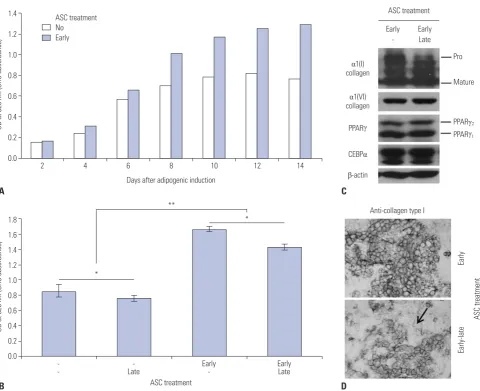
8 days after adipogenic induction, and ascorbic acid increased the accumulation of triglycerides in these cells, especially dur-ing the early period of adipogenesis. In this study, to show the effect of ascorbic acid on long-term culture, 3T3-L1 cells were maintained for up to 14 days for differentiation into mature adi-pocytes. The accumulation of triglycerides in cells sharply in-creased up to 8 days after adipogenic induction. Then, it
sh-owed a plateau without ascorbic acid, but steadily increased with ascorbic acid treatment during the early period (Fig. 1A). However, adding ascorbic acid during the late period of adipo-genesis, with or without ascorbic acid treatment during the early period, interestingly, reduced the ORO staining of triglyc-erides (p<0.05) (Fig. 1B). Although specific markers for lipoly-sis were not examined, this reduction of ascorbic acid during the late period could be regarded as lipolysis of adipocytes, be-cause most 3T3-L1 preadipocytes had differentiated into ma-ture adipocytes by 8 days after adipogenic induction.
In our previous study, we also showed that the differential expression of type I and VI collagen enhanced the accumula-tion of lipid during adipogenesis of 3T3-L1 preadipocytes.6
Thus, to investigate the effect of ascorbic acid as a cofactor of collagen synthesis on this reduction, type I collagen was detect-ed in cell extracts and fixdetect-ed cells. In the immunoblotting of the cell extracts, the expression of mature α1(I) collagen was in-creased by adding ascorbic acid during the late period, whereas procollagen decreased. However, the α1(VI) collagen and ad-ipogenic markers, CEBPα and PPARγ, were similarly expressed, indicating that ascorbic acid could not affect the expression of well-known adipogenic agents during the late period (Fig. 1C). These immunoblotting results suggest that the reduction of lipid in adipocytes may not be induced by the collagen syn-thesis activity of ascorbic acid on early adipogenesis period.
In immunocytochemical examination, the staining pattern of type I collagen in formalin-fixed cells was not different be-tween the two groups, with and without ascorbic acid treat-ment during the late period, while the number of type I collagen unstained cells (arrow) increased in the group that was treat-ed with ascorbic acid during the late period (Fig. 1D). This sug-gests that a weakened cell barrier enabled the loss of lipid drop-lets of adipocytes and explains the reduced triglyceride staining in Fig. 1B.
Antioxidant activity could cause the reduction of adipogenesis
was treated during the early period with ascorbic acid, rama-lin significantly decreased the strong adipogenesis induced by ascorbic acid, which suggests that the antioxidant activity of ramalin might weaken the adipogenesis induced by ascorbic acid through collagen synthesis.
Ascorbic acid reduced in vivo adipogenesis after ovariectomy
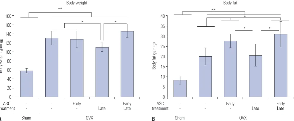
To apply this effect of ascorbic acid on adipogenesis in in vivo experiment, ascorbic acid was injected orally into the ovariec-tomized rat during the early or late period after OVX. At 6 weeks after the removal of both ovaries, the ovariectomized rat gained more body weight and body fat than sham control, confirming that the cessation of ovarian hormones in females induces weight gain through fat accumulation (Fig. 3A and B).
Inter-estingly, when ascorbic acid was injected during 2 weeks of the late period after OVX, total body weight was significantly reduced, compared to other ovariectomized rat groups (Fig. 3A). For the body fat gain among ovariectomized rats, the in-crease of fat upon late injection of ascorbic acid was much less than that for early injections or early-late injections of ascorbic acid and similar to the OVX control (Fig. 3B).
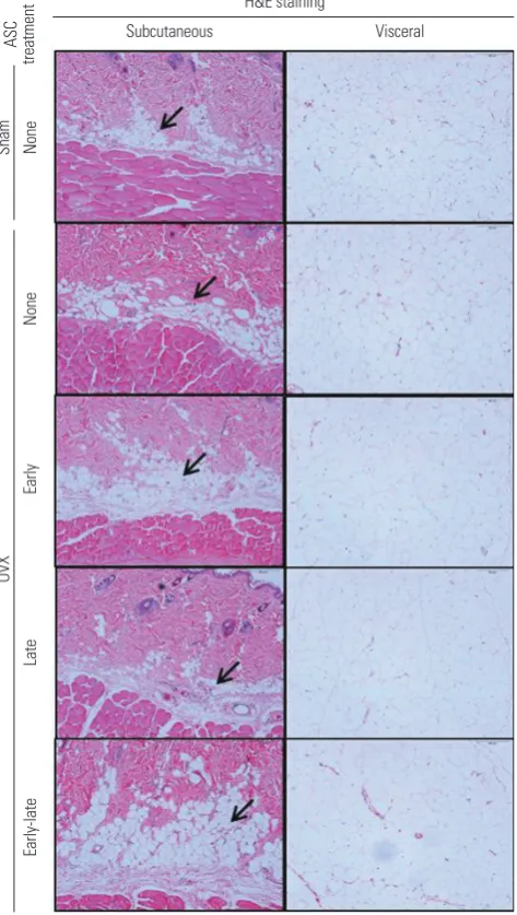
Histologically, the thickness of subcutaneous fat layer in-creased at 6 weeks after OVX (sham vs. OVX none) (Fig. 4). Like the in vitro result, injection of ascorbic acid during the early period after surgery expanded the subcutaneous fat layer, com-pared to sham control or OVX control (OVX early vs. sham or OVX none) (Fig. 4). Intriguingly, the thickness of the subcuta-neous fat layer was reduced to the extent of sham control when ascorbic acid was injected only during the late period after
ASC treatment
ASC treatment
Anti-collagen type I Early
-CEBPα
β-actin
PPARγ PPARγ2
Pro
PPARγ1
Mature
α1(VI)
collagen α1(I) collagen
Early Late
Early-late
[image:4.595.48.529.67.458.2]Early
Fig. 1. Long-term effect of ASC on adipogenesis of 3T3-L1 mouse preadipocytes. (A) Graphical demonstration of long-term adipogenesis with or with-out ASC by ORO staining. Adipogenic induction on Day 0 and ASC treatment during the early period (Day -2, Day 0, and Day 2). (B) Effect of ASC on long-term adipogenesis depending on the treatment period. ORO staining was measured on Day 14. ASC treatment during the early period, the late
period (Day 8, Day 10, and Day 12), or both (n=9). (C) Western blotting analysis of ASC treated adipocytes on Day 14 against collagens, α1(I) and
α1(VI), and adipogenic markers, PPARγ and CEBPα. ASC treatment during the early period or both early and late periods. (D) Immunocytochemical
features of ASC treated adipocytes against type I collagen at Day 14. ASC treatment during the early period, or both early and late periods. Arrow in-dicates the area of unstained cells. *p<0.05, **p<0.01. ASC, ascorbic acid; ORO, Oil Red O; OD, optical density.
1.4
1.2
1.0
0.8
0.6
0.4
0.2
0.0
2 4 6 8 10 12 14
Days after adipogenic induction
OD at 520 nm (ORO absorbance)
ASC treatment No Early
1.8 1.6 1.4 1.2 1.0 0.8 0.6 0.4 0.2 0.0
-- Late- Early- EarlyLate
ASC treatment
OD at 520 nm (ORO absorbance)
*
* **
B D
surgery (OVX late vs. sham) (Fig. 4). However, even though ascorbic acid was injected during the late period, subcutane-ous fat layer did not decrease when ascorbic acid was injected during the early period (OVX early vs. OVX early-late) (Fig. 4). The H&E staining of visceral adipocytes in greater omentum in sham control showed diverse sizes of adipocytes, whereas those in OVX presented enlarged adipocytes (OVX vs. sham) (Fig. 4).
DISCUSSION
Ascorbic acid is a cofactor in the enzymatic process of colla-gen synthesis and enhances the adipocolla-genesis of 3T3-L1 cells
with type IV and VI collagen as structural proteins of cells.6,7 In
the culture of human skin, ascorbic acid also increased the formation of multiple lipid lamellar structures.16 In this study,
[image:5.595.60.553.71.246.2]ascorbic acid showed the significant increase of adipogenesis and lipid accumulation of 3T3-L1 cells, which was maintained for 14 days when it was applied during the induction period. These effects of ascorbic acid in in vitro experiments were also observed here in the animal obesity model induced by bilat-eral OVX. The ascorbic acid that was orally administered right after OVX for 2 weeks tended to increase body fat mass, body fat ratio, subcutaneous fat (depth), and visceral fat (size of adipo-cytes), although the differences were statistically insignificant. Meanwhile, lipid accumulation of adipocytes is another pro-cess, the balance of lipogenesis and lipolysis. After OVX, rats Fig. 2. Inhibitory effect of the antioxidant ramalin on adipogenesis of 3T3-L1 mouse preadipocytes. Adipogenesis was induced with or without ASC during the early period (Day -2, Day 0, and Day 2). Ramalin treatment during the early or late period (Day 8, Day 10, and Day 12). (A) ORO staining pic-ture of ramalin treated adipocytes on Day 14. (B) Graphical demonstration of adipogenesis in ramalin-treated adipocytes with ORO staining on Day 14 (n=9). *p<0.05, **p<0.01. ASC, ascorbic acid; ORO, Oil Red O; OD, optical density.
B A
1.8 1.6 1.4 1.2 1.0 0.8 0.6 0.4 0.2 0.0
- Early
Ramalin Late - Early Late
Induction without ASC Induction with ASC
OD at 520 nm (ORO absorbance)
*
**
**
Ramalin treatment
Induction with ASC
Induction without ASC
- Early Late
Fig. 3. Body composition analysis of ovariectomized rats with or without ASC injection. ASC injection during the early period (Week 1 and 2), the late
period (Week 5 and 6), or both after OVX (n=8). (A) Body weight difference 6 weeks after OVX. (B) Body fat difference 6 weeks after OVX. *p<0.05,
**p<0.01. ASC, ascorbic acid; OVX, ovariectomy.
B A
180 160 140 120 100 80 60 40 20 0
40
35
30
25
20
15
10
5
0
Sham OVX Sham OVX
Body weight gain (g) Body fat gain (g)
* * *
* *
-- -
-ASC
treatment -- Early- Late- EarlyLate treatmentASC -- Early- Late- EarlyLate
** **
[image:5.595.60.554.307.511.2]and mice gained body weight, showing an increase in both total adipose tissue mass and lean body mass associated with hyperphagia.17 Although the mechanism was not clear, the lack
of estradiol did not inhibit the decrease of eating in the estrus cycle and induced a tonic increase of eating probably due to the decrease of satiating hormones of CCK, GLP-1, and gluca-gon.17 In this study, body weight rapidly increased until 4 weeks
after OVX and then maintained, as has been shown in previous papers.18,19 Interestingly, the administration of ascorbic acid in
this maintenance period after OVX significantly reduced body weight gain and subcutaneous fat layer, while the administra-tion of ascorbic acid right after OVX resulted in a slight in-crease therein. Similar to the in vivo experiment, lipid
accumu-OVX
Early-late
Early
None
Late
Subcutaneous
H&E staining
Visceral
Sham
ASC
treatment
[image:6.595.45.282.77.496.2]None
Fig. 4. Histological evaluation of ovariectomized rats with or without ASC injection. ASC injection during the early period (Week 1 and 2), late period (Week 5 and 6), or both after OVX. H&E staining was performed with subcutaneous fat and visceral fat. Arrows indicate subcutaneous fat layer. ASC, ascorbic acid; OVX, ovariectomy; H&E, hematoxylin and eosin.
lation was significantly reduced by ascorbic acid, which was treated to well-differentiated 3T3-L1 adipocytes. This reduc-tion of lipid accumulareduc-tion might result from the antioxidant effect of ascorbic acid rather than the action as a cofactor and be reproduced with another antioxidant substance, ramalin. Ramalin has been recently discovered to be a potent non-toxic antioxidant from Antarctic lichen and is 1.2 times more potent than ascorbic acid in scavenging superoxide radicals.20
Rama-lin has been reported to reduce lipid accumulation in 3T3-L1 preadipocyte culture.20,21 In this study, ramalin significantly
reduced lipid accumulation in well-differentiated 3T3-L1 adipo-cytes, both with and without ascorbic acid treatment. In com-parison, as seen in Fig. 1A and 2B, the addition of ramalin dur-ing the late period decreased lipid accumulation more than that of ascorbic acid in 3T3-L1 cells. This suggested that, if a more potent antioxidant was used, a higher level of body fat re-duction would occur. However, contrary to ascorbic acid, ra-malin is unable to facilitate the synthesis of collagen ability, and for this reason, ramalin did not increase adipogenesis when administered during the early period (Fig. 2B). Therefore, we suggest that ascorbic acid would have two different functions, a cofactor of collagen synthesis for adipogenesis and an anti-oxidant for lipolysis.
While it is evident that ascorbic acid has a potent antioxi-dant function, there is a debate concerning the effect of ascor-bic acid on body weight reduction clinically. It has been report-ed that ascorbic acid supplementation rreport-educreport-ed body weight, insulin resistance and atherosclerosis12,22 and that there was an
inverse relationship between serum ascorbic acid and body weight and cardiovascular diseases.23,24 However, cohort study
about the relationship between body mass index and dietary intake of ascorbic acid has shown that ascorbic acid might be weakly related to a reduction in body weight and waist circum-ference only in obese people who are genetically predisposed to a high waist-hip-ratio.25
It is clinically important to clarify whether ascorbic acid plays a beneficial or harmful role in obesogenic processes related with the menopausal transition in women. Weight gain as a cardiovascular risk factor is a frequent feature in menopause women. The adipogenic effect of ascorbic acid shown in pre-vious reports6,7 could aggravate obesity in the menopausal
and female-specific obesity.
ACKNOWLEDGEMENTS
This study was supported by the National Research Founda-tion of Korea (NRF-2014R1A1A2058114) and was also support-ed by a Korea University Grant.
ORCID
Byoungjae Kim https://orcid.org/0000-0002-7186-4290
Min-Goo Lee https://orcid.org/0000-0003-2822-4973
REFERENCES
1. Parker WH, Broder MS, Liu Z, Shoupe D, Farquhar C, Berek JS. Ovarian conservation at the time of hysterectomy for benign dis-ease. Clin Obstet Gynecol 2007;50:354-61.
2. Rivera CM, Grossardt BR, Rhodes DJ, Brown RD Jr, Roger VL, Melton LJ 3rd, et al. Increased cardiovascular mortality after early bilateral oophorectomy. Menopause 2009;16:15-23.
3. McCarthy AM, Menke A, Ouyang P, Visvanathan K. Bilateral oo-phorectomy, body mass index, and mortality in U.S. women aged 40 years and older. Cancer Prev Res (Phila) 2012;5:847-54. 4. Singh PN, Haddad E, Knutsen SF, Fraser GE. The effect of
meno-pause on the relation between weight gain and mortality among women. Menopause 2001;8:314-20.
5. Barnes MJ. Function of ascorbic acid in collagen metabolism. Ann N Y Acad Sci 1975;258:264-77.
6. Kim B, Choi KM, Yim HS, Lee MG. Ascorbic acid enhances adi-pogenesis of 3T3-L1 murine preadipocyte through differential ex-pression of collagens. Lipids Health Dis 2013;12:182.
7. Ono M, Aratani Y, Kitagawa I, Kitagawa Y. Ascorbic acid phos-phate stimulates type IV collagen synthesis and accelerates adi-pose conversion of 3T3-L1 cells. Exp Cell Res 1990;187:309-14. 8. Padayatty SJ, Katz A, Wang Y, Eck P, Kwon O, Lee JH, et al. Vitamin
C as an antioxidant: evaluation of its role in disease prevention. J Am Coll Nutr 2003;22:18-35.
9. Zhu LL, Cao J, Sun M, Yuen T, Zhou R, Li J, et al. Vitamin C prevents hypogonadal bone loss. PLoS One 2012;7:e47058.
10. Sugiura M, Nakamura M, Ogawa K, Ikoma Y, Yano M. High Vita-min C intake with high serum β-cryptoxanthin associated with lower risk for osteoporosis in post-menopausal Japanese female subjects: Mikkabi Cohort Study. J Nutr Sci Vitaminol (Tokyo) 2016;62:185-91.
11. Kim DE, Cho SH, Park HM, Chang YK. Relationship between bone mineral density and dietary intake of β-carotene, vitamin C,
zinc and vegetables in postmenopausal Korean women: a cross-sectional study. J Int Med Res 2016;44:1103-14.
12. Garcia-Diaz DF, Lopez-Legarrea P, Quintero P, Martinez JA. Vita-min C in the treatment and/or prevention of obesity. J Nutr Sci Vitaminol (Tokyo) 2014;60:367-79.
13. Hasegawa N, Niimi N, Odani F. Vitamin C is one of the lipolytic substances in green tea. Phytother Res 2002;16 Suppl 1:S91-2. 14. Ji H, Om AD, Yoshimatsu T, Umino T, Nakagawa H, Sakamoto S.
Effect of dietary ascorbate on lipogenesis and lipolysis activities in black sea bream, Acanthopagrus schlegelii. Fish Physiol Bio-chem 2010;36:749-55.
15. Campión J, Milagro FI, Fernández D, Martínez JA. Diferential gene expression and adiposity reduction induced by ascorbic acid supplementation in a cafeteria model of obesity. J Physiol Bio-chem 2006;62:71-80.
16. Ponec M, Weerheim A, Kempenaar J, Mulder A, Gooris GS, Bou-wstra J, et al. The formation of competent barrier lipids in recon-structed human epidermis requires the presence of vitamin C. J Invest Dermatol 1997;109:348-55.
17. Lutz TA, Woods SC. Overview of animal models of obesity. Curr Protoc Pharmacol 2012;Chapter 5:Unit5.61.
18. Chen Y, Heiman ML. Increased weight gain after ovariectomy is not a consequence of leptin resistance. Am J Physiol Endocrinol Metab 2001;280:E315-22.
19. Wegorzewska IN, Walters K, Weiser MJ, Cruthirds DF, Ewell E, Larco DO, et al. Postovariectomy weight gain in female rats is re-versed by estrogen receptor alpha agonist, propylpyrazoletriol. Am J Obstet Gynecol 2008;199:67.e1-5.
20. Paudel B, Bhattarai HD, Koh HY, Lee SG, Han SJ, Lee HK, et al. Ra-malin, a novel nontoxic antioxidant compound from the Antarc-tic lichen Ramalina terebrata. Phytomedicine 2011;18:1285-90. 21. Kim SY, Jang YJ, Park B, Yim JH, Lee HK, Rhee DK, et al. Ramalin
inhibits differentiation of 3T3-L1 preadipocytes and suppresses adiposity and body weight in a high-fat diet-fed C57BL/6J mice. Chem Biol Interact 2016;257:71-80.
22. Naylor GJ, Grant L, Smith C. A double blind placebo controlled trial of ascorbic acid in obesity. Nutr Health 1985;4:25-8.
23. Johnston CS, Beezhold BL, Mostow B, Swan PD. Plasma vitamin C is inversely related to body mass index and waist circumference but not to plasma adiponectin in nonsmoking adults. J Nutr 2007; 137:1757-62.
24. Kurl S, Tuomainen TP, Laukkanen JA, Nyyssönen K, Lakka T, Sive-nius J, et al. Plasma vitamin C modifies the association between hypertension and risk of stroke. Stroke 2002;33:1568-73.