RESEARCH ARTICLE
Nkx genes establish second heart field cardiomyocyte progenitors
at the arterial pole and pattern the venous pole through
Isl1 repression
Sophie Colombo1, Carmen de Sena-Tomás1, Vanessa George1, Andreas A. Werdich2, Sunil Kapur2, Calum A. MacRae2and Kimara L. Targoff1,*
ABSTRACT
NKX2-5is the most commonly mutated gene associated with human congenital heart defects (CHDs), with a predilection for cardiac pole abnormalities. This homeodomain transcription factor is a central regulator of cardiac development and is expressed in both the first and second heart fields (FHF and SHF). We have previously revealed essential functions of nkx2.5 and nkx2.7, two Nkx2-5 homologs expressed in zebrafish cardiomyocytes, in maintaining ventricular identity. However, the differential roles of these genes in the specific subpopulations of the anterior (aSHF) and posterior ( pSHF) SHFs have yet to be fully defined. Here, we show that Nkx genes regulate aSHF and pSHF progenitors through independent mechanisms. We demonstrate that Nkx genes restrict proliferation of aSHF progenitors in the outflow tract, delimit the number of pSHF progenitors at the venous pole and pattern the sinoatrial node acting through Isl1 repression. Moreover, optical mapping highlights the requirement for Nkx gene dose in establishing electrophysiological chamber identity and in integrating the physiological connectivity of FHF and SHF cardiomyocytes. Ultimately, our results may shed light on the discrete errors responsible for NKX2-5-dependent human CHDs of the cardiac outflow and inflow tracts.
KEY WORDS:nkx2.5,nkx2.7, Arterial pole, Venous pole, Second heart field, Zebrafish
INTRODUCTION
Cardiomyocytes (CMs) are derived from at least two distinct cardiac progenitor populations located in the splanchnic mesoderm. First heart field (FHF) cells differentiate early in the lateral plate mesoderm to give rise to the linear heart tube (Buckingham et al., 2005; Vincent and Buckingham, 2010). Subsequently, second heart field (SHF) cells migrate to the pharyngeal mesoderm, proliferate and differentiate into CMs at the arterial and venous poles. The anterior SHF (aSHF) subpopulation gives rise to the right ventricle and outflow tract (OFT), whereas the posterior SHF ( pSHF) contributes primarily to the atria and inflow tract (IFT) (Buckingham et al., 2005; Cai et al., 2003; Dominguez et al., 2012; Dyer and Kirby, 2009; Galli et al., 2008; Rana et al., 2014;
Vincent and Buckingham, 2010; Zaffran et al., 2004). Errors in specification and differentiation of the SHF cells under these precise spatial and temporal constraints can lead to a variety of congenital heart defects (CHDs), specifically conotruncal (OFT) and atrial (IFT) abnormalities. These malformations of the cardiac poles are found in 10% and 30%, respectively, of individuals with CHDs and result in significant neonatal morbidity and mortality (Gelb and Chung, 2014; Hoffman and Kaplan, 2002; Hoffman et al., 2004; Loffredo, 2000; Nemer, 2008; Payne et al., 1995; Pierpont et al., 2007; Supino et al., 2006). Despite this clinical impact, the mechanisms underlying the development of the arterial and venous poles of the heart are incompletely understood.
NKX2-5, a homeodomain transcription factor expressed in cells of the FHF and SHF (Stanley et al., 2002), and a regulator of cardiac development (Bruneau, 2008), is associated with a myriad of human CHDs. Moreover, individuals carryingNKX2-5mutations demonstrate a propensity to develop malformations involving the poles of the heart, such as Tetralogy of Fallot, double outlet right ventricle and atrial septal defects (Abou Hassan et al., 2015; Benson et al., 1999; Chung and Rajakumar, 2016; Elliott et al., 2003; Goldmuntz et al., 2001; McElhinney et al., 2003; Nakajima, 2010; Schott et al., 1998; Srivastava and Olson, 2000). Investigation of the genetic basis of CHDs in model systems has yielded insights into functions of Nkx2-5 in progenitor specification in Drosophila, Xenopusand mouse (Azpiazu and Frasch, 1993; Bodmer, 1993; Grow and Krieg, 1998; Prall et al., 2007), and in cardiac morphogenesis in mouse and zebrafish (Barth et al., 2010; Lyons et al., 1995; Prall et al., 2007; Tanaka et al., 1999; Targoff et al., 2013, 2008; Tu et al., 2009). Furthermore,Nkx2-5mutants, whether inside or outside the DNA-binding domains (Benson et al., 1999; Gutierrez-Roelens et al., 2002; Ikeda et al., 2002), regulate wild-type targets and also ‘off-targets’, destabilizing the cardiac transcriptional hierarchy in this context (Bouveret et al., 2015). Although previous work has revealed specific roles forNkx2-5in the SHF (Cambier et al., 2014; Clark et al., 2013; Dorn et al., 2014; George et al., 2015; Guner-Ataman et al., 2013; Prall et al., 2007; Watanabe et al., 2012; Witzel et al., 2012; Zhang et al., 2014), the distinct cellular and molecular mechanisms operating in the aSHF and pSHF subpopulations remain largely unknown.
Recent studies have revealed essential functions of Nkx2-5 transcriptional regulation in OFT development. In mouse, Nkx2-5 has been shown to drive arterial pole formation via a Bmp2/Smad1 negative-feedback loop (Prall et al., 2007) and through a direct repressive effect on a 1.7 kb Fgf10 enhancer element (Watanabe et al., 2012). In zebrafish,nkx2.5plays a role in aSHF proliferation (Guner-Ataman et al., 2013) and stabilization of Nkx2.5 by Ajuba protein regulates progenitor specification in this population (Witzel et al., 2012). Moreover, the expression patterns ofltbp3andmef2cb,
Received 16 November 2017; Accepted 4 December 2017
1Division of Cardiology, Department of Pediatrics, College of Physicians and Surgeons, Columbia University, New York, NY 10032, USA.2Brigham and Women’s Hospital/Harvard Medical School, Cardiovascular Division, 75 Francis Street, Thorn 11, Boston, MA 02115, USA.
*Author for correspondence ([email protected])
S.C., 0000-0001-8945-5631; V.G., 0000-0003-1990-4651; A.A.W., 0000-0002-1222-1180; C.A.M., 0000-0001-5181-2664; K.L.T., 0000-0002-6066-6002
DEVEL
O
contributing to atrioventricular (AV) septation (Goddeeris et al., 2008; Hoffmann et al., 2009, 2014; Mommersteeg et al., 2006; Snarr et al., 2007a,b; Xie et al., 2012). Interestingly, Nkx2-5 is important in establishing a balance between sinoatrial node (SAN) and atrial myocardial identities (Espinoza-Lewis et al., 2011; Mommersteeg et al., 2007; Nakashima et al., 2014). Another homeodomain transcription factor, Shox2, is also essential for SAN development, in part through repression of Nkx2-5 (Blaschke et al., 2007; Espinoza-Lewis et al., 2009). This Shox2-Nkx2-5 antagonistic mechanism inhibits pacemaker properties that define a ‘default’ primitive cellular phenotype (Ye et al., 2015). Furthermore, studies have shown that Shox2 directly regulates Isl1 (Hoffmann et al., 2013) and Nkx2-5 inhibitsIsl1(Dorn et al., 2014; Prall et al., 2007; Watanabe et al., 2012), a key cardiac transcription factor expressed in the pSHF (Cai et al., 2003; Snarr et al., 2007a; Sun et al., 2007; van den Berg et al., 2009). Variation in ISL1 confers genetic susceptibility for CHDs (Stevens et al., 2010), thus highlighting a crucial role in human disease. Moreover, data have illuminated a pivotal role forIsl1in SAN development and function (Liang et al., 2015; Tessadori et al., 2012; Vedantham et al., 2015), and these findings offer explanations for CCS abnormalities in individuals harboringNKX2-5 mutations. Given embryonic stem cell work suggesting that direct Isl1 repression by Nkx2-5 is necessary to establish the ventricular lineage (Dorn et al., 2014) and our findings revealing essential roles of Nkx genes in ventricular identity maintenance (George et al., 2015; Targoff et al., 2013), we sought to uncover novel Nkx-Isl1 interactions that are required to pattern the IFT.
Our previous studies demonstrate that the Nkx2-5 homologs expressed in zebrafish CMs,nkx2.5andnkx2.7(Lee et al., 1996), act redundantly in cardiac morphogenesis (Targoff et al., 2008; Tu et al., 2009) and are crucial in maintaining ventricular identity by repressing atrial fate (Targoff et al., 2013). Furthermore, temporally controlled expression of nkx2.5 is essential to ensure chamber-specific identity maintenance in both the FHF and SHF during explicit developmental time windows (George et al., 2015). Despite these insights, little is known about the mechanisms by which Nkx genes control the specification and differentiation of SHF progenitors at the cardiac poles. In this study, we find that Nkx genes positively regulate aSHF progenitors at the arterial pole and restrict pSHF-derived CM differentiation at the venous pole. At the arterial pole, Nkx genes are required to promote the aSHF progenitors, within the proliferating pharyngeal region, to differentiate and assemble the OFT. In nkx2.5−/−;nkx2.7−/− embryos, unrestricted expression patterns of SAN genes, bmp4, tbx2b, hcn4and shox2, highlight severe abnormalities in venous pole development. Furthermore, our findings reveal that Nkx genes
shed light on the mechanisms underlying conotruncal and venous pole malformations, and conduction system defects in individuals carryingNKX2-5mutations.
RESULTS
OFT size is restricted innkxmutants
Although our previous studies revealed synergistic functions of nkx2.5 and nkx2.7 in OFT development (George et al., 2015; Targoff et al., 2013), the discrete cellular and temporal mechanisms operating downstream of Nkx genes in the aSHF remain unclear. Previous studies have identified SHF progenitors in the anterior lateral plate mesoderm (ALPM) that expressnkx2.5and contribute to the OFT (Guner-Ataman et al., 2013; Paffett-Lugassy et al., 2017). Thus, we examined the roles of nkx2.5 and nkx2.7 in regulating the spatiotemporal contribution of the aSHF to arterial pole formation. Employing a developmental timing assay to assess the differences in protein maturation time in embryos expressing Tg(myl7:EGFP)andTg(-5.1myl7:nDsRed2)(de Pater et al., 2009; Huang et al., 2003; Mably et al., 2003) (Fig. 1A-I), we visualized the accretion of the aSHF-derived CMs in the OFT of wild-type and nkx2.5−/−; nkx2.7+/+,nkx2.5−/−; nkx2.7+/−andnkx2.5−/−; nkx2.7−/−
(hereafter referred to asnkxmutant) embryos. Progressive loss of Nkx gene function highlights substantial reduction in the volume of OFT myocardium (Fig. 1D-I). Moreover, we detected a statistically significant reduction in aSHF-derived CMs, or EGFP+ DsRed− cells, when comparing wild-type (24 cells±3 cells; n=12) with nkx2.5−/−;nkx2.7−/− (16 cells±4 cells; n=3) embryos (P<0.005). Taken together, these findings underscore thatnkx2.5and nkx2.7 positively regulate the contribution of aSHF progenitors at the zebrafish arterial pole.
Nkx genes are dispensable for OFT cardiomyocyte proliferation
Prior work has elucidated an important role for SHF CM proliferation in zebrafish arterial pole development (Guner-Ataman et al., 2013; Lazic and Scott, 2011; Nevis et al., 2013; Schindler et al., 2014; Zeng and Yelon, 2014; Zhou et al., 2011). Given the decreased number of aSHF-derived CMs in the nkx mutant OFT, we tested whether diminished proliferation is responsible for this observation. We pulsed embryos carrying Tg(-5.1myl7:nDsRed2) at 24 hpf, when aSHF-derived CMs proliferate, with the thymidine analog 5-ethynyl-2′-deoxyuridine (EdU) (Guner-Ataman et al., 2013; Schindler et al., 2014; Zeng and Yelon, 2014). At 48 hpf, we counted EdU+DsRed+MF20+cells at the arterial pole to quantify proliferating nuclei in the myocardium (Fig. S1). We detected no statistically significant differences in the proliferation indices between wild-type (1.0%±1.3%; n=15) and
DEVEL
O
nkxmutant (1.5%±1.2%;n=8) embryos, indicating that a reduced rate of OFT proliferation is not responsible for the decrease in CMs accrued at the arterial pole.
Nkx genes delimit the OFT progenitor pool
We then postulated that a shrunken arterial pole size in Nkx mutant embryos could represent an earlier influence of Nkx genes on the reservoir of undifferentiated aSHF progenitors. The SHF-derived population at the OFT can be divided into two cell types: differentiated CMs that strongly express myl7,vmhc and nkx2.5, and a progenitor pool that faintly expresses these same markers and strongly expressesmef2cbandltbp3(Lazic and Scott, 2011; Zeng and Yelon, 2014; Zhou et al., 2011). Indeed, our findings reveal diminished mef2cb expression in the arterial pole of wild-type compared with nkx mutant embryos (Fig. 2A-E). Specifically, mef2cbexpression is slightly decreased innkx2.5−/−;nkx2.7+/+and nkx2.5−/−;nkx2.7+/−embryos (Fig. 2C,D), and further diminished in nkx2.5−/−;nkx2.7−/− embryos (Fig. 2E) when compared with wild-type and nkx2.5+/+;nkx2.7−/− embryos (Fig. 2A,B). These
results corroborate our previously published studies illustrating a progressive decline inltbp3expression with successive reduction of Nkx gene dose (Targoff et al., 2013). Altogether, our findings suggest that Nkx gene function is required to establish the proper size of the aSHF progenitor pool. However, in assessing this population through gene expression profiles of SHF markers, we may fail to identify all cells destined to become aSHF-derived CMs. Given the importance of the temporal regulation of aSHF precursor accretion to the arterial pole (Cambier et al., 2014; Jahangiri et al., 2016; Zeng and Yelon, 2014), we sought to quantify aSHF progenitors by tracking these late-differentiating cells during OFT recruitment. Recent studies have confirmed the contribution of distalTg(nkx2.5:ZsYellow)-expressing progenitor cells to the OFT
myocardium usingTg(nkx2.5:kaede)(Guner-Ataman et al., 2013; Zeng and Yelon, 2014). Employing Tg(nkx2.5:ZsYellow) and Tg(myl7:EGFP), we examined aSHF progenitor cells in the pharyngeal region distal to the arterial pole where they proliferate prior to differentiation and addition to heart (Hutson et al., 2010; Tirosh-Finkel et al., 2010; van den Berg et al., 2009; Zeng and Yelon, 2014). We observed dispersion and lower levels of expression of Tg(nkx2.5:ZsYellow) in the distal nkx2.5+ OFT
progenitors in nkx mutant compared with wild-type embryos at 36 hpf (Fig. 2F,G). We quantified this alteration in expression patterns by counting ZsYellow+EGFP−cells distal to the arterial pole and between the branchial arches (Fig. 2H). Our results demonstrate a dramatic decrease in the number of distalnkx2.5+
progenitors contributing to the OFT in nkx mutant embryos. Together, these data suggest that loss of Nkx gene function limits the number of undifferentiated aSHF cells prior to their deployment to the arterial pole.
Nkx genes regulate OFT progenitor proliferation timing and differentiation
From our findings demonstrating that Nkx genes are required for OFT formation by early regulation of the aSHF progenitor pool size, as opposed to later influence on SHF-derived CM production, we postulated that the decreased number of OFT progenitors in Nkx mutants could be due to diminished progenitor proliferation. To investigate this hypothesis, we performed an EdU incorporation assay in embryos carryingTg(nkx2.5:ZsYellow)at 18 hpf to identify dividing progenitor cells prior to their migration to the arterial pole. At 40 hpf, we performed MF20 immunostaining to delineate proliferative ZsYellow+ cells that had been incorporated into the OFT in wild-type (Fig. 3A,B) andnkxmutant (Fig. 3C,D) embryos. Surprisingly, we found no significant difference in the proliferation index of the total OFT progenitor population when comparing wild-type withnkxmutant embryos (Fig. 3E). We next tested whether there might be differences in OFT progenitor proliferation indices between wild-type andnkxmutant embryos in the proximal early-differentiating (ZsYellow+ MF20+) and distal late-differentiating (ZsYellow+ MF20−) populations. The proximal OFT progenitor population is recruited to the arterial pole while the distal OFT progenitor population remains in a proliferative state in the pharyngeal region (van den Berg et al., 2009). We found that our results from the EdU incorporation assay are comparable between wild-type and nkx mutant embryos in the proximal precursor population contributing to the arterial pole (Fig. 3F). However, there is a statistically significant increase in the proliferation index of those OFT progenitors that reside in the distal pharyngeal region when comparing wild-type and nkx mutant embryos (Fig. 3B compared with D; Fig. 3G). In summary, our data suggest that Nkx genes regulate OFT size by restricting the proliferation of aSHF progenitors in the distal pharyngeal region and promoting differentiation as they migrate to form the arterial pole.
[image:3.612.51.299.56.305.2]isl1functions downstream of Nkx genes at the venous pole In light of our evidence suggesting that Nkx genes positively regulate the aSHF OFT progenitor pool, we examined possible roles in pSHF development given the relatively high incidence of atrial septal defects in individuals carrying NKX2-5 mutations. In the absence of clearly defined regulators of the IFT in zebrafish, we investigated Isl1, a LIM homeodomain transcription factor, as a potential candidate downstream of Nkx genes at the venous pole. Extensive studies elucidated thatisl1is essential for normal cardiac development across species (Brade et al., 2007; Cai et al., 2003; de Fig. 1. OFT size and CM numbers are reduced innkxmutants.(A-I) Lateral
view, anterior towards the right, at 52 hpf. Representative reconstructions of fourz-stacks from a developmental timing assay show diminished cell numbers in the OFT of wild-type (A-C),nkx2.5−/−;nkx2.7+/+(D-F) and
nkx2.5−/−;nkx2.7−/−(G-I) embryos carryingTg(myl7:EGFP)andTg(-5.1myl7:
nDsRed2).
DEVEL
O
Pater et al., 2009; Pandur et al., 2013; Witzel et al., 2012), delineates the SHF at both arterial and venous poles (Cai et al., 2003; Hami et al., 2011; Snarr et al., 2007a; Stoyek et al., 2015; Witzel et al., 2012), and is required for proper pacemaker development and function (Liang et al., 2015; Tessadori et al., 2012; Vedantham et al., 2015). In zebrafish, using an antibody that recognizes both Isl1 and Isl2 proteins (D’Amico et al., 2007; Hutchinson and Eisen, 2006; Witzel et al., 2012), Isl1/2+cells are found near the OFT and IFT (de Pater et al., 2009; Hami et al., 2011; Witzel et al., 2012), and co-expression of Nkx2.5 in this population is evident during
[image:4.612.101.515.58.227.2]somitogenesis (Witzel et al., 2012). However, Isl2b appears to play a role in arterial pole development (Witzel et al., 2017) and Isl1 in venous pole development with a loss of late-differentiating CMs in the IFT in isl1−/− embryos (de Pater et al., 2009). Thus, we postulated that Nkx genes inhibit isl1 expression at the SV. In support of our model, we found that Isl1 immunostaining is localized specifically to the IFT of wild-type embryos (Fig. 4A), while Isl1+ CMs extend throughout the atrium (S46+ region) in nkx2.5−/−;nkx2.7+/+ embryos (Fig. 4C) and reach the OFT in nkx2.5−/−;nkx2.7−/−embryos (Fig. 4E). We quantified Isl1+nuclei Fig. 2. Nkx genes delimit the OFT progenitor pool.(A-E) Dorsal view, anterior towards the top, at 26 hpf.In situhybridization depicts diminished expression of mef2cb, which labels aSHF-derived CM progenitors, in representativenkx2.5−/−;nkx2.7+/+(n=6) (C),nkx2.5−/−;nkx2.7+/−(n=3) (D) andnkx2.5−/−;nkx2.7−/−(n=5)
(E) embryos compared with wild-type (n=15) (A) andnkx2.5+/+;nkx2.7−/−(n=5) (B) embryos. (F,G) Ventral view of the arterial pole region at 36 hpf. Confocal
projections of immunohistochemistry for ZsYellow (green) and EGFP (red) in embryos carryingTg(nkx2.5:ZsYellow)andTg(myl7:EGFP)illustrate decreased contribution ofnkx2.5+aSHF-derived CM progenitors (ZsYellow+EGFP−) to the arterial pole innkx2.5−/−;nkx2.7−/−(G) compared with wild-type
(F) embryos (boxed areas). (H) Quantification of ZsYellow+EGFP−cells at the arterial pole in wild-type (n=9) andnkxmutant (n=8) embryos reveals a statistically
significant decrease in this population following the loss of Nkx gene function. Student’st-test was used to determine statistical significance. Mean and s.e.m. of each data set are shown (**P<0.0001).
Fig. 3. Nkx genes regulate OFT progenitor proliferation timing and differentiation.(A-D) Ventral view of the arterial pole region at 40 hpf. Confocal projections of immunohistochemistry for EdU (red), ZsYellow (green) and MF20 (gray) in embryos carryingTg(nkx2.5:ZsYellow)following EdU incubation at 18 hpf in wild-type (A,B) andnkx2.5−/−;nkx2.7+/+(C,D) embryos. White arrows indicate EdU+ZsYellow+cells. The boxed areas in A,C are shown at higher magnification in B,D. (E-G) Proliferation indices showing a statistically significant increase in undifferentiated, ZsYellow+MF20−progenitors innkxmutant (n=6) compared with
wild-type (n=6) embryos (G); the proliferation index is comparable in the proximal, ZsYellow+MF20+CM population contributing to the arterial pole (F) as well as in the total OFT, ZsYellow+ MF20+/−CM population (E). Student’st-test was used to
determine statistical significance. The mean and s.e.m. of each data set are shown (*P<0.005).
DEVEL
O
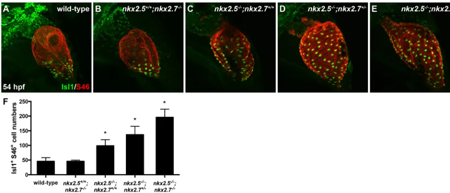
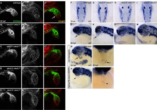
[image:4.612.51.341.456.735.2]and detected a progressive increase in the number of cells expressing this transcription factor as Nkx gene dose is diminished (Fig. 4F). Notably, the number of Isl1+CMs in thenkx2.5−/−;nkx2.7−/−heart is approximately equivalent to the total number of CMs in wild-type embryos (Targoff et al., 2013). Next, we investigated Nkx2.5 and Isl1 expression patterns employing immunohistochemistry in embryos carrying Tg(nkx2.5:ZsYellow)at 52 hpf. This transgene faithfully recapitulates the endogenousnkx2.5expression observed byin situhybridization (data not shown). We find that Nkx2.5 and Isl1 are virtually mutually exclusive at the venous pole in wild-type embryos (Fig. 5A-A″). Nkx2.5 is expressed in a decreasing gradient from the OFT to the inflow of the atrium, whereas Isl1 expression is confined to SV with only a few cells co-expressing Nkx2.5 and Isl1 (Fig. 5A″). Alternatively, in nkx mutants, Isl1 expression is derepressed in all atrial CMs that normally express Nkx2.5 (Fig. 5B-E″). Taken together, our studies strongly support the in vivosuppression of Isl1+cell fate by Nkx genes in the SV during cardiac development.
We then explored the timing of the Nkx-Isl1 interaction in further detail. We evaluatedisl1expression during early stages of cardiac differentiation in the ALPM. Our data illustrate that specification of isl1+ progenitors occurs normally at 10 somites
and 15 somites (Fig. 5F-I and data not shown), butisl1expression is enhanced at 26 hpf innkxmutant compared with wild-type embryos (Fig. 5J-M). These findings indicate that Nkx genes are first required to inhibit isl1 at the heart tube stage. Using a novel transgene generated in our lab for ubiquitousnkx2.5overexpression, Tg(hsp70l:nkx2.5-EGFP) (George et al., 2015), we tested the sufficiency of nkx2.5 to inhibit isl1 expression (Fig. 5N-Q). Following heat-shock activation of this inducible transgene at 21 somites, isl1 expression was repressed specifically in the inflow region at both 24 hpf and 52 hpf (Fig. 5P,Q). Altogether, these results demonstrate that Nkx2.5 is not required forisl1+progenitor
specification, but activation of Nkx2.5 transcription prior to heart tube elongation is sufficient to repressisl1at the venous pole during chamber emergence.
Nkx genes regulate differentiation of pSHF progenitors Although recent work has shown the contribution of late-differentiating CMs to the venous pole in zebrafish (de Pater et al., 2009; Mosimann et al., 2015), little is known about the transcriptional regulatory networks guiding accretion and differentiation of this pSHF progenitor population. Given the ability of nkx2.5 to restrict Isl1 to the IFT (Fig. 5N-Q), we hypothesize that Nkx genes are essential in patterning the pSHF-derived CMs in this region. To test this hypothesis, we first performed a developmental timing assay to delineate the kinetics of pSHF progenitor accumulation using embryos expressingTg(myl7: EGFP)andTg(-5.1myl7:nDsRed2)(as in Fig. 1A-I). Intriguingly, our data reveal a statistically significant increase in pSHF-derived CMs, or EGFP+DsRed− CMs, at the venous pole innkx2.5−/−; nkx2.7+/+ embryos along with further enhancement innkx2.5−/−; nkx2.7−/− embryos when compared with wild-type embryos (Fig. 6A-I,N). However, the source of the increased late-differentiating CMs at the IFT remains unknown. This finding may reflect either increased proliferation of pSHF progenitors or enhanced accrual of pSHF-derived CMs.
[image:5.612.70.531.57.255.2]Consistent with our data illustrating normal CM production at the arterial pole following the loss of Nkx gene function (Fig. S1), we observed no difference in proliferation indices at the venous pole following EdU incorporation at 24 hpf betweennkx mutant and wild-type embryos (data not shown). Thus, we hypothesized that Nkx genes are required for pSHF progenitor proliferation prior to accretion to the IFT. We performed an EdU incorporation assay at 18 hpf in embryos carrying Tg(-5.1myl7:nDsRed2) to detect dividing CMs at the venous pole. Quantification of the proliferation indices for EdU+DsRed+MF20+cells in the IFT region at this time point also revealed no statistically significant difference between wild-type and nkx mutant embryos (Fig. 6J-M,O). These data support the conclusion that the IFT expansion in nkx mutant embryos is most likely due to enhanced pSHF-derived CM differentiation.
Fig. 4. Nkx genes inhibit Isl1 expression at the sinus venosus.(A-E) Lateral view, anterior towards the top, at 54 hpf in wild-type (A),nkx2.5+/+;nkx2.7−/−(B),
nkx2.5−/−;nkx2.7+/+(C),nkx2.5−/−;nkx2.7+/−(D) andnkx2.5−/−;nkx2.7−/−(E) embryos. Representative confocal images of immunofluorescence for Isl1 (green)
and S46 (red) demonstrate that Isl1 is restricted to the venous pole of wild-type (A) andnkx2.5+/+;nkx2.7−/−(B) embryos. However, Isl1 expression expands
throughout the atrium innkxmutants (C-E). (F) Quantification of Isl1+S46+CMs is depicted showing a gradual increase in cell number with a progressive loss of
nkxalleles. Mean and s.e.m. of each data set are shown (*P<0.005) in wild-type (n=16),nkx2.5+/+;nkx2.7−/−(n=2),nkx2.5−/−;nkx2.7+/+(n=6),nkx2.5−/−;nkx2.7+/−
(n=10) andnkx2.5−/−;nkx2.7−/−(n=4) embryos. Student’st-test was used to determine statistical significance.
DEVEL
O
Nkx genes pattern specialized nodal tissue in the sinus venosus viaisl1
Although recent studies have elucidated the existence of specialized conduction tissue in the zebrafish heart (Arrenberg et al., 2010; Chi et al., 2008; Milan et al., 2006) and have identifiedisl1+as molecular
marker of this population (Tessadori et al., 2012), the pathways controlling the development of the sinoatrial node (SAN) are still under investigation. Therefore, from evidence of conduction system abnormalities in individuals withNKX2-5mutations (Benson et al., 1999; Elliott et al., 2003; Gutierrez-Roelens et al., 2002; Schott et al., 1998) and from our findings revealing that Nkx genes regulate isl1 expression (Figs 4 and 5), we posited that Nkx genes are fundamental in patterning the pSHF-derived CMs that contribute to the specialized nodal tissue in the SV. To address this issue, we explored the expression of a number of pacemaker cell genes:bmp4, tbx2b, hcn4andshox2(Blaschke et al., 2007; Espinoza-Lewis et al., 2009; Tessadori et al., 2012). In wild-type embryos,bmp4,tbx2b andhcn4expression is restricted to the OFT, atrioventricular canal
(AVC) and IFT at 52 hpf (Fig. 7A,F,K). Intriguingly, all three genes are expressed throughout the myocardium innkxmutant embryos (Fig. 7C-E,H-J,M-O). We observe a similar expression pattern for shox2when comparing wild-type andnkxmutant embryos (Fig. S2). Taken together, our findings illustrate that Nkx genes regulate regionalization of SAN markers to the IFT.
Next, we postulated that Isl1 acting downstream of Nkx genes is responsible for patterning the SAN and establishing the pacemaker activity of the zebrafish heart. Using a previously validated morpholino (MO) to knockdown Isl1 function (Hutchinson and Eisen, 2006), we observed thatbmp4expression is absent in wild-type and nkx2.5+/+;nkx2.7−/− embryos specifically at the IFT
followingislet1E2MO injection, whereas normal AVC and OFT expression ofbmp4persists (compare Fig. 8A,B with F,G). These findings are consistent with evidence of selective inhibition ofbmp4 expression in the IFT of theisl1sa29mutant (de Pater et al., 2009).
[image:6.612.52.558.55.410.2]Interestingly, inhibition of isl1 splicing in nkx mutant embryos reveals specific downregulation of ectopic atrial and IFT bmp4 Fig. 5. Nkx2.5 is not required forisl1+progenitor specification, but is sufficient to repressisl1at the venous pole.(A-E″) Representative confocal images, lateral view, anterior towards the top, at 52 hpf. Immunostaining for Isl1 (red) in embryos carryingTg(nkx2.5:ZsYellow)demonstrates that Isl1 is restricted to the venous pole and Nkx2.5 expression is absent in this region in wild-type (white arrow) (A-A″) andnkx2.5+/+;nkx2.7−/−(B-B″) embryos. However, Isl1 expression is
derepressed in the atrium ofnkx2.5−/−;nkx2.7+/+(C-C″) andnkx2.5−/−;nkx2.7+/−(D-D″) embryos, and expands towards the outflow tract innkx2.5−/−;nkx2.7−/−
(E-E″) embryos. (F-I) Dorsal view, anterior towards the top, at 10 somites.In situhybridization shows no variation in ALPMisl1expression between wild-type (F) and nkxmutant (G-I) embryos. (J-M) Lateral view, anterior towards the left, at 26 hpf.In situhybridization demonstrates thatisl1expression is restricted to a ring at the venous pole of the heart tube in wild-type (J) andnkx2.5+/+;nkx2.7−/−(K) embryos. However,isl1expression expands to the atrium innkx2.5−/−;nkx2.7+/+ embryos (L) and throughout the entire heart tube innkx2.5−/−;nkx2.7−/−embryos (M). Black arrows indicate the ring ofisl1expression near the IFT (J,K). (N-Q) Lateral
view, anterior towards the left, at 24 hpf (N,P) and ventral view, anterior towards the top, at 52 hpf (O,Q) in non-transgenic (N,O) andTg(hsp70l:nkx2.5-EGFP) (P,Q) embryos.In situhybridization demonstrates thatisl1expression is restricted to a ring at the venous pole (black arrows) at 24 hpf (n=9) (N) and 52 hpf (n=7) (O), and that overexpression ofnkx2.5inhibitsisl1expression specifically in this region (dotted arrows) at 24 hpf (n=9/11) (P) and 52 hpf (n=8/9) (Q).
DEVEL
O
expression (compare Fig. 8H-J with C-E) in the context of otherwise normal development. Together, our results demonstrate that the misregulation of bmp4 expression in nkx mutant embryos is mediated by isl1 overexpression, ultimately placing isl1 downstream of Nkx genes in defining the pSHF-derived specialized conduction cells of the IFT.
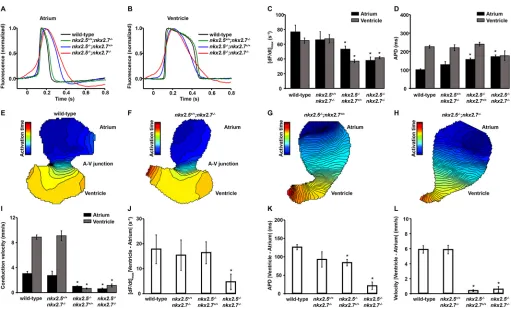
Nkx genes regulate electrophysiological patterning
Finally, we explored the effects of the derepression of SAN genes on the electrophysiological phenotype in nkx mutant hearts. The establishment of electrical gradients enables the transition from peristaltic to sequential contraction in the developing heart and this patterning is regulated by differential contributions of FHF and SHF progenitor populations (Mosimann et al., 2015). We hypothesized that the loss of Nkx gene function could lead to a disruption in regional patterns of intercellular coupling given both the absence of ventricular identity (Targoff et al., 2013, 2008) and the expanded SAN gene expression domain innkxmutant compared with wild-type embryos (Fig. 7).
To explore these issues, we measured action potential characteristics and conduction properties in isolated wild-type and nkxmutant hearts using high-resolution optical mapping (Fig. 9, Fig. S3). We did not detect any electrophysiological phenotypes in embryos carrying single-allele deficiencies in one or bothnkxgenes (nkx2.5+/−;nkx2.7+/+,nkx2.5+/+;nkx2.7+/−andnkx2.5+/−;nkx2.7+/−).
When comparing wild-type and nkx2.5+/+;nkx2.7−/− embryos,
there is no significant difference in the detailed action potential morphology (Fig. 9A,B), maximum upstroke velocity, [dF/dt]max (Fig. 9C), or atrial and ventricular action potential durations (Fig. 9D). Furthermore, there are no observed differences between
the atrial and ventricular conduction velocities in wild-type and nkx2.5+/+;nkx2.7−/−embryos (Fig. 9E,F,I). In contrast, disruption
ofnkx2.5gene function affects multiple components of the action potential with significant slowing of the upstroke phase in both chambers (Fig. 9A-C), marked diminution of the maximum slope of the action potential upstroke (Fig. 9C), as well as a substantial prolongation of atrial action potential duration and accentuation of phase 4 depolarization in atrial CMs (Fig. 9A,D). Moreover, homozygous loss of nkx2.5 function leads to severe slowing in action potential propagation, as demonstrated by closer spacing of the isochrones and significantly lower mean conduction velocities measured across both cardiac chambers (Fig. 9G-I). Interestingly, although there is failure of the normal specification of physiological AV delay in the hearts ofnkx2.5−/−;nkx2.7+/+embryos (Fig. 9K,L),
there remain significant differences in action potential characteristics between the ventricle and atrium (Fig. 9J,K and Fig. S3A-C,E-G). Although the hearts from nkx2.5+/+;nkx2.7−/− embryos did not
[image:7.612.94.518.57.306.2]exhibit any detectable electrophysiological phenotype (Fig. 9F), combined loss of function of both nkx homologs in nkx2.5−/−; nkx2.7−/− embryos leads to complete loss of electrophysiological chamber identity and further slowing of the action potential upstroke phase in the atrium (Fig. 9A,C,J and Fig. S3D). The loss of action potential duration differences between chambers is also evident in nkx2.5−/−;nkx2.7−/−embryos (Fig. 9D,K and Fig. S3H), including the emergence of a pacemaker phenotype in ventricular cells (Fig. 9B) and slowing of the conduction velocity (Fig. 9H,I,L). Interestingly, there are subtle, but significant, paradoxical effects on absolute atrial and ventricular conduction velocities innkx2.5−/−;nkx2.7−/− when compared with nkx2.5−/−;nkx2.7+/+ embryos. In the atrium, the nkx2.5−/−;nkx2.7−/−embryos exhibit even slower conduction than the Fig. 6. Nkx genes regulate cardiomyocyte accretion to the atrium.(A-I) Lateral images, anterior towards the top, at 52 hpf. Representative reconstructions of 10z-stacks from a developmental timing assay highlight the venous pole of wild-type (A-C),nkx2.5−/−;nkx2.7+/+(D-F) andnkx2.5−/−;nkx2.7−/−(G-I) embryos
carryingTg(myl7:EGFP)(green) andTg(-5.1myl7:nDsRed2)(red). Asterisks indicate EGFP+DsRed−cells. (J-M) Ventral view, anterior towards the top, at 40 hpf
of wild-type (J,K) andnkx2.5−/−;nkx2.7+/+(L,M) embryos carryingTg(-5.1myl7:nDsRed2).Confocal projections (J,L) of immunohistochemistry for EdU (red), DsRed (green) and MF20 (gray) following EdU incubation at 18 hpf and fourz-stack reconstructions (K,M) were used to count EdU+DsRed+MF20+CMs in the atrium. The boxed areas in J,L are shown at higher magnification in K,M, respectively. White arrows indicate EdU+DsRed+CMs. (N,O) Quantification of EGFP+DsRed−cells at the venous pole for wild-type (n=12),nkx2.5−/−;nkx2.7+/+(n=5) andnkx2.5−/−;nkx2.7−/−(n=4) embryos, and proliferation indices in
wild-type (n=5) andnkxmutant (n=3) embryos. Student’st-test was used to determine statistical significance. Mean and s.e.m. of each data set are shown (**P<0.0001). There is no statistically significant difference between proliferation indices in the DsRed+MF20+populations of wild-type andnkxmutant embryos.
DEVEL
O
nkx2.5−/−;nkx2.7+/+embryos. However, in the ventricle, there is an
increase in the conduction velocity in the nkx2.5−/−;nkx2.7−/− embryos, although conduction remains markedly slowed and chamber identity is lost from an electrophysiological standpoint. Although these electrophysiological attributes of the nkx mutant embryos reflect severe defects in intercellular coupling, the individual CM properties exhibit a phenotype that is more advanced developmentally than that observed in the primitive heart tube. Altogether, these data highlight the independent regulation of discrete
CM parameters throughout early cardiac development and chamber identity maintenance.
DISCUSSION
[image:8.612.99.517.58.309.2]Together, these findings offer new insights into the roles of Nkx genes in SHF development, highlighting the differential regulation at the arterial and venous poles. We show that Nkx genes promote the establishment of the SHF CM progenitor pool and differentiation of this population at the OFT. Our studies also reveal that Nkx genes are Fig. 7. Nkx genes regulate SAN markers.(A-O) Ventral view, anterior towards the top, at 52 hpf. Dotted white lines illustrate the cardiac silhouettes.In situ hybridization forbmp4(A-E),tbx2b(F-J) andhcn4(K-O). Expression ofbmp4is restricted to the OFT (white arrow), AVC (yellow arrow) and IFT (black arrow) in wild-type (n=7/7) (A) andnkx2.5+/+;nkx2.7−/−(n=4/4) (B) embryos. However, its expression is expanded throughout the cardiac chambers innkx2.5−/−;nkx2.7+/+ (n=3/4) (C),nkx2.5−/−;nkx2.7+/−(n=3/3) (D) andnkx2.5−/−; nkx2.7−/−(n=3/3) (E) embryos. Similar findings are observed in the wild-type (n=6/7) (F),nkx2.5+/+;
nkx2.7−/−(n=3/4) (G),nkx2.5−/−;nkx2.7+/+(n=3/3) (H),nkx2.5−/−;nkx2.7+/−(n=2/2) (I) andnkx2.5−/−;nkx2.7−/−(n=2/2) (J) embryos using thetbx2bprobe, and in
wild-type (n=8/8) (K),nkx2.5+/+;nkx2.7−/−(n=5/6) (L),nkx2.5−/−;nkx2.7+/+(n=9/11) (M),nkx2.5−/−;nkx2.7+/−(n=3/3) (N) andnkx2.5−/−;nkx2.7−/−(n=3/3) (O)
embryos using thehcn4probe.
Fig. 8.isl1acts downstream of Nkx genes at the venous pole.(A-J) Ventral view, anterior towards the top, at 52 hpf. Dotted white lines illustrate the cardiac silhouettes.In situhybridization forbmp4(A-J) in wild-type (A,F),nkx2.5+/+;nkx2.7−/−(B,G),nkx2.5−/−;nkx2.7+/+(C,H),nkx2.5−/−;nkx2.7+/−(D,I) andnkx2.5−/−;
nkx2.7−/−(E,J) embryos. Inhibition ofisl1expression usingislet1E2MO in wild-type (n=4/4) (compare A with F) andnkx2.5+/+;nkx2.7−/−(n=4/4) (compare
B and G) embryos leads to downregulation ofbmp4expression specifically at the venous pole of the heart (black arrow) without affecting the AVC (yellow arrow) and OFT (white arrow) expression. Innkx2.5−/−;nkx2.7+/+(n=2/2),nkx2.5−/−;nkx2.7+/−(n=5/6) andnkx2.5−/−;nkx2.7−/−(n=3/3) embryos,isl1inhibition rescues
the misregulation ofbmp4expression in the atrium (dotted arrows), yet AVC and OFT expression patterns remain unchanged (compare C-E with H-J).
DEVEL
O
[image:8.612.97.514.511.676.2]essential for the proper patterning of the venous pole by regulating the maintenance of working myocardial properties through repression of isl1. Following the loss ofNkxgene function, the entire myocardium adopts an Isl1+identity associated with activation of the SAN genes bmp4,tbx2b, hcn4 and shox2. Misregulation of these SAN genes is associated with electrophysiological evidence of expansion of the pacemaker phenotype across both chambers, with cellular characteristics suggesting diminished sodium channel availability at the membranes, features of calcium-dependent action potentials, and diastolic phase 4 potentials. Together, our findings implicate Nkx genes in combinatorial roles for anterior-posterior patterning of chamber identity, regional integration of FHF and SHF territories, and maturation of discrete cassettes of physiological CM differentiation within the heart tube. Moreover, these cellular abnormalities induced by the loss of Nkx gene function in the two-chambered zebrafish heart offer a potential window into the morphological and conduction system defects in individuals with CHD that harborNKX2-5mutations.
Through illustration of the differential functions of Nkx factors in the aSHF and pSHF, our studies complement and extend work in other model organisms. Previous studies have revealed a role for Nkx2-5 in limiting cardiac progenitor specification and favoring SHF proliferation (Prall et al., 2007). Yet in zebrafish, early specification of SHF progenitors appears normal given the expression patterns ofnkx2.5,nkx2.7,hand2(Targoff et al. 2013)
andisl1 (Fig. 5F-I) innkxmutant embryos. However, nkx2.5has been shown to regulate SHF progenitor proliferation at the proximal OFT via ltbp3 (Guner-Ataman et al., 2013). Through detailed dissection of the proliferative potential of the proximal and distal OFT progenitors, our results further elucidate the role that Nkx genes play in limiting the aSHF-derived CM population by regulating the timing of progenitor differentiation at the arterial pole. In view of studies illuminating a common progenitor of heart and head muscle derivatives in other models (Harel et al., 2012; Lescroart et al., 2015, 2010; Nagelberg et al., 2015; Wang et al., 2013), we postulate that aSHF progenitors could also adopt a head muscle fate in the absence of Nkx gene function. Future experiments would be required to test this hypothesis, which may offer additional insights into the diminished late-differentiating cells at the OFT of nkxmutant embryos. Taken together, although other mechanisms may play a role, our findings highlight a novel function of Nkx genes in establishing an aSHF progenitor pool necessary to assemble an arterial pole of the proper size and morphology.
[image:9.612.51.562.53.363.2]Our data also illustrates that, in the absence ofnkx2.5, the atrial myocardium adopts a venous pole identity characterized by misexpression of isl1 and SAN genes associated with failure of specification of the cardiac conduction system (CCS) tissue at the AV boundary. Further graded expansion of this abnormal CM differentiation, including manifestation of increased cellular Fig. 9. Nkx genes regulate electrophysiological patterning.(A,B) Representative atrial (A) and ventricular (B) action potentials showing the time courses of cellular depolarization and repolarization in wild-type,nkx2.5+/+;nkx2.7−/−,nkx2.5−/−;nkx2.7+/+andnkx2.5−/−;nkx2.7−/−hearts. (C) Mean maximum slope of the
atrial and ventricular action potentials measured as the maximum of the time derivative of the action potential upstroke phase, [dF/dt]max, in wild-type,nkx2.5+/+;
nkx2.7−/−,nkx2.5−/−;nkx2.7+/+andnkx2.5−/−;nkx2.7−/−hearts. (D) Action potential duration (APD) in wild-type,nkx2.5+/+;nkx2.7−/−, nkx2.5−/−;nkx2.7+/+and
nkx2.5−/−;nkx2.7−/−hearts. (E-H) Representative isochronal maps illustrating the positions of the depolarizing wave front in 5 ms time intervals for wild-type (E),
nkx2.5+/+;nkx2.7−/−(F),nkx2.5−/−;nkx2.7+/+(G) andnkx2.5−/−;nkx2.7−/−(H) hearts. (I) Mean atrial and ventricular action potential propagation velocities in
wild-type,nkx2.5+/+;nkx2.7−/−, nkx2.5−/−;nkx2.7+/+andnkx2.5−/−;nkx2.7−/−hearts. (J-L) Absolute differences in the maximum slope of the upstroke phase, [dF/dt]
max (J), APD (K) and the conduction velocity (L) between the ventricle and the atrium in wild-type,nkx2.5+/+;nkx2.7−/−,nkx2.5−/−;nkx2.7+/+andnkx2.5−/−;nkx2.7−/−
hearts. Student’st-test was used to determine statistical significance. The mean and s.e.m. of each data set are shown (*P<0.05) for wild-type (n=5),nkx2.5+/+;
nkx2.7−/−(n=5),nkx2.5−/−;nkx2.7+/+(n=6) andnkx2.5−/−;nkx2.7−/−(n=3) hearts.
DEVEL
O
and SAN identity could explain progressive AV conduction defects into adulthood that are exhibited in individuals with NKX2-5 mutations (Jay et al., 2003).
We have previously shown that ventricular CMs transdifferentiate into atrial CMs, demonstrating plasticity of the differentiated myocardium in nkx mutant embryos (Targoff et al., 2013). It is possible that flexibility of the differentiated CMs results in sarcomeric rearrangement that allows for fate transformation in the nkx2.5−/−;nkx2.7−/− embryos. In support of this concept, a recent report demonstrates that ablation of ventricular CMs in the zebrafish embryos leads to atrial-to-ventricular transdifferentiation with associated sarcomeric restructuring (Zhang et al., 2013). Our electrophysiological studies further substantiate this hypothesis through their demonstration that graded loss ofnkxalleles regulates multiple elements of the functional identity of the atrial and ventricular myocardium. Although atrial and ventricular action potential features and conduction properties are distinct in wild-type,nkx2.5+/+;nkx2.7−/−andnkx2.5−/−;nkx2.7+/+hearts, each of
the individual null alleles exhibits discrete effects on resting membrane potentials, CM activation, action potential morphology and conduction velocity. Moreover, the electrophysiological differences between atrial and ventricular CMs are completely abolished innkx2.5−/−;nkx2.7−/−embryos. Despite these findings, there are discordances in the effects of individual genes on electrophysiology in the atrium and ventricle that implicate additional regulatory partners in the specification of the complex regional electrical function necessary for a vertebrate heart.
Taken together, these studies on the functions of Nkx genes in the aSHF and pSHF highlight important concepts in our understanding of CM differentiation. Changes in the expression of individual transcription factors, signaling molecules and ion channels reveal discrete physiological modules and can remodel nuanced functional characteristics in CMs. Given this malleability, the notion of a stable differentiated state does not fully reflect the resolution of modern cellular biology, which has revealed phenotypes that are a result of combinations of discrete and overlapping protein repertoires. These data parallelin vitrostudies in iPS-derived differentiated cell types. Despite a physiological resemblance to mature CMs, these populations demonstrate remarkable cellular heterogeneity in detailed electrophysiological phenotypes and transcriptional profiles that more closely resemble one of the parent fibroblast than of the CM being studied. Our data suggest that individual proteins may be sufficient and necessary to induce and maintain plastic states where the regenerative potential in the heart might be realized. Thus, it would be valuable to address the fundamental nature of cardiomyocyte differentiation at the level of individual physiologic functions in order to obtain a better understanding of
3′) (Hutchinson and Eisen, 2006). Uninjected sibling embryos were processed as controls.
Heat shock conditions
Embryos from outcrosses of fish carryingTg(hsp70l:nkx2.5-EGFP)were maintained at 28.5°C and exposed to heat shock at 37°C at desired stages (21 somites to 24 somites). To implement heat shock, 50 embryos were placed in 2.5 ml of embryo medium in a 35 mm Petri dish on top of a covered heat block for 1 h. Three hours after the initiation of heat shock, transgenic embryos were identified by visualization of ubiquitous EGFP expression. Non-transgenic sibling embryos exposed to heat shock served as controls.
Depigmentation of embryos
When necessary, embryos were depigmented after overnight fixation in 4% paraformaldehyde at 4°C. They were washed twice in PBT (PBS-0.1% Tween) then incubated in 3% H2O2, 0.5% KOH and 0.1% Tween for 8 min
at 26 h post fertilization (hpf ) and 18 min at 52 hpf. Subsequently, the embryos were washed twice again in PBT and stored in methanol at−20°C.
In situhybridization
We performed whole-mountin situhybridization as previously described (Yelon et al., 1999) formef2cb(ZDB-GENE-040901-7),isl1 (ZDB-GENE-980526-112), bmp4 (ZDB-GENE-980528-2059), tbx2b (ZDB-GENE-990726-27), hcn4 (ZDB-GENE-050420-360) and shox2 (ZDB-GENE-040426-1457).
Immunofluorescence
Whole-mount double immunofluorescence was conducted using a protocol described previously (Alexander et al., 1998) for the following primary antibodies: anti-Islet1/2 (1:50; 39.4D5, Developmental Studies Hybridoma Bank), anti-atrial myosin heavy chain (1:20; S46, Developmental Studies Hybridoma Bank), anti-RCFP (1:50; Clontech, 632475) and anti-GFP (1:100; Life Technologies, A11122). When employing the following primary antibodies, anti-Eln2 (1:500; Miao et al., 2007) and anti-sarcomeric myosin heavy chain (1:20; MF20, Developmental Studies Hybridoma Bank), another published protocol was used (Zhou et al., 2011). The following secondary antibodies were employed: goat anti-mouse IgG2b Alexa Fluor 488 or 568, goat anti-mouse IgG1 Alexa Fluor 488 or 568, goat anti-mouse IgG2a Alexa Fluor 488, goat anti-rabbit IgG Alexa Fluor 488 or 568, and goat anti-mouse IgG Cy5 (all at 1:200; Invitrogen).
Proliferation assay
Cardiomyocyte proliferation in embryos expressingTg(-5.1myl7:nDsRed2)f2
orTg(nkx2.5:ZsYellow)fb7was assessed using the Click-iT EdU Imaging Kit
(Life Technologies) with reported protocols (Cheesman et al., 2011; Mahler et al., 2010; Schindler et al., 2014). Following EdU incubation for 30 min on ice, embryos were chased for 24 h. Embryos were then fixed in 2% formaldehyde, washed once in PBS and washed twice in PBS-0.2% saponin. Subsequently, immunostaining was performed as described above followed by the Click-iT staining reaction. EdU+ZsYellow+MF20+/−and
EdU+ DsRed+ MF20− cells were counted using ImageJ software, going
through allz-stacks. We calculated the proliferation indices as the percentage
DEVEL
O
of cells that incorporated EdU. Student’st-test (homoscedastic, two-tailed distribution) determined statistical significance between the means of cell number data sets.
Imaging
Images were captured with a Zeiss M2Bio microscope and a Zeiss AxioCam digital camera, prior to processing with Zeiss AxioVision and Adobe Creative Suite software. Confocal imaging was performed with a Nikon A1R MP confocal microscope andz-stacks were analyzed with ImageJ.
Developmental timing assay and cell counting
For the developmental timing assay, embryos carrying Tg(-5.1myl7:
nDsRed2)f2andTg(myl7:EGFP)twu277 were fixed at 52 hpf for 1 h in 1%
formaldehyde, washed once in PBS, washed three times in PBS-0.2% saponin, deyolked and flat-mounted. Newly differentiated CMs were identified as green, but not red cells (EGFP+DsRed−CMs) and were counted onz-stacks
using ImageJ software. To count Isl1+cells, we performed immunostaining for
Isl1 and S46 (as described above) and quantified individual CMs that were positive for Isl1 using ImageJ software on reconstructedz-stacks. Student’s
t-test (homoscedastic, two-tailed distribution) determined statistical significance between the means of cell number data sets.
Genotyping
PCR genotyping was performed on genomic DNA extracted from individual embryos followingin situhybridization, immunofluorescence or live imaging. Detection ofnkx2.5vu179 was performed using primers 5′-CAAACTCACCTCCACACAGG-3′and 5′ -TTACCATCCCGAACCA-AAAC-3′to generate a 144 bp fragment. Digestion of the mutant PCR product with Hinf1 generates 30 bp and 114 bp fragments. Analysis of
nkx2.5vu179in embryos carryingTg(hsp70l:nkx2.5-EGFP)was performed
as previously described (George et al., 2015). Detection ofnkx2.7vu413was executed using primers 5′-TTGTTGCAAATGGACACGTT-3′and 5′ -G-GAGTGTCTCGCCGTTTTTA-3′to generate a 158 bp fragment. Digestion of the mutant PCR product withMseI creates 28 bp and 130 bp fragments.
Optical mapping
Embryonic zebrafish hearts were isolated at 72 hpf and stained with the transmembrane potential-sensitive dye, di-8-ANEPPS (Invitrogen), to measure action potentials using high-speed optical mapping as previously described (Mosimann et al., 2015; Panáková et al., 2010). Fluorescence intensities were recorded with a high-speed CCD camera (CardioCCD-SMQ, Redshirt Imaging) at 2000 frames per second. Activation times were determined as the times at which action potentials reached 50% of their amplitude during the depolarization phase at every location. This criterion for defining electrical activation was found in computer simulations to correlate with the time at which the maximum depolarizing Na+ current
occurs during propagation (Fast and Kleber, 1995). Action potentials were temporally (800 Hz low-pass cutoff ) and spatially (3×3 pixel average) filtered to enhance signal-to-noise ratios. To quantify the slope of the action potential upstroke phase, the maximum time derivative of the normalized action potential, [dF/dt]max, was calculated. Action potential duration (APD)
was defined as the absolute time difference between the decay phase and the upstroke phase of the local action potential at 50% depolarization. By fitting a second-order polynomial hypersurface to the three-dimensional image plane-activation time relationship, conduction velocity vectors were subsequently derived using an established algorithm (Bayly et al., 1998) with custom scripts. Conduction velocity vector fields were estimated from the local action potentials (Panáková et al., 2010). All electrophysiological measurements were averaged across the relevant anatomic segments for each heart.
Acknowledgements
We are grateful to Joshua Barber for expert zebrafish care and members of the Targoff Laboratory for their constructive input. We appreciate the critical reading of the manuscript by Lilianna Solnica-Krezel and Joshua S. Waxman. We also thank C. Geoffrey Burns and Caroline E. Burns for sharing the
Tg(nkx2.5:ZsYellow)line.
Competing interests
S.C. is currently a consultant for Pairnomix, but the work presented in this paper has no connection with this company.
Author contributions
Conceptualization: S.C., K.L.T.; Methodology: S.C., C.A.M., K.L.T.; Validation: S.C., C.d.S.-T., A.A.W., C.A.M., K.L.T.; Formal analysis: S.C., C.d.S.-T., V.G., A.A.W., S.K., C.A.M., K.L.T.; Investigation: S.C., C.d.S.-T., V.G., A.A.W., S.K., C.A.M., K.L.T.; Resources: S.C., C.A.M., K.L.T.; Data curation: S.C., C.d.S.-T., V.G., A.A.W., S.K.; Writing - original draft: S.C., A.A.W., C.A.M., K.L.T.; Writing - review & editing: C.A.M., K.L.T.; Visualization: K.L.T.; Supervision: C.A.M., K.L.T.; Project administration: K.L.T.; Funding acquisition: C.A.M., K.L.T.
Funding
This work was supported by grants from the National Institutes of Health (K12HD043389, K08HL088002 and R01HL131438 to K.L.T., and R24OD017870 to C.A.M.] and the Burroughs Wellcome Fund [IRSA to C.A.M.]. Deposited in PMC for release after 12 months.
Supplementary information
Supplementary information available online at
http://dev.biologists.org/lookup/doi/10.1242/dev.161497.supplemental
References
Abou Hassan, O. K., Fahed, A. C., Batrawi, M., Arabi, M., Refaat, M. M., DePalma, S. R., Seidman, J. G., Seidman, C. E., Bitar, F. F. and Nemer, G. M.
(2015). NKX2-5 mutations in an inbred consanguineous population: genetic and phenotypic diversity.Sci. Rep.5, 8848.
Alexander, J., Stainier, D. Y. R. and Yelon, D.(1998). Screening mosaic F1
females for mutations affecting zebrafish heart induction and patterning.Dev. Genet.22, 288-299.
Arrenberg, A. B., Stainier, D. Y. R., Baier, H. and Huisken, J.(2010). Optogenetic control of cardiac function.Science330, 971-974.
Azpiazu, N. and Frasch, M.(1993). tinman and bagpipe: two homeo box genes that determine cell fates in the dorsal mesoderm of Drosophila.Genes Dev.7, 1325-1340.
Barth, J. L., Clark, C. D., Fresco, V. M., Knoll, E. P., Lee, B., Argraves, W. S. and Lee, K.-H.(2010). Jarid2 is among a set of genes differentially regulated by Nkx2.5 during outflow tract morphogenesis.Dev. Dyn.239, 2024-2033.
Bayly, P. V., KenKnight, B. H., Rogers, J. M., Hillsley, R. E., Ideker, R. E. and
Smith, W. M. (1998). Estimation of conduction velocity vector fields from
epicardial mapping data.IEEE Trans. Biomed. Eng.45, 563-571.
Benson, D. W., Silberbach, G. M., Kavanaugh-McHugh, A., Cottrill, C., Zhang, Y., Riggs, S., Smalls, O., Johnson, M. C., Watson, M. S., Seidman, J. G. et al.
(1999). Mutations in the cardiac transcription factor NKX2.5 affect diverse cardiac developmental pathways.J. Clin. Invest.104, 1567-1573.
Blaschke, R. J., Hahurij, N. D., Kuijper, S., Just, S., Wisse, L. J., Deissler, K., Maxelon, T., Anastassiadis, K., Spitzer, J., Hardt, S. E. et al.(2007). Targeted mutation reveals essential functions of the homeodomain transcription factor Shox2 in sinoatrial and pacemaking development.Circulation115, 1830-1838.
Bodmer, R.(1993). The gene tinman is required for specification of the heart and visceral muscles in Drosophila.Development118, 719-729.
Bouveret, R., Waardenberg, A. J., Schonrock, N., Ramialison, M., Doan, T., de Jong, D., Bondue, A., Kaur, G., Mohamed, S., Fonoudi, H. et al.(2015). NKX2-5 mutations causative for congenital heart disease retain functionality and are directed to hundreds of targets.Elife4, e06942.
Brade, T., Gessert, S., Kühl, M. and Pandur, P.(2007). The amphibian second heart field: Xenopus islet-1 is required for cardiovascular development.Dev. Biol.
311, 297-310.
Briggs, L. E., Takeda, M., Cuadra, A. E., Wakimoto, H., Marks, M. H., Walker, A. J., Seki, T., Oh, S. P., Lu, J. T., Sumners, C. et al.(2008). Perinatal loss of Nkx2-5 results in rapid conduction and contraction defects.Circ. Res. 103, 580-590.
Bruneau, B. G.(2008). The developmental genetics of congenital heart disease.
Nature451, 943-948.
Buckingham, M., Meilhac, S. and Zaffran, S.(2005). Building the mammalian
heart from two sources of myocardial cells.Nat. Rev. Genet.6, 826-837.
Cai, C.-L., Liang, X., Shi, Y., Chu, P.-H., Pfaff, S. L., Chen, J. and Evans, S.
(2003). Isl1 identifies a cardiac progenitor population that proliferates prior to differentiation and contributes a majority of cells to the heart.Dev. Cell5, 877-889.
Cambier, L., Plate, M., Sucov, H. M. and Pashmforoush, M.(2014). Nkx2-5
regulates cardiac growth through modulation of Wnt signaling by R-spondin3.
Development141, 2959-2971.
Cheesman, S. E., Neal, J. T., Mittge, E., Seredick, B. M. and Guillemin, K.(2011). Epithelial cell proliferation in the developing zebrafish intestine is regulated by the Wnt pathway and microbial signaling via Myd88.Proc. Natl. Acad. Sci. USA108
Suppl. 1, 4570-4577.
DEVEL
O
Dobreva, G., Schiemann, M., Dirschinger, R. et al. (2014). Direct Nkx2-5 transcriptional repression of Isl1 controls cardiomyocyte subtype identity.Stem Cells
33, 1113-1129.
Dyer, L. A. and Kirby, M. L.(2009). The role of secondary heart field in cardiac development.Dev. Biol.336, 137-144.
Elliott, D. A., Kirk, E. P., Yeoh, T., Chandar, S., McKenzie, F., Taylor, P., Grossfeld, P., Fatkin, D., Jones, O., Hayes, P. et al.(2003). Cardiac homeobox gene NKX2-5 mutations and congenital heart disease: associations with atrial septal defect and hypoplastic left heart syndrome. J. Am. Coll. Cardiol. 41, 2072-2076.
Espinoza-Lewis, R. A., Yu, L., He, F., Liu, H., Tang, R., Shi, J., Sun, X., Martin, J. F., Wang, D., Yang, J. et al.(2009). Shox2 is essential for the differentiation of cardiac pacemaker cells by repressing Nkx2-5.Dev. Biol.327, 376-385.
Espinoza-Lewis, R. A., Liu, H., Sun, C., Chen, C., Jiao, K. and Chen, Y. P.(2011). Ectopic expression of Nkx2.5 suppresses the formation of the sinoatrial node in mice.Dev. Biol.356, 359-369.
Fast, V. G. and Kleber, A. G.(1995). Cardiac tissue geometry as a determinant of unidirectional conduction block: assessment of microscopic excitation spread by optical mapping in patterned cell cultures and in a computer model.Cardiovasc. Res.29, 697-707.
Galli, D., Dominguez, J. N., Zaffran, S., Munk, A., Brown, N. A. and Buckingham, M. E.(2008). Atrial myocardium derives from the posterior region of the second heart field, which acquires left-right identity as Pitx2c is expressed.Development
135, 1157-1167.
Gelb, B. D. and Chung, W. K.(2014). Complex genetics and the etiology of human congenital heart disease.Cold Spring Harb. Perspect. Med.4, a013953.
George, V., Colombo, S. and Targoff, K. L.(2015). An early requirement for nkx2.5 ensures the first and second heart field ventricular identity and cardiac function into adulthood.Dev. Biol.400, 10-22.
Goddeeris, M. M., Rho, S., Petiet, A., Davenport, C. L., Johnson, G. A., Meyers, E. N. and Klingensmith, J.(2008). Intracardiac septation requires hedgehog-dependent cellular contributions from outside the heart. Development 135, 1887-1895.
Goldmuntz, E., Geiger, E. and Benson, D. W.(2001). NKX2.5 mutations in
patients with tetralogy of fallot.Circulation104, 2565-2568.
Grow, M. W. and Krieg, P. A.(1998). Tinman function is essential for vertebrate heart development: elimination of cardiac differentiation by dominant inhibitory mutants of the tinman-related genes, XNkx2-3 and XNkx2-5. Dev. Biol.204, 187-196.
Guner-Ataman, B., Paffett-Lugassy, N., Adams, M. S., Nevis, K. R., Jahangiri, L., Obregon, P., Kikuchi, K., Poss, K. D., Burns, C. E. and Burns, C. G.(2013). Zebrafish second heart field development relies on progenitor specification in anterior lateral plate mesoderm and nkx2.5 function.Development140, 1353-1363.
Gutierrez-Roelens, I., Sluysmans, T., Gewillig, M., Devriendt, K. and Vikkula, M.
(2002). Progressive AV-block and anomalous venous return among cardiac anomalies associated with two novel missense mutations in the CSX/NKX2-5 gene.Hum. Mutat.20, 75-76.
Hami, D., Grimes, A. C., Tsai, H.-J. and Kirby, M. L.(2011). Zebrafish cardiac development requires a conserved secondary heart field. Development 138, 2389-2398.
Harel, I., Maezawa, Y., Avraham, R., Rinon, A., Ma, H.-Y., Cross, J. W., Leviatan, N., Hegesh, J., Roy, A., Jacob-Hirsch, J. et al.(2012). Pharyngeal mesoderm regulatory network controls cardiac and head muscle morphogenesis.Proc. Natl. Acad. Sci. USA109, 18839-18844.
Hoffman, J. I. E. and Kaplan, S.(2002). The incidence of congenital heart disease.
J. Am. Coll. Cardiol.39, 1890-1900.
Hoffman, J. I. E., Kaplan, S. and Liberthson, R. R. (2004). Prevalence of
congenital heart disease.Am. Heart J.147, 425-439.
Hoffmann, A. D., Peterson, M. A., Friedland-Little, J. M., Anderson, S. A. and Moskowitz, I. P.(2009). sonic hedgehog is required in pulmonary endoderm for atrial septation.Development136, 1761-1770.
Sumiyoshi, T., Takano, H., Nagai, R. et al.(2002). Novel point mutation in the cardiac transcription factor CSX/NKX2.5 associated with congenital heart disease.Circ. J.66, 561-563.
Jahangiri, L., Sharpe, M., Novikov, N., Gonzalez-Rosa, J. M., Borikova, A., Nevis, K., Paffett-Lugassy, N., Zhao, L., Adams, M., Guner-Ataman, B. et al.
(2016). The AP-1 transcription factor component Fosl2 potentiates the rate of myocardial differentiation from the zebrafish second heart field.Development143, 113-122.
Jay, P. Y., Berul, C. I., Tanaka, M., Ishii, M., Kurachi, Y. and Izumo, S.(2003). Cardiac conduction and arrhythmia: insights from Nkx2.5 mutations in mouse and humans.Novartis Found. Symp.250, 227-238; discussion 238-241, 276-229.
Jay, P. Y., Harris, B. S., Maguire, C. T., Buerger, A., Wakimoto, H., Tanaka, M., Kupershmidt, S., Roden, D. M., Schultheiss, T. M., O’Brien, T. X. et al.(2004). Nkx2-5 mutation causes anatomic hypoplasia of the cardiac conduction system.
J. Clin. Invest.113, 1130-1137.
Lazic, S. and Scott, I. C.(2011). Mef2cb regulates late myocardial cell addition from a second heart field-like population of progenitors in zebrafish.Dev. Biol.354, 123-133.
Lee, K.-H., Xu, Q. and Breitbart, R. E.(1996). A new tinman-related gene, nkx2.7, anticipates the expression of nkx2.5 and nkx2.3 in zebrafish heart and pharyngeal endoderm.Dev. Biol.180, 722-731.
Lescroart, F., Kelly, R. G., Le Garrec, J.-F., Nicolas, J.-F., Meilhac, S. M. and Buckingham, M.(2010). Clonal analysis reveals common lineage relationships between head muscles and second heart field derivatives in the mouse embryo.
Development137, 3269-3279.
Lescroart, F., Mohun, T., Meilhac, S. M., Bennett, M. and Buckingham, M.
(2012). Lineage tree for the venous pole of the heart: clonal analysis clarifies controversial genealogy based on genetic tracing.Circ. Res.111, 1313-1322.
Lescroart, F., Hamou, W., Francou, A., Théveniau-Ruissy, M., Kelly, R. G. and
Buckingham, M.(2015). Clonal analysis reveals a common origin between
nonsomite-derived neck muscles and heart myocardium.Proc. Natl. Acad. Sci. USA112, 1446-1451.
Liang, X., Zhang, Q., Cattaneo, P., Zhuang, S., Gong, X., Spann, N. J., Jiang, C., Cao, X., Zhao, X., Zhang, X. et al.(2015). Transcription factor ISL1 is essential for pacemaker development and function.J. Clin. Invest.125, 3256-3268.
Loffredo, C. A.(2000). Epidemiology of cardiovascular malformations: prevalence and risk factors.Am. J. Med. Genet.97, 319-325.
Lyons, I., Parsons, L. M., Hartley, L., Li, R., Andrews, J. E., Robb, L. and Harvey, R. P.(1995). Myogenic and morphogenetic defects in the heart tubes of murine embryos lacking the homeo box gene Nkx2-5.Genes Dev.9, 1654-1666.
Mably, J. D., Mohideen, M. A., Burns, C. G., Chen, J.-N., Fishman, M. C. and Mohideen, M.-A. P. K.(2003). heart of glass regulates the concentric growth of the heart in zebrafish.Curr. Biol.13, 2138-2147.
Mahler, J., Filippi, A. and Driever, W.(2010). DeltaA/DeltaD regulate multiple and temporally distinct phases of notch signaling during dopaminergic neurogenesis in zebrafish.J. Neurosci.30, 16621-16635.
McElhinney, D. B., Geiger, E., Blinder, J., Benson, D. W. and Goldmuntz, E.
(2003). NKX2.5 mutations in patients with congenital heart disease.J. Am. Coll. Cardiol.42, 1650-1655.
Miao, M., Bruce, A. E. E., Bhanji, T., Davis, E. C. and Keeley, F. W.(2007).
Differential expression of two tropoelastin genes in zebrafish.Matrix Biol.26, 115-124.
Milan, D. J., Giokas, A. C., Serluca, F. C., Peterson, R. T. and MacRae, C. A.
(2006). Notch1b and neuregulin are required for specification of central cardiac conduction tissue.Development133, 1125-1132.
Mommersteeg, M. T. M., Soufan, A. T., de Lange, F. J., van den Hoff, M. J., Anderson, R. H., Christoffels, V. M. and Moorman, A. F.(2006). Two distinct pools of mesenchyme contribute to the development of the atrial septum.Circ. Res.99, 351-353.
Mommersteeg, M. T. M., Brown, N. A., Prall, O. W. J., de Gier-de Vries, C., Harvey, R. P., Moorman, A. F. M. and Christoffels, V. M.(2007). Pitx2c and