http://dx.doi.org/10.4236/jct.2016.76046
How to cite this paper: Sharma, S. and Gangenahalli, G. (2016) Gene Expression Profiling of Human c-Kit Mutant D816V. Journal of Cancer Therapy, 7, 439-454. http://dx.doi.org/10.4236/jct.2016.76046
Gene Expression Profiling of Human c-Kit
Mutant D816V
Shilpa Sharma, Gurudutta Gangenahalli*
Division of Stem Cell & Gene Therapy Research, Institute of Nuclear Medicine & Allied Sciences (INMAS), New Delhi, India
Received 15 May 2016; accepted 20 June 2016; published 23 June 2016
Copyright © 2016 by authors and Scientific Research Publishing Inc.
This work is licensed under the Creative Commons Attribution International License (CC BY).
http://creativecommons.org/licenses/by/4.0/
Abstract
The tyrosine kinase receptor III, c-Kit/stem cell factor receptor and its ligand, human stem cell factor (huSCF) are the predominant regulator of mitogenesis in the hematopoietic stem and pro-genitor cells. However, gain-of-function mutations alter c-Kit auto-regulatory mechanisms to ab-errant c-Kit signaling, leading to the onset or progression of cancerous transformations. The most common mutation of c-Kit is the substitution of aspartic acid residue in position 816 to valine (D816V), which is majorly responsible for its ligand-independent constitutive activation, and is implicated in hematopoietic malignancies. Currently, molecular targeted therapy is increasingly becoming a hot spot due to its specificity and low toxicity. As the molecular mechanisms responsi-ble for D816V-c-Kit mediated tumorogenicity are largely unknown, in this study, we aimed to in-vestigate the D816V-c-Kit signaling mediated downstream molecular targets. Specifically, we created c-Kit active mutant form D816V and performed inducible gene expression of mutant D816V-c-Kit in monomyelocytic cell line U937. Mutant D816V-c-Kit expressing cells revealed sig-nificantly enhanced cellular mitogenic activity compared to wild-type c-Kit expressing cells inde-pendent of huSCF. To examine the molecular targets regulating tumorogenic proliferation, we evaluated the consequences of mutant D816V-c-Kit expression on downstream gene expression profile by high throughput microarray technology. The levels of some of the relevant genes (PIK3CB, eIF4B, PRKCDBP, MOAP1) were validated by quantitative polymerase chain reaction. SLA, STAT5B, MAP3K2 and MAPK14 emerged as important downstream molecular targets of mutant D816V-c-Kit. Further, by dissecting the signaling pathways, we also demonstrated that the D816V- c-Kit mediated hematopoietic cell proliferation is dependent on molecular target p38 MAP kinase.
Keywords
c-Kit Mutant, Hematopoietic Cells, Microarray Gene Expression, Proliferation
1. Introduction
c-Kit and huSCF are encoded at the white spotting and steel loci of the mouse, respectively. Mutations at both the W and the Sl locus cause deficiencies in hematopoiesis, gametogenesis and melanogenesis [1]. In humans, c-Kit encodes a transmembrane glycoprotein that belongs to platelet-derived growth factor and macrophage growth factor receptor subfamily [2]. Activation of c-Kit by huSCF binding results in receptor dimerization and activation of intrinsic tyrosine kinase activity. Specific tyrosine residues are autophosphorylated, which results in the activation of downstream signaling pathways, including the Ras/Erk pathway and the phosphoinositide- 3-kinase (PI3K) pathway [3]-[5].
Though, physiologically regulated tyrosine kinase activity of c-Kit is necessary for the controlled proliferation of hematopoietic stem and progenitor cells, mutational activations disturb c-Kit dynamic regulatory mechanisms, resulting in oncogenic transformation [6] [7]. Gain of independence of external growth stimuli is a crucial step. This can be achieved in several different ways, including mutations that render receptor tyrosine kinases consti-tutively active in the absence of ligand stimulation [8] [9]. In the case of c-Kit, these mutations most commonly occur either in exon 11 (encoding the juxta-membrane region), predominantly in gastrointestinal stromal tumors or in exon 17 (encoding the activation loop of the kinase domain). This is exemplified by codon 816 mutations found in several human malignancies including acute myeloid leukemia, mastocytosis, germ cell tumors of the seminoma or dysgerminoma types, sinonasal natural killer/T-cell lymphomas, and in intracranial teratomas [10] [11]. D816V mutation leads to the conversion of an aspartic acid residue to a valine, tyrosine, phenylalanine, asparagine or histidine residue [11] [12]. The acquired activating point mutation D816V significantly impairs the efficacy of targeted cancer therapies, thus limiting the treatment options for therapies [13]. Efforts to inhibit mutant c-Kit with tyrosine kinase inhibitors have been unsatisfactory, indicating a need for preclinical ap-proaches to identify alternative molecular targets. Therefore, in this study, we sought to develop a human hema-topoietic cell line model expressing D816V-c-Kit as a platform to dissect the molecular mechanisms underlying contributions of mutant D816V-c-Kit to myeloid leukaemia development. To unravel unknown molecular tar-gets driving mutant D816V-c-Kit for leukemogenicity, we used the advanced approach of microarray technolo-gy. We used inducible gene expression system for the regulated expression of wild-type c-Kit and mutant D816V-c-Kit in huSCF/c-Kit null U937 cells and assessed the cells’ mitogenic potential. We performed micro-array profiling of huSCF activated wild-type c-Kit expressing cells (induced) and mutant D816V-c-Kit express-ing cells (induced). To the best of our knowledge, this is the first report that identifies the putative molecular targets triggered by mutant D816V-c-Kit using high throughput microarray in human myeloid leukemic U937 cells. Overall, the results of this study indicate that mutant D816V-c-Kit change the transcript levels of few genes responsible for the cell proliferation. Furthermore, the current data provide novel candidate genes, such as PIK3R1, eIF4B, STAT5B, MAPK14, elicited by mutant D816V-c-Kit in hematopoietic cells. Further, inhibition of proliferative capacity of mutant D816V-c-Kit expressing cells using p38 MAP kinase inhibitor indicates that p38 MAP kinase plays an essential role in D816V-c-Kit mediated tumorogenicity.
2. Materials and Methods
2.1. Creation of Recombinant Wild-Type pc-Kit-TRE2hyg and Its invitro Site-Directed Mutagenesis
Figure 1. Restriction analysis of subcloned c-Kit. Restriction-digestion analysis of recombinant vector wild-type c-Kit- pTRE2hyg using HindIII give specific fragments of size 4.9 Kb, 2.5 Kb and 1.1 Kb (HindIII has two sites in c-Kit insert and one site in pTRE2hyg).
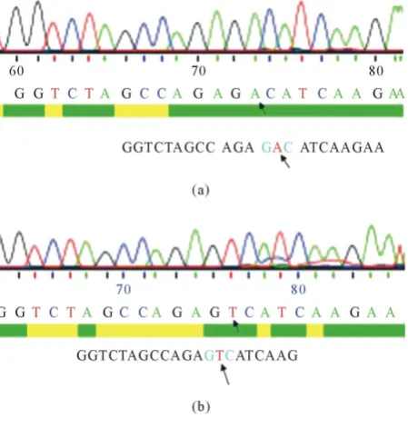
Figure 2. DNA sequencing of D816V-c-Kit-pTRE2hyg
[image:3.595.97.539.87.362.2] [image:3.595.203.428.412.648.2]2.2. Cell Lines and Stable Transfection
Human myelomonocytic leukemic cell line U937 was obtained from National Centre for Cell Science (NCCS, Pune, India). U937 cells were maintained in complete medium (RPMI-1640 medium supplemented with 1.0% sodium pyruvate, 10% FCS, 2 mM glutamine, 100 U/ml penicillin and 100 μg/ml streptomycin) at 37˚C in a fully humidified atmosphere of 95% room air and 5% CO2. The culture was maintained thrice in a week in fresh
complete medium. Exponentially growing cells with ≥94% viability were used for transfection. Approximately, 3 × 10⁵/ml number of cells were transfected with p Tet-off plasmid DNA. Transfected cells were selected using puromycin (0.5 ng/µl) containing media. The Tet-off stable U937 cells were maintained by periodically adding doxycycline (1 µg/µl) after every two days. Tet-off transfected U937 cells were subsequently transfected with recombinant vectors-wild type c-Kit-TRE2hyg and D816V-c-Kit-TRE2hyg. Double transfected cells were se-lected using hygromycin containing culture medium and stable transfected positive clones were screened by li-miting dilution. All transfections in U937 cells were performed using hilymax transfection reagent (Dojindo, Japan).
3. Results
3.1. Generation of Inducible Wild-Type and Mutant D816V-c-Kit U937 Cells
3.1.1. Flow Cytometry
Transfection of wild-type and mutant D816V-c-Kit stable transfectants were assessed by flow cytometry. Brief-ly, one million exponentially growing transfectants were washed and incubated for one hour with 0.5 μg of pri-mary mouse anti-human monoclonal c-Kit antibody (Santa Cruz, USA) at a 1:200 dilution on ice. After washing three times with staining buffer (2% FBS and 0.1% sodium azide in phosphate-buffered saline (PBS), cells were subsequently incubated with secondary FITC-labelled goat anti-mouse antibody (Santa Cruz Biotechnology, USA). These cells were washed to remove unbound secondary antibodies and analyzed by flow cytometry. Non-specific binding was assessed by using isotype-negative controls that were added at the same concentra-tions. After washing and resuspension, samples were analyzed on a Becton Dickinson (San Jose, CA) FACS Ca-libur flow cytometer.
3.1.2. Western Blotting
Wild-type and mutant D816V-c-Kit were stimulated with and without huSCF (100 ng/ml) for 24 hrs. Cells were lysed in an appropriate amount of RIPA lysis buffer (50 mM Tris-HCl, pH 7.4, 150 mM sodium chloride, 0.25% deoxycholic acid, 1% NP-40, 1 mM EDTA), supplemented with 1 mM phenylmethylsulfonyl fluoride, 1 mM dithiothreitol, and 1× protease inhibitor. Lysates were incubated for 30 min on ice and then cleared by centrifu-gation at 14,000 xg for 15 min at 4˚C. Protein content was determined by BCA method (Thermo Scientific, USA). Immunoblotting was performed following a standard protocol. Briefly, samples were separated by SDS-polyacrylamide gel electrophoresis (SDS-PAGE) and transferred onto nitrocellulose membrane (Pall Life Science). The membrane was blocked using 3% BSA dissolved in TBS buffer containing 0.1% tween-20 (TBST) for 1hr at room temperature and incubated overnight at 4˚C with primary antibodies: primary anti-human mouse monoclonal p-c-Kit antibody (Santa Cruz Biotechnology, USA), primary anti-human mouse monoclonal p-P38 MAP kinase antibody (Santa Cruz Biotechnology, USA), and anti-β-actin (Santa Cruz Biotechnology, USA). After three 5 min washes with TBST, membranes were incubated with HRP-conjugated secondary antibodies (1:2000) for 1 hr at RT and washed three times with TBST buffer. Blots were developed using TMB stabilized substrate (Promega, Wisconsin). Membranes were incubated with horseradish peroxidase-conjugated secondary antibodies (Santa Cruz Biotechnology, USA) and bands were visualized using enhanced chemiluminescence method (Amersham Pharmacia Biotech Piscataway).
3.2. Proliferation Assay
de-termined by plating cells (2 × 10⁴/well) in a 96-well plate for 24 hrs at the indicated concentrations. Wild-type c-Kit cells and D816V-c-Kit cells were pre-incubated in the presence or absence of p38 MAP kinase inhibitor at concentration of 1 µM for 24 hrs. This was followed by the addition of 100 μl media with or without huSCF at 100 ng/ml.
3.3. Microarray
Wild-type c-Kit expressing cells (induced) stimulated with huSCF (100 ng/ml) for 24 hrs in serum-starved me-dia and mutant D816V-c-Kit expressing cells (induced) were harvested and stored in RNAlater (Ambion Inc., USA). RNA isolation, quality control, and hybridization were performed by Genotypic Technologies Pvt. Ltd (Bangalore, India) according to the manufacturer’s instructions (Affymetrix, Santa Clara, USA). Briefly, RNA was isolated using an RNeasy mini-kit (Qiagen, USA) and quantified in a Nanodrop spectrophotometer. The RNA sample purity ratios were more than 1.9 for ratios of 260/280 nm and 260/230 nm; the RNA Integrity Number (RIN) values were greater than 7.1 as evaluated on a bio-analyzer 2100 (Agilent Technologies, India). Following reverse transcription of RNA in each replicate into respective cDNA, Cy3-labelled cRNA was pro-duced by in vitro transcription and hybridized to a whole genome array chip (human whole gene expression mi-croarray, 8 × 60 K array, Agilent Technologies).
3.4. Microarray Expression Data Analysis
Transcripts which were reliably detected in all the replicates were considered for subsequent data analysis. The data was normalized by percentile shift normalization method using GeneSpring GX11 (Agilent technologies, India). Following normalization, average signal intensity of the probes showing at least 30% change in expres-sion across the 3 donors were computed and ratios (treated/untreated) were log2 transformed. Statistical analysis
of the data was performed using Cyber-T regularized t statistic [14] due to small sample size (n = 3) since it takes into account Bayesian estimate of variance by pooling across genes with similar intensities [15]. The func-tional annotation tool (DAVID Bioinformatics Resources 6.7) was used to determine the biological relevance of the data and molecular functions represented by differentially regulated genes [16], enabling us to explore and clarify the biological process, by considering a p-value ≤0.05 as significant. Further, the signaling pathways re-gulated by significantly altered genes were identified using the Pathway Miner tool [17], which provides annota-tion from the Kyoto Encyclopedia of Genes and Genomics (KEGG), Biocarta and GenMAPP, taking Fisher ex-act p-value ≤0.05 as significant due to the small number of replicates [18]. Genes shown by microarray were classified with a significant change in expression level if they met the criteria of average log2 ratio ≥ ± 0.6 (±1.5
fold) and p-value ≤0.05 in case of mutant D816V-c-Kit expressing cells as compared to wild-type c-Kit ex-pressing cells.
3.5. cDNA Synthesis, PCR and RT-PCR
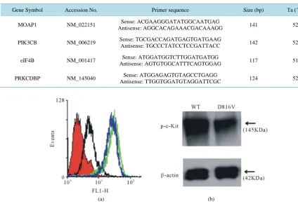
Specific genes and their expression levels were validated using real-time PCR (RT-PCR) [19]. Total RNA was isolated using GenElute Mammalian Total RNA kit (Sigma, USA). cDNA synthesis was performed using Super Script III cDNA synthesis kit (Invitrogen, USA) as per the manufacturer’s instructions. RT-PCR was done using SYBR green quantitative RT-PCR kit (Sigma, USA) following manufacturer protocol. Gene specific primers were designed by Beacon designer software. Amplification of target genes were performed with thermal cycling condition of 95˚C for 10 min followed by amplification cycles of 95˚C for 30 seconds, 52˚C for 60 seconds, 72˚C for 30 seconds in a spectrofluorometric thermal cycler (Stratagene). Data was analyzed by using the comparative ΔΔCt method [20] by normalizing the ΔCT values for each gene to the ΔCT values of the housekeeping gene, actin. No template control (without cDNA template) was taken as negative control for RT-PCR. Data was nor-malized by the amount of β-actin mRNA. The primer sets and length of the PCR products are listed in Table 1.
3.6. Treatment with p38MAPK Inhibitor (SB202190)
4. Results
4.1. Generation of Stable Mutant D816V-c-Kit Cells Using Doxycycline Regulated Gene Expression
Mutant D816V-c-Kit construct was validated by sequencing across the manipulated region. To study signaling pathways of D816V-c-Kit in progenitor cells, we used U937 cells transfected with wild-type and mutant D816V-c-Kit. Both wild-type c-Kit and the D816V mutant of c-Kit were stably transfected into the hematopoie-tic cell line U937 using the Tet-off inducible gene expression system [22]-[24] (Figure 3(a)). Flow cytometry analysis showed no change in fluorescence intensity between wild-type c-Kit expressing (induced) and mutant D816V-c-Kit (induced) cells. Induced mutant D816V-c-Kit cells showed significant shift in fluorescence inten-sity compared to uninduced mutant D816V-c-Kit cells (with doxycycline in media) and isotype control. By western blotting, wild-type c-Kit expressing cells and mutant D816V-c-Kit expressing cells showed receptor expression using c-Kit monoclonal antibody (Figure 3(b)).
4.2. Mutant D816V-c-Kit Mutant Revealed Enhanced Proliferation as Compared to Wild Type c-Kit
[image:6.595.87.512.342.636.2]The directed proliferation potential of D816V-c-Kit expressing (induced) cells as compared to wild-type c-Kit expressing (induced) cells was analyzed in vitro by MTS assay. As shown Figure 4, D816V-c-Kit expressing cells showed significantly higher proliferation than huSCF (100 ng/ml) stimulated wild-type c-Kit expressing
Table 1. Details of primers used for real-time PCR in induced c-Kit mutant D816V expressing cells.
Gene Symbol Accession No. Primer sequence Size (bp) Ta (˚C)
MOAP1 NM_022151 Sense: ACGAAGGGATATGGCAATGAG
Antisense: AGGCACAGAAACGACAAAGG 141 52
PIK3CB NM_006219 Sense: TGCGACCAGATGAGTGATGAAG
Antisense: TGCCCTATCCTCCGATTACC 142 52
eIF4B NM_001417 Sense: ATGGATGGTCTTGGATGATGG
Antisense: AGTGTGGCATTTCAGTGGAG 117 51
PRKCDBP NM_145040 Sense: ATGGAGAGTGTAGCCTGAGG
Antisense: TTGGTGGATGTAGGATTCGC 124 52
(a) (b)
Figure 4. Mitogenic assay. Wild-type-c-Kit and D816V-c-Kit expressing cells number were as-sessed by MTS assay and expressed as relative absorbance units. Results are expressed as mean ± SD of triplicate and representative of at least 4 independent experiments (*p < 0.05).
(induced) cells. The wild-type c-Kit expressing cells showed response to growth factor, huSCF by showing their higher proliferation in presence of huSCF. However, mutant D816V-c-Kit expressing cells displayed no signifi-cant change in proliferation in the presence or absence of huSCF.
4.3. Transcriptional Response of Mutant D816V-c-Kit in Mutant Cells
Among the differentially regulated genes, genes involved in cell proliferation, PIK3R1 (0.82 fold, p = 0.02), FES (1.06 fold, p = 0.0353), eIF4B (1.11 fold, p = 0.02), ABL2 (0.67 fold, p = 0.0009), MAPK family, MAP2K3 (0.87 fold, p = 0.02), MAPK14 (1.49 fold, p = 0.00), Ras gene family, RAB7B (1.24 fold, p = 0.01), RAB3GAP1 (1.07 fold, p = 0.01), cell adhesion, ABL2 (0.67 fold, p = 0.00), LY9 (0.72 fold, p = 0.03), apopto-sis, MOAP1 (−1.42 fold, p = 0.04), transcription factors, TAF5L/PCAF (1.09 fold, p = 0.01), nuclear receptors, U2AF (0.85 fold, p = 0.00), and metabolism, CYP2B6 (0.8 fold, p = 0.02), HMGCS1 (0.87 fold, p = 0.02) were altered in mutant D816V-c-Kit expressing cells. Expression of a number of other signaling molecules involved in mutant D816V-c-Kit mediated ligand-independent proliferation modulated were SLA (1.14 fold, p = 0.155) and STAT5 (2.32 fold, p = 0.103).
4.4. Validation of Microarray data
To verify expression levels of transcripts obtained from microarray, real-time PCR analysis was performed. Up-regulated genes chosen were PIK3R1 (0.82 fold) and eIF4B (1.11 fold); down-regulated genes were MOAP1 (−1.42 fold, p < 0.04) and PRKCDBP (−1.29 fold, p < 0.01). These genes depicted an identical pattern of alteration as seen by microarray expression analysis (Figure 5(a)). Further, we also confirmed expression of few genes which did not qualify selection criteria but are of relevance to the current study such as Src like adap-tor, SLA (1.14 fold, p < 0.155) and Signal transducer and activator of transcription 5, STAT5B (2.32 fold, p < 0.103) (Figure 5(b)).
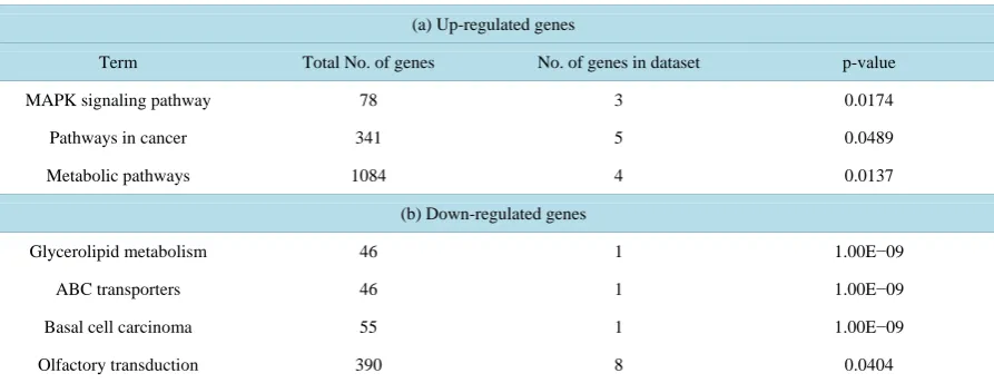
4.5. Gene Ontology Analysis
Further evaluation of biological processes and functions affected by mutant D816V-cKit expressing cells using DAVID bioinformatics resources identified upregulated genes to be functionally involved in MAPK signaling pathway (MAP2K3, MAPK14), mTOR signaling pathway (PIK3R1, EIF4B), cell cycle (eIF4B, ABL2), cell proliferation (INSIG1, FES, PIK3R1), transcription factors (TAF5L/PCAF, STAT5B) and cell adhesion (LY9). The down-regulated genes consisted of glyceroid metabolism (PPAP2C), basal cell carcinoma (MYCL1), cell differentiation (NDRG4, PDLIM7) and PCLKC (involved in negative regulation for cell growth). Significant (p < 0.04) over represented Gene Ontology terms (molecular function) in down and up-regulated genes is shown in
[image:7.595.222.405.84.231.2]Figure 5. Validation of mRNA levels of genes from microarray data. (a) qPCR assessment of genes showing statistically significant difference on the microarray. mRNA levels of proliferative genes (MOAP1, PI3KCB, eIF4B and PRKCDBP) in mutant D816V-c-Kit expressing U937 cells by Real-Time PCR; (b) qPCR assessment of genes (SLA and STAT5B) not showing statistically significant difference in microarray. Data of 3 independent experiments presented as mean ± SD (*p < 0.05).
Table 2. Gene ontology (biological processes) terms over-represented (p≤ 0.05) in the set of genes altered by c-Kit mutant form D816V.
(a) Up-regulated genes
Term Total No. of genes No. of genes in dataset p-value
MAPK signaling pathway 78 3 0.0174
Pathways in cancer 341 5 0.0489
Metabolic pathways 1084 4 0.0137
(b) Down-regulated genes
Glycerolipid metabolism 46 1 1.00E−09
ABC transporters 46 1 1.00E−09
Basal cell carcinoma 55 1 1.00E−09
Olfactory transduction 390 8 0.0404
4.6. Dose-Dependent Cytotoxic Effect of p38 MAP Kinase in Mutant D816V-c-Kit Cells Analysis of %viability in U937 cells in the presence of p38 MAP kinase inhibitor (SB202190) at the indicated concentrations showed that p38 MAP kinase inhibitor showed no cytotoxicity at concentration of 1 µM in 24 hrs (Figure 6(a)) but showed reduction in p38 MAP kinase phosphorylation compared to the vehicle, DMSO as shown by immunoblotting using phospho-p38 MAP kinase antibody (Figure 6(b)).
4.7. Generation of Stable Mutant D816V-c-Kit Cells Using Doxycycline Regulated Gene Expression
[image:8.595.91.538.369.541.2](a) (b)
Figure 6. Effects of p38MAP kinase inhibitor on cell viability in U937 cells. (a) U937 cells were cultured with various concentrations of p38MAP Kinase inhibitor (0.1 - 7 μM). U937 cells were treated with the indicated concentrations of p38MAP kinase inhibitor for 2 hrs. The cell viability was measured by the metabolic-dye-based MTS assay. Results represent the mean ± SD of three experiments performed in triplicate. The significance was determined by Student’s t-test (*p < 0.05 vs vehicle control); (b) Immunoblot using phospho-p38MAP kinase antibody. Western blot analysis of activated phospho-p38 in the lysates of U937 cells either pretreated with p38MAP kinase inhibitor (0.5 μM and 1 μM) or vehicle control. Blotted proteins were probed with an-ti-phospho-p38 and then with anti-p38 antibodies, each followed by peroxidase-conjugated sec-ondary antibody. The level of β-actin is shown at the bottom as a loading control. One represent-ative experiment of three independent experiments is shown.
D816V-c-Kit (induced) cells. Induced mutant D816V-c-Kit cells showed significant shift in fluorescence inten-sity compared to uninduced mutant D816V-c-Kit cells (with doxycycline in media) and isotype control. By western blotting, wild-type c-Kit expressing cells and mutant D816V-c-Kit expressing cells showed receptor expression using c-Kit monoclonal antibody (Figure 3(b)).
4.8. Mutant D816V-c-Kit Mutant Revealed Enhanced Proliferation as Compared to Wild Type c-Kit
The directed proliferation potential of D816V-c-Kit expressing (induced) cells as compared to wild-type c-Kit expressing (induced) cells was analyzed in vitro by MTS assay. As shown Figure 4, D816V-c-Kit expressing cells showed significantly higher proliferation than huSCF (100 ng/ml) stimulated wild-type c-Kit expressing (induced) cells. The wild type c-Kit expressing cells showed response to growth factor, huSCF, by showing their higher proliferation in presence of huSCF. However, mutant D816V-c-Kit expressing cells displayed no signifi-cant change in proliferation in the presence or absence of huSCF.
4.9. Transcriptional Response of Mutant D816V-c-Kit in Mutant Cells
Among the differentially regulated genes, genes involved in cell proliferation, PIK3R1 (0.82 fold, p = 0.02), FES (1.06 fold, p = 0.0353), eIF4B (1.11 fold, p = 0.02), ABL2 (0.67 fold, p = 0.0009), MAPK family, MAP2K3 (0.87 fold, p = 0.02), MAPK14 (1.49 fold, p = 0.00), Ras gene family, RAB7B (1.24 fold, p = 0.01), RAB3GAP1 (1.07 fold, p = 0.01), cell adhesion, ABL2 (0.67 fold, p = 0.00), LY9 (0.72 fold, p = 0.03), apopto-sis, MOAP1 (−1.42 fold, p = 0.04), transcription factors, TAF5L/PCAF (1.09 fold, p = 0.01), nuclear receptors, U2AF (0.85 fold, p = 0.00), and metabolism, CYP2B6 (0.8 fold, p = 0.02), HMGCS1 (0.87 fold, p = 0.02) were altered in mutant D816V-c-Kit expressing cells. Expression of a number of other signaling molecules involved in mutant D816V-c-Kit mediated ligand-independent proliferation modulated were SLA (1.14 fold, p = 0.155) and STAT5 (2.32 fold, p = 0.103).
4.10. Validation of Microarray Data
Up-regulated genes chosen were PIK3R1 (0.82 fold) and eIF4B (1.11 fold); down-regulated genes were MOAP1 (−1.42 fold, p < 0.04) and PRKCDBP (−1.29 fold, p < 0.01). These genes depicted an identical pattern of alteration as seen by microarray expression analysis (Figure 5(a)). Further, we also confirmed expression of few genes which did not qualify selection criteria but are of relevance to the current study such as Src like adap-tor, SLA (1.14 fold, p < 0.155) and Signal transducer and activator of transcription 5, STAT5B (2.32 fold, p < 0.103) (Figure 5(b)).
4.11. Gene Ontology Analysis
Further evaluation of biological processes and functions affected by mutant D816V-cKit expressing cells using DAVID bioinformatics resources identified upregulated genes to be functionally involved in MAPK signaling pathway (MAP2K3, MAPK14), mTOR signaling pathway (PIK3R1, EIF4B), cell cycle (eIF4B, ABL2), cell proliferation (INSIG1, FES, PIK3R1), transcription factors (TAF5L/PCAF, STAT5B) and cell adhesion (LY9). The down-regulated genes consisted of glyceroid metabolism (PPAP2C), basal cell carcinoma (MYCL1), cell differentiation (NDRG4, PDLIM7) and PCLKC (involved in negative regulation for cell growth). Significant (p < 0.04) over represented Gene Ontology terms (molecular function) in down and up-regulated genes is shown in
Table 2.
4.12. Dose-Dependent Cytotoxic Effect of p38 MAP Kinase in Mutant D816V-c-Kit Cells Analysis of %viability in U937 cells in the presence of p38 MAP kinase inhibitor (SB202190) at the indicated concentrations showed that p38 MAP kinase inhibitor showed no cytotoxicity at concentration of 1 µM in 24 hrs (Figure 6(a)) but showed reduction in p38 MAP kinase phosphorylation compared to the vehicle, DMSO as shown by immunoblotting using phospho-p38 MAP kinase antibody (Figure 6(b)).
4.13. p38 MAP Kinase Inhibitor Mediated Reduction of Proliferation in Mutant D816V-c-Kit Cells
[image:10.595.221.404.500.664.2]The vehicle, DMSO did not modify any investigated parameter in comparison with control culture. Investigation of the effect of p38 MAP kinase inhibitor in mutant D816V-c-Kit cells revealed significant reduction in cell pro-liferation. Mutant D816V-c-Kit expressing cells without huSCF showed more inhibition as compared to huSCF induced growth of mutant cells. Wild-type c-Kit expressing cells showed no effect on proliferation by p38 MAP kinase inhibitor either in presence or absence of huSCF. U937 cells showed no significant reduction in overall survival in the presence or absence of inhibitor, suggesting no cytotoxic effects on cell survival at dose of 1 µM at 24 hrs (Figure 7).
5. Discussion
Receptor tyrosine kinases are one of the most important targets underlying oncogenesis as their deregulation in-creases cellular proliferation in hematological malignancies [5]. D816V-c-Kit mutation is considered to be the most common gaof-function mutation in case of mastocytosis, a myelo-proliferative neoplasm, and is in-volved to a lesser extent in germ-cell tumors, acute myeloid leukemia and mucosal melanoma [11]. Though, c-Kit inhibitor, imatinib is widely used in treatment of these diseases, however, imatinib fails to inhibit cells that exhibit the D816V mutation [12] [13]. New paradigm of cancer therapy involves the molecular characteristics of tumor to guide the therapeutic regimens. Biological significance of D816V mutation of c-Kit needs to be defined at molecular level to target causative kinase proteins for anti-cancer therapies.
Therefore, in the present study, we tried to understand the molecular response of mutant form of c-Kit at D816V residue c-Kit for the following reasons: 1) c-Kit mutation at residue D816V significantly impairs the ef-ficacy of cancer therapies, limiting the treatment options for therapies; 2) the signaling pathways activated by D816V-c-Kit remain largely unidentified and 3) this will aid in developing novel molecular inhibitors with the potential to overcome resistance mutations. We investigated the downstream signaling events in D816V-c-Kit expressing cells compared to wild-type c-Kit using high-throughput gene expression profiling. Specifically, we developed a stable mutant D816V-c-Kit expressing cellular system and studied its correlation with proliferation and altered signal transduction compared to wild-type c-Kit expressing cells. We selected human hematopoietic progenitor U937 cells as this cell population is huSCF and c-Kit null at mRNA transcript level, which would eliminate the interference of host endogenous c-Kit gene expression [25]. Also, human U937 cells resource of-fers the opportunity to explore the study of c-Kit mutant expression and its function in the complex human con-ditions, thus would enable support for translational medical research in future. Expression of c-Kit gene could be done through viral transduction; however, transduction with viral vectors involves the risk of malignant trans-formation [26]. Furthermore, it has been reported that low efficiency of viral transduction of HSCs in human clinical trials severely decrease the level of chimerism or long-term repopulating ability of hematopoietic cells [27]. Further, to avoid undesirable oncogenic effects by constitutive activated signaling, we first made an effort to develop a system for transgene expression of c-Kit (wild-type and mutant) in a regulated manner using Tet-off inducible gene expression vector system. Additionally, this also offers the opportunity to precisely ex-plore and study the transgene expression and its function in mammalian cell system. In this system, transacti- vator (tTA) is prevented from interacting with its binding site on DNA (tetO) by the effector substance, doxycy-cline, at low non-toxic concentrations [28]. Consequently, induced mutant D816V-c-Kit expressing cells (with-out doxycycline) showed much higher expression than uninduced mutant D816V-c-Kit cells (with doxycycline in media) by flow cytometry. Unexpectedly, mutant D816V-c-Kit expressing cells showed similar expression to wild type c-Kit expressing cells instead of showing higher receptor expression. This is due to increased rate of degradation of surface receptor mutant D816V-c-Kit and subsequently rapid receptor turnover [29].
After achieving the doxycycline inducible regulated transgene surface expression of c-Kit genetic construct in the hematopoietic progenitor cell line U937, we further analyzed the proliferation of these cells towards huSCF
in vitro. We used serum-free media to avoid non-specific activation of c-Kit by other serum proteins, thus to re-duce signal-to-noise ratio. Our results revealed significantly enhancedproliferation of the mutant D816V-c-Kit expressing U937 cells as compared to wild-type c-Kit expressing cells. D816V-c-Kit expressing cells showed similar proliferation in presence or absence of huSCF (ligand-independent) whereas wild-type c-Kit cells showed proliferation in presence of huSCF only (ligand-dependent). huSCF showed no contribution for D816V mediated leukemogenicity in D816V-c-Kit expressing U937 cells unlike in mast cells [30]. This signifies for difference in mutant D816V-c-Kit downstream signaling compared to wild-type c-Kit, which ultimately regu-lates the cell proliferation.
as the PI3K signaling, STAT, or RAS/MAPK pathway [31]. Quantitative PCR validated upregulation of PIK3R1. Earlier studies have also shown that PIK3R1 encoded protein, p85 subunit ofPI3K, is downstream target of D816V-c-Kit [32]. Rather, PI3K is constitutively associated with D816V-c-Kit receptor [32]. PI3K has been earlier shown to be implicated as participant in signaling pathways regulating multiple cellular functions, in-cluding proliferation, differentiation, anti-apoptosis and tumorigenesis [33] [34]. Another proliferative gene, FES, which encodes protein with tyrosine kinase activity, was also discerned up-regulated in mutant D816V-c- Kit expressing cells. This is in accordance with previous studies which have shown that FES is an essential ef-fector of D816V-c-Kit proliferation signal. Reduction of FES expression leads to decrease in proliferation of D816V-c-Kit expressing cells [35]. Another up-regulated gene, eukaryotic translation initiation factor 4B (EIF4B) encode for proteins that are involved in the early initiation of protein synthesis. PI3K signaling pathway has also been shown to be related to EIF4B [36].
MOAP1 and PRKCDBP down-regulation in mutant D816V-c-Kit expressing cells compared to wild-type c-Kit expressing cells were also validated by quantitative PCR. MOAP1 gene encodes protein which function-ally mediates caspase-dependent apoptosis by its interaction with apoptosis regulator protein [37]. The gene en-coding protein kinase C delta binding protein, PRKCDBP expression has been shown to induce the G1 cell-cycle arrest and increased cellular sensitivity to various apoptotic stresses [38]. The expression of this pro-tein was found to be down-regulated in various cancer cell lines, suggesting the possible tumor suppressor func-tion of this protein.
Few up-regulated microarray outcome genes, selectively, SLA (1.14 fold, p < 0.15) and STAT5B (2.32 fold, p < 0.10), did not meet the selection criteria, however, exhibit statistically significant change in expression when assessed by quantitative RT-PCR. This difference arises because of the sensitivities of the two techniques and use of different statistical analysis methods. It has been reported that SLA recruits ubiquitin ligases, which tag mutant D816V-c-Kit for degradation, contributing to its lower surface expression compared to wild-type c-Kit [39]. This is the most common mechanism through which SLA protein regulate receptor stability as well as downstream signaling [40]. STAT5 up-regulation by mutant D816V-c-Kit expressing cells as downstream target is consistent with STAT5 up-regulation as downstream target in D816V-c-Kit expressing HMC-1 cells impor-tant for cell proliferation. STAT activation can be aberrant as it is involved with other pathological situations [31] [41]. The up-regulation of BCR/ABL in D816V-c-Kit expressing cells, which encodes for kinase active fusion protein, opens the possibility that mutant D816V phosphorylates additional substrate molecules that are not phosphorylated by wild-type c-Kit and is expected to constitute novel targets for selective therapy. Earlier stu-dies reported that Bcr/Abl kinase is an oncogenic fusion which protects hematopoietic progenitor cells from spontaneous apoptosis [42].
potential target for therapeutic intervention.
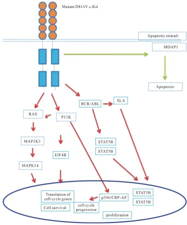
Overall, the results of this study indicate that mutant D816V-c-Kit expressing cells show upregulation of gene expression of BCR-ABL, PI3K, SLA, p38 MAPK and STAT5 pathways for cellular proliferation (Figure 8). Further in-depth broadband research of mutant D816V-c-Kit receptor would unveil an approach to manipulate HSCs with molecular targeted drugs to maximize efficacy with minimal dose.
[image:13.595.124.498.155.604.2]6. Conclusion
Present study demonstrated that D816V-c-Kit mutant is a key molecular player displaying efficacy for factor- independent hematopoietic cellular hyper-proliferation. Genes (PIK3R1, eIF4B, SLA, MAPK14, STAT5) mod-ulated upon mutant D816V-c-Kit induction reveals transformation of huSCF-dependent cells to factor indepen-dent proliferation. A better understanding of the relationship between MAP kinase signal transduction system and the regulation of cell proliferation is essential for the rational design of novel pharmaco-therapeutic ap-proaches [47]. Study provides a better fundamental understanding for cellular and molecular mechanism of mu-tant D816V-c-Kit, by revealing the up-regulation of group of genes which are known as crucial regulators of hematopoietic proliferation and survival. Deciphering molecular targets and functional role of dozens of uncha-racterized genes identified needs to be undertaken along with identification of signaling pathways that would help pharmacologists and molecular biologists to design novel drugs targeting at site responsible for mutation D816V mediated tumorous transformation of c-Kit.
Acknowledgements
We are thankful to Dr. R.P. Tripathi, Director of INMAS for providing us necessary facilities. Authors also thank the Ministry of Defence, Department of Defence Research and Development for research grant. We thank Mrs. Namita Kalra for help during data acquisition by flow cytometry. Shilpa Sharma in particular thanks Indian Council of Medical Research (ICMR) for the award of Senior Research Fellow and Research Associateship.
References
[1] Yarden, Y., Kuang, W.J., Yang, F.T., Coussens, L., Munemitsu, S., Dull, T.J., et al. (1987) Human Proto-Oncogene c-Kit: A New Cell Surface Receptor Tyrosine Kinase for an Unidentified Ligand. EMBO Journal, 6, 3341-3351. [2] Qiu, F., Ray, P., Brown, K., Barker, P.E., Jhanwar, S., Ruddle, F.H. and Besmer, P. (1988) Primary Structure of c-Kit:
Relationship with the CSF-1/PDGF Receptor Kinase Family Oncogenic Activation of v-Kit Involves Deletion of Extracellular Domain and C Terminus. EMBO Journal, 7, 1003-1011.
[3] Ashman, L.K., Cambareri, A.C., To, L.B., Levinsky, R.J. and Juttner, C.A. (1991) Expression of the YB5.B8 Antigen (c-Kit Proto-Oncogene Product) in Normal Human Bone Marrow. Blood, 78, 30-37.
[4] Papayannopoulou, T., Brice, M., Broudy, V.C. and Zsebo, K.M. (1991) Isolation of c-Kit Receptor-Expressing Cells from Bone Marrow, Peripheral Blood, and Fetal Liver: Functional Properties and Composite Antigenic Profile. Blood, 78, 1403-1412.
[5] Sharma, S., Gurudutta, G.U., Satija, N.K., Pati, S, Afrin, F., Gupta, P., et al. (2006) Stem Cell c-KIT and HOXB4 Genes: Critical Roles and Mechanisms in Self-Renewal, Proliferation, and Differentiation. Stem Cells and Develop-ment, 15, 755-778. http://dx.doi.org/10.1089/scd.2006.15.755
[6] Ashman, L.K. (1999) The Biology of Stem Cell Factor and Its Receptor c-Kit. International Journal of Biochemistry &
Cell Biology, 31, 1037-1051. http://dx.doi.org/10.1016/S1357-2725(99)00076-X
[7] Linnekin, D. (1999) Early Signaling Pathways Activated by c-Kit in Haematopoietic Cells. International Journal of
Biochemistry & Cell Biology, 31, 1053-1074. http://dx.doi.org/10.1016/S1357-2725(99)00078-3
[8] Longley, B.J., Reguera, M.J. and Ma, Y. (2001) Classes of c-Kit Activating Mutations: Proposed Mechanisms of Ac-tion and ImplicaAc-tions for Disease ClassificaAc-tion and Therapy. Leukemia Research, 25, 571-576.
http://dx.doi.org/10.1016/S0145-2126(01)00028-5
[9] Rodrigues, G.A. and Park, M. (1994) Oncogenic Activation of Tyrosine Kinases. Current Opinion in Genetics &
De-velopment, 4, 15-24. http://dx.doi.org/10.1016/0959-437X(94)90086-8
[10] Moriyama, Y., Tsujimura, T., Hashimoto, K., Morimoto, M., Kitayama, H., Matsuzawa, Y., et al. (1996) Role of As-partic Acid 814 in the Function and Expression of c-Kit Receptor Tyrosine Kinase. Journal of Biological Chemistry, 271, 3347-3350. http://dx.doi.org/10.1074/jbc.271.7.3347
[11] Agarwal, S., Kazi, J.U., Mohlin, S., Pahlman, S. and Ronnstrand, L. (2015) The Activation Loop Tyrosine 823 Is Es-sential for the Transforming Capacity of the c-Kit Oncogenic Mutant D816V. Oncogene, 34, 4581-4590.
http://dx.doi.org/10.1038/onc.2014.383
[12] Tsujimura, T., Hashimoto, K., Kitayama, H., Ikeda, H., Sugahara, H., Matsumura, I., et al. (1999) Activating Mutation in the Catalytic Domain of c-Kit Elicits Hematopoietic Transformation by Receptor Self-Association Not at the Li-gand-Induced Dimerization Site. Blood, 93, 1319-1329.
http://dx.doi.org/10.1021/jm500413g
[14] Baldi, P. and Long, A.D. (2001) A Bayesian Framework for the Analysis of Microarray Expression Data: Regularized t-Test and Statistical Inferences of Gene Changes. Bioinformatics, 17, 506-519.
http://dx.doi.org/10.1093/bioinformatics/17.6.509
[15] Murie, C., Woody, O., Lee, A.Y. and Nadon, R. (2009) Comparison of Small n Statistical Tests of Differential Expres-sion Applied to Microarrays. BMC Bioinformatics, 10, 45. http://dx.doi.org/10.1186/1471-2105-10-45
[16] Huang, D.W., Sherman, B.T. and Lempicki, R.A. (2009) Systematic and Integrative Analysis of Large Gene Lists Us-ing DAVID Bioinformatics Resources. Nature Protocols, 4, 44-57. http://dx.doi.org/10.1038/nprot.2008.211
[17] Pandey, R., Guru, R.K. and Mount, D.W. (2004) Pathway Miner: Extracting Gene Association Networks from Mole-cular Pathways for Predicting the Biological Significance of Gene Expression Microarray Data. Bioinformatics, 20, 2156-2158. http://dx.doi.org/10.1093/bioinformatics/bth215
[18] Curtis, R.K., Oresic, M. and Vidal-Puig, A. (2005) Pathways to the Analysis of Microarray Data. Trends in
Biotech-nology, 23, 429-435. http://dx.doi.org/10.1016/j.tibtech.2005.05.011
[19] Heid, C.A., Stevens, J., Livak, K.J. and Williams, P.M. (1996) Real Time Quantitative PCR. Genome Research, 6, 986-994. http://dx.doi.org/10.1101/gr.6.10.986
[20] Livak, K.J. and Schmittgen, T.D. (2001) Analysis of Relative Gene Expression Data Using Real-Time Quantitative PCR and the 2-ΔΔCT Method. Methods, 25, 402-408. http://dx.doi.org/10.1006/meth.2001.1262
[21] Warrior, U., Chiou, X.G., Sheets, M.P., Sciotti, R.J., Parry, J.M., Simmer, R.L., et al. (1999) Development of a p38 Kinase Binding Assay for High Throughput Screening. Journal of Biomolecular Screening, 4, 129-135.
http://dx.doi.org/10.1177/108705719900400306
[22] Gossen, M., Freundlieb, S., Bender, G., Müller, G., Hillen, W. and Bujard, H. (1995) Transcriptional Activation by Te-tracyclines in Mammalian Cells. Science, 268, 1766-1769. http://dx.doi.org/10.1126/science.7792603
[23] Gossen, M., Bonin, A.L., Freundlieb, S. and Bujard, H. (1994) Inducible Gene Expression Systems for Higher Euka-ryotic Cells. Current Opinion in Biotechnology, 5, 516-520. http://dx.doi.org/10.1016/0958-1669(94)90067-1
[24] Mayerhofer, M., Gleixner, K.V., Hoelbl, A., Florian, S., Hoermann, G., Aichberger, K.J., et al. (2008) Unique Effects of KIT D816V in BaF3 Cells: Induction of Cluster Formation, Histamine Synthesis, and Early Mast Cell Differentia-tion Antigens. The Journal of Immunology, 180, 5466-5476. http://dx.doi.org/10.4049/jimmunol.180.8.5466
[25] Chandrashekran, A., Gordon, M.Y. and Casimir, C. (2004) Targeted Retroviral Transduction of c-Kit+ Hematopoietic Cells Using Novel Ligand Display Technology. Blood, 104, 2697-2703.
http://dx.doi.org/10.1182/blood-2003-10-3717
[26] Sadelain, M. (2004) Insertional Oncogenesis in Gene Therapy: How Much of a Risk? Gene Therapy, 11, 569-573. http://dx.doi.org/10.1038/sj.gt.3302243
[27] Schuster, J.A., Stupnikov, M.R., Ma, G., Liao, W., Lai, R., Ma, Y. and Aguila, J.R. (2012) Expansion of Hematopoie-tic Stem Cells for Transplantation: Current Perspectives. Experimental Hematology & Oncology, 1, 12.
http://dx.doi.org/10.1186/2162-3619-1-12
[28] Woods, S.L. and Bishop, J.M. (2011) A New Transgenic Mouse Line for Tetracycline Inducible Transgene Expression in Mature Melanocytes and the Melanocyte Stem Cells Using Dopachrome tautomerase Promoter. Transgenic
Re-search, 20, 421-428. http://dx.doi.org/10.1007/s11248-010-9421-6
[29] Sun, J., Pedersen, M. and Ronnstrand, L. (2009) The D816V Mutation of c-Kit Circumvents a Requirement for Src Family Kinases in c-Kit Signal Transduction. c-Kit Expression in Human Megakaryoblastic Leukemia Cell Lines, The
Journal of Biological Chemistry, 284, 11039-11047. http://dx.doi.org/10.1074/jbc.M808058200
[30] Amagai, Y., Tanaka, A., Matsuda, A., Jung, K., Ohmori, K. and Matsuda, H. (2013) Stem Cell Factor Contributes to Tumorigenesis of Mast Cells via an Autocrine/Paracrine Mechanism. Journal of Leukocyte Biology, 93, 245-250. http://dx.doi.org/10.1189/jlb.0512245
[31] Chaix, A., Lopez, S., Voisset, E., Gros, L., Dubreuil, P. and De Sepulveda, P. (2011) Mechanisms of STAT Protein Activation by Oncogenic KIT Mutants in Neoplastic Mast Cells. The Journal of Biological Chemistry, 286, 5956-5966. http://dx.doi.org/10.1074/jbc.M110.182642
[32] Chian, R., Young, S., Danilkovitch-Miagkova, A., Ronnstrand, L., Leonard, E., Ferrao, P., Ashman, L. and Linnekin, D. (2001) Phosphatidylinositol 3 Kinase Contributes to the Transformation of Hematopoietic Cells by the D816V c-Kit Mutant. Blood, 98, 1365-1373. http://dx.doi.org/10.1182/blood.V98.5.1365
[33] Blume-Jensen, P., Janknecht, R. and Hunter, T. (1998) The Kit Receptor Promotes Cell Survival via Activation of PI 3-Kinase and Subsequent Akt-Mediated Phosphorylation of Bad on Ser136. Current Biology, 8, 779-782.
http://dx.doi.org/10.1016/s0960-9822(98)70302-1
and Therapeutic Implications. Current Genomics, 8, 271-306. http://dx.doi.org/10.2174/138920207782446160
[35] Voisset, E., Lopez, S., Dubreuil, P. and De Sepulveda, P. (2007) The Tyrosine Kinase FES Is an Essential Effector of KITD816V Proliferation Signal. Blood, 110, 2593-2599. http://dx.doi.org/10.1182/blood-2007-02-076471
[36] Gabillot-Carre, M., Lepelletier, Y., Humbert, M., de Sepuvelda, P., Hamouda, N.B., Zappulla, J.P., et al. (2006) Ra-pamycin Inhibits Growth and Survival of D816V-Mutated c-Kit Mast Cells. Blood, 108, 1065-1072.
http://dx.doi.org/10.1182/blood-2005-06-2433
[37] Foley, C.J., Freedman, H., Choo, S.L., Onyskiw, C., Fu, N.Y., Yu, V.C., et al. (2008) Dynamics of RASSF1A/MOAP- 1 Association with Death Receptors. Molecular and Cellular Biology, 28, 4520-4535.
http://dx.doi.org/10.1128/MCB.02011-07
[38] Lee, J.H., Kang, M.J., Han, H.Y., Lee, M.G., Jeong, S., Ryu, B.K., et al. (2011) Epigenetic alteration of PRKCDBP in Colorectal Cancers and Its Implication in Tumor Cell Resistance to TNFα-Induced Apoptosis. Clinical Cancer
Re-search, 17, 7551-7562. http://dx.doi.org/10.1158/1078-0432.CCR-11-1026
[39] Kazi, J.U., Agarwal, S., Sun, J., Bracco, E. and Ronnstrand, L. (2014) Src-Like-Adaptor Protein (SLAP) Differentially Regulates Normal and Oncogenic c-Kit Signaling. Journal of Cell Science, 127, 653-662.
http://dx.doi.org/10.1242/jcs.140590
[40] Dragone, L.L., Shaw, L.A., Myers, M.D. and Weiss, A. (2009) SLAP, a Regulator of Immunoreceptor Ubiquitination, Signaling, and Trafficking. Immunological Reviews, 232, 218-228.
http://dx.doi.org/10.1111/j.1600-065X.2009.00827.x
[41] Bibi, S., Arslanhan, M.D., Langenfeld, F., Jeanningros, S., Cerny-Reiterer, S., Hadzijusufovic, E., et al. (2014) Coope-rating STAT5 and AKT Signaling Pathways in Chronic Myeloid Leukemia and Mastocytosis: Possible New Targets of Therapy. Haematologica, 99, 417-429. http://dx.doi.org/10.3324/haematol.2013.098442
[42] Yu, C., Krystal, G., Varticovksi, L., McKinstry, R., Rahmani, M., Dent, P., et al. (2002) Pharmacologic Mitogen-Act- ivated Protein/Extracellular Signal Regulated Kinase Kinase/ Mitogen Activated Protein Kinase Inhibitors Interact Synergistically with STI571 to Induce Apoptosis in Bcr/Abl-Expressing Human Leulemia Cells. Cancer Research, 62, 188-199.
[43] Koul, H.K., Pal, M. and Koul, S. (2013) Role of p38 MAP Kinase Signal Transduction in Solid Tumors. Genes &
Cancer, 4, 342-359. http://dx.doi.org/10.1177/1947601913507951
[44] Maher, P. (1999) p38 Mitogen-Activated Protein Kinase Activation Is Required for Fibroblast Growth Factor-2-Sti- mulated Cell Proliferation but Not Differentiation. The Journal of Biological Chemistry, 274, 17491-17498.
http://dx.doi.org/10.1074/jbc.274.25.17491
[45] Kanda, Y., Nishio, E., Kuroki, Y., Mizuno, K. and Watanabe, Y. (2001) Thrombin Activates p38 Mitogen-Activated Protein Kinase in Vascular Smooth Muscle Cells. Life Sciences, 68, 1989-2000.
http://dx.doi.org/10.1016/S0024-3205(01)00990-0
[46] Adam, R.M., Roth, J.A., Cheng, H.L., Rice, D.C., Khoury, J., Bauer, S.B., et al. (2003) Signaling Through Pi3k/Akt Mediates Stretch and PDGF-Bb-Dependent DNA Synthesis in Bladder Smooth Muscle Cells. Journal of Urology, 169, 2388-2393.http://dx.doi.org/10.1097/01.ju.0000063980.99368.35
[47] Zhang, W. and Liu, H.T. (2002) MAPK Signal Pathways in the Regulation of Cell Proliferation in Mammalian Cells.
Cell Research, 12, 1989-2000.http://dx.doi.org/10.1038/sj.cr.7290105