Dominant-negative effect of the heterozygous
C104R TACI mutation in common variable
immunodeficiency (CVID)
Lilit Garibyan, … , Kai W. Wucherpfennig, Raif S. Geha
J Clin Invest.
2007;
117(6)
:1550-1557.
https://doi.org/10.1172/JCI31023
.
B cells from patients with common variable immunodeficiency (CVID) who are
heterozygous for transmembrane activator and CAML interactor (TACI) mutation C104R,
which abolishes ligand binding, fail to produce Igs in response to TACI ligand. It is not
known whether this is due to haploinsufficiency or dominant interference. Using in vitro
transfection assays, here we demonstrate that C104R and the corresponding murine TACI
mutant, C76R, which also does not bind ligand, dominantly interfere with TACI signaling.
This effect was dependent on preassociation of the mutants with WT TACI in the absence of
ligand. The mutants did not interfere with ligand binding by WT TACI, suggesting that they
may act by disrupting ligand-induced receptor rearrangement and signaling. This work
demonstrates that TACI preassembles as an oligomeric complex prior to ligand binding and
provides a mechanistic insight into how the heterozygous C104R TACI mutation can
potentially lead to CVID.
Research Article
Immunology
Find the latest version:
Dominant-negative effect of the heterozygous
C104R TACI mutation in common variable
immunodeficiency (CVID)
Lilit Garibyan,1,2 Adrian A. Lobito,3 Richard M. Siegel,3 Matthew E. Call,4
Kai W. Wucherpfennig,4,5 and Raif S. Geha1,2
1Division of Immunology, Children’s Hospital Boston, Boston, Massachusetts, USA. 2Department of Pediatrics, Harvard Medical School, Boston,
Massachusetts, USA. 3Immunoregulation Unit, Autoimmunity Branch, National Institute of Arthritis and Musculoskeletal and Skin Diseases, NIH,
Bethesda, Maryland, USA. 4Department of Cancer Immunology and AIDS, Dana-Farber Cancer Institute, Boston, Massachusetts, USA. 5Department of Neurology, Harvard Medical School, Boston, Massachusetts, USA.
B cells from patients with common variable immunodeficiency (CVID) who are heterozygous for
transmem-brane activator and CAML interactor (TACI) mutation C104R, which abolishes ligand binding, fail to produce
Igs in response to TACI ligand. It is not known whether this is due to haploinsufficiency or dominant
inter-ference. Using in vitro transfection assays, here we demonstrate that C104R and the corresponding murine
TACI mutant, C76R, which also does not bind ligand, dominantly interfere with TACI signaling. This effect
was dependent on preassociation of the mutants with WT TACI in the absence of ligand. The mutants did not
interfere with ligand binding by WT TACI, suggesting that they may act by disrupting ligand-induced
recep-tor rearrangement and signaling. This work demonstrates that TACI preassembles as an oligomeric complex
prior to ligand binding and provides a mechanistic insight into how the heterozygous C104R TACI mutation
can potentially lead to CVID.
Introduction
Common variable immunodeficiency (CVID) is the most common primary immunodeficiency disease that comes to medical atten-tion. CVID is characterized by markedly reduced serum levels of IgG, IgA, and often IgM (1). In addition to having low levels of Igs, patients with CVID fail to produce specific antibodies after vaccination or exposure to antigens (2). CVID patients suffer from recurrent sinopulmonary, ear, and gastrointestinal infections and have increased risk of autoimmune and lymphoproliferative diseases (1). Most cases of CVID are sporadic, but about 10% are familial with a predominantly autosomal dominant inheritance (3). It has been recently found that TNFRSF13B, which encodes transmembrane activator and calcium-modulating ligand (CAML) interactor (TACI), is mutated in a small subgroup (approximately 5%–10%) of patients with CVID (4, 5). Most of these patients are heterozygous with respect to TACI mutation.
TACI is a cell-surface receptor that belongs to the TNF receptor (TNFR) superfamily and is mainly expressed on B cells (6). Struc-turally, TACI is a type III transmembrane protein. The extracellular (EC) domain contains 2 cysteine-rich domains (CRDs), which are hallmarks of the TNFR superfamily (6). Each CRD contains 6 highly conserved cysteines that participate in the formation of 3 intrachain disulfide bonds (Figure 1). Both human TACI (hTACI)
and murine TACI (mTACI) have 2 CRD domains that are con-served between the 2 species (Figure 1). The TNF family members BAFF (B cell–activating factor) and APRIL (a proliferation-induc-ing ligand) are ligands for TACI. Recently, TACI was found to bind to proteoglycans including syndecan-2 (7, 8). Functionally, TACI has been shown to be important in Ig class switching, Ig produc-tion, and regulation of B cell homeostasis (9–12). TACI-deficient mice have low levels of serum IgA and IgM and impaired antibody response to the type II T-independent antigens pneumococcal vac-cine polyvalent (Pneumovax) and TNP-Ficoll (11). In addition, as they age, these mice develop fatal disorders characterized by auto-immune nephritis and lymphoproliferation, leading to lymphoma in 15% of the mice (10).
One of the most common TACI mutations in patients with CVID is C104R, which is located in the EC domain of the receptor (13). The mutated C104 residue is located in the second CRD of TACI (Figure 1) and forms a disulfide bond with C93. This resi-due is highly conserved together with its surrounding sequence in all species examined (our unpublished observations). It has been demonstrated that the C104R mutation abolishes binding of BAFF and APRIL to TACI (4, 5). Most CVID patients with a C104R muta-tion are heterozygous. Furthermore, we have reported that B cells from such patients fail to produce Igs in response to APRIL (4). Two explanations have been proposed to explain the deleterious effect of the heterozygous C104R mutation on B cell function. One invokes haploinsufficiency because one normal copy of TACI may not be sufficient for proper function. However, mice with a single copy of the TACI gene have a normal phenotype (11), strongly argu-ing that TACI haploinsufficiency may not cause B cell dysfunction. Alternatively, the C104R mutant may act as a dominant negative and interfere with the function of the normal allele.
Given the variable age of onset of CVID, which extends into the seventh decade of life, and the genetic heterogeneity of the human
Nonstandard abbreviations used: APRIL, a proliferation-inducing ligand; BAFF, B cell–activating factor; CAML, calcium-modulating ligand; CD, cytoplasmic domain; CFP, cyan fluorescent protein; CRD, cysteine-rich domain; CVID, common variable immunodeficiency; DM, double mutant; DTSSP, 3,3′ -dithiobis[sulfosuccinimidyl-propionate]; EC, extracellular; FRET, fluorescence resonance energy transfer; IC, intracellular; PC, protein C–derived peptide; SA, streptavidin; SBP, SA-binding peptide; snIP, sequential nondenaturing IP; TACI, transmembrane activator and CAML interactor; TNFR, TNF receptor; YFP, yellow fluorescent protein.
Conflict of interest: The authors have declared that no conflict of interest exists.
research article
The Journal of Clinical Investigation http://www.jci.org Volume 117 Number 6 June 2007 1551
population, we concluded that the causality of TACI missense mutations is best established in a mouse model where mutations in mTACI that correspond to hTACI mutations found in CVID patients are introduced into a uniform genetic background. Prior to the generation of such a model, it was important to confirm that the mouse mutants behave similarly to the human mutants. We have taken advantage of the fact that the C104 residue in hTACI and its surrounding sequence are highly conserved in the mouse to test whether mutation of the corresponding residue C76 in mTACI abolishes ligand binding. We show that the C76R muta-tion abolishes the capacity of mTACI to bind ligand. More impor-tantly, both the human and mouse mutants preassociate with WT TACI in a ligand-independent manner and act as dominant negative. Coexpression of the mutant with WT TACI did not inter-fere with ligand binding, suggesting that the mutant may disrupt ligand-induced receptor rearrangement and signaling.
Results
TACI monomers engage in ligand-independent homotypic interaction in transfected 293T cells. We first sought to determine whether TACI molecules transfected into 293T cells associate in the absence of ligand. 293T cells were cotransfected with vectors that encode Myc-tagged WT mTACI (Myc-WT mTACI) and Flag-tagged WT mTACI (Flag-WT mTACI). Figure 2A shows that Myc-WT mTACI was coprecipitated with Flag-WT mTACI (Figure 2A, lane 2). This interaction was not dependent on the presence of the cytoplasmic domain (CD), as it was still retained when the CD of TACI was replaced by cyan fluorescent protein (CFP) to generate TACIΔCD-CFP, as Myc-WT mTACI was able to coimmunopre-cipitate TACIΔCD-CFP (Figure 2B, lane 3). However, deletion of CRD1, encoded by aa 1–40 (Figure 1), to generate TACIΔCRD1, abrogated association with TACIΔCD-CFP (Figure 2B, lane 5). The homotypic TACI interaction detected in 293T cells was spe-cific, as another TNFR family receptor, CD40, did not interact with TACI under the same conditions (Figure 2B, lane 2). These results suggest that TACI monomers engage in a ligand-inde-pendent homotypic interaction that is deligand-inde-pendent on the CRD1 domain located at the N terminus of the receptor.
TACI assembles at least into trimers in ER microsomal membranes. The above data indicate that TACI monomers interact but do not define the stoichiometry of the interaction. To examine how many TACI monomers preassemble prior to ligand binding, we used sequential nondenaturing IP (snIP), a technique that has been used to elucidate the molecular mechanisms for the assembly of the T cell receptor–CD3 complex and NK cell receptors (14–16). snIP allows the isolation of intact radiolabeled receptor complexes that have been assembled in vitro using ER microsomes purified from lymphocytes. Specialized affinity tags are used that permit the elution of assembled complexes under nondenaturing condi-tions after the IP such that the eluate can be used for subsequent IPs. The 12-aa protein C–derived peptide (PC) tag allows elution from the calcium-dependent anti-PC mAb by EDTA, and the 47-aa streptavidin-binding (SA-binding) peptide (SBP) tag allows elution from SA with free biotin.
[image:3.585.44.377.80.213.2]To our knowledge, the snIP technique has never been used to study assembly of receptors from the TNFR superfamily. To determine whether snIP is suitable for studying the composition of TACI oligomers, we first cloned TACI into PC- and SBP-tagged
Figure 1
[image:3.585.322.507.465.746.2]Aligned aa sequences of human and murine TACI. Cysteine residues that make up the TNFR motif have an asterisk above them; the CRD1 and CRD2 domains are under-lined. The vertical lines indicate identical residues and the dots, similar residues. The open triangle denotes the human C104R mutation found in CVID and its correspond-ing mutation C76R in mTACI.
Figure 2
Self association of mTACI monomers in transfectants. (A and B) 293T
vectors. Equal amounts of mRNA encoding the tagged proteins were used in an in vitro translation system in the presence of 35S-labeled aa and ER microsomes. Figure 3A shows that individu-ally translated PC-tagged and SBP-tagged TACIs were successfully precipitated from ER membranes (lanes 1 and 2). When PC-tagged and SBP-tagged TACIs were cotranslated together, anti-PC beads captured predominantly PC with a minor amount of SBP (lane 3), and the converse was true for SA beads (lane 4). More impor-tantly, snIP, using first anti-PC followed by EDTA elution and SA bead precipitation of the elutant, resulted in the recovery of both PC-tagged and SBP-tagged TACI (lane 5). When PC-tagged and SBP-tagged TACIs were assembled separately and then mixed together, no TACI-SBP was coprecipitated with TACI-PC (lane 6), ruling out nonspecific association of the tagged proteins. The same results were obtained when the SDS-PAGE was run under nonreducing conditions (data not shown), excluding the possi-bility that covalent complexes were being immunoprecipitated by this technique. Thus, these results suggest that noncovalent, ligand-independent preassociation of TACI monomers can occur during assembly in ER microsomes.
We generated a third TACI monomer tagged with yellow fluores-cent protein (TACI-YFP) to examine whether preassembled TACI trimers can be detected in ER microsomes. Assembly reactions were set up with TACI-PC, TACI-SBP, and TACI-YFP. Single IP
with anti-PC captured predominantly PC-tagged TACI along with SBP- and YFP-tagged TACI (Figure 3B, line 1), while single IP with SA beads captured predominantly SBP-tagged TACI along with PC- and YFP-tagged TACI (Figure 3B, line 2). More importantly, 2-step snIP that targeted first the PC and then the SBP epitope (PC→SBP) and vice versa (SBP→PC) coprecipitated all 3 tagged molecules, including TACI-YFP (Figure 3B, lanes 3 and 4). These results demonstrate that TACI monomer can assemble at least into trimers in the absence of ligand and that this assembly can take place in ER microsomes.
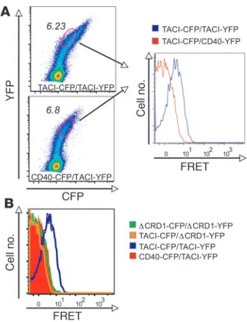
TACI assembles independently of ligand in living cells. To confirm TACI homotypic interaction in living cells, we used a flow-cyto-metric approach to analyze fluorescence resonance energy transfer (FRET) between TACI monomers in which the CD was replaced with CFP or YFP. Energy transfer was detected between TACIΔ CD-CFP and TACIΔCD-YFP in the absence of ligand, supporting the notion of a preassembled receptor complex (Figure 4A). More importantly, there was no FRET signal between WT TACI and TACIΔCRD1 and no detectable FRET with CD40 control (Figure 4B). Thus, mTACI molecules are in close proximity in living cells, and this is dependent on the CRD1 domain.
Endogenous TACI exists as an oligomeric complex on the surface of B cells. We next examined whether endogenous TACI forms trimers or higher-order oligomers on the cell surface in the absence of ligand. We treated A20 murine B cells with
membrane-imperme-Figure 3
snIP analysis of TACI receptor assembly in ER microsomes. (A and B) Assembly of differentially tagged TACI monomers was examined in
an in vitro translation system using ER microsomes. Membranes were solubilized with digitonin, and complexes were isolated by snIP using the protein C (PC) and SA-binding protein (SBP) affinity tags on the
[image:4.585.73.249.80.365.2]C terminus of the TACI chains. SBP-tagged complexes were captured by SA beads and PC tagged complexes were captured by anti-PC mAb. Asterisk (lane 6) represents mixing control in which TACI-SBP and TACI-PC monomers were translated separately and mixed prior to solubilization and IP.
Figure 4
TACI self-associates through CRD1 in living cells. (A) Flow
cytomet-ric analysis of 293T cells cotransfected with CFP- or YFP-tagged mTACI and CFP- or YFP-tagged CD40 control. The dual-expressing cells were electronically gated, and more than 20,000 events were acquired into the FRET gate and used for subsequent analysis. (B)
Overlay of FRET plots derived as in A for interactions between
[image:4.585.332.511.508.741.2]research article
The Journal of Clinical Investigation http://www.jci.org Volume 117 Number 6 June 2007 1553
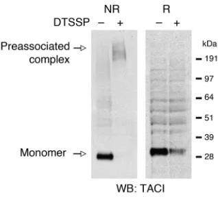
ant, thiol-cleavable chemical cross-linker 3,3′-dithiobis[sulfosucci nimidyl-propionate] (DTSSP) to covalently cross-link cell-surface molecules that are closely associated, with less than 12 Å distance from each other. Detergent-solubilized lysates were run on a gel under nonreducing conditions or under reducing conditions (100 mM DTT) and probed for TACI. In the absence of DTSSP, under nonreducing conditions a strong band that corresponds to mono-meric TACI was visualized (Figure 5). In the presence of DTSSP, under nonreducing conditions monomeric TACI was no longer detected; instead, a high-molecular-weight complex that exhib-ited a molecular size greater than 191 kDa became evident. The presence of DTT in the same sample resulted in the disappearance of the higher-molecular-weight complex and appearance of TACI monomer. The decreased recovery of monomeric TACI under reducing conditions in DTSSP cross-linked sample seen in Figure 5 was due to decreased affinity of the monoclonal anti-TACI to modified TACI monomer, as no difference in band intensity was observed when polyclonal antibody was used for Western blotting (data not shown). These data show that native TACI monomers assemble as oligomers on the surface of B cells.
The murine C76R mutant fails to bind ligand but preassociates with WT TACI. Having established that TACI receptor monomers preassoci-ate in a ligand-independent manner, we next determined whether the C76R mTACI mutant can preassociate with WT TACI. First we established that the C76R mTACI mutant behaves similarly to the C104R hTACI mutant, in that it is expressed on the cell surface but fails to bind the ligand. Figure 6A shows that the C76R mTACI mutant is expressed on the cell surface. However, like its hTACI C104R counterpart, it was unable to bind BAFF. There was con-sistently a slight difference in the intensity of expression of C76R
[image:5.585.80.239.81.225.2]TACI compared with WT TACI, possibly because the missense mutation alters the epitope recognized by the Ab. However, this difference was small and cannot account for the complete inability of the mutant to bind BAFF. When 293T cells were cotransfected with Myc-tagged C76R mTACI and Flag-tagged WT mTACI, the Myc-tagged mutant coprecipitated with Flag-WT mTACI (Figure 2A, lane 3). This interaction was independent of the presence of the CD, as Myc-tagged C76R mTACI coprecipitated with CFP-tagged TACIΔCD (Figure 2B, lane 4). The ligand-independent preassocia-tion of the C76R mTACI mutant with WT TACI was confirmed with the in vitro translation and assembly system using PC-tagged C76R mutant and SBP- and YFP-tagged WT TACI. Assembly reactions were set up with C76R TACI-PC, WT TACI-SBP, and WT TACI-YFP. Single IP with anti-PC captured predominantly PC-tagged C76R TACI with a minor amount of SBP- and YFP-tagged WT TACI (Figure 6B, lane 3), while single IP with SA beads captured predominantly SBP-tagged WT TACI with a minor amount of C76R TACI-PC and WT TACI-YFP (Figure 6B, lane 4). More importantly, 2-step snIP that targeted the SBP and PC epit-opes (SBP→PC) brought down all 3 tagged molecules (Figure 6B, lane 5). This preassociation occurred in the ER and was specific, as no complex was observed in mixing control, in which WT TACI-YFP, WT TACI-SBP, and C76R TACI-PC were translated in sepa-rate reactions, the contents of which were combined before solubi-lization and IP (Figure 6B, lane 6). These results demonstrate that WT TACI and C76R TACI monomers can assemble at least into trimers in the absence of ligand in ER microsomes. Association
Figure 6
C76R mTACI associates with WT TACI. (A) Surface expression and
recombinant human BAFF (rhBAFF) binding of WT and mutant mTACI
in transfected 293T cells. (B) snIP analysis of the association of WT and
mutant mTACI in ER microsomes. Asterisk (lane 6) represents mixing control in which TACI-SBP, TACI-PC, and TACI-YFP monomers were translated separately and mixed prior to solubilization and IP. (C) FRET
analysis of the association of WT and mutant mTACI in 293T cells.
Figure 5
[image:5.585.301.541.399.735.2]of the C76R mutant and WT TACI was further demonstrated in living cells by FRET analysis (Figure 6C).
The murine C76R mutant inhibits signaling by WT TACI. It has been previously reported that engagement of TACI by its ligands BAFF and APRIL leads to activation of NF-κB in transfected 293T cells (6, 10, 17, 18). To understand the functional significance of ligand-independent preassociation of the C76R mutant and WT TACI, a quantitative luciferase assay was used to measure signaling via TACI in 293T cells transfected with WT TACI, C76R mutant, or equiva-lent amounts of both. Simultaneous expression of both mutant and WT TACI on the surface of cotransfected cells was confirmed using differentially tagged constructs (data not shown). Figure 7A shows that ligation of WT TACI with BAFF caused NF-κB acti-vation. As expected, the C76R mutant, which fails to bind BAFF, was unable to activate NF-κB. NF-κB activation was significantly reduced (P < 0.001) in 293T cells cotransfected with equal amounts of WT and mutant TACI. These results demonstrate that the C76R mutant dominantly interferes with signaling by WT TACI.
To ascertain that the dominant-negative effect of the mutant is dependent on its ability to preassociate with WT TACI, we examined the effect of CRD1-deleted C76R mTACI double mutant (DM) on WT TACI signaling. Simultaneous expression of the DM and WT TACI on the surface of cotransfected cells was demonstrated using differentially tagged constructs (data not shown). Figure 7B shows that the DM had no effect on signaling by WT TACI. These data suggest that the ability of the C76R mutant to act as a dominant negative is dependent on its ability to preassociate with WT TACI.
Preassociation of C76R with WT TACI could interfere with ligand binding by the oligomeric complex. Alternatively, C76R may not interfere with ligand binding but could still interfere with sig-naling if all subunits of the receptor oligomer need to bind ligand
for signaling to proceed. To test whether C76R TACI mutant inter-feres with ligand binding to WT TACI, we examined the binding of Flag-tagged BAFF to 293T cells transfected with equal amounts of WT and C76R mutant TACI. Figure 7C shows that the presence of C76R mutant did not decrease ligand binding to WT TACI. No interference by the mutant with ligand binding to WT TACI was observed either under limiting concentrations of BAFF or when the mutant was used at a concentration 4-fold that of WT TACI (data not shown). These data indicate that the C76R mutant does not interfere with ligand binding by WT TACI.
[image:6.585.51.285.82.297.2]C104R mutant hTACI preassembles with WT hTACI.We examined the association of WT hTACI and C104R hTACI mutant in 293T cells. 293T cells were cotransfected with full-length Flag-WT hTACI in
Figure 7
C76R mTACI dominantly interferes with signaling, but not ligand bind-ing, by WT mTACI. (A) 293T cells were transfected with 0.1 μg WT mTACI alone, 0.1 μg C76R mTACI, or 0.1 μg of each along with 0.1 μg of NF-κB–luc reporter plasmid and 10 ng control pRL-TK plasmid. Equal amounts of DNA were used for each transfection by using empty vector DNA when needed. After 4 hours, recombinant mBAFF (0.1
μg/ml) was added, and reporter gene activity was determined 20 hours later. NF-κB luciferase induction was normalized to Renilla luciferase intensity and total protein of each sample. Fold induction was calculat-ed by dividing the normalizcalculat-ed NF-κB luciferase result of BAFF-treated samples by the NF-κB luciferase result of untreated samples. n = 7. (B)
The same assay as described in A was performed, with C76R mTACI
that lacked the CRD1 domain (DM) substituting for C76R mTACI. Val-ues represent the fold increase by BAFF stimulation compared with unstimulated sample and are the mean ± SEM from 6 independent experiments. (C) TACI surface expression and rhBAFF binding by
[image:6.585.331.512.482.744.2]293T cells transfected with 1 μg of WT mTACI, 1 μg of C76R mTACI alone, or 1 μg of each.
Figure 8
C104R hTACI associates with WT hTACI. (A and B) 293T cells were
research article
The Journal of Clinical Investigation http://www.jci.org Volume 117 Number 6 June 2007 1555
combination with Myc-WT hTACI or Myc-C104R hTACI. As shown in Figure 8, hTACI monomers were found to homotypically associ-ate (Figure 8A, lane 2). More importantly, C104R hTACI was able to associate with WT hTACI (Figure 8A, lane 3). This homotypic interaction was not dependent on the intracellular (IC) domain, as it was still retained when the IC domain of WT hTACI was replaced by CFP to generate hTACIΔCD-CFP. Figure 8B shows that WT hTACIΔCD-CFP associated with Myc-WT hTACI (lane 3) as well as with Myc-C104R hTACI (lane 4). These associations were specific because there was no association of CD40ΔCD-CFP with Myc-WT hTACI (Figure 8B, lane 2).
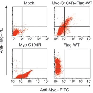
The C104R mutant inhibits signaling by WT hTACI. Previously we have demonstrated that B cells from 3 CVID patients heterozygous for the C104R hTACI mutation failed to secrete IgG and IgA in response to APRIL, indicating that TACI-mediated signaling was impaired (4). One can argue that this could possibly be due to the ability of the mutant to interfere with WT hTACI surface expres-sion. However, simultaneous expression of mutant and WT hTACI on the surface of cotransfected cells was confirmed using differen-tially tagged constructs (Figure 9). C104R did not interfere with the expression of WT hTACI, and more importantly, it did not interfere with ligand binding by WT hTACI in cotransfected cells (Figure 10, A and B). To get a better understanding of the molec-ular mechanism by which C104R could be interfering with WT hTACI function, we used a quantitative luciferase assay to mea-sure signaling via TACI in 293T cells transfected with WT hTACI, C104R mutant, or equivalent amounts of both. Figure 10C shows
that ligation of WT hTACI with BAFF caused NF-κB activation, while the C104R mutant, which fails to bind BAFF, was unable to activate NF-κB. TACI signaling was significantly reduced in 293T cells cotransfected with equal amounts of WT and mutant hTACI. These results demonstrate that the C104R mutant dominantly interferes with signaling by WT hTACI.
Discussion
Our data clearly demonstrate that endogenous mTACI exists as a pre-associated oligomeric complex and that a missense C76R mutation that abolishes ligand binding functions as a dominant negative.
Our work provides new insight into the structure and function of TACI receptor by showing that it preassociates into an oligo-meric complex prior to ligand binding. Homotypic interaction of TACI molecules was demonstrated by coimmunoprecipitation of differentially tagged TACI molecules (Figure 2) and was confirmed in living cells by FRET (Figure 4). We used a novel snIP method to explore the stoichiometry of the TACI oligomeric complex. Using 3 differentially tagged TACI molecules, we demonstrated that a minimum of 3 TACI monomers preassociate in the absence of ligand. However, with only 3 differentially tagged monomers, we could not determine, using this method, whether TACI can pre-associate into an oligomer of a higher order than a trimer. The results of chemical cross-linking studies on A20 B cells, which express endogenous TACI, suggest that the TACI receptor exist on the cell surface as a preassociated higher-order oligomer (Figure 5), although the presence of other molecules in this complex can-not be ruled out. Thus, TACI joins the TNFR family members Fas,
[image:7.585.73.250.83.261.2]TNFR1 (p60), and TNFR2 (p80) as a receptor with the ability to preassociate independently of ligand (19, 20).
Figure 9
[image:7.585.306.533.532.741.2]C104R mutant does not interfere with surface expression of WT hTACI. 293T cells were cotransfected with the indicated hTACI plasmids, and surface expression of Flag-tagged WT hTACI and Myc-tagged C104R hTACI was analyzed 24 hours after transfection using anti-Myc–FITC and anti-Flag–PE antibodies. Mock, untransfected.
Figure 10
C104R hTACI dominantly interferes with signaling, but not ligand bind-ing, by WT hTACI. hTACI surface expression (A) and rhBAFF binding
(B) by 293T cells transfected with 1 μg of WT hTACI, 1 μg of C104R hTACI alone, or 1 μg of each together. (C) 293T cells were transfected
Recently published data demonstrate that BAFF and APRIL can form higher-order oligomers under physiologic conditions. BAFF has been reported to assemble into a virus-like cage complex of 60 monomers (21–23). It has been suggested that the increased mul-tivalency of the 60-mer affects the quality of the signal transduced rather than the avidity of binding (23). APRIL has been shown to bind to heparan sulfate proteoglycans and oligomerize (24). This oligomerization was shown to be essential for proper signaling (25). Given these observations and our data, it is logical to posit that TACI exists on the cell surface in a preassociated oligomeric state ready to efficiently bind its oligomeric ligands in order to transduce a rapid and strong signal. Higher-order clustering with simultaneous engagement of oligomeric receptors may be required to transduce a signal above a certain threshold, and the inclusion of non–ligand-binding subunits in preassociated oligomers may inhibit this process.
The demonstration that WT TACI monomers preassociate prior to ligand binding is critical to understanding the molecular mecha-nisms by which heterozygous mutations in TACI that affect ligand binding can cause B cell dysfunction. The prototype of such a muta-tion is C104R, one of the most common TACI mutamuta-tions associ-ated with CVID. According to the haploinsufficiency explanation, a single copy of WT TACI is insufficient to transduce a signal. The alternative explanation is that C104R TACI acts as a dominant-negative mutant that interferes with signaling. Our demonstra-tion that the C104R hTACI mutant, and the C76R mTACI mutant that corresponds to it, associates with WT TACI and functions as a dominant negative favors the second hypothesis. Preassociation of the C76R mutant with WT mTACI was demonstrated by several techniques that included coimmunoprecipitation, snIP, and FRET (Figures 2 and 6). Coexpression of the mutant with WT mTACI significantly inhibited ligand-induced activation of NF-κB (Figure 7A). This inhibition was strictly dependent on the assembly of the mutant with WT mTACI, as a C76R mutant that lacked the CRD1 domain and failed to associate with WT mTACI also failed to inhib-it signaling by WT mTACI (Figure 7B).
These results are strikingly similar to the findings with EC Fas mutations cloned from patients with autosomal dominant auto-immune lymphoproliferative syndrome (ALPS). Those Fas muta-tions also failed to bind FasL and preassociated with wild-type receptors, impairing FasL-mediated apoptosis (19). One possible explanation for the dominant effect of the C76R mutant is that inclusion of the mutant in the preassociated TACI oligomeric complex would interfere with ligand binding to the complex. This explanation was ruled out because coexpression of the mutant with WT mTACI had no detectable effect on ligand binding (Fig-ure 7C). This result is consistent with the previously reported observation that B cells from CVID patients heterozygous for the C104R hTACI mutations show normal APRIL binding (5). These data raise the possibility that the mutant may interfere with signal-ing by disruptsignal-ing ligand-induced conformational changes in the receptor that allow proper recruitment of downstream signaling molecules. Further biophysical and structural studies are neces-sary to prove this hypothesis.
The present work provides new insight into the structure of the mTACI receptor by demonstrating that it preassociates as an oligo-mer prior to ligand binding. We have also shown that the hTACI mutant C104R, which is associated with CVID, preassociates with WT hTACI (Figure 8A). As with mTACI, this preassociation is independent of the IC domain (Figure 8B). More importantly, the
hTACI mutant C104R can interfere with signaling by WT hTACI without interfering with the surface expression of WT hTACI or with its ability to bind ligand (Figures 9 and 10). Thus, ligand-independent preassociation of C104R mutant with WT hTACI provides a potential explanation for how the heterozygous C104R mutation may disrupt TACI signaling in B cells, illustrated by the impaired ability of B cells from CVID patients heterozygous for C104R to produce IgG and IgA in response to APRIL (4). Final proof that this B cell dysfunction results in CVID awaits the analy-sis of mice carrying the C76R mutation.
Methods
Antibodies and reagents. SA, and Myc coupled to agarose beads and
Flag antibody were purchased from Sigma-Aldrich. Anti-myc (9B11) anti-body was purchased from Cell Signaling Technology. Calcium-dependent anti-PC (mouse mAb HPC4) was purchased from Roche. Anti-GFP (Living Colors A.v. Monoclonal Antibody JL-8) was purchased from Clontech. Dig-itonin was purchased from Biosynth International. mTACI surface expres-sion was analyzed using rat monoclonal anti-TACI–PE antibody (R&D Sys-tems), and hTACI surface expression was analyzed using goat polyclonal anti-TACI antibody (R&D Systems) as described previously (4).
cDNA constructs and in vitro transcription. mTACI sequence was
ampli-fied from mouse B cells by RT-PCR. hTACI sequence was ampliampli-fied from human PBMCs by RT-PCR. We obtained blood by venipuncture from a healthy adult control after obtaining informed consent. The study pro-tocol was approved by the Ethics Committee/Internal Review Board of Children’s Hospital, Boston, Massachusetts, USA. PBMCs were isolated from the blood and used to obtain mRNA. BALB/c wild-type mouse spleen was used to purify B cells and obtain mRNA. The procedure performed on the mouse was reviewed and approved by the Animal Care and Use Committee of the Children’s Hospital Boston. The sequences were cloned into pCMVTag3A (Myc tag), pCMVTag2A (Flag tag), pcDNA6, and modi-fied pSP64 vector with PC and SBP tags described in ref. 14. For FRET, TACI was cloned into modified YFP and CFP vectors, which have muta-tions introduced into CFP and YFP that inhibit their inherent tendency to form hetero- or homodimers as described in ref. 26 (gift from C. Car-man, Harvard Medical Center and Beth Israel Deaconess Medical Center, Boston, Massachusetts, USA). Mutation was introduced by QuikChange Mutagenesis Kit (Stratagene). In vitro transcription was performed from cDNA constructs using T7 RiboMAX Large-Scale RNA Production Kit and m7G cap analog (Promega).
In vitro translation and assembly. The in vitro translation and
assem-bly reactions followed by IP and analysis were performed as previously described (14, 15). All in vitro translation and assembly reactions were
performed at 30°C. Each 25-μl reaction contained 17.5 μl
nuclease-treat-ed rabbit reticulocyte lysate (Promega), 0.5 μl amino acid mixture minus
methionine (Promega), 0.5 μl SUPERase-In RNase inhibitor (Ambion),
1–2 μl 35S-labeled methionine (Amersham), equimolar amounts of each
RNA (300 ng), and 2.0 μl ER microsome from murine hybridoma (IVD12)
as previously described (14). Reactions were performed with an initial translation period of 30 minutes under reducing conditions, followed by a 2-hour assembly period after addition of oxidized glutathione to
4 mM. Reaction volumes were 25–100 μl as required for optimal signal
with multistep snIP procedures.
IP and electrophoretic analysis. Translation assembly reactions were stopped by dilution with 1 ml of ice-cold PBS with 10 mM iodoacetamide, and
microsomes were pelleted and rinsed. Pellets were lysed in 400 μl of IP
research article
The Journal of Clinical Investigation http://www.jci.org Volume 117 Number 6 June 2007 1557 and primary captures were performed overnight at 4°C. IPs were washed
twice with 500 μl of IP buffer. Nondenaturing elution of SA-captured
com-plexes was performed by incubation with 400 μl of IP buffer with 100 μM
free biotin for 1 hour at 4°C, and eluted complexes were incubated with PC antibody and protein G beads for 2 hours at 4°C and washed as described above. Nondenaturing elution of PC-captured complexes was performed
by incubation with 400 μl of IP buffer with EDTA for 1 hour at 4°C.
Cross-linking experiment. A20 B cell lymphoma cells were washed and
resuspended in PBS. Cells were then treated with 1 mM solution of the cross-linker DTSSP (Pierce) for 30 minutes at room temperature, and the reaction was quenched with 50 mM Tris-Cl (pH 7.5) for 20 minutes on ice. The cells were lysed in 1% Triton, 10 mM HEPES, 150 mM NaCl, 1 mM EDTA, 100 mM PMSF, and protease inhibitors. Equal amounts of lysates were subjected to electrophoresis under nonreducing (without DTT) or reducing (with 100 mM DTT) conditions and were analyzed for TACI complexes with monoclonal rat anti-TACI or polyclonal goat anti-TACI antibodies (R&D Systems).
293T coimmunoprecipitation. 293T cells were transfected using FuGENE 6
(Roche). Forty-eight hours after transfection, cells were harvested and lysed in IP buffer: 0.5% NP-40 or 1% Triton, 50 mM HEPES, 10 mM iodo-acetamide, 250 mM NaCl, 5 mM EDTA, 100 mM PMSF, and complete protease inhibitor cocktail (Sigma-Aldrich). After preclearing with protein G–agarose beads (Upstate), proteins were immunoprecipitated with either anti-Myc or anti-Flag agarose beads (Sigma-Aldrich). Immune complexes were washed 3–5 times with lysis buffer. Proteins were electrophoresed on Tris/glycine gels, transferred to nitrocellulose membranes, and blotted with the indicated antibodies.
FRET.TACI receptors with C-terminal in-frame fusions (at position 156
in place of the IC domain) to monomeric CFP and YFP were generated by standard PCR cloning techniques. After sequence and protein expres-sion of these constructs were confirmed, they were transfected into 293T cells, and flow cytometric detection of FRET was performed as previously described (19, 27) using a CyAn cytofluorimeter (Dako) and 408/488 nm dual laser excitation. Data was analyzed by FlowJo software (version 8.3; Tree Star) and fluorescence plotted on biexponential axes.
Statistics. Significance of experimental values was analyzed by using the
2-tailed Student’s t test.
Acknowledgments
The authors thank Chris Carman for the modified CFP and YFP vectors and members of the Geha laboratory for useful discus-sions. L. Garibyan was funded by the Medical Scientist Training Program grant to Harvard Medical School. This work was sup-ported by NIH grants P01-AI-031541 and T32-AI-007512 to R.S. Geha and R01 AI054520 to K.W. Wucherpfennig.
Received for publication November 21, 2006, and accepted in revised form February 27, 2007.
Address correspondence to: Raif S. Geha, Children’s Hospital Boston, Division of Immunology, 1 Blackfan Circle, Karp Family Research Building, 10th floor, Boston, Massachusetts 02115, USA. Phone: (617) 919-2482; Fax: (617) 730-0528; E-mail: raif.geha@ childrens.harvard.edu.
1. Cunningham-Rundles, C., and Bodian, C. 1999. Common variable immunodeficiency: clinical and immunological features of 248 patients. Clin. Immunol.92:34–48.
2. Kokron, C.M., et al. 2004. Clinical and laboratory aspects of common variable immunodeficiency. An. Acad. Bras. Cienc.76:707–726.
3. Schroeder, H.W., Jr., Schroeder, H.W., 3rd, and Sheikh, S.M. 2004. The complex genetics of com-mon variable immunodeficiency. J. Investig. Med.
52:90–103.
4. Castigli, E., et al. 2005. TACI is mutant in common variable immunodeficiency and IgA deficiency. Nat. Genet.37:829–834.
5. Salzer, U., et al. 2005. Mutations in TNFRSF13B encoding TACI are associated with common vari-able immunodeficiency in humans. Nat. Genet.
37:820–828.
6. von Bulow, G.U., and Bram, R.J. 1997. NF-AT acti-vation induced by a CAML-interacting member of the tumor necrosis factor receptor superfamily. Sci-ence.278:138–141.
7. Mackay, F., Schneider, P., Rennert, P., and Brown-ing, J. 2003. BAFF and APRIL: a tutorial on B cell survival. Annu. Rev. Immunol.21:231–264. 8. Bischof, D., et al. 2006. Selective activation of TACI
by syndecan-2. Blood.107:3235–3242.
9. Castigli, E., et al. 2005. TACI and BAFF-R mediate isotype switching in B cells. J. Exp. Med.201:35–39. 10. Seshasayee, D., et al. 2003. Loss of TACI causes fatal
lymphoproliferation and autoimmunity,
establish-ing TACI as an inhibitory BLyS receptor. Immunity.
18:279–288.
11. von Bulow, G.U., van Deursen, J.M., and Bram, R.J. 2001. Regulation of the T-independent humoral response by TACI. Immunity.14:573–582. 12. Yan, M., et al. 2001. Activation and accumulation
of B cells in TACI-deficient mice. Nat. Immunol.
2:638–643.
13. Hymowitz, S.G., et al. 2005. Structures of APRIL-receptor complexes: like BCMA, TACI employs only a single cysteine-rich domain for high affinity ligand binding. J. Biol. Chem.280:7218–7227. 14. Call, M.E., Pyrdol, J., Wiedmann, M., and
Wucherp-fennig, K.W. 2002. The organizing principle in the formation of the T cell receptor-CD3 complex. Cell.
111:967–979.
15. Feng, J., Garrity, D., Call, M.E., Moffett, H., and Wuch-erpfennig, K.W. 2005. Convergence on a distinctive assembly mechanism by unrelated families of activat-ing immune receptors. Immunity.22:427–438. 16. Garrity, D., Call, M.E., Feng, J., and
Wucherpfen-nig, K.W. 2005. The activating NKG2D receptor assembles in the membrane with two signaling dimers into a hexameric structure. Proc. Natl. Acad. Sci. U. S. A.102:7641–7646.
17. Marsters, S.A., et al. 2000. Interaction of the TNF homologues BLyS and APRIL with the TNF receptor homologues BCMA and TACI. Curr. Biol.
10:785–788.
18. Yan, M., et al. 2000. Identification of a receptor for BLyS demonstrates a crucial role in humoral
immunity. Nat. Immunol.1:37–41.
19. Siegel, R.M., et al. 2000. Fas preassociation required for apoptosis signaling and dominant inhibition by pathogenic mutations. Science.288:2354–2357. 20. Chan, F.K., et al. 2000. A domain in TNF receptors
that mediates ligand-independent receptor assem-bly and signaling. Science.288:2351–2354. 21. Kim, H.M., et al. 2003. Crystal structure of the
BAFF-BAFF-R complex and its implications for receptor activation. Nat. Struct. Biol.10:342–348. 22. Cachero, T.G., et al. 2006. Formation of virus-like
clusters is an intrinsic property of the tumor necro-sis factor family member BAFF (B cell activating factor). Biochemistry.45:2006–2013.
23. Liu, Y., et al. 2002. Crystal structure of sTALL-1 reveals a virus-like assembly of TNF family ligands. Cell.108:383–394.
24. Hendriks, J., et al. 2005. Heparan sulfate proteogly-can binding promotes APRIL-induced tumor cell proliferation. Cell Death Differ.12:637–648. 25. Ingold, K., et al. 2005. Identification of
proteogly-cans as the APRIL-specific binding partners. J. Exp. Med.201:1375–1383.
26. Zacharias, D.A., Violin, J.D., Newton, A.C., and Tsien, R.Y. 2002. Partitioning of lipid-modified monomeric GFPs into membrane microdomains of live cells. Science.296:913–916.