Altered wound healing in mice lacking a
functional osteopontin gene (spp1).
L Liaw, … , J M Davidson, B L Hogan
J Clin Invest.
1998;
101(7)
:1468-1478.
https://doi.org/10.1172/JCI2131
.
Osteopontin (OPN) is an arginine-glycine-aspartate (RGD)- containing glycoprotein
encoded by the gene secreted phosphoprotein 1 (spp1). spp1 is expressed during
embryogenesis, wound healing, and tumorigenesis; however, its in vivo functions are not
well understood. Therefore, OPN null mutant mice were generated by targeted mutagenesis
in embryonic stem cells. In OPN mutant mice, embryogenesis occurred normally, and mice
were fertile. Since OPN shares receptors with vitronectin (VN), we tested for compensation
by creating mice lacking both OPN and VN. The double mutants were also viable,
suggesting that other RGD-containing ligands replace the embryonic loss of both proteins.
We tested the healing of OPN mutants after skin incisions, where spp1 was upregulated as
early as 6 h after wounding. Although the tensile properties of the wounds were unchanged,
ultrastructural analysis showed a significantly decreased level of debridement, greater
disorganization of matrix, and an alteration of collagen fibrillogenesis leading to small
diameter collagen fibrils in the OPN mutant mice. These data indicate a role for OPN in
tissue remodeling in vivo, and suggest physiological functions during matrix reorganization
after injury.
Research Article
Find the latest version:
The Journal of Clinical Investigation
Volume 101, Number 7, April 1998, 1468–1478 http://www.jci.org
Altered Wound Healing in Mice Lacking a Functional Osteopontin Gene (
spp1
)
Lucy Liaw,* David E. Birk,i Christopher B. Ballas,§ Jeffrey S. Whitsitt,§ Jeffrey M. Davidson,§¶ and Brigid L.M. Hogan*‡
*Department of Cell Biology and ‡Howard Hughes Medical Institute, §Department of Pathology, Vanderbilt University Medical Center,
Nashville, Tennessee 37232; iDepartment of Anatomy and Cellular Biology, Tufts University School of Medicine, Boston, Massachusetts 02111; and ¶Department of Veterans Affairs Medical Center, Nashville, Tennessee 37212-2637
Abstract
Osteopontin (OPN) is an arginine-glycine-aspartate (RGD)-containing glycoprotein encoded by the gene secreted phos-phoprotein 1 (spp1). spp1 is expressed during embryogene-sis, wound healing, and tumorigenesis; however, its in vivo functions are not well understood. Therefore, OPN null mu-tant mice were generated by targeted mutagenesis in embry-onic stem cells. In OPN mutant mice, embryogenesis oc-curred normally, and mice were fertile. Since OPN shares receptors with vitronectin (VN), we tested for compensation by creating mice lacking both OPN and VN. The double mutants were also viable, suggesting that other RGD-con-taining ligands replace the embryonic loss of both proteins. We tested the healing of OPN mutants after skin incisions, where spp1 was upregulated as early as 6 h after wounding. Although the tensile properties of the wounds were un-changed, ultrastructural analysis showed a significantly de-creased level of debridement, greater disorganization of ma-trix, and an alteration of collagen fibrillogenesis leading to small diameter collagen fibrils in the OPN mutant mice. These data indicate a role for OPN in tissue remodeling in vivo, and suggest physiological functions during matrix re-organization after injury. (J. Clin. Invest. 1998. 101:1468– 1478.) Key words: gene targeting • matrix • vitronectin •
col-lagen • skin
Introduction
Osteopontin (OPN)1 is a secreted phosphoprotein containing
the arginine-glycine-aspartate (RGD) tripeptide integrin bind-ing motif. Early isolation of OPN from bone (1) and subse-quent cloning (2) indicated a function in the adhesion of bone cells to the mineralized matrix. However, the gene encoding OPN was soon found to be identical to a gene induced upon
cell transformation (3, 4), and later was shown to have a more widespread distribution, suggesting relevance in other tissues as well. The gene was also independently described as eta1, a T cell product induced after cell activation (5). Further work related to immune cell function has provided evidence that OPN acts as a cytokine, and may play important roles in T cell and macrophage function during responses to bacterial infec-tion, pathogen resistance, and wound healing (6). In vitro stud-ies using purified protein have verified that OPN can act as an adhesion substrate and migration stimulus by interaction with several integrins, leading to downstream signaling events that include calcium mobilization, protein phosphorylation, and regulation of gene expression and cell differentiation (7).
A variety of studies have shown that the gene secreted phosphoprotein 1 (spp1) is expressed in selected tissues during embryonic development. Transcripts for spp1 are first detect-able during gastrulation in the notochord (8). Secreted factors from the notochord are known to affect neural and somitic cell differentiation (9), raising the possibility that OPN contributes to notochord elongation or sclerotomal cell migration. The second region of high spp1 expression is at the embryonic/ma-ternal interface. OPN is produced by invading trophoblasts, by the metrial gland cells of the deciduum, and in the placenta (10), correlating temporally with blastocyst invasion and later, placentation. Experiments addressing the functions of OPN during embryogenesis have not been described previously.
Of note, another RGD-containing protein, vitronectin (VN), has been localized during embryonic development to re-gions overlapping with spp1 expression. Although limited in sites of synthesis primarily to hepatocytes and some areas of the central nervous system, circulating plasma levels of VN are on the order of 200–400 mg/ml, and deposition of the protein occurs in several tissues (11). As a consequence, embryonic tis-sues, including the notochord, placenta, deciduum, and some connective tissues, both produce OPN and are sites of VN dep-osition (12). Targeted mutation of the gene encoding VN, vtn, has demonstrated that normal embryonic development oc-curs in the absence of VN protein (13). Therefore, we were interested in the interactions of VN and OPN during embryo-genesis.
To address the in vivo roles of OPN, we have generated a null mutation in the murine spp1 gene by homologous recom-bination in embryonic stem (ES) cells. We report that the loss of OPN or both OPN and VN is permissive for embryonic de-velopment, and the OPN mutants as well as OPN/VN double mutants were also fertile. These observations suggest that ad-ditional RGD-containing proteins may be acting in a compen-satory manner. The viability of the OPN mutant allows us to address the roles of this protein in the adult, where despite nu-merous studies, its in vivo roles are not well established.
In the adult, OPN production is normally restricted, but upregulation of spp1 occurs during cell injury and pathology. spp1 is highly expressed in transformed cells, and OPN is found in a variety of tumors (14). The presence of OPN at sites
Address correspondence to Lucy Liaw, Center for Molecular Medi-cine, Maine Medical Center Research Institute, 125 John Roberts Rd., Suite #8, South Portland, ME 04106. Phone: 207-761-9090; FAX: 207-761-2130; E-mail: [email protected]
Received for publication 25 October 1997 and accepted in revised form 26 January 1998.
of wound healing, particularly associated with leukocytes (15, 16), is concordant with a model where OPN is an important ef-fector of the immune response. The necessity of a robust im-mune response for subsequent healing has been demonstrated by macrophage depletion studies (17) and stimulation of mac-rophages using immunomodulators (18). A postulated role for OPN at wound sites is to function as a mediator of immune cell function and subsequent repair. Paradigms of tissue injury as-sociated with spp1 upregulation are as diverse as cardiovascu-lar injury and disease (19), renal tubulointerstitial fibrosis (20), and kidney stone formation (21).
Using full-thickness skin incisions to study immune cell function and wound matrix remodeling, we report significant differences in the ability of OPN mutant animals to remodel wound matrix in a process that involves fibrillar collagen orga-nization. Thus, our studies demonstrate the novel findings that mice lacking OPN and VN develop normally and are fertile, and that OPN functions during dermal tissue repair and col-lagen fibrillogenesis.
Methods
Construction of spp1 targeting vector
A 129/SvJ genomic library (Stratagene, Inc., La Jolla, CA) was screened using a 39 196-bp fragment of the spp1 cDNA. Two of the largest positive clones were characterized by restriction and partial sequence analysis. The replacement targeting vector was constructed in the vector pPNT (22) using a 4-kb XhoI/SpeI fragment for the 59 arm, and a 2.2-kb SmaI/ NotI fragment for the 39 arm. After homologous recombination, exons 4–7 of spp1 are thus replaced by phosphoglycerokinase (PGK) neomycin resistance gene (neor), with an hsv-tk cassette (for herpes simplex virus– thymidine kinase) outside the region of homology. Transcription of PGKneo and hsv-tk occur in the opposite direction to the spp1 locus.
Generation of recombinant ES cell lines and chimeric mice
TL-1 ES cells (20 3 106 cells, at passage 10 [23]) were electroporated with 50 mg linearized targeting vector with a single pulse at 800 V, 3 mF. Cells were fed daily with DME containing 1,000 U/ml leukemia-inhibitory factor (ESgro; GIBCO BRL, Gaithersburg, MD). The next day, selection was started with 300 mg/ml active G418 (GIBCO BRL), followed 2 d later with the addition of gancyclovir to a final 2
mM concentration. After 8 d, double resistant clones were picked. Six correctly targeted ES cell clones were identified. Three of these clones were injected into C57BL/6 blastocysts to generate chimeric animals. Chimeric males from each line were bred to Black Swiss or 129/SvJ females (Taconic Farms, Inc., Germantown, NY) to obtain heterozygous pups. No differences were noted between animals from different ES cell clones. The analyses described were done with ani-mals on a 129 3 Black Swiss hybrid background. The mutant allele has been designated spp1tm1blh in accordance with standard nomenclature (24), and has been submitted to TBASE under the accession number TG-000-04-502 (http://www.bis.med.jhmi.edu/Dan/tbase/tbase.html).
Genotyping of OPN mice
Southern blotting. Genomic DNA was isolated and digested with XhoI, SphI, or SpeI. For initial screening, DNA was digested with XhoI, and blots were probed with probe C (see Fig. 2 A). Genotypes were verified by probing SpeI-digested samples with probes A or B, which are outside the region of homology.
PCR analysis. The following primers were used: OPIn3, 59 CCA-TACAGGAAAGAGAGACC39; OPIn4, 59 AACTGTTTTGCTTGC-ATGCG39; Neo1360, 59CGTCCTGTAAGTCTGCAGAA39. OPIn3 and OPIn4 amplify an endogenous 600-bp fragment, and OPIn3 and Neo1360 amplify a 500-bp fragment from the recombined mutant al-lele. The three primers were used together at an annealing tempera-ture of 538C.
Reverse transcription (RT) PCR analysis for spp1 transcript
Total RNA from cells was reverse transcribed with avian reverse transcriptase using oligo dT primers. Primers for spp1 were Ex6 and Ex7, which amplify a 410-bp cDNA and a 1.1-kb genomic product at an annealing temperature of 508C, while control primers were from murine hypoxanthine phosphoribosyltransferase (HPRT), which were used at an annealing temperature of 508C. Ex6, 59 AATCG-TCCCTACAGTCGATG39; Ex7, 59 TAGGGTCTAGGACTAGCT-TG39; HPRT1, 59GCTGGTGAAAGGGACCTCT39; and HPRT2, 59CACAGGACTAGAACACCTGC39.
Immunohistochemistry
Immunostaining for OPN was performed with purified goat IgG of an antibody generated against rat OPN, OP199 (25). Antibody was used at a concentration of 10–18 mg/ml. Detection was done using either a peroxidase-conjugated anti–goat IgG at 1:200 dilution (Jackson Im-munoResearch Laboratories, West Grove, PA) followed by incuba-tion with the chromogen diaminobenzidine with nickel, or a biotinyl-ated anti–goat IgG followed by detection using the ABC Elite method (Vector Laboratories, Inc., Burlingame, CA) with diami-nobenzidine as the substrate. Macrophage immunostaining in inci-sional wounds was performed with the anti–murine pan-macrophage antibody BM8 (Accurate Chemical and Science Corp., Westbury, NY) at 1:100 dilution, followed by a biotinylated anti–rat IgG (Vector Laboratories, Inc.) at 1:100 dilution. Detection was performed using the ABC Elite kit (Vector Laboratories, Inc.) with diaminobenzidine as the substrate. For analysis of cell number, positive cells were counted as number/area of high power (360) field. Fields in the inci-sion plane in the subepithelial regions were quantitated, and sections from five animals per group were analyzed.
Skin incisional wounding
Groups of 3-mo-old mice that were either wild-type or OPN null mu-tant (129 3 Black Swiss) were anesthetized with ketamine/xylazine and shaved. A single longitudinal full-thickness incision was made in the dorsal skin, and the margins were closed with wound clips. At the given time after wounding, skin samples were collected. For histology and immunohistochemistry, skin wounds were fixed in 4% parafor-maldehyde and paraffin embedded. Wounds were either prepared fresh (or previously frozen) or after fixation in 10% buffered forma-lin for tensile strength analysis using a tensiometer (Instron Corp., Canton, MA). Tensile strength was calculated as force per volume of tissue. The numbers of animals used per group were as follows: 7-d wild-type and mutant mice, n5 4 each, 14-d wild-type mice, n5 38, and mutant mice, n5 52. For normal skin, the number of animals used per group were wild-type mice, n5 23, mutant mice, n5 31. Statistical analysis was performed using the Student’s t test.
In situ hybridization
In situ hybridization was performed as described (26) using [a-35 S]-UTP riboprobes synthesized from the spp1 cDNA. Skin incisions were analyzed for expression using the sense probe as a negative con-trol, where no signal was detected.
Transmission electron microscopy
Analyses of dermal wounds. Three mutant and three wild-type male mice were analyzed, and the results were consistent within both groups. The wounds were analyzed using a series of overlapping mi-crographs. First, a subepithelial series was taken from the unwounded region through the wound to the opposite unwounded region. An-other series was taken perpendicular to the epidermis at midwound
and again in the peripheral unwounded region. For purposes of anal-ysis, the dermis was divided into three zones as diagrammed in Fig. 1. Zone 1 is directly subepithelial, zone 2 is middermal, and zone 3 is re-ferred to as the deep dermal region.
Fibril diameter measurements.Micrographs were chosen randomly from each of the three zones in the wound and the unwounded tissue. The fibril diameters were measured blind from calibrated micro-graphs. A grid was placed over the micrographs, and all fibrils within a given area were measured. For presentation, measurements were normalized using the internal 67-nm repeat of the fibrils. The normal-ization factor was constant in all groups studied.
Results
Targeted mutation of spp1 generates viable homozygous mu-tant mice. The genomic structure of the murine spp1 gene has been characterized (28), and consists of seven exons, with the translational start site in exon 2. A targeting construct was de-signed to delete exons 4–7 of the gene and replace them with a neor cassette (pGKneo; Fig. 2 A). The recombination was
pre-dicted to create a null mutation of the gene, since the majority of the coding region is deleted. Six correctly recombined ES clones were identified, and three were used to generate lines of mice on the 129/SvJ 3 Black Swiss hybrid and 129/SvJ inbred backgrounds. Heterozygous mice on both backgrounds were healthy, with a wild-type phenotype. Breeding pairs were es-tablished from spp1tm1 heterozygous mice generated from two
[image:4.612.67.297.59.188.2]of the ES cell clones. Genotyping (Fig. 2 B) of several litters at
Figure 1. Analysis of incisional wounds by transmission electron mi-croscopy. Depicted is a cross section of the skin indicating the region of the wound plane (arrow). Dermal areas examined were directly subepithelial (region 1, 100 mm into the dermis), middermal (region 2, from 100 to 400 mm into the dermis), and deep dermal (region 3, 400–500 mm into the dermis). Corresponding regions outside of the wound area were also examined (Unwounded region).
[image:4.612.58.550.390.650.2]weaning demonstrated the expected Mendelian ratio of ho-mozygous mutant offspring. The spp1tm1 homozygous mutant
mice were phenotypically normal, and no abnormalities were noted in growth, external morphology, or behavior compared with wild-type littermates. In addition, both male and female mutants were completely fertile, giving birth to normal sized litters, and raising pups successfully to weaning.
spp1 mutagenesis results in an OPN null mutation. Several methods were used to verify that the targeted mutation of spp1 inactivated the gene. RT-PCR was performed using RNA col-lected from mouse embryonic fibroblasts (MEF) derived from heterozygous matings, and also from adult kidneys from wild-type or mutant animals. No spp1 transcript was detected in ho-mozygous samples, although a product was amplified in sam-ples from heterozygous or wild-type animals (Fig. 2 C). The absence of transcript in OPN mutant mice was confirmed by in situ hybridization using a riboprobe generated from the full-length spp1 cDNA. In the OPN mutant, transcripts were not detected in regions where spp1 is normally highly expressed (data not shown). Immunostaining was performed using an OPN antibody (25) in adult kidneys of wild-type or spp1tm1
ho-mozygous mutant animals. In the normal adult kidney, OPN was produced in a subset of epithelial cells of the distal tubules (Fig. 2 D). In contrast, OPN protein was not detected in kid-ney tubules from OPN mutant animals (Fig. 2 D). Sections of bone from OPN mutant animals were also analyzed using anti-OPN antibodies, and the results verified the absence of protein in the bone of mutant animals although bone structure was un-altered (M. McKee, data not shown). The complete absence of spp1 mRNA and OPN protein in homozygous mutant animals demonstrates that our targeting scheme resulted in a null mu-tation.
Generation of mice lacking both OPN and VN. One expla-nation for the normal embryonic and reproductive phenotype
of the OPN mutant animals is the presence of compensatory proteins. Of other RGD-containing proteins, VN shares bind-ing specificity to at least three OPN receptors: avb1, avb3, and
avb5 (29). A null mutation of the gene encoding VN (vtn) also
results in viable and fertile mice (13). Therefore, it was of great interest to test the hypothesis that VN and OPN were func-tionally replacing each other in the respective mutant mice. Using the BSS interspecific backcross panel (The Jackson Laboratory, Bar Harbor, ME) (30), we confirmed (31) and ex-tended the localization of the vtn gene to central chromosome 11, (data available at www.jax.org/resources/documents/cmdata). Since spp1 is located on chromosome 5 (32), the double muta-tion was accomplished by breeding. Animals that were double homozygous for the spp1 and vtn mutations were born at the predicted ratios, and no defects were observed in their fertility rates.
Expression of spp1 during incisional wound healing. Since spp1 is transiently expressed at high levels in the inflammatory cells entering skin wounds (16), we tested the healing of inci-sional wounds by histological, ultrastructural, and physiologi-cal analyses in the OPN mutant mice. To establish the tempo-ral expression of spp1 during the healing process, in situ hybridization was performed on incisions from wild-type mice collected at 6, 12, 24, and 48 h, and at 3, 6, 9, and 12 d after in-jury. Whereas no expression was noted in nonwounded skin, spp1 expression was upregulated as early as 6 h after injury, primarily in regions rich in infiltrating inflammatory cells at the wound margin (Fig. 3 A). This expression persisted during the early inflammatory response and remained high at 12–72 h after wounding (Fig. 3, B–E), but decreased by 6 d (Fig. 3 F). Expressing cells were localized to the dermal stroma, and at no time did keratinocytes of the epidermis express spp1. At higher magnification (Fig. 3, G and H), we observed that posi-tive cells included polymorphonuclear leukocytes, and
immu-Figure 3. Expression of spp1 after skin in-cisional injury. Full-thickness skin inci-sions were collected at various times after injury, and in situ hybridization was carried out using an antisense [35S]UTP-labeled
[image:5.612.57.403.465.740.2]nostaining with an antimacrophage antibody in parallel sec-tions identified spp1-expressing cells as macrophages as well. Although most positive cells appeared to correspond to in-flammatory cells at the wound margin and adjacent to the es-char, it was not possible to exclude the possibility that dermal
fibroblasts also expressed spp1. In situ hybridization of wounds from OPN mutant animals did not give signal above background (data not shown).
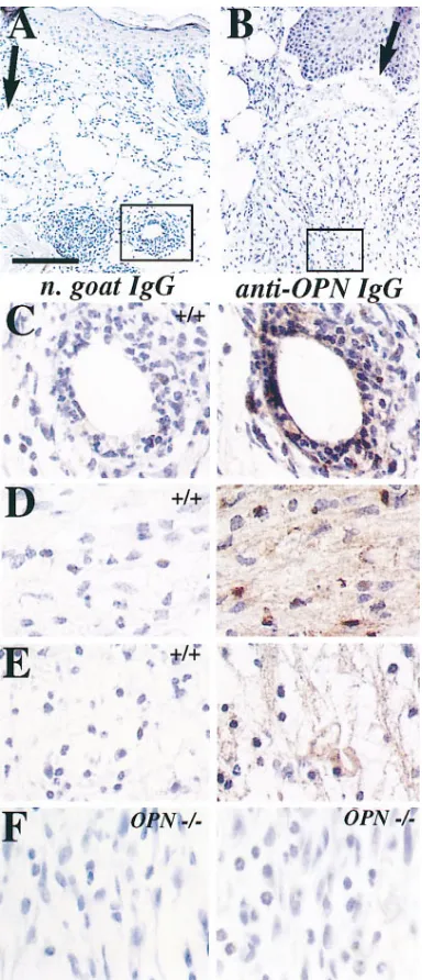
We further examined the protein localization of OPN using immunostaining of wounds from wild-type animals. As ex-pected, protein was detected in the dermal stroma in 4-d wounds (Fig. 4). Protein was localized to regions directly in the incision wound plane (Fig. 4 B, arrow indicates region of inci-sion), as well as in adjacent regions (Fig. 4 A). In addition to cell-associated staining (Fig. 4 C), abundant extracellular stain-ing was also seen (Fig. 4, D and E), reflectstain-ing deposition of OPN in the matrix of the wound. Comparable sections of wounds from OPN null mutant animals were also immu-nostained, and no OPN was detectable in any mutant animals (Fig. 4 F).
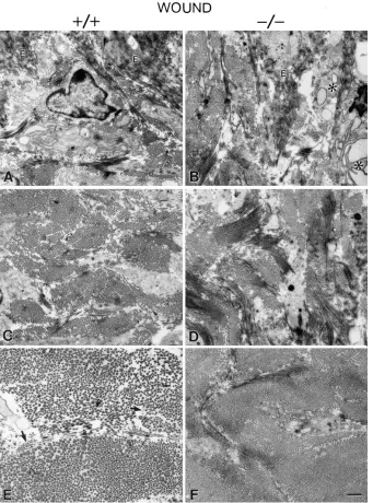
Morphological analysis of wounds from wild-type and OPN mutant mice. Healing wounds were compared between wild-type and OPN null mutant mice during the repair process. The extent of reepithelization and general wound organization was similar between mutant and wild-type skin when analyzed us-ing light microscopy (data not shown). However, ultrastruc-tural analysis demonstrated several distinct differences be-tween the mutant and wild-type wounds at 2 wk after injury. First, the wound regions in the OPN mutant mice contained more cell debris than corresponding regions in wild-type mice. The increased cell debris seen in mutant mice was most strik-ing in the subepithelial dermis (zone 1; Fig. 5, A and B). How-ever, this difference was obvious throughout both the superfi-cial and middermal zones (Fig. 5, C and D). Since OPN has been shown to be a chemoattractant for macrophages, which are primarily responsible for debridement at the wound site, incisions were immunostained with a pan-macrophage anti-body to test for differences in macrophage influx in wounds of normal or OPN mutant mice. Incisions at 4 d were chosen, since at this time macrophages have begun to appear in large numbers at the wound site. In both groups, there were high levels of macrophages throughout the incision, and the num-bers were not significantly different between groups (means6 SEM of wild-type mice 5 43.867.8, of OPN mutant mice 5 41.564.6). However, the apparent lower level of debridement in the OPN mutants suggests that the activity of these mac-rophages may be compromised.
The second major difference between resolving wounds in wild-type and OPN mutant animals was that in the OPN mu-tant, the reforming matrix was less organized than in corre-sponding regions of wild-type mice. Specifically, in the subepi-thelial and middermal zones, the organization of collagen fibrils and fibers was not as distinct in the mutant compared with wild-type mice (Fig. 5, A and C, wild-type, vs. Fig. 5, B and D, mutant). Fibers were less organized and more difficult to define in the mutant mice. Finally, in the deep reticular der-mis, the collagen fibrils in the OPN mutant mice were signifi-cantly smaller than fibrils in the corresponding region of the wild-type mice (Fig. 5, E and F). This difference in fibril diam-eter in the wound is shown at higher magnification in Fig. 6 (left columns).
[image:6.612.57.249.57.503.2]The differences in fibril diameters seen in the deep reticu-lar dermis (zone 3) of the wound area in the OPN mutants compared with wild-type mice were significant. In the mutants, fibrils remained small, with a homogenous distribution at all wound levels (Fig. 6, second column, and Table I). In contrast, in the wild-type mice, fibrils became significantly larger, and
Figure 4. Localization of OPN in skin wounds. (A and B) Low mag-nification views of skin incisions 4 d after injury in wild-type animals, showing position of C and D (boxed regions) relative to the incision plane (black arrows). (C–E) Serial sections of skin incisions 4 d after injury in wild-type animals stained with anti-OPN IgG (right) or nor-mal goat IgG (left). (C) Higher magnification of region boxed in A, showing that positive cells were localized to regions surrounding ves-sels, possibly reflecting inflammatory cells entering the wound site. (D) Higher magnification of boxed region in B, showing that OPN protein was both cell- and matrix-associated (E). (F) Two compara-ble sections from 4-d wounds of OPN null mutant animals were stained with anti-OPN IgG. No OPN was detectable in mutant ani-mals. Sections were counterstained with hematoxylin. Scale bar, 400
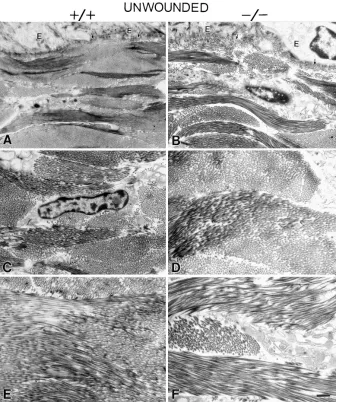
there was a heterogeneous distribution of fibril diameters. The difference was readily apparent in the deep dermal region (Fig. 6, zone 3). Small diameter fibrils comparable to those seen in the deep dermis of the mutant mice were also present in the wild-type mice (Fig. 5 E, arrows). Although the reorga-nization of the matrix was significantly different in the wounds of OPN mutant mice compared with the wild-type controls, the normal unwounded skin in both groups was generally simi-lar in terms of both tissue architecture and cell organization in the subepithelial (Fig. 7, A and B), middermal (Fig. 7, C and D), and deep dermal regions (Fig. 7, E and F). Collagen fibril diameters were also comparable in the unwounded dermis of both wild-type and OPN mutants (Fig. 6, right columns, and Table I). In contrast to the wound regions in the OPN mutants, there was an increase in mean fibril diameter as well as devel-opment of a heterogeneous distribution of fibril diameters in unwounded dermis from the superficial to deep regions. There
was no difference between the wild-type and mutant mice in this respect. Therefore, the differences in dermal organization between the OPN mutant and wild-type mice only appear to be significant in the context of the healing process.
[image:7.612.57.398.58.518.2]Tensile strength of healing incisional wounds. We tested whether the differences in collagen organization in the OPN mutant mice led to changes in tensile strength, a measure thought to be determined predominantly by fibrillar collagen deposition and remodeling. Samples were collected at 7 and 14 d after injury and tested either fresh or after fixation. Fig. 8 A shows that no significant differences in tensile strength were apparent at 7 or 14 d between normal and mutant mice. One consistent finding was that regardless of genotype, there was a significantly greater fresh tensile strength of male versus fe-male skin 2 wk after injury (Fig. 8 B). In normal unwounded skin, the same results were noted. There was no significant difference in normal skin strength as a function of genotype,
but there was a very large difference as a function of gender (Fig. 8 C).
Discussion
Functional compensation between OPN and VN-related pro-teins during embryogenesis. Targeted mutation of genes has been accomplished for several adhesive proteins and their re-ceptors, including RGD-containing proteins and members of the integrin family (33). In many cases, the phenotypes of the mutants are unexpected, with animals presenting with no obvi-ous defects, or surviving to later stages of development than the expression pattern or in vitro evidence would predict. One
interpretation of these findings is that proteins with shared functional domains or receptors compensate for each other’s absence during embryogenesis. Our findings reported here of a normal phenotype in spp1 mutant mice suggest that there are complex functional interactions among RGD-containing in-tegrin ligands during embryogenesis. In addition, the in vivo roles of OPN may also be mediated through nonintegrin re-ceptors. One possible explanation for the normal embryonic phenotype even in the spp1/vtn double mutant mice is a func-tional compensation by other extracellular proteins. The avb3
[image:8.612.57.394.55.396.2]integrin is a predominant receptor for both OPN and VN, and can also bind thrombospondin, tenascin, fibronectin, and oth-ers. Interestingly, single mutations in the genes encoding
Figure 6. Alterations in collagen fibril for-mation after a full-thickness dermal wound in OPN mutant mice. Collagen fibril diam-eters remained small, with a homogenous distribution at all wound levels in the OPN mutant mouse (WOUND 2/2), while in the wild-type mouse, fibril diameters be-came significantly larger, with a heteroge-nous distribution of diameters (WOUND
1/1) (compare zone [Z] 3, WOUND 1/1 vs. 2/2). In contrast, fibril formation was comparable in unwounded dermis in wild-type (UNWOUNDED 1/1) versus OPN mutant (UNWOUNDED 2/2) mice. Sec-tions are 100–125 nm thick and stained with uranyl acetate/phosphotungstic acid. Bar, 100 nm.
Table I. Collagen Fibril Diameters in Wild-type versus OPN Mutant Skin
1/1 Zone 1
1/1 Zone 2
1/1 Zone 3
2/2 Zone 1
2/2 Zone 2
2/2 Zone 3
Wounded
Mean6SD (nm) 65611 63612 107651 5668 5867 5469
Range (nm) 16–91 (n 5 359) 22–93 (n 5 354) 26–310 (n 5 399) 23–82 (n 5 270) 29–82 (n 5 345) 21–73 (n 5 264) Unwounded
Mean6SD (nm) 87627 101633 126639 80618 110637 116629
Range (nm) 37–308 (n 5 287) 30–280 (n 5 297) 42–283 (n 5 275) 37–149 (n 5 187) 28–322 (n 5 298) 45–252 (n 5 314)
[image:8.612.58.556.597.702.2]thrombospondin-1, tenascin-C, OPN, and VN do not lead to developmental defects. However, inactivation of the av
integ-rin, a component of their shared receptors, causes embryonic and perinatal lethality (33). These data are consistent with the idea that loss of one or two of these proteins can be compen-sated for by another ligand that signals through a common re-ceptor.
Role of OPN during wound healing—inflammatory phase. We have studied wound healing in skin incisions, where the ini-tial component of repair is hemostasis and an inflammatory re-sponse. During the inflammation phase, spp1 was expressed at high levels in regions of infiltrating leukocytes. This expression was seen as early as 6 h after wounding with the first influx of neutrophils, and was also seen in macrophages as they entered the wound site. Since all cell types expressing spp1 could not be identified definitively in the stroma, it is likely that fibro-blasts may also be a source of OPN. Previous in vivo data have demonstrated that injection of OPN led to an accumulation of macrophages and, to a lesser extent, polymorphonuclear leu-kocytes (15). In addition, OPN affects B cell and macrophage function (34), and thus is postulated to act as a cytokine for T cells, as well as a chemoattractant for macrophages (6). This
[image:9.612.57.397.58.460.2]latter hypothesis has been validated in a model of renal tubu-lointerstitial disease in vivo, where macrophage influx into the injury site is decreased significantly in the OPN null mutant mouse (Ophascharoensuk, V., C.M. Giachelli, L. Liaw, K. Gordon, R. Schmidt, C.E. Alpers, S.J. Shankland, W.G. Couser, and R.J. Johnson, manuscript submitted for publica-tion). In the case of the cutaneous wound model, although the numbers of macrophages were similar in wild-type and mutant animals, our findings support a role for OPN in the activation or function of these macrophages. Incisions in OPN mutants showed a marked decrease in the level of debridement, sug-gesting that the phagocytes are less active in the removal and degradation of damaged connective tissue. We have also ex-amined the expression of macrophage markers modulated upon cell activation, including the mannose receptor (35). Our preliminary findings indicate that macrophages in wounds of OPN mutant animals have a higher level of mannose receptor compared with wild-type animals. Since a decrease in mannose receptor in macrophages correlates with their activation (36), this suggests that a greater proportion of macrophages in the OPN mutant wounds are not activated. Taken together, these findings support a model where the processes of macrophage
chemotaxis, activation, or phagocytic activity are mediated in part by OPN at the wound site. A similar idea has been pro-posed during wound healing in bone, where OPN secreted by macrophages is postulated to be required for cell adhesion and phagocytosis (37). Thus, one explanation for the high levels of OPN at wound sites is that the protein stimulates immune ac-tivity. Data from several labs posit that this model may be ap-plicable to dermal wound repair, cardiovascular remodeling, and wound healing of bone.
OPN and collagen fibrillogenesis. After the inflammatory phase, granulation tissue forms, and there is maturation and remodeling of the matrix. There was a significant difference in matrix organization and collagen fibrillogenesis in the OPN mutants compared with controls. In the incisions of wild-type animals at 2 wk, collagen fibrils were organized into well-defined fibers composed of a heterogeneous mixture of fibrils of varying diameter, including coarse fibrils in the deep dermis. However, the incisions in OPN mutant mice had a more disor-ganized matrix overall, with less collagen organization into fi-bers. The fibers that were present were composed only of ho-mogeneous smaller diameter fibrils. One possibility for these differences is that OPN may affect the synthesis and/or turn-over of matrix components involved in the regulation of fibril formation, either by affecting the types of collagens synthe-sized, or by association with proteins present in the remodeling wound. Regulation of fibril formation at this step involves het-erotypic interactions of fibrillar collagens and the extent of amino-terminal processing. Collagen type III is a marker of the immature wound (38), and heterotypic interaction of dif-ferent collagens is thought to regulate fibril size. In support of this idea, mice with a targeted mutation of the gene encoding type III procollagen (Col3a1) formed dermal collagen fibers of greater diameter than wild-type animals (39), and a point mu-tation in Col3a1 likewise led to matrix disorganization and lagen fibril diameter heterogeneity (40, 41). The loss of col-lagen type III during tendon development also correlates with increases in collagen fibril diameter (42), and analysis of hu-man skin has shown that large collagen fibrils consist mostly of type I, while smaller fibrils contain type III collagen.
There-fore, one possible cause of the persistence of smaller fibrils in the OPN mutant wounds is the disregulation of matrix syn-thesis.
Presumably as the wound resolves itself, the collagen fibrils mature. It has been suggested that this involves the destabiliza-tion of the immature fibrils followed by fusion, yielding longer and larger diameter fibrils (43, 44). Collagen-binding mole-cules that have been demonstrated to regulate this step include the small leucine-rich proteoglycans decorin, fibromodulin, and lumican. These proteoglycans have been implicated in the regulation of several parts of collagen fibrillogenesis (45), a role supported by the phenotype of the decorin null mutant mice, where uncontrolled lateral fusion of collagen fibrils oc-curs (46). The absence of OPN in the wounds of the mutant mice could alter synthesis of molecules that are involved in the stabilization or turnover of these components, such as the leu-cine-rich proteoglycans or matrix metalloproteinases, which are also known to be produced during skin injury (reference 47, and Crawford, H.C., J.H. Wright, J. Mudgett, and L.M. Matrisian, manuscript submitted for publication). A mainte-nance of stable immature fibrils would explain the wound phe-notype observed in the OPN mutant mice.
[image:10.612.67.523.62.209.2]OPN may also interact directly with collagen or the other proteins discussed above. OPN has been shown to bind di-rectly to collagen type I (48), and to interact with collagen types II, III, IV, and V (49), and fibronectin (50), although it is not certain when such interactions occur in connective tissues in vivo. In mineralized tissues such as the tooth, OPN has been localized by immunocytochemistry to specific regions of col-lagen fibrils (51). Whether by a direct or indirect mechanism, lack of OPN in the wound appears to alter matrix assembly, leading to disorganization in the resolution of the wound. Since matrix organization was normal in the unwounded skin of OPN mutants, these functions may be replaced by other proteins during development. Alternatively, different proces-ses may be involved during embryogenesis versus adult wound healing. Long-term observation of residual scar tissue is needed to determine whether continued remodeling in the OPN mutants eventually normalizes matrix architecture.
In summary, we report the first establishment of OPN null mutant mice, which are viable, thus providing the opportunity to determine the in vivo functions of OPN during disease or tissue remodeling. Further experiments are necessary to un-derstand fully the functions of OPN during incisional wound healing, but we provide evidence for a role in stimulation of macrophage function, and regulation of collagen fibrillogene-sis. These findings indicate an in vivo role of OPN in the struc-tural remodeling and resolution of dermal wounds.
Acknowledgments
We are grateful to Xianxian Zheng and David Ginsburg (University of Michigan, Ann Arbor, MI) for providing the VN mutant mice. We also thank members of the laboratories of Richard Johnson and Charlie Alpers (University of Washington, Seattle) for their contribu-tions to immunostaining and ultrastructural analysis of kidney, re-spectively; Marc McKee (University of Montreal, Montreal, Canada) for ultrastructural analysis of bone; Virginia Shepherd (Vanderbilt University, Nashville, TN) for macrophage expertise; and Julie Blackwell-Fowler for excellent technical assistance.
L. Liaw is the recipient of a Developing Scientist Award from the American Heart Association. This work was supported in part by Na-tional Institutes of Health grant AG-06528 and the Department of Veterans Affairs (J.M. Davidson). B.L.M. Hogan is an Investigator of the Howard Hughes Medical Institute.
References
1. Franzén, A., and D. Heinegård. 1985. Isolation and characterization of two sialoproteins present only in bone calcified matrix. Biochem. J. 232:715– 724.
2. Oldberg, A., A. Franzén, and D. Heinegård. 1986. Cloning and sequence analysis of rat bone sialoprotein (osteopontin) cDNA reveals an arg-gly-asp cell-binding sequence. Proc. Natl. Acad. Sci. USA. 83:8819–8823.
3. Senger, D.R., D.F. Wirth, and R.O. Hynes. 1980. Transformation-specific secreted phosphoproteins. Nature. 286:619–621.
4. Smith, J.H., and D.T. Denhardt. 1987. Molecular cloning of a tumor pro-moter-inducible mRNA found in JB6 mouse epidermal cells: induction is stable at high, but not at low, cell densities. J. Cell. Biochem. 34:13–22.
5. Patarca, R., G.J. Freeman, R.P. Singh, F.-Y. Wei, T. Durfee, F. Blattner, D.C. Regnier, C.A. Kozak, B.A. Mock, J.C. Morse III, et al. 1989. Structural and functional studies of the early T-lymphocyte activation 1 (Eta-1) gene. Def-inition of a novel T cell-dependent response associated with genetic resistance to bacterial infection. J. Exp. Med. 170:145–161.
6. Weber, G.F., and H. Cantor. 1996. The immunology of Eta-1/osteopon-tin. Cytokine Growth Factor Rev. 7:241–248.
7. Denhardt, D.T., C.A. Lopez, E.E. Rollo, S.M. Hwang, X.R. An, and S.E. Walther. 1995. Osteopontin-induced modifications of cellular functions. Ann.
NY Acad. Sci. 760:127–142.
8. Thayer, J.M., C.M. Giachelli, P.E. Mirkes, and S.M. Schwartz. 1995. Ex-pression of osteopontin in the head process late in gastrulation in the rat. J.
Exp. Zool. 272:240–244.
9. Fan, C.M., and M. Tessier-Lavigne. 1994. Patterning of mammalian somites by surface ectoderm and notochord: evidence for sclerotome induction by a hedgehog homolog. Cell. 79:1175–1186.
10. Nomura, S., A.J. Wills, D.R. Edwards, J.K. Heath, and B.L.M. Hogan. 1988. Developmental expression of 2ar (osteopontin) and SPARC (osteonec-tin) RNA as revealed by in situ hybridization. J. Cell Biol. 106:441–450.
11. Seiffert, D., T.J. Podor, and D.J. Loskutoff. 1993. Distribution of vit-ronectin. In Biology of Vitronectins and Their Receptors. K.T. Preissner, S. Rosenblatt, C. Kost, J. Wegerhoff, and D.F. Mosher, editors. Elsevier Science B.V., Amsterdam. 75–81.
12. Cooper, S., W. Bennett, S. Roach, and M.F. Pera. 1993. Vitronectin ex-pression in human germ cell tumours and normal mouse development. In Biol-ogy of Vitronectins and Their Receptors. K.T. Preissner, S. Rosenblatt, C. Kost, J. Wegerhoff, and D.F. Mosher, editors. Elsevier Science B.V., Amsterdam. 83– 90.
13. Zheng, X., T.L. Saunders, S.A. Camper, L.C. Samuelson, and D. Gins-burg. 1995. Vitronectin is not essential for normal mammalian development and fertility. Proc. Natl. Acad. Sci. USA. 92:12426–12430.
14. Brown, L.F., A. Papadopoulos-Sergiou, B. Berse, E.J. Manseau, K. Tognazzi, C.A. Perruzzi, H.F. Dvorak, and D.R. Senger. 1994. Osteopontin ex-pression and distribution in human carcinomas. Am. J. Pathol. 145:610–623.
15. Singh, R.P., R. Patarca, J. Schwartz, P. Singh, and H. Cantor. 1990. Def-inition of a specific interaction between the early T lymphocyte activation 1 (Eta-1) protein and murine macrophages in vitro and its effect upon macro-phages in vivo. J. Exp. Med. 171:1931–1942.
16. Murry, C.E., C.M. Giachelli, S.M. Schwartz, and R. Vracko. 1994. Mac-rophages express osteopontin during repair of myocardial necrosis. Am. J.
Pathol. 145:1450–1462.
17. Leibovich, S.J., and R. Ross. 1975. The role of the macrophage in wound repair: a study with hydrocortisone and anti-macrophage serum. Am. J. Pathol. 78:71–91.
18. Portera, C.A., E.J. Love, L. Memore, L. Zhang, A. Müller, W. Browder, and D.L. Williams. 1997. Effect of macrophage stimulation on collagen biosyn-thesis in the healing wound. Am. Surg. 63:125–130.
19. Giachelli, C.M., S.M. Schwartz, and L. Liaw. 1995. Molecular and cellu-lar biology of osteopontin: potential role in cardiovascucellu-lar disease. Trends
Car-diovasc. Med. 5:88–95.
20. Giachelli, C.M., R. Pichler, D. Lombardi, D.T. Denhardt, C.E. Alpers, S.M. Schwartz, and R.J. Johnson. 1994. Osteopontin expression in angiotensin II-induced tubulointerstitial nephritis. Kidney Int. 45:515–524.
21. Kohri, K., S. Nomura, and Y. Kitamura. 1993. Structure and expression of the mRNA encoding urinary stone protein (osteopontin). J. Biol. Chem. 15: 180–184.
22. Tybulewicz, V.L.J., C.E. Crawford, P.K. Jackson, R.T. Bronson, and R.C. Mulligan. 1991. Neonatal lethality and lymphopenia in mice with a ho-mozygous disruption of the c-abl proto-oncogene. Cell. 65:1153–1163.
23. Zhao, G.-Q., K. Deng, P.A. Labosky, L. Liaw, and B.L.M. Hogan. 1996. The gene encoding bone morphogenetic protein 8B is required for the initiation and maintenance of spermatogenesis in the mouse. Genes Dev. 10:1657–1669.
24. Davisson, M.T. 1995. Genetic nomenclature guide (mouse). In Trends in Genetics Nomenclature Guide. A. Stewart, editor. Elsevier Trends Journals, Oxford. 35–38.
25. Liaw, L., M. Almeida, C.E. Hart, S.M. Schwartz, and C.M. Giachelli. 1994. Osteopontin promotes vascular cell adhesion and spreading and is chemotactic for smooth muscle cells in vitro. Circ. Res. 74:214–224.
26. Zhao, G.-Q., and B.L.M. Hogan. 1996. Evidence that mouse Bmp8a (Op2) and Bmp8b are duplicated genes that play a role in spermatogenesis and placental development. Mech. Dev. 57:159–168.
27. Birk, D.E., and R.L. Trelstad. 1984. Extracellular compartments in ma-trix morphogenesis: collagen fibril, bundle, and lamellar formation by corneal fibroblasts. J. Cell Biol. 99:2024–2033.
28. Craig, A.M., and D.T. Denhardt. 1991. The murine gene encoding se-creted phosphoprotein 1 (osteopontin): promoter structure, activity, and induc-tion in vivo by estrogen and progesterone. Gene (Amst.) 100:163–171.
29. Liaw, L., M.P. Skinner, E.W. Raines, R. Ross, D.A. Cheresh, S.M. Schwartz, and C.M. Giachelli. 1995. The adhesive and migratory effects of os-teopontin are mediated via distinct cell surface integrins. Role of avb3 in smooth muscle cell migration to osteopontin in vitro. J. Clin. Invest. 95:713–724. 30. Rowe, L.B., J.H. Nadeau, R. Turner, W.N. Frankel, V.A. Letts, J.T. Ep-pig, M.S. Ko, S.J. Thurston, and E.H. Birkenmeier. 1994. Maps from two inter-specific backcross DNA panels available as a community genetic mapping re-source. Mamm. Genome. 5:253–274.
31. Segre, J.A., J.L. Nemhauser, B.A. Taylor, J.H. Nadeau, and E.S. Lander. 1995. Positional cloning of the nude locus: genetic, physical, and tran-scription maps of the region and mutations in the mouse and rat. Genomics. 28: 549–559.
32. Fet, V., M.E. Dickinson, and B.L.M. Hogan. 1989. Localization of the mouse gene for secreted phosphoprotein 1 (Spp1) (2ar, osteopontin, bone sialoprotein1, 44-kDa bone phosphoprotein, tumor-secreted phosphoprotein) to chromosome 5, closely linked to Ric (Rickettsia resistance). Genomics. 5:375– 377.
33. Hynes, R.O. 1996. Targeted mutations in cell adhesion genes: what have we learned from them? Dev. Biol. 180:402–412.
34. Lampe, M.A., R. Patarca, M.V. Iregui, and H. Cantor. 1991. Polyclonal B cell activation by the Eta-1 cytokine and the development of systemic au-toimmune disease. J. Immunol. 147:2902–2906.
35. Shepherd, V.L., R. Abdolrasulnia, M. Garrett, and H.B. Cowan. 1990. Down-regulation of mannose receptor activity in macrophages after treatment with lipopolysaccharide and phorbol esters. J. Immunol. 145:1530–1536.
36. Noorman, F., E.A.M. Braat, M. Barrettbergshoeff, E. Barbe, A. Van-leeuwen, J. Lindeman, and D.C. Rijken. 1997. Monoclonal antibodies against the human mannose receptor as a specific marker in flow cytometry and immu-nohistochemistry for macrophages. J. Leukocyte Biol. 61:63–72.
37. McKee, M.D., and A. Nanci. 1996. Secretion of osteopontin by mac-rophages and its accumulation at tissue surfaces during wound healing in miner-alized tissues: a potential requirement for macrophage adhesion and phagocy-tosis. Anat. Rec. 245:394–409.
38. Clore, J.N., I.K. Cohen, and R.F. Diegelmann. 1979. Quantitation of the collagen types I and III during wound healing in rat skin. Proc. Soc. Exp. Biol.
Med. 161:337–340.
40. Toman, D., L. Garrett, T. Mascara, J. Bateman, and B. de Crombrugghe. 1993. Transgenic mice harboring a Lys→Met cross-linking mutation in type III collagen. Matrix. 13:50. (Abstr.)
41. Quaglino, D., Jr., D. Toman, B. de Crombrugghe, and J.M. Davidson. 1994. Transgenic mice expressing a col3a1 mutation at a crosslinking position exhibit structural alterations at sites of wound healing. Matrix Biol. 14:512. (Ab-str.)
42. Birk, D.E., and R. Mayne. 1997. Localization of collagen types I, III and V during tendon development. Changes in collagen types I and III are corre-lated with changes in fibril diameter. Eur. J. Cell Biol. 72:352–361.
43. Birk, D.E., M.V. Nurminskaya, and E.I. Zycband. 1995. Collagen fibril-logenesis in situ: fibril segments undergo post-depositional modifications result-ing in linear and lateral growth durresult-ing matrix development. Dev. Dyn. 202:229– 243.
44. Birk, D.E., E.I. Zycband, S. Woodruff, D.A. Windelmann, and R.L. Trelstad. 1997. Collagen fibrillogenesis in situ: fibril segments become long fibrils as the developing tendon matures. Dev. Dyn. 208:291–298.
45. Iozzo, R.V. 1997. The family of small leucine-rich proteoglycans: key regulators of matrix assembly and cellular growth. Crit. Rev. Biochem. Mol.
Biol. 32:141–174.
46. Danielson, K.G., H. Baribault, D.F. Holmes, H. Graham, K.E. Kadler, and R.V. Iozzo. 1997. Targeted disruption of decorin leads to abnormal col-lagen fibril morphology and skin fragility. J. Cell Biol. 136:729–743.
47. Okada, A., C. Tomasetto, Y. Lutz, J.P. Bellocq, M.C. Rio, and P. Bas-sett. 1997. Expression of matrix metalloproteinases during rat skin wound heal-ing: evidence that membrane type-1 matrix metalloproteinase is a stromal acti-vator of pro-gelatinase A. J. Cell Biol. 137:67–77.
48. Chen, Y., B.S. Bal, and J.P. Gorski. 1992. Calcium and collagen binding properties of osteopontin, bone sialoprotein, and bone acidic glycoprotein-75 from bone. J. Biol. Chem. 267:24871–24878.
49. Butler, W.T. 1995. Structural and functional domains of osteopontin.
Ann. NY Acad. Sci. 760:6–11.
50. Mukherjee, B.B., M. Nemir, S. Beninati, E. Cordella-Miele, K. Singh, I. Chackalaparampil, V. Shanmugan, M.W. DeVouge, and A.B. Mukherjee. 1995. Interaction of osteopontin with fibronectin and other extracellular matrix mole-cules. Ann. NY Acad. Sci. 760:201–212.

![Figure 3. Expression of cisional injury. Full-thickness skin inci-sions were collected at various times after injury, and in situ hybridization was carried out using an antisense [spp1 after skin in-35S]UTP-labeled spp1 riboprobe](https://thumb-us.123doks.com/thumbv2/123dok_us/8215882.820086/5.612.57.403.465.740/expression-cisional-thickness-collected-hybridization-antisense-labeled-riboprobe.webp)