i
FETAL ELECTROCARDIOGRAM EXTRACTION AND
ANALYSIS
Thesis submitted in partial fulfillment
for the award of the degree of
Bachelor of Technology
In
Electronics and Communication Engineering
Submitted by
AJIT KUMAR SAHU (110EC0180)
JYOTIRANJAN RAJ (110EI0052)
Under the guidance of
Dr. Samit Ari
Assistant Professor
Department of Electronics & Communication Engineering
National Institute of Technology, Rourkela
Odisha-769008
INDIA
ii
DECLARATION
I hereby declare that the work presented in the thesis entitled “
Fetal
Electrocardiogram Extraction And Analysis”
is a bonafide record of the
research work done by me under the
supervision of Dr. Samit Ari,
Department of Electronics & Communication Engineering,
National
Institute of Technology, Rourkela, India and that this thesis work has not
been presented for the award of any other degree.
AJIT KUMAR SAHU
JYOTIRANJAN RAJ
Dept. of Electronics & Comm. Engg.
National Institute of Technology,Rourkela
Odisha-769008
INDIA
iii
CERTIFICATE
Certified that this project thesis on
“
FETAL ELECTROCARDIOGRAM
EXTRACTION AND ANALYSIS”
is a bonafide work of
“
AJIT KUMAR SAHU
”
and
“
JYOTIRANJAN RAJ
”
who carried out the research project
under my supervision and guidance during Aug 2013-May
2014(7
th& 8
thSemester).
This thesis has not been
submitted for any degree or academic award elsewhere.
Date : Dr. Samit Ari
Place:
Assistant Professor
Dept. of Electronics & Comm. Engg.
National Institute of Technology, Rourkela
Odisha-769 008
INDIA
iv
ACKNOWLEDGEMENT
Firstly, we would like to express our deep sense of respect and gratitude towards our advisor and guide Prof. Samit Ari, who has been the guiding force behind this work. We want to thank him for introducing us to the field of Signal Processing and giving us the opportunity to work under him. It is our good fortune to have got an opportunity to work under such a wonderful person. We sincerely appreciate his inspiration; support and guidance. He has been instrumental in making this project a success.
We also express our sincere gratitude to Prof S. Meher, Head of Department, Electronics and Communication Engineering for allowing access to all valuable facilities which were a necessity for us. We would also like to convey our gratitude to all the research scholars in our department who assisted us during the completion of this project work and their well-timed and valuable guidance that helped us to finish the work in the predetermined period of time.
We are obliged to staff members of department of Electronics and Communication Engineering, for the valuable information provided by them in their respective fields. We are grateful for their cooperation during the period of our assignment.
Our thanks and appreciations also go to our friends and batch mates in developing the project and people who have willingly helped us out with their abilities.
Ajit Kumar Sahu Jyotiranjan Raj
v ABSTRACT
Diagnosis of mother’s and child’s heart beat is very necessary during pregnancy and hence we use Fetal electrocardiogram (FECG) extraction for the same. The signal contains precise informations that can help doctors during pregnancy and labor. In this thesis, an easy-to use method has been implemented using adaptive noise canceller (ANC). Using the ANC, an effective algorithm has been proposed. The algorithm uses ANC, Least Mean Square (LMS) method and a Simulink model for the extraction of FECG. The FECG extraction method has been implemented using an algorithm implemented on MATLAB using Simulink models. The extracted FECG signal is a noise free signal. The QRS complex has been detected using another algorithm that counts the R-R peaks. The simulation result shows that heart rate of the FECG signal can be counted using the detection algorithm.
This project evokes a complete model of the FECG extraction with the implementation of effective algorithms and adaptive filters and finally gives the heart rate of the FECG signal.
Keywords:
Electrocardiogram, Adaptive Noise Cancellation, Maternal Heart Beat, LMS Algorithm, QRS Complex, Peak Detection
vi CONTENTS Declaration……….ii Certificate………..iii Acknowledgement……….iv Abstract………...v List of Figures……….viii 1. INTRODUCTION……….1 1.1Extraction………...2 1.2Fetal Heart………..3 1.3Heart Signal………...4 1.4Literature Survey………...5 1.5Objective………5 1.6Database……….6 1.7Thesis Outline………6
2. EXTRACTION OF THE FETAL ECG SIGNAL………...…...8
2.1Methodology…………...……….11
2.2Algorithm……….12
2.3Proposed Simulink Model………...13
2.4Least Mean Square Algorithm……….14
vii 2.6Experimental Results……….………..19 2.7Conclusion………...22 3 QRS COMPLEX………23 3.1Methodology………...……….25 3.2Experimental Results…….………...28 3.3Conclusion………...…29
4 CONCLUSION AND FUTURE WORKS………..….……..…..30
4.1Conclusions………..30
4.2Future Works………...30
viii
LIST OF FIGURES
Fig 1.1 Measurement of Fetal ECG……….3
Fig 2.1 FECG Extraction Algorithm………..12
Fig 2.2 Simulink Model 1………...13
Fig 2.3 Simulink Model 2……….…….14
Fig 2.4 Maternal Heart Beat Signal……….…..16
Fig 2.5 Fetal ECG Signal………...………16
Fig 2.6 Measured Signal ……….……….….………....17
Fig 2.7 Adaptive Noise Cancellation System………..………….………..……...18
Fig 2.8 Measured Fetal ECG Signal………..……20
Fig 2.9 Output of the Adaptive Noise Canceller (ANC)………...21
Fig 2.10 Peak Detection……….21
Fig 3.1 QRS waveform………...………...24
Fig 3.2 Original ECG signal………..………26
Fig 3.3 First Order Derivative having a smooth and rectified signal………26
Fig 3.4 Second Order Derivative having a smooth and rectified signal………26
Fig 3.5 Sum of First and Second Order Derivative………...26
Fig 3.6 Square Pulse Output for each QRS complex………26
Fig 3.7 Differentiation Technique implemented on MATLAB…...28
1
CHAPTER 1
2 1.1 FETAL ECG EXTRACTION
Every year, one out of hundred babies born with some form of heart defects. This occurs due to genetic syndrome, inherited disorder or environmental factors like misuse of drugs. In any case regular monitoring of heart of the baby is necessary before it is born. Hence, Fetal ECG (FECG) signals are necessary to monitor the heart condition of the baby, so that if any abnormalities found then it can be solved clinically by the concerned doctors [1].
Fetal ECG monitoring is a widely used technique for diagnosis and to find out fetal abnormalities. The physician can easily prepare himself/herself for the possible abnormalities in a fetus by taking diagnosis of the fetal ECG signal during the pregnancy stage. It’s the simplest non-invasive method to diagnose various heart diseases. The various electrical activity of the heart is represented by the Fetal ECG (FECG) and hence it provides valuable information about its physiological state. The FECG signal can be easily obtained from the abdomen of a pregnant women and the chest gives the maternal electrocardiogram (MECG) signal. The addition of the MECG signal with the FECG signal is typically annoying one. By placing electrodes on the maternal abdomen, the FECG signal, thus generated, gives minute details about the fetal condition which are very useful during diagnosis. The abdominal ECG signal contains various unwanted interferences, maternal ECG (MECG) signal and electromyogram (EMG) and the FECG signal is corrupted by various noise and the skin impedance [2].
The electrical activity of the heart is described by the ECG. The ECG signal consist of three types of wave. The details about QRS complex have been discussed in Chapter 2. The peaks of the QRS complex give the information about the heart rate of the abdominal ECG (AECG) signal. Hence, it is very crucial for the doctors to detect the heart defects before it causes any damage to the fetus or the mother. When the ECG signal is taken from the abdominal leads, a composite signal is picked [3].
3
There are various methods to extract FECG like wavelet transform, Doppler ultrasound, adaptive filtering, correlation methods, blind source separation technique and a combination of blind source separation methods and wavelet analysis. The heart rate of the FECG can be determined by calculating the R-R peaks of the QRS complex [4]. But, as the FECG signal is merged with MECG signal and various interferences, it is very difficult to calculate the heart rate from the raw signal. Hence, the FECG is extracted from the raw signal to get the proper heart rate of the fetal signal.
1.2 HEART OF THE FETUS
There are various organs that grows slowly inside a fetus. Amongst that aone of them is the fetal heart. It goes through various changes with time during the early stages of pregnancy. After 22 days of pregnancy the heart states beating. From 18th - 20th week of conception the FECG signals
can be measured from the abdomen of the mother. In the maternal abdomen the skin and the fat have very conductivity. Combining them forms a volume conductor which helps in the propagation of the ECG signals to the surface. Fig 1.2 shows the measurement of ECG signal from the fetus.
4 1.3 HEART SIGNAL
Action Potential
It is the membrane potential of the cell. When an electrical current is stimulated to a cell then the cell goes through various mechanical contraction to give a potential known as action potential [6].
Resting Potential
It is the static membrane potential of the cell. When the cells are at rest the membrane readily allows ions like K+ and Cl- to pass into them because they are permeable membranes. The
outside of cell is positively charged than the inside of the cell. Hence, K+ ions flows into the cell to balance the charge. Hence, a potential difference gets developed between the inside and outside of the cell at the state of equilibrium. A resting cell is a polarised cell [7].
Depolarisation
Initially the cell doesn’t allow Na+ ions inside due to higher permeability of the K+ ions. But
when the cell is excited it starts allowing the Na+ ions to move inside. This movement of Na+
ions contain the ionic currents. Hence, the membrane barrier reduces to Na+ ions. The rushing of Na+ ions into the cell is called “Avalanche effect”. Due to higher concentration of the K+ ions in the cell it tries to move out of the cell but can’t move as fast as the Na+ ions. Hence, the inside of
the cell becomes positive with respect to the outside of the cell due to imbalance of the K+ ions. This process is called depolarisation [8].
Repolarisation
The repolarisation of cells depends on the time dependent and voltage dependent of the membrane permeability changes for K+ ions compared with that for the Na+ ions. During
5
depolarisation the Na+ ions moment get slower and hence repolarisation occurs due to the increase in moment of the K+ ions at the end of depolarisation [9].
1.4 LITERATURE SURVEY
Seeing a typical fetal ECG (FECG) signal, the doctor rectifies the fetal heart abnormalities. The ECG device extract the FECG signal from the Abdominal ECG (AECG) signal and shows them on the monitor. The heart rate of the fetal heart comes to be greater than the maternal heart rate. The motivation behind this project are challenges like the extraction of a low amplitude and high frequency signal from the high amplitude and low frequency signal. In this work, we have ensured extraction of fetal ECG signal but we haven’t processed it in an efficient manner as done in research work by Prasanth K and Baby Paul [10]. We have taken 4000 samples of the AECG signal with peak amplitude of 3.5 millivolts. And then the fetal ECG signal rate comes in the range 120-160 heart beats per minute. . And hence the fetal heart beats faster than the maternal heart and is very sensitive to heart abnormalities.
Our project ensures the peaks of the FECG is detected properly but not as efficiently done by Butler Pyke [11], it uses Pan-Tompkins algorithm for the detection of the QRS complex. In this project, we have used the differentiation technique for the detection of the R-R peaks in a QRS complex. Though the method is not so efficient, it gives us the required output as i.e. almost accurate FECG heart rate.
1.5 OBJECTIVE
Two ECG signals were taken; one from the chest of the mother and another from the abdomen of the mother that contains both the fetus heartbeat signal and maternal heartbeat. The objective of our study is to separate both these parts and thus extract the fetus heartbeat signal and then detect the R-peaks in order to determine the fetus heart rate.
6
Regenerating the waveforms of both ECG signals in MATLAB using the data taken from the ECG machine.
Separating both signals and reducing any noise present in the signal to obtain the fetus heartbeat signal using ANC.
ii) To count the R-peaks and calculating the heart rate of the fetus by using the R-R separation.
Applying a differentiation technique to the QRS-complex to differentiate the extracted signal up to 3rd order to get the peaks.
Setting a threshold and comparing the peaks with it to detect the R-peaks.
Finally counting the R-peaks per one minute to obtain the heart rate of the baby.
1.6 DATABASE
Abdominal and direct fetal electrocardiogram database included 38 and 41 weeks of gestation between research materials, labor derived from 5 different girls’ multichannel fetal electrocardiogram (FECG) has records. Entries fetal electrocardiogram (Itam company, Zabrze, Poland), Department of Obstetrics, Medical University of Silesia, through acquisition and analysis system was purchased KOMPOREL. Each record in the stomach and the fetal head to the maternal fetal electrocardiogram obtained directly from the record contains four different signals.
1.7 THESIS OUTLINE
Chapter 1 of the thesis explains the background of the fetal electrocardiogram extraction and also give details about literature survey on FECG extraction. Next it gives detail about the online data used in this thesis and the experimental setup to implement the extraction process and get the FECG signal as output.
7
Chapter 2 of the thesis explains the algorithm of fetal ECG extraction. Steps that has been taken and the database that has been used to extract the FECG signal from the Abdominal ECG (AECG) signal. Then the algorithm is implemented on MATLAB and the output is taken as plots.
Chapter 3 explains the method used for the detection of R-R peaks from the QRS complex of the FECG signal.
Chapter 4 describes the extraction output which is final output of the whole thesis work and gives the concluding remark and future work.
8
CHAPTER 2
EXTRACTION OF THE
FETAL ECG SIGNAL
9
Several methods are used for the extraction of fetal ECG signal from maternal ECG signal which is actually a mixture of both mother and fetus heartbeat signals. Some of the most widely used methods are Principal Component Analysis (PCA), Independent Component Analysis (ICA) and Adaptive Noise Canceller (ANC). Both PCA and ICA methods come under a category known as Blind Source Separation (BSS) which is due to the lack of any prior knowledge about the source or mixing type. The above mentioned methods are briefly described below.
ANC (Adaptive Noise Canceller)
This method is used for non-stationary type noise or interference which is not necessarily a random process. Adaptive filters are systems with a linear filter that uses a transfer function which is controlled by different variable parameters and these parameters can be adjusted according to an optimization algorithm [12]. Most adaptive filters are digital filters owing to the complexity of the optimization algorithms used. In case of an ANC no info about the signal and noise characteristic is available and both the noise and signal are uncorrelated. Another reference signal is obtained from a second source which is strongly correlated with the noise but uncorrelated with the signal. When the signal and noise are stationary, an adaptive filter acts as a fixed filter whereas it acts as a notch filter or comb filter for a periodic interference. Least Mean Squares (LMS) and Recursive Least Squares (RLS) filters are a few examples of adaptive filters and all these filters described above can only be applicable for additive noise.
PCA (Principal Component Analysis)
In this method the axis is rotated corresponding to the direction of maximum covariance in order to encode the second-order dependencies but the high order dependencies in the data are not addressed by it. Basically PCA can be termed as a statistical procedure to obtain a set of linearly uncorrelated variables called principal components from a set of observations of possibly
10
correlated variables using some kind of orthogonal transformation technique. In other words it can be described as an orthogonal linear transformation used to transform the data into a new coordinate system in which the greatest variance of the data lies on the first coordinate otherwise known as the first principal component and the second greatest variance on the second principal component and so on. The dimensionality in a data can be reduced while preserving the characteristics contributing the most to its variance by eliminating the higher principal components using PCA [13].
ICA (Independent Component Analysis)
This technique is used to recover a set of independent signals from the measured signals or it can also be termed as a computational method that separates multivariate signals into a number of additive subcomponents [14]. Here the number of independent signals obtained is same as the number of measured signals where each measured signal is represented as a linear combination of the independent signals obtained from it.
Mathematically,
Yi = k1X1 + k2X2 + … + knXn (2.1)
Here Yi is the ith measured signal and Xi is the ith independent signal derived from the set of
measured signals and hence the entire system can be expressed as,
Y = KX (2.2) INC is perhaps the most widely used statistical technique used to deal with BSS problems which decomposes a multivariate signal into non-Gaussian independent signals. Maximum Entropy Method is an example of ICA [15] [16].
11
Out of the three methods stated above the Adaptive Noise Canceller (ANC) method is going to be used in this thesis. Hence, the proposed fetal ECG signal extraction process is described in the next few topics as follows,
2.1 METHODOLOGY
The electrocardiogram (ECG) signal for both the mother and fetus are retrieved from the given data that is sampled at 5000 Hz using a smoothing filter to give the discrete data a somewhat smoother shape. The filter used here is a digital filter known as the Savitzky-Golay Filter. The main purpose of using this filter is to smooth the signal by increasing the signal-to-noise ratio (SNR) without distorting the signal significantly. This filter uses a convolution method to achieve the goal. The heart rate of a fetus is significantly higher than that of the maternal heart rate which is in this case approximately a rate of 85 beats per minute whereas for the fetus it is approximately 132 beats per minute. In general, the fetus heart beats faster than that of the mother, with a range of rates from 120 to 160 beats per minute. The fetal electrocardiogram signal has a much weaker amplitude as compared to its maternal counterpart [17] [18]. For example here the mother ECG signal has a peak of 3.5 millivolts whereas the fetal ECG signal has a peak of just 0.25 millivolts. ECG signals are taken from two different locations of the mother’s body, the chest and the abdomen. The chest signal gives the original mother ECG signal whereas the signal obtained from the abdomen is a mixture of both mother and fetus heartbeat signals usually dominated by the maternal component propagated from the chest cavity to the abdomen. A linear FIR filter can be used with 10 randomized coefficients in order to describe this path. There may be some additional broadband interference associated with both mother and fetus signals that can be eliminated by the addition of a small amount of uncorrelated Gaussian noise. The task performed by the adaptive filter is to adaptively remove maternal
12
component from the fetus heartbeat signal. To do so it needs a reference signal which is nothing but the signal generated from the maternal ECG itself. Like the fetal ECG signal the maternal ECG signal is also expected to contain some additive broadband noise [19].
2.2 ALGORITHM USED FOR EXTRACTION
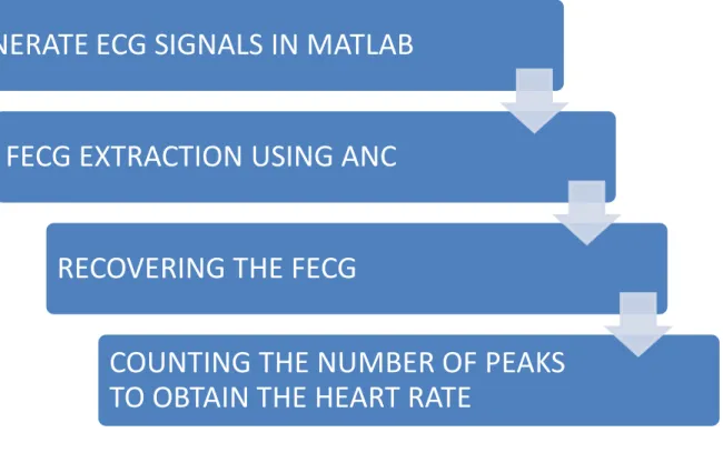
The extraction method described in this thesis is based on a MATLAB code that consists of 4 simple steps as described in the flowchart of the proposed algorithm shown below in Fig. 2.1.
Fig 2.1 FECG Extraction Algorithm
GENERATE ECG SIGNALS IN MATLAB
FECG EXTRACTION USING ANC
RECOVERING THE FECG
COUNTING THE NUMBER OF PEAKS
TO OBTAIN THE HEART RATE
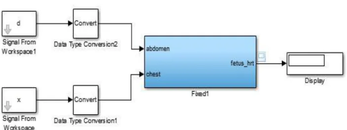
13 2.3 PROPOSED SIMULINK MODEL
To identify the heart rate of the fetus based on sensor data from two electrodes a suitable Simulink model is proposed. Both the primary signals i.e. the mother’s and baby’s heart beat signal have some superimposed noise associated with them. The goal of this model is to filter out everything except the baby’s heartbeat and calculate the period of the signal. The proposed Simulink model is shown below in Fig. 2.2 and Fig 2.3.
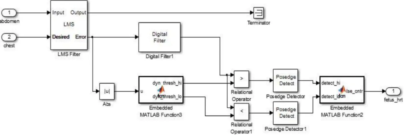
14
Fig 2.3 Simulink Model 2 2.4 LEAST MEAN SQUARE (LMS) ALGORITHM
Calculate the error and find the least mean of it. This is what the algorithm does. The error signal is the difference of the desired output signal and the actual output signal. For deriving this algorithm, the steepest descent algorithm is used [20]. Now, what a steepest descent algorithm does? It finds out the gradient vector by calculating the derivative of the error. It is a recursive algorithm and helps in finding out the wiener filter.
Now, what LMS algorithm does? It just minimizes the 𝐸[𝑒(𝑛)2] i.e. the mean of the mean of the square of the error. The LMS algorithm is based on the Steepest Descent (SD) algorithm but it is somehow different because it estimates the value continuously.
The SD algorithm is a deterministic gradient method whereas the LMS algorithm is a stochastic gradient method [21] [22].
Using LMS algorithm we cannot find the exact values and hence we can’t compute the optimal weights. The weight values are never updated to the optimal values. But it converges the mean of
15
the square of the error, hence, it changes the optimal weight although there is no changes in the weights.
The weight update equation has been given below:
W𝑛+1 = 𝑊𝑛− 𝜇∇𝜀[𝑛] (2.3)
2.5 IMPLEMENTATION OF THE ALGORITHM
The different steps proposed in the algorithm are briefly described below,
I. Generation of the ECG signals using MATLAB
MATLAB is an easy-to use tool which is very helpful in the extraction of the Fetal ECG (FECG) signal from the Abdominal ECG (AECG). Using MATLAB we generate the signal on which the task can be performed and implemented easily [23]. MATLAB contains a function Savitzky-Galoy filter function and using this command the required signals are generated.
MATLAB coding is used to simulate the shapes of the ECG signals for both the mother and the baby. The maternal ECG signal generated from mother’s heart using the given data in previous section is shown in Fig. 2.4. The peak amplitude of this signal is 3.5 millivolts and the heart rate being 85 beats per minute. But the fetus heart beats noticeably faster than that of the mother’s ranging between 120 to160 beats per minute as stated previously [24] [25]. The FECG signal generated is shown in Fig. 2.5 and the measured signal taken from the mother’s abdomen which is usually dominated by the mother’s heartbeat signal is shown in Fig. 2.6.
16
Fig 2.4 Measured ECG signal from the abdomen of the mother i.e. Abdominal ECG (AECG) signal
17
The above signal shows the Maternal Heartbeat Signal being generated by using a Savitzky-Galoy filter. 4000 samples of the signal has been taken with peak voltage of 3.5 millivolts.
Fig 2.6 Fetal Electrocardiogram Signal
II. FECG Signal Extraction Methods Used
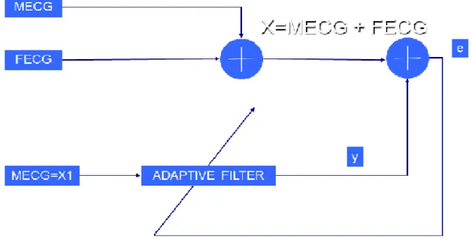
1) Adaptive Noise Canceller (ANC):
Two signals are needed to remove background artefacts including different noises or interference already present in the FECG signal by using adaptive filtering. The first one is the FECG signal added with MECG signal and the second being the reference signal which is also the signal to be cancelled to get the FECG signal [26] [27]. The reference signal is nothing but equal to the MECG signal itself. Noises present in both the signals must be well-correlated i.e. the noise in the primary and secondary signals. In fetal ECG extraction procedure adaptive filters are used to adaptively remove a mother’s heartbeat signal from the
18
fetal ECG signal to get the heartbeat signal of the baby or the fetus. When the operating environment is stationary, these filters possess constant shape and orientation for the error-performance surface. But for operation in an environment that is not stationary, the bottom of this surface moves continually along with the chances of changing orientation and curvature. Hence, for non-stationary inputs, the filter seeks the bottom of the error performance surface along with continually tracking it down. The block diagram for a basic ANC system is shown below in Fig. 2.7.
Fig 2.7 Adaptive Noise Cancellation System
By making exact measurements of the gradient vector at each iteration and choosing the step-size parameter suitably, the tap-weight vector computed would converge to the optimum Wiener solution using the method of steepest-descent. Several recursive algorithms can be used to change the tap-weight vector after each iteration. Least Mean Square (LMS) algorithm is one of such algorithms that has a certain advantage as it
19
doesn’t includes any steps regarding matrix inversion [28] [29]. This can be stated mathematically as,
Wk+1 = Wk + 2µ εk Xk (2.4)
where Wk is the tap-weight vector, εk is the error and Xk is the input signal vector at kth
iteration whereas μ is the step size parameter.
2) High Frequency Removal Using Digital Filter:
FIR and IIR filters are the two primary types of filters widely used in Digital Signal Processing (DSP). Among those FIR filters can be easily designed in linear phase and some of the calculations can be omitted by using FIR filters, thus providing some important computational efficiency. In order to count the fetal heart rate the R-peaks of the FECG signal must be extracted [30] [31]. An FIR filter with appropriate filter coefficients is used to remove high frequencies in order to achieve this goal. Then a threshold value is set and the peaks with higher values than the threshold are detected as the R-peaks of the FECG signal. The number of R-peaks are counted and from the R-R interval the fetal heart rate is calculated.
2.6 EXPERIMENTAL RESULTS
Heart rate is calculation of the beating of the heart. Heart beats in a fixed time duration and the calculation of number of beats i.e. number of R peaks per minute gives the heart rate of the ECG signal.
20
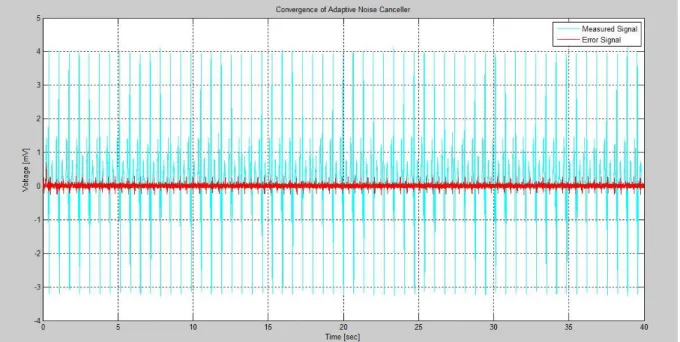
Finally the signal is cleaned up and a threshold level is set so that any value above it can be considered as a peak and hence the number of peaks in the signal can be counted. The heart rate counting has been performed using the QRS detection techniques that has been discussed in Chapter 3. The most optimized technique has been to count the R peaks of the FECG signal. Counting the R peaks the heartbeat of the Fetus is calculated. The heart beat comes to be 135 beats per minute. This is comes in the range exactly [32]. As the fetal heart rate is greater than the maternal heart rate and it comes in the range 120-160 beats per minute, this experiment verifies the same aptly. Finally, all the plots are shown below that explains the signal forms of the fetal ECG (FECG) signal.
Heartbeat calculation is never a difficult task if the fetal signal has been extracted from the abdominal ECG (AECG) signal. Because, the first step is always to extract the FECG signal and then applying optimized QRS complex detection techniques heartbeat can be calculated.
21
Fig 2.9 Output of the Adaptive Noise Canceller (ANC)
22 2.7 CONCLUSION
In this chapter the fetal ECG (FECG) extraction algorithm has been implemented successfully using the adaptive noise canceller (ANC). The algorithm is implemented on MATLAB and then output is taken using different plots. Then the peaks were detected using the differentiation technique which has been explained in the Chapter 2 of this thesis. Finally, the heartbeat of the fetus is measured successfully.
23
CHAPTER 3
QRS COMPLEX
DETECTION
24
Now a days it has been a trend to simulate ECG signals using microcomputers and various signal processing devices. According to a survey the processing of ECG signals using systems based on microcomputers is extremely efficient. There are many systems which has already been designed to perform such tasks on ECG signals. And various signal processing has been done on them to know the completed details about the ECG signals. But the ECG devices require a precise detection of the QRS complex so as to run a diagnosis and detect the abnormalities. Hence, QRS peak detection is a very crucial part of ECG signal processing. The figure below shows a QRS waveform [33].
Fig 3.1 QRS waveform [30]
The transfer of stimuli or signal to atria and ventricles and then the return of the same stimuli or signal from the atria and ventricles is what forms an Electrocardiogram (ECG) signal. This signal is called Electrical activity occurring in the muscles of a human being. Fig 3.1 shows some kind of ECG wave which consists of various parts from P and it goes alphabetically to U. Details about all the parts have been explained below.
P wave – This part of the signal is generated due to the depolarisation of the atria. It is called activation.
25
QRS Complex- This part of the signal is generated due to the depolarisation of the ventricles. It is also called activation.
The other parts of the waveform forms the part of depolarisation or recovery. The parts include ST wave, T wave and U wave [34].
Hence, for conclusion the PQRSTU wave represents the complete electrical activity of the heart due to the atria and ventricles. It is one kind of a cycle process that is repeated again after the recovery of the heart. As long as this process continues it gives us a continuous ECG signal which also time varying.
3.1 METHODOLOGY
Differentiation Technique for QRS Detection
In a cardiac cycle, the QRS complex has the largest slope i.e. larger rate of change of voltage. This is due to the rapid conduction and depolarisation characteristics of the ventricles. The derivative gives the rate of change. So in an attempt to develop an algorithm to detect the QRS complex the 𝑑𝑡𝑑 operation would be the most logical starting point.
There are various techniques like Pan-Tompkins model, bandpass filtering technique, template matching technique etc. that can be implemented to detect a QRS wave. Amongst them the differentiation technique is also an efficient technique detect the R peaks of a QRS complex. The basis of many detection techniques is differentiation. It is basically a high pass filter. Whenever the derivative of a signal is taken the higher frequencies characteristic of the QRS complex gets amplified while attenuating the lower frequencies of the P and T wave [35].
An algorithm based on the first order, second order and third order derivative has been used to detect the peaks. The figures below shows the steps involved in the signal processing.
26
Fig 3.2 Original ECG signal [22]
Fig 3.3 First Order Derivative having a smooth and rectified signal [22]
Fig 3.4 Second Order Derivative having a smooth and rectified signal [22]
Fig 3.5 Sum of First and Second Order Derivative [22]
27
The absolute values of the first and second order derivative can be calculated from the ECG signal as
𝑦0(𝑛𝑇) = | 𝑥(𝑛𝑇) − 𝑥(𝑛𝑇 − 2𝑇) | (3.1) 𝑦1(𝑛𝑇) = | 𝑥(𝑛𝑇) − 2𝑥(𝑛𝑇 − 2𝑇) + 𝑥(𝑛𝑇 − 4𝑇) | (3.2) These two data buffers, 𝑦0(𝑛𝑇) and 𝑦1(𝑛𝑇), are first scaled and then summed to give the following output
𝑦2(𝑛𝑇) = 1.3𝑦0(𝑛𝑇) + 1.1𝑦1(𝑛𝑇) (3.3) The data buffer 𝑦2(𝑛𝑇) is properly scanned until a certain threshold is met or exceeded.
𝑦2(𝑖𝑇) ≥ 1.0 (3.4)
Once this condition is met for a data point in 𝑦2(𝑖𝑇), the next eight points are needed to compared to the threshold. The segment might be part of the QRS complex. This algorithm detects the pulse and in addition to that it has the advantage that it produces a pulse which is proportional in width to the complex. It has a particular disadvantage that it is sensitive to high-frequency noise.
28 3.2 EXPERIMENTAL RESULTS
The following figures show the MATLAB implementation of the code with peak detection.
Fig 3.7 Differentiation Technique implemented on MATLAB
The above figure shows the first order, second order and cumulative order differentiation of an ECG signal. The peaks in the first signal are not visible properly but once it reaches the cumulative order differentiation the peaks are quite visible enough to find out the R peaks.
29
Fig 3.8 QRS Peaks
The above figure shows the QRS peaks being detected by thresholding and locating the maximum which is then converted into beats. In one epoch, number beats in 12 second has been calculated and thereby heart rate has been computed.
3.3 CONCLUSION
In this chapter a technique has been explained for the detection of R-R peaks from the QRS complex. Hence, the algorithm for the technique has been implemented successfully. And then the peaks of the FECG are detected and the heartbeat of the fetus is measured.
30
CHAPTER 4
CONCLUSIONS AND
FUTURE WORKS
31 4.1 CONCLUSIONS
The outputs of the extracted signal were recorded on MATLAB. Finally, the fetal ECG signal is extracted and heartbeat of the signal is calculated. The algorithm based on Adaptive Noise Canceller (ANC) is proposed and implemented successfully. The performance of the algorithm has been verified successfully on MATLAB and the algorithm is found to be highly efficient. It is found after successful implementation that fetal ECG (FECG) signal can be successfully extracted by using Least Mean Square (LMS) algorithm for tap-weight vectors. The LMS algorithm is implemented by MATLAB codes and hence the LMS algorithm implements ANC successfully. R peaks were also detected successfully giving the final heart rate of the signal.
4.2 FUTURE WORKS
The results obtained matches with the actual FECG signals significantly and will suffice to prove correct. The work done here is on-going and it has many scopes in future because it hasn’t been detailed yet. The future work would include designing an ECG with in-build software implementing the above extraction algorithm. Then the technique can be used widely in product development. But the extraction techniques may change with time and hence more efficient and error free techniques will be developed in future. In this way the project can move forward in future.
32
REFERENCES
[1] International Journal of Advanced Research in Electrical, Electronics and Instrumentation Engineering Vol. 2, Issue 4, April 2013
[2] http://en.wikipedia.org/wiki/Electrocardiography last accessed on Jan, 2014
[3] http://zone.ni.com/reference/en-XX/help/372357A-01/lvaftconcepts/aft_lms_algorithms/ last accessed on Feb, 2014
[4] http://en.wikipedia.org/wiki/Least_mean_squares_filter last accessed on Mar, 2014
[5] http://www.cs.cmu.edu/~aarti/pubs/ANC.pdf last accessed on April, 2014
[6] http://ijater.com/files/288abb76-cc40-4bdc-ba4b-b9f4772fab8c_ijater_10_14.pdf
[7] American National Standard for Ambulatory Electrocardiographs, publication ANSI/AAMI EC38-1994, Association for the Advancement of Medical Instrumentation, 2004
[8] AHA ECG database. Available from Emergency Care Research Institute, 5200 Butler Pike, Plymouth Meeting, PA 19462, 2010
[9] Fahim Sufi, Qiang Fang, Ibrahim Khalil, and Seedahmed S. Mahmoud, “Novel Methods of faster Cardiovascular Diagnosis In Wireless Telecardiology,” IEEE Journal on Selected Areas In Communications, Vol. 27, No. 4 2005
[10] Rachid Merzougui, Mohammed Feham, “Algorithm of remote monitoring ECG using mobile phone: Conception and implementation”, Third International Conference on Broadband Communications, Information Technology & Biomedical Applications. 2006
[11] Turker Ince, Serkan Kiranyaz, and Moncef Gabbouj, “A Generic and Robust System for
Automated Patient-Specific Classification of ECG Signals,” IEEE Trans. Biomed. Eng. vol.56, pp.1415-1426 2007
[12] L. Biel, O. Pettersson, L. Philipson, and P. Wide.”Ecg analysis: an approach in human identification,”.IEEE Trans. Instrum. Meas., 50(3):808–812 2008
33
[13] X.Afonso, W.J. Tompkins, T.Nguyen, S.Luo, “ECG beat detection using filter,” IEEE Trans. Biomed. Eng. vol. 46, pp. 230-236 Vikram Goyal, “J2ME Tutorial, Part 1: Creating MIDlets,” 2002
[14] Vikram Goyal ,”J2ME Tutorial, Part 2: User Interfaces with MIDP 2.0,” 2001 .
[15] S. Akselrod, D. Gordon, F. A. Ubel, D. C. Shannon, A. C. Barger, and R. J. Cohen.”Power spectrum analysis of heart rate fluctuation: A quantitative probe of beat to beat cardiovascular control”. Science, 213(1981):220–222 1999
[16] MIT-BIH Database distribution, Massachusetts Institute of Technology, 77 Massachusetts Avenue, Cambridge, MA 02139, 1998. http://www.physionet.org/physiobank/database/mitdb/ 2000
[17] R.M. Rangayyan,”Biomedical Signal Analysis: A Case-study Approach,” Wiley– Interscience, New York, 2001, pp.18-28. 2001
[18] P. S. Hamilton and W. J. Tompkins.”Quantitative investigation of qrs detection rules using the mit/bih arrhythmia database,”Biomedical Engineering, IEEE Transactions on, BME-33(12):1157–1165, 2004
[19] P. S. Hamilton and W. J. Tompkins.”Quantitative investigation of qrs detection rules using the mit/bih arrhythmia database,”Biomedic, 2000
[20] Ahlstrom, M. L. and Tompkins, W. J. 1983. Automated high-speed analysis of Holter tapes with microcomputers. IEEE Trans. Biomed. Eng., BME-30: 651–57, 1999
[21] Ahlstrom, M. L. and Tompkins, W. J. 1985. Digital filters for real-time ECG signal processing using microprocessors. IEEE Trans. Biomed. Eng., BME-32: 708–13, 2006
[22] Balda R. A., Diller, G., Deardorff, E., Doue, J., and Hsieh, P. 1977. The HP ECG analysis program. Trends in Computer-Processed Electrocardiograms. J. H. vanBemnel and J. L. Willems, (eds.) Amsterdam, The Netherlands: North Holland, 197–205, 2008
[23] Dobbs, S. E., Schmitt, N. M., Ozemek, H. S. 1984. QRS detection by template matching using real-time correlation on a microcomputer. Journal of Clinical Engineering, 9: 197–212, 2003
34
[24] Friesen, G. M., Jannett, T. C., Jadallah, M. A., Yates, S. L., Quint, S. R., Nagle, H. T. 1990. A comparison of the noise sensitivity of nine QRS detection algorithms. IEEE Trans. Biomed. Eng., BME-37: 85–97, 2002
[25] V. Zarzoso and A. K. Nandi, Noninvasive fetal ECG extraction: blind separation versus adaptive noise cancellation, IEEE Trans. on Biomed. Eng., vol. 48 (1), pp.12-18, Jan. 2001.
[26] Khamene, A and Negahdaripour, A New Method for the Extraction of Fetal ECG from the Composite Abdominal Signal, IEEE Trans. Biomed. Engineering 47 2000.
[27] C.W. Li, C.X.Zheng, C.F.Tai, Detection of ECG Characteristic Points Using Wavelet Transforms; IEEE Transaction on Biomedical Eng., 42.No.1:22-28, January 1995.
[28] P. P. Kanjilal, S. Palit, and G. Saha, “Fetal ECG extraction from single-channel maternal ECG using singular value decomposition,” IEEE Transactions on Biomedical Engineering, Vol. 44, pp. 51–59, 1997.
[29] T. Solum, I. Ingermarsson, and A. Nygren, “The accuracy of abdominal ECG for fetal electronic monitoring,” Journal of Perinatal Medicine, Vol. 8, No 3, pp. 142–149, 1980.
[30] K. Karlsso, H. Lilja, K. Lindecrantz, and K. G. Rosen, “Microprocessor based waveform analysis of the fetal electrocardiogram during labor,” International Journal of Gynaecol and Obstetrics, Vol. 30, No. 2, pp. 109–16, 1989.
[31] M. B. I. Reaz and L. S. Wie, “Adaptive linear neural network filter for fetal ECG extraction,” Proceedings of International Conference on Intelligent Sensing and Information Processing, Chennai, India, pp. 321–324, January 2004.
[32] MIT-BIH Database distribution, Massachusetts Institute of Technology, 77 Massachusetts Avenue, Cambridge, MA 02139, 1998. http://www.physionet.org/physiobank/database/mitdb/ 2000
[33] V. Zarzoso and A. K. Nandi, Noninvasive fetal ECG extraction: blind separation versus adaptive noise cancellation, IEEE Trans. on Biomed. Eng., vol. 48 (1), pp.12-18, Jan. 2001.
[34] http://zone.ni.com/reference/en-XX/help/372357A-01/lvaftconcepts/aft_lms_algorithms/ last accessed on Feb, 2014
35
[35] Friesen, G. M., Jannett, T. C., Jadallah, M. A., Yates, S. L., Quint, S. R., Nagle, H. T. 1990. A comparison of the noise sensitivity of nine QRS detection algorithms. IEEE Trans. Biomed. Eng., BME-37: 85–97, 2002
![Fig 1.1 Measurement of Fetal ECG [5]](https://thumb-us.123doks.com/thumbv2/123dok_us/10015105.2493803/11.918.343.577.712.944/fig-measurement-of-fetal-ecg.webp)


![Fig 3.1 QRS waveform [30]](https://thumb-us.123doks.com/thumbv2/123dok_us/10015105.2493803/32.918.253.668.425.741/fig-qrs-waveform.webp)
