The Role of Epigenetic Changes in The Development of
Diabetes Mellitus
Wardhana, Djoko W. Soeatmadji
Department of Internal Medicine, Faculty of Medicine, Brawijaya University - Saiful Anwar Hospital, Malang, Indonesia.
Corresponding Author:
Prof. Djoko Wahono Soeatmadji, MD., PhD. Division of Endocrinology, Metabolic, and Diabetes, Department of Internal Medicine, Faculty of Medicine, Brawijaya University - Saiful Anwar Hospital. Jl. Veteran Malang 65145, Indonesia. email: [email protected].
ABSTRAK
Diabetes melitus (DM) adalah masalah kesehatan yang paling penuh dengan tantangan dan dipercaya merupakan hasil antara interaksi gen dan paparan lingkungan. Terdapat hipotesis mekanisme epigenetik, menggunakan dasar molekular untuk menjelaskan mekanisme terjadinya DM. Karena paparan lingkungan termasuk status gizi dan keadaan hiperglikemia, risiko terjadinya DM telah dimulai sejak pre-konsepsi, yang berlangsung hingga dewasa, dan diwariskan antar-generasi. Terdapat 3 mekanisme utama epigenetik yang berperan pada DM. Mekanisme-mekanisme epigenetik juga memiliki peran dalam memori metabolik dimana komplikasi-komplikasi DM dapat tetap terjadi walaupun kadar gula darah dalam tubuh sudah normal. Pembatasan kalori dapat membantu menunda proses dan terjadinya penyakit-penyakit degeneratif termasuk DM dengan cara menstabilkan genom melalui mekanisme-mekanisme epigenetik.
Kata kunci: diabetes, mekanisme epigenetik, memori metabolik. ABSTRACT
Diabetes mellitus (DM) is one of the most abundant diseases in the 21st century and believed as result of interaction between genes and environment exposure. There is a hypotesis of epigenetic mechanisms, using molecular basis to explain about the mechanism of DM. Because of the enviromental exposure including nutrition status and hyperglycemia state, the risk of DM has started since pre-conception, last until adulthood and will be inhireted trans-generational . Mainly, there are 3 epigenetic mechanisms that have role in DM. Epigenetic mechanisms are also have role in the metabolic memory that the DM complications may still developed although the blood glucose level is already normal. The restriction of calory intake may help delaying the development and onset of degerative diseases including DM by stabilizing genome through epigenetic mechanisms.
Keywords: diabetes, epigenetic mechanisms, metabolic memory. INTRODUCTION
Diabetes mellitus (DM) is one of the abundant diseases in the 21st century. The approximated prevalence of DM among adults in the world was 366 million in 2011, and in 2030 this will become 552 million.1 DM is a disease
caused by metabolic dysregulation, marked by chronic high blood glucose level causing many complications that may reduce life expectancy and quality of life. The disease mechanism of DM is not well understood until now, but it is known that it is the result of interaction between
genes and environment exposure.1,2
There is a hypothesis regarding epigenetic mechanisms explaining the association between imbalanced nutrition and altered disease risk, using molecular basis to explain the link between perinatal nutrition intake and degenerative diseases, including DM.2 The classical definition of
epigenetic was first stated by Conrad Waddington
in the 1950s, that mentioned the epigenetic trait as a stable phenotype changes that may be inherited, as the result from changes in chromosomes but with no alternation in DNA sequence.3 Through epigenetics, the organism can alter its pattern of development to cope with environment stress, and this may pass intergenerationally as the
accumulation of epigenetic modifications.4
INFLUENCES OF CONCEPTION, PRE-NATAL, AND POST-NATAL NUTRITIONAL EXPOSSURES AS RISK OF DIABETES MELLITUS TO THE OFFSPRINGS
Early life exposures bring a long period of life increased the risks for DM, and may cause persistent changes including
metabolism. Intrauterine malnutrition influences organogenesis, which has bad effects on organ
morphology and function.5 The period of embriogenic development has been known as a critical window in establishing the epigenome.6
The exposures may cause trans-generationally
inherited traits and epigenetic modifications.
Trans-generational has a meaning that the the changes in phenotype must last for at least 3 generations after the first exposure.7 The inheritance mechanism may happen through mother or father (P0). A pregnant woman (P0) can carry genetic and epigenetic information until F2 generation. Her fetus (F1) and its germ cell (F2)
may be affected by enviromental expossures. The
changes in F1 phenotype or epigenetic state to be considered trans-generational if it lasts until F3 generation. In males (P0), the expossure may change his germ cells (F1), but since the F2 germ cells are not expossed, every phenotype
or epigenetic changes in F2 are defined as
trans-generational.7,8
Paternal’s nutrition intake can influence offspring’s risk of diseases through sperm epigenome which is included heritable
informations, like: DNA methylation. Paternal obesity has relationship with changes in sperm numbers, concentration, motility, morphology, and result in sperm DNA damage.8 The paternal impaired fasting glucose and glucose intolerance, as manfifestation of pre-diabetes, will alter gene expression patterns in pancreatic islets, by downregulating several genes involved in glucose metaboism and insulin signaling pathway. It is hypothesized that nutrition supply as methyl donor may alter epigenetic reprogramming in sperm.9
Maternal pregnant obesity in combination with gestational DM may lead to newborn with
hyperinsulinemia state and increased offspring’s
fat mass until the 6th week, and this is strongly
suggest as an important influence of gestational DM to the offsprings. Maternal obesity during
pregnancy will become a serious danger to
offsprings in adulthood. It will start trascriptional
programs that may increase the expression of
inflammatory molecules and immune cells, which reflect the epigenetic proccess. Hyperinsulinemia
state in response to maternal obesity is mediated by the Insulin Receptor Substrate-1 (IRS-1), which is regulated by epigenetic mechanism through short noncoding miRNA-126.9,10
Maternal nutritional deficiency or famine may also give influence to the offspring depending on
the timing of their prenatal exposures. During the Dutch famine in the years 1944 until 1945, those who were concieved in the midst of famine had
shown to have hypomethylation at CpG (Cytosine-phopshate-Guanine) islands inside important
promoter site and it still can be observed until even 60 years later.11 The intrauterine condition with poor nutrition will epigenetically program the
offsprings to survive in a low nutritional postnatal
environment, revealing ‘catch-up growth’ in postnatal period, and when they are exposed to nutrition which are high in energy, they will have obesity as a cause of insulin resistance state.12
EPIGENETIC MECHANISMS TO DEVELOP DIABETES MELLITUS
The epigenetic mechanisms are primarily occuring through three mechanisms. There are: DNA methylation, post-translational histone
DNA Methylation
The adult somatic cells consist of one haploid set of chromosome, which is inherited half from mother and the other half from father. Epigenetic
patterns are at first deleted and reprogrammed two
times to correct the imprinting sites with
allele-specific methylation, to form tissue allele-specific gene
methylation pattern in postnatal periode. The 1st phase of deleting and reprogramming happened during gametogenesis. The DNA methylation is
deleted and later reestablished by taking specific
imprints in oocytes and in spermatozoa. The 2nd phase of epigenetic deleting and reprogramming happened during pre-implantation. During this
proccess, the genome with possible exception of imprinted genes and some retrotransposons, is now demethylated. The DNA methylation is restored after implantation, and then forming cell
descent specific patterns in rapid way to oversee its differentiation.13 (Figure 1)
The DNA methylation is defined as the
attachment a of methyl group to a cytosine (C) nucleotide at position 5 (5mC) and specifically
happened when a cytosine’s position is next to guanine and joined by phosphate in DNA, which
is called by CpG dinucleotide. The regions of genome consists lots of CpG dinucleotide are called by CpG islands.13 The frequent epigenetic
Figure 1. Exposure throughout steps of life and the epigenetic dysregulation. Environmental exposures may cause epigenetic changes, increasing the risk of diseases
including DM. These changes are heritable to the offsprings.13
Figure 2. Landmark of regulatory regions and histone. The DNA methylation may take place at the CpG islands in the promoter region. An illustration of histones that consist of 4 types of histone with its tail.5
modification of DNA is in the demethylated
CpG islands in promoter region. The promoter
methylation during development and diseases proccess is related to gene trancriptional silencing.5 (Figure 2)
The specific methylation of CpG dinucleotides
in tissue is performed by DNA methyltransferase or DNMTs (1,3a,3b, and 3B) that has activity in de novo methylation (except for DNMT1 that has
function to maintance the methylation of CpG),
making the covalent binding of methyl group to
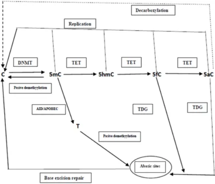
form 5mC.13,14 The demethylation proccess starts by the ten-eleven translocation methylcytosine dioxygenase1 (TET1), which is then oxydized
5mC to form 5-hydroxymethycytosine (5hmC),then into 5-formylcytosine (5fC), and then into 5-carboxylcytosin (5aC). The 5mC, 5hmC, 5fC, and 5aC may have passive demethylation to form C. The 5f and 5aC can be cut by thymine-DNA glycosylase (TDG), then
followed by DNA repair (base excision repair or
BER) to form C. As an alternative, the 5aC may react with decarboxylase to form C. The 5mC can
be deaminated by activation-induced deaminase (AID)/apolipoprotein B mRNA editing complex
(APOBEC) to become 5-hydroxymethyl Urasil (5hmU) then exicised by TDG or
methyl-CpG-binding domain protein 4 (MBD4), and at the
last followed by DNA repair to form C again.14 (Figure 3)
The hyperglycemia state may induce global demethylation at all genomic loci including the promoter, intergenic, and intragenic sites. The
most methylated CpG sites of diabetic islets
show decrease in methylation.15 This may be caused by the reduction of activity of DNMT1 during cell proliferation or a reduction in the activity of DNMT3a and 3b which responsible for methylation. There is also an increase in
the action of Growth arrest and DNA-damage-inducible protein GADD45A (GADD45A)
which may also cause demethylation in human pancreatic cells. These changes are risk factor for DM. Another explaination for hypomethylation in hyperglycemia state is due to the decrease in methyl donor.16,17 The hyperglycemia state that may induce global demethylation is largely maintain in the metabolic memory state.15 (Table
1)
Post-translational Histone Modifications
Histones (H1, H2A, H2B, H3, and H4) are globular protein which exist around DNA, packed to form chromatin. Post-translational histone conformational changes may happen
Figure 3. Model of DNA demethylation pathways initiated by TET. An illustration of the enzymatic changes among C, 5mC, 5hmC, 5fC, and 5aC.14
as the result of enzymes that modify lysine and arginine residues in amino terminus. The modifications are mainly acetylation and methylation. The acetylation may increase DNA accessability,meanwhile the methylation may result in either increased or decreased of DNA accessability, depend on the type of methylationing and histone.14,18
The histone methylations may happen at the lysine residue using SET- and non-SET domain histone lysine methyltransferase (HMTase) that regulates transcription and repair; and at arginine residue using protein arginine methyltransferase (PRMT) or coactivator-associated arginine
methyltransferase1 (CARM1) that regulates
transcription. The demethylation proccess may happen using lysine specific demethylase1 (LSD1). The acetylation may happen by using histone acetyl transferase (HAT) that
regulates transcription, repair, replication, and condensation; and the deacetylation proccess
using histone deacetylase (HDAC).18-20 The active genes can be methylated at H3K4, H3K36, and H3K79, and the silenced or repressed genes are methylated at H3K9, H3K27, and H4K20.21 The reversible acetylation of N-terminal lysine residues are at positions 9,14,18, and 23 of H3 and at 5,8,12, and 16 of H4. These reversible acetylation can mediate the decondensation of nucleosome structure, alters the link between histone and DNA, and also may give access for the binding of transcription factors.18
The miRNA Expression
The miRNA may work by blocking and altering the genes expression.4 The target of miRNA is the gene promoter, through the
recruitment of specific argonaute proteins to
form epigenetic remodeling complexes. It can reduce gene expression by keeping histone deacetylation, histone methylation (H3K9 and H3K27), and DNA methylation.22 The miRNA can bind to 3’untranslated region of target mRNA, reveling in degradation or inhibit protein translation.4
In the nucleus, RNA polymerase II is transforming miRNA to form long segmens of coding or noncoding RNA (pri-miRNA,consist of 70-100 nucleotides) which are covered up and polyadenylated, then captured and also extracted by a complex consists of RNase type III, drosha,
and dsRNA binding protein, DiGeorge syndrome critical region gene 8 (DGCR8)’ to form
pre-miRNA, then to form a complex with exportin5
and RanGTP, and then it is translocated to
cytoplasm. The dicer in cytoplasm can cleave the pre-miRNAs near hairpin loop to produce a short ~22 base pair long imperfect RNA duplexes. This short imperfect RNA dupleces are further added into mature RNA-induced silencer
complex (mature RISC or MiRISC), and has a
role as a template for capturing target mRNAs.
The MiRISC can rend the target mRNAs through
catalytic domain (RNA III domain) of argonaute
protein (as the core component of RISC complex)
and then degradating them.23 The MiRISC may also prevent protein translation through several
different mechanisms. These mechanisms are
including the inhibition of trancription at level
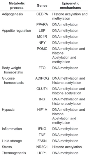
Table 1. Genes with expressions on metabolic process which are related to obesity and type 2 DM
Metabolic
process Genes mechanismsEpigenetic Adipogenesis CEBPA Histone aceylation and
methylation PPARA DNA methylation Appetite regulation LEP DNA methylation MC4R DNA methylation NPY DNA methylation POMC DNA methylation and
histone Acetylation and methylation Body weight
homeostatis FTO DNA methylation Glucose
homeostasis ADIPOQ DNA methylation and histone acetylation GLUT4 DNA methylation and
histone acetylation INS DNA methylation and
histone acetylation Hypoxia HIF1A DNA methylation and
histone Acetylation and methylation
Inflammation IFNG DNA methylation TNF DNA methylation Lipid storage FASN DNA methylation Stress NR3C1 Histone acetylation Thermogenesis UCP1 DNA methylation These genes which have expressions on metabolic process that controlled by epigenetic mechanisms.
of initiation and elongation, and the degradation of mRNA or the immature protein products of mRNA. The immature protein products of RNA are then segregated into P bodies to make inhibition of translational proccess and or
deadenylation of mRNA. The final result is the
destabilization of mRNA.24
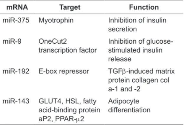
The exposure of islet cells to palmitate can cause the increament of miR-34a and miR-146 which activate p53 and reduce vesicle-associated membrane protein 2. These proccess may lead to islet cells apoptosis and the reduction of insulin secretion. The exposure of proinflammatory cytokines to islet cells can activate miR-21,miR-34a, and miR-146, that may also lead to the reduction of insulin secretion and the increament of the apoptosis of the cells. The upregulated miR-29a and -29b in insulin target organs under hyperglycemic state may reduce insulin mediated glucose uptake.25 (Table 2)
reactive oxygen species (ROS) production.26 Hyperglycemia state can reduced the activity of sirtuin1 (SIRT1) that can lead to the acetylation of p53, nuclear factor-kappa beta
(NF-κβ) subunit p65, and histone 3 bound to
p66shc promoter. The activation of p53 can lead to the increament of p66shc transcription. The reduction in the role of hyperglycemia
state-induced GCN (an acetyltransferase) can cause
the acetylation of H3 and the subsequent p66shc transcription through chromatin remodeling. The p53 protein that upregulates the p66shc can lead to the persistence of mitochondrial ROS production. The high ROS production may result in the endothelial dysfunction, vascular
inflammation, and apoptosis proccess of cells.27 Moreover, the high production of ROS may reduce the signalling proccess from insulin receptor substrate1 (IRS1) to phosphatidyl inositol 3 kinase (PI3K), and also activates c-jun N-terminal kinase that translocate pancreatic and duodenal homeobox-1 (PDX-1) from nucleus to cytoplasm and induces the degradation of mafA, resulting in the decreased of insulin gene expression,insulin production, and insulin secretion.28,29
Restriction of calory intake has already shown to give a positive correlation with disease prevention. This restriction may increase lifespan and delay cardiovascular disease, DM, and cancer, through chromatin remodeling which lead to increase in age and lifespan, and delaying the onset of degenerative diseases by stabilization of genome through epigenetic mechanisms.9
CONCLUSION
Diabetes mellitus is a metabolic disease characterized by high blood glucose levels, as the result of genes and environmental interaction. It is now accepted about the concept of epigenetic mechanisms by molecular basis to explain the development of DM. There are 3 main epigenetic mechanisms: DNA methylation, post-translational histone modification, and micro RNA (miRNA) expression. The epigenetic changes on the metabolic status are already there since pre-conception depending on the maternal and paternal epigenetic changes, and it will last
Table 2. The miRNA targets in type 2 DM
mRNA Target Function
miR-375 Myotrophin Inhibition of insulin secretion
miR-9 OneCut2
transcription factor Inhibition of glucose-stimulated insulin release
miR-192 E-box repressor TGFb-induced matrix protein collagen col a-1 and -2
miR-143 GLUT4, HSL, fatty acid-binding protein aP2, PPAR-m2
Adipocyte
differentiation
METABOLIC MEMORY
The long-(heritable) or short-acting (non heritable) environmental exposure may result in long- or short-term epigenetic changes.26
The Diabetes Control and Complications Trial (DCCT) and Epidemiology of Diabetes Intervention and Complications (EDIC) come
with a conclusion that in DM patients who had improved glycemic control may still developed complications as result of prior poor glycemic control.20 In 1990, Roy and colleagues had described the mechanism as metabolic memory,
which is defined as epigenetic changes caused
until adulthood, and the epigenetic changes will be inherited trans-generational. The epigenetic mechanisms are also involved in the metabolic memory, the condition where the complications of DM may still happen although the blood sugar
level is already normal. Calories restriction may be benefit on delaying the development and onset
of degenerative diseases including DM through epigenetic mechanisms to stabilize the genome.
REFFERENCES
1. Gouti A, Dekaken A. Epigenetic pathways in
type 2 diabetes and its complications. Med Pub J. 2013;1(11):1-5.
2. Zheng J, Xiao X, Zhang Q, Yu M. DNA methylation: the pivotal interaction between early-life nutrition and glucose metabolism in later life. Br J Nutr. 2014;112:1850-7.
3. Moosavi A, Ardekani AM. Role of epigenetics in biology and human diseases. Iran Biomed. 2016;20(5):246-58.
4. Day J, Savani S, Krepley BD, et al. Influence of
paternal preconception exposures on their offspring: though epigenetics to phenotype. Am J Stem Cells.
2016;5(1):11-8.
5. Hajj NE, Schneider E, Lehnen H, Haaf T. Epigenetics and life-long consequences of an adverse nutritional and diabetic intrauterine environment. Reproduction. 2014;148:R111-R120.
6. Dijk SV, Molloy PL, Varinli H, et al. Epigenetics and human obesity. Int J Obes.2015;39:85-97.
7. Rissman EF, Adli M. Transgenerational epigenetic inheritance: focus on endocrine disrupting compounds. Endocrinol. 2014;155:2770-80.
8. Ding GL, Huang HF. Role for Tet in
hyperglycemia-induced demethylation:a novel mechanism of diabetic metabolic memory. Diabetes. 2014;63:2906-8.
9. Grayson SE, de Leon FAP, Muscoplat CC. Epigenetics:
understanding how our choices lead to our diseases. J
Clin Case Rep. 2014;4(11):1-6.
10. Rehan MK. Epigenetics and diabetes mellitus. The Egypt J Intern Med.2016;28:39-51.
11. Smith CJ, Ryckman KK. Epigenetic and developmental influences on the risk of obesity, diabets, and metabolic
syndrome. Diabetes, Metabolic Syndrome and Obesity: Targets and Therapy. 2015;8:295-302.
12. Sterns JD, Smith CB, Steele JR, et al. Epigenetics and
type II diabetes mellitus:underlying mechanisms of
prenatal predisposition. Frontiers Cell Develop Biol.
2014;2:article 15.
13. Martinez JA, Milagro FI, Claycombe KJ, Schalinske
KL. Epigenetics in adipose tissue,obesity,weight loss, and diabetes. Adv Nutr. 2014;5:71-81.
14. Fleisch AF, Wright RO, Baccarelli AA. Environmental
epigenetics: a role in endocrine disease? J Mol Endocrinol. 2012;49:R61-R67.
15. Li En, Zhang Y. DNA methylation in mammals. Cold
Spring Harb Perspect Biol. 2014;6:a019133. 16. Intine RV, Sarras Jr MP. Metabolic memory and chronic
diabetes complications:potential role for epigenetic
mechanisms. Curr Diab Rep. 2012;12(5):551-9. 17. Dayeh T, Volkov P, Salo S, et al. Genome-wide DNA
methylation analysis of human pancreatic islets from
type 2 diabeteic and non-diabetic donors identifies candidate genes that influence insulin secretion. PLOS Genetic. 2014;10(3):e1004160.
18. Choi SW, Friso S. Epigenetics: a new bridge between
nutrition and health. Adv Nutr. 2010;1:8-16.
19. Kouzarides T. Chromatin modifications and their function. Cell. 2007;128:693-705.
20. Gurusamy N, Sukumaran V, Alhazzani A, et al. Complications of diabetes: an unsolicited epigenetic
memory. Diabesity. 2015;1:3-6.
21. Miao F, Wu X, Zhang L, et al. Genome-wide analysis
of histone lysine methylation variations caused by diabetic conditions in human monocytes. J Niological
Chemist. 2007;282(10):13854-63.
22. Handy DE, Castro R, Loscalzo J. Epigenetic
modifications basic mechanisms and role in
cardiovascular disease. Circulation. 2011;123:2145-56.
23. Sato F, Tsuchiya S, Melzer SJ, Shimizu K. MicroRNAs and epigenetics. FEBS J. 2011;278:1598-609. 24. Muhonen P, Holthofer H. Epigenetic and
microRNA-mediated regulation in diabetes. Nephrol Dial Transplant. 2009;24:1088-96.
25. Joglekar MV, Parekh VS, Hardikar AA. Islet-specific
microRNAs in pancreas development, regeneration and diabetes. Indian J Experiment Biol. 2011;49:401-8.
26. Togliatto G, Dentelli P, Vrizzi MF. Skewed epigenetics:
an alternative therapeutic option gor diabetes complications. J Diab Res. 2015;article ID 373708:7 pages.
27. Paneni F, Volpe M, Luscher TF, et al. SIRT1, p66shc, and Set7/9 in vascular hyperglycemic memory. Diabetes. 2013;62:1800-7.
28. Bender SB, McGraw AP, Jaffe IZ, Sowers JR.
Mineralocorticoid receptor-mediated vascular insulin resistance. An early contributor to diabetes-related vascular disease? Diabetes. 2013;63:313-9.
29. Jia L, Xing J, Ding Y, et al. Hyperuricemia causes
pancreatic β-cell death and dysfunction through NF-κβ