Ex vivo and in vivo studies to assess chemical effects
on steroidogenesis in fish: development and
application of methods
Inauguraldissertation
zur
Erlangung der Würde eines Doktors der Philosophie
vorgelegt der
Philosophisch-Naturwissenschaftlichen Fakultät
der Universität Basel
von
Maria Luz a Marca Pereira
aus Ourense, Spanien
Genehmigt von der Philosophisch-Naturwissenschaftlichen Fakultät
auf Antrag von
Prof. Dr. Patricia Holm
und
Prof. Dr. Helmut Segner
Basel, den 29. März 2011
Prof. Dr. Martin Spiess
Dekan
Original document stored on the publication server of the University of Basel
edoc.unibas.ch
This work is licenced under the agreement „Attribution Non-Commercial No Derivatives – 2.5 Switzerland“. The complete text may be viewed here:
Attribution-Noncommercial-No Derivative Works 2.5 Switzerland
You are free:
to Share — to copy, distribute and transmit the work
Under the following conditions:
Attribution. You must attribute the work in the manner specified by the author or licensor (but not in any way that suggests that they endorse you or your use of the work).
Noncommercial. You may not use this work for commercial purposes.
No Derivative Works. You may not alter, transform, or build upon this work.
• For any reuse or distribution, you must make clear to others the license terms of this work. The best way to do this is with a link to this web page.
• Any of the above conditions can be waived if you get permission from the copyright holder. • Nothing in this license impairs or restricts the author's moral rights.
Quelle: http://creativecommons.org/licenses/by-nc-nd/2.5/ch/deed.en Datum: 3.4.2009 Your fair dealing and other rights are in no way affected by the above.
This is a human-readable summary of the Legal Code (the full license) available in German: http://creativecommons.org/licenses/by-nc-nd/2.5/ch/legalcode.de
Disclaimer:
The Commons Deed is not a license. It is simply a handy reference for understanding the Legal Code (the full license) — it is a human-readable expression of some of its key terms. Think of it as the user-friendly interface to the Legal Code beneath. This Deed itself has no legal value, and its contents do not appear in the actual license. Creative Commons is not a law firm and does not provide legal services. Distributing of, displaying of, or linking to this Commons Deed does not create an attorney-client relationship.
Table of contents
Summary
1
1.
Introduction
4
1. Endocrine disruption in the aquatic environment 4
2. Endocrine control of reproductive function and interference of 5 environmental toxicants with the hypothalamic-pituitary-gonadal axis
3. Morphology, endocrinology and environmental modulation 7
of sex differentiation in teleost fish
1.3.1. Sexual differentiation and gonadal development 7
1.3.2. Endocrine control of sexual differentiation and development 9 1.3.3. Fish early sexual development: a sensitive period of life 10
4. Chemicals interfering with the steroidogenesis 11
1.4.1. 1,4,6-androstatriene-3,17-dione, an aromatase inhibitor 14
1.4.2. Prochloraz, an imidazole fungicide 15
1.4.3. Tributyltin, a persistent industrial compound 16
5. Fish test methods for endocrine disrupting chemicals 17
1.5.1. Fish in vivo testing 17
1.5.2. Fish in vitro testing 19
1.5.3. In vitro screening assays for endocrine disrupting chemicals 19 interfering with steroidogenesis
1.5.4. Ex vivo organ cultures 20
6. Test fish species 21
1.6.1. Fathead minnow (Pimephales promelas) and 21
zebrafish (Danio rerio)
1.6.2. Brown trout (Salmo trutta fario) 22
2.
Paper 1
32
Development of an ex vivo brown trout (Salmo trutta fario) gonad culture for assessing chemical effects on steroidogenesis
1. Introduction 34
2. Material and methods 37
3. Results and discussion 42
4. Conclusion 56
3.
Paper 2
62
Comparative ex vivo and in vivo effects of prochloraz and tributyltin on sex hormone biosynthesis in juvenile brown trout (Salmo trutta fario)
1. Introduction 64
2. Material and methods 65
3. Results 70
4. Discussion 74
5. Conclusion 77
4.
Paper 3
82
Molecular and cellular effects of chemicals disrupting steroidogenesis during early ovarian development of brown trout (Salmo trutta fario)
1. Introduction 84
2. Material and methods 85
3. Results 89
4. Discussion 91
Table of contents
5.
Paper 4
98
Mode of sexual differentiation and its influence on the gonadal response of the Fathead minnow and Zebrafish exposed to an endocrine disruptor during early-life
1. Introduction 100
2. Material and methods 101
3. Results 105
4. Discussion 113
6.
Final discussion
120
1. The ex vivo gonad assay as tool to identify steroidogenesis disruptors 120 2. The ex vivo gonad assay as potential tool to inform on in vivo effects 122 3. Impaired sexual development potentially caused by
steroidogenesis disruption 124
4. Evaluation of the Fish Sexual Development Test 126
Je tiens à remercier toutes les personnes qui ont contribué, de près ou de loin, à l’avancée de ce travail de thèse.
I thank the Prof. Dr. Holm for allowing me to do my PhD at the Programm MGU and fo letting me develop and realize my PhD project. I thank also Prof. Dr. Segner for accepting the task as co-referee for this thesis and for his perspicacious remarks during my PhD defense.
I thank particularly Dr. KL Thorpe, my co-supervisor and friend, for supporting, helping and motivating me in the most difficult times of my PhD thesis. Her skills as a research scientist and her professional knowledge were an example for me and allowed me to learn a lot in the scientific research field.
I am also very grateful to Dr. James Wheeler from Syngenta for the partial financing of my PhD thesis, and I thank him for his interest in my research and for his distant, but helpful supervision.
I thank Prof. Dr. Elisabeth Eppler, Natallia Shved, Giorgi Berishvili and Karl Link for their collaboration and their nice assistance in their laboratory of the Anatomy Institute of the University of Zürich.
No se cuanto agradecer a Patrick Schwartz, alias Dr. Don Negro, que me ayudó muchísimo con todos sus consejos sobre la organización y la presentación de mi trabajo y que supo estar a mi escucha. Tambien quiero agradecerle los buenos momentos que pasamos juntos, y sin los cuales no habría sobrevivido durante esta tesis.
Ich danke Sara Schubert, Oliver Körner und Katja Knauer für ihre Freundschaft, ihr freundliches Verständniss und ihren moralischen Rückhalt, besonders im Jahr meiner Verteidigung.
Ich danke Nadja Häfeli, Christian Michel und Heidi Schiffer für ihre Freundschaft und die nette Zusammenarbeit bei MGU. Insbesondere danke ich Christian für das Lesen meinen Artikeln und seine fachkundigen Korrenturen.
J’aimerais encore remercier toutes les personnes qui sont passées à un moment donné par le Programm MGU et qui m’ont toutes, d’une certaine façon, aidé pendant ma
thèse de doctorat. Je garderai un très bon souvenir de Helge Abicht, Lukas Zimmermann, Gabi Thoma, Marion Mertens, Steffi Knauert, Andrea Leimgruber, Felicitas Maeder, Barbara Colucci, Martha Grajales, Sophia Bloch, Alexandra Sauer, Yvonne Scherrer, Catherine Fehlmann, Barbara Berli, Nadia von Moos, Angela Solothurnmann, Claudio Gamboni, Jürgen Hottinger et Viktor Mislin.
Et finalement, je remercie mon mari, François a Marca, d’avoir accepté et supporté, durant ces 5 dernières années, mon manque de disponibilité et de considération à cause du stress et de l’importante masse de travail à fournir dans le contexte de cette thèse.
1 The aquatic environment receives many chemical substances of natural or anthropogenic origin, which can influence the endocrine functions and health of wildlife. Various examples of endocrine disruption in wildlife were documented in aquatic organisms, for which associations between reproductive and developmental effects and exposure to endocrine-disrupting chemicals (EDCs) have been demonstrated. Since most of the endocrine-disrupting effects reported appear to be a consequence of feminization of males, most ecotoxicological research has been directed to identify estrogenic chemicals. However, the endocrine-disrupting effects exerted by EDCs can result from different mechanisms such as agonism or antagonism of endogenous steroid hormones via interaction with steroid hormone-receptors, or interference with the sex steroid hormone synthesis. Given the potential threat of these EDCs for wildlife, effective testing methods are required by regulatory agencies and industry to identify and assess the different mechanisms of action by which the EDCs exert their adverse effects. Testing strategies for endocrine disruption are being developed, in particular with fish test assays. These strategies are based on tiered approach, starting with fish in vitro and in vivo screening assays that identify and inform on potential endocrine mechanisms and effects. The results of the screening assays have then to be confirmed by higher tiered fish in vivo assays that characterize any apical adverse effect resulting from endocrine mode of action. Although the assays to screen for chemicals interacting with sex steroid receptor are widely available, tests to identify and inform on effects of chemicals that act via disrupting sex steroid biosynthesis still need to be developed. The aim of this thesis was therefore to develop and evaluate the potential of different fish test methods focused on chemicals that may interfere with the sex steroid biosynthesis.
In a first step, an ex vivo gonad assay from juvenile brown trout (Salmo trutta fario) was developed to specifically identify substances that disrupt the activity of enzymes involved in the sex steroid biosynthesis. The ex vivo gonad assay was applied to test model chemicals, known or suspected to inhibit sex steroid biosynthesis: 1,4,6-androstatriene-3,17-dione (ATD), an aromatase inhibitor pharmaceutical; prochloraz, an imidazole fungicide; tributyltin (TBT), an organotin compound and persistent organic pollutant. Their effects in the ex vivo gonad assay were assessed by measuring 17-estradiol and testosterone
Summary
2 concentrations from the culture medium. The different profile of sex steroid concentrations obtained for each chemical exposure showed that the ex vivo gonad assay cannot only identify the chemicals disrupting the steroidogenesis, but has also the potential to inform on their specific mechanism of action.
To further evaluate the ex vivo gonad assay and its potential to inform on in vivo
effects, the responses to prochloraz and TBT exposure were compared in the ex vivo and in vivo exposure assays of juvenile brown trout. The effects were again assessed by measuring 17-estradiol and testosterone concentrations, and also by analyzing somatic indices and histopathology of gonads from fish exposed in vivo to the test chemicals. The results of this study demonstrated that the ex vivo gonad assay has the potential to inform on in vivo
effects of chemicals disrupting the steroidogenesis and accordingly on their potential to affect sexual development of fish. This study highlights the potential of the ex vivo gonad assay to be a sensitive and informative tool for such EDCs.
The ex vivo gonad assay was then used to further analyze the potential of the steroidogenic inhibitors to impair the regulation of early sexual development of fish. This was investigated by comparing cellular and molecular effects of ex vivo and in vivo exposures to ATD, prochloraz and TBT. The ex vivo 17-estradiol and testosterone concentrations were
measured and ex vivo/in vivo gene expression of the aromatase and insulin-like growth factors (IGFs), involved in the regulation of sexual development, were compared. It was shown that the test chemicals could interfere with both the sex steroid and IGF systems and potentially lead to altered sexual development.
Finally, to confirm the potential of steroidogenic inhibitors to impair sex differentiation and development, a higher tier fish in vivo test, a Fish Sexual Development Test (FSDT), was applied. Two model fish species, zebrafish (Danio rerio) and fathead minnow (Pimephales promelas) were exposed, from embryo to sexual maturity, to prochloraz and the effects on their sexual differentiation were compared by assessing the sex ratios, the histology of gonads, and the vitellogenin concentration. The results of this last study demonstrated that, although the different strategies of sexual differentiation of zebrafish and fathead minnow influence the response of their gonad morphology and their sensitivity to prochloraz exposure, the exposure to steroidogenic inhibitors has the potential to alter their sexual development and subsequently the reproductive success and population structure of fish.
3 To conclude, we suggest that the evaluation of the ex vivo and in vivo methods in our different studies are sensitive and valuable tools for application in environmental risk assessment of chemicals interfering with the sex steroid biosynthesis. Although further characterization and validation studies of the ex vivo gonad and FSDT assays are still required, the combination of both ex vivo and in vivo assays represents a good testing approach.
4
1.
Introduction
1.1. Endocrine disruption in the aquatic environment
The endocrine system of vertebrates regulates vital life processes, such as development, growth, metabolism and reproduction (Di Giulio and Hinton, 2008; Lintelmann et al., 2003; Pait and Nelson, 2002) and is very sensitive toward disturbing influences. Research over the last two decades has established at the global scale that certain environmental contaminants have the potential to interfere and modulate endocrine functions of humans and wildlife (Kavlock et al., 1996; Tyler et al., 1998). Compounds that interfere with the endocrine system are termed endocrine disrupting chemicals (EDCs) and include a diverse group of synthetic industrial and agricultural chemicals and even some naturally occurring compounds (Jobling et al., 2003; Pait and Nelson, 2002). EDCs have been defined as exogenous substances or mixtures that alter function(s) of the endocrine system and consequently cause adverse effects in an intact organism, or its progeny, or even subpopulations (Kavlock et al., 1996; Mills and Chichester, 2005; Vos et al., 2000). According to the growing evidence of the possible harmful effects of EDCs for both wildlife and humans (Colborn et al., 1993), research on endocrine disruption has become a major area of environmental research (Sumpter, 2005).
The aquatic environment is especially vulnerable to chemical pollution due to its tendency to receive and accumulate relatively high concentrations of chemicals, which enter via a variety of point and non-point sources, e.g. direct domestic and industrial discharges, soil and pavement runoff, or atmospheric deposition (Di Giulio and Hinton, 2008; Sumpter, 2002). The ability of EDCs to affect endocrine functions of aquatic organisms depends on a number of factors, including the endocrine-disrupting potency, the concentration and duration of exposure, the bioaccumulative potential, the life stage of the exposed organism, and/or the presence of other environmental stressors such as temperature, salinity, and other contaminants (Pait and Nelson, 2002). In the aquatic environment, fish are organisms susceptible to be exposed and harmed by EDCs. They can take up EDCs either by ingestion of contaminated food or by contact of their gills and skin with contaminated water (Di Giulio and Hinton, 2008).
Many field and laboratory studies support the hypothesis that EDCs can impair endocrine functions and reproductive health of various fish species. Biological effects in freshwater fish that have been attributed to EDC exposure include impaired gonadal
5 development, abnormal blood steroid concentrations, altered sexual behavior, impaired reproductive output, reduced hatching success and/or larval survival, or altered growth and development (Jobling and Tyler, 2003; Mills and Chichester, 2005; Pait and Nelson, 2002; Scholz and Klüver, 2009). Although it is not yet clear whether EDCs can impact reproductive success and structure of wild fish populations (Mills and Chichester, 2005; Jobling and Tyler, 2003), there is strong evidence that EDCs can disrupt reproductive health of individual fish.
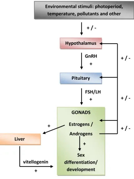
1.2. Endocrine control of reproductive function and interference of environmental toxicants with the hypothalamic-pituitary-gonadal axis
Interference of EDCs with the endocrine system may disrupt the reproductive, thyroid or adrenocordical functions that are responsible for the maintenance of homeostasis, reproduction, development, and/or behavior of all vertebrate species. To date, most evidence for endocrine disruption has been obtained in the reproductive function of vertebrates, especially of teleost fish (Di Giulio and Hinton, 2008; Pait and Nelson, 2002). The development of reproductive tissue and the coordination of the complex processes occurring during the annual reproductive cycle of fish are controlled by hormones secreted along the hypothalamic-pituitary-gonadal (HPG) axis (Norris, 2007; Fig. 1). The hypothalamus integrates environmental stimuli (e.g. photoperiod and temperature), which results in secretion of various neurotransmitters and neuropeptides that influence the action of the gonadotropin-releasing hormone (GnRH). This neurohormone in turn regulates directly the synthesis and secretion of gonadotropins (follicle-stimulating hormone, FSH and luteinizing hormone, LH) from the pituitary gland. These gonadotropins, distributed via the bloodstream, cause alterations in the synthesis of sex steroid hormones, particularly estrogens and androgens (Bone et al., 1995). Estrogens are primarily secreted by follicle cells surrounding the oocytes of the ovaries (i.e. theca and granulosa cells), while androgens are primarily secreted by the interstitial Leydig cells of the testes. Sex steroid hormones may display their actions by binding to specific cell-surface receptors, but principally by binding to nuclear steroid receptors (i.e. estrogen (ER), androgen (AR) receptors), resulting in alteration of gene expression (Tyler et al., 1998).
Estrogens and androgens have a variety of functions, many of which are similar in various classes of vertebrates, including sex determination and differentiation, sexual development, control of the reproductive cycle and behavior, and development of
Chapter 1 Introduction
6 secondary sex characteristics (Bone et al., 1995). The response of target tissues (mainly gonads, brain and liver) to sex steroid hormones is largely dependent on the expression of steroid hormone receptors, which in turn is regulated by steroid hormones and therefore fluctuates in response to alterations of the sex steroid biosynthesis (Tyler et al., 1998). The concentration of steroids and their receptors change during the reproductive life of fishes; consequently, the degree of adverse effects caused by exposure to EDCs varies depending on the developmental and reproductive stage of fish (Di Giulio and Hinton, 2008).
Figure 1. Schematic representation of the HPG axis controlling reproduction in fish. Feedback effects of steroids and other hormonal positive or negative effects (+ or -), illustrated by arrows, maintain a dynamic control of the reproductive endocrine system.
Environmental stimuli: photoperiod, temperature, pollutants and other
stressors Hypothalamus + / - GnRH Pituitary + FSH/LH + GONADS Estrogens / Androgens Sex differentiation/ development + Liver + + vitellogenin + / - + / - + / -
7
The HPG axis provides several possible points of interaction for EDCs that may result in interference with the reproductive endocrine function (Scholz and Klüver, 2009). Chemicals may act in the hypothalamus to influence GnRH secretion, which would secondarily affect pituitary gonadotropin secretion in response to GnRH (Khan et al., 2001). Other chemicals can act directly on the pituitary by influencing GnRH signaling pathways and gonadotropin secretion (Thomas, 1993). Exposure to these chemicals may result in alterations of gonadal development and function, for example in impaired steroid hormone or gamete production. In addition, some chemicals exert their effect directly on the gonads by influencing the activities of gonadotropin pathways and enzymes involved in the synthesis of sex steroid hormones (Ankley et al., 2005; Benninghoff et al., 2005). Alterations in the steroid hormone synthesis can dramatically influence steroid levels that target tissues such as gonads, and other organs important for reproduction, like the liver and the brain. Steroid levels may also be influenced by increases in metabolic clearance rate of steroids due to chemical induction of hepatic cytochrome P450 enzymes (Qian et al., 2007). Furthermore, steroid hormone action can also be affected in target tissues by direct interaction of chemicals with steroid receptors, resulting in agonism and antagonism of steroid hormone action. The agonist or antagonist mechanisms of EDCs may impair reproductive function for example by influencing the steroid feedback system (Harris et al., 2001), or the vitellogenin (precursor protein of the egg yolk) production in the liver (Jobling and Sumpter, 1993). Therefore, chemicals that can interact with the HPG axis could have serious effects not only on the development and well being of individual organisms, but also more importantly on the ability of these organisms to reproduce, and their offspring to survive and eventually reproduce (Lintelmann et al., 2003).
1.3. Morphology, endocrinology and environmental modulation of sex differentiation in teleost fish
1.3.1. Sexual differentiation and gonadal development
The sex differentiation refers to gonadal development once sex has been determined, i.e. when the undifferentiated gonad follows the ovarian or testicular differentiation pathway (Guerrero-Estevez and Moreno-Mendoza, 2010; Piferrer and Guigen, 2008). Among all different fish species, there is a large variety of patterns of sexual differentiation, from hermaphroditism to gonochorism. In most gonochoristic species, the
Chapter 1 Introduction
8 undifferentiated bipotential gonad develops directly into ovary or testis (an exception is juvenile hermaphroditism for which gonads are initially hermaphroditic, but later develop as ovaries or testes; Devlin and Nagahama 2002), while hermaphroditic species may be synchronic (ovarian and testicular tissue evolving simultaneously) or sequential (protandric, inversion from male to female; or protogynic, inversion from female to male; Sadovy and Shapiro, 1987). The different processes of sex differentiation are highly plastic and certain environmental factors, such as temperature or pH, or exposure to EDCs, may influence the sex differentiation towards male or female tissue development (Devlin and Nagahama, 2002; Nakamura, 1998).
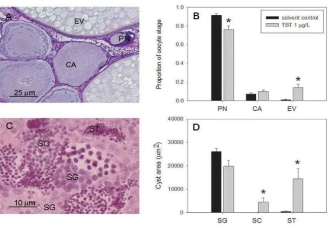
Sex differentiation has been shown to begin first in the gonads of females than in males in the majority of the gonochoristic teleosts examined to date. The onset of ovarian differentiation and oogenesis starts with differentiation of somatic and germ cells and intensive proliferation of germ cells (mitosis), which is followed by their entry into meiosis to form follicles, composed of the oocyte and a layer of granulosa cells surrounded by an external layer of theca cells (Strüssman and Nakamura, 2002). The differentiation of the primary ovarian follicle into an egg is a process accompanied by its growth and massive structural and functional changes. The oogenesis starts with the transformation of primordial germ cells into oogonia, followed by transformation into primary oocytes; the meiosis starts at this moment and is followed by a massive growth of the oocyte during vitellogenesis, whereby the oocyte accumulates nutritional reserves needed for the development of the embryo (Patiño and Sullivan, 2002). During this time the oocyte remains in meiotic arrest till the maturation process, which is characterized by the resumption of meiosis. Finally ovulation takes place at the end of the maturation process (Lubzens et al., 2010). During oogenesis, the oocytes can be divided into different stages of maturity (Wolf et al., 2004): perinucleolar, cortical alveolar, early vitellogenic, late vitellogenic, and mature oocytes (Fig. 2).
In juvenile hermaphrodites, where gonads of genotypic males develop first female characteristics, the first sign of male differentiation is the regression of female tissue. In differentiated gonochorists, the onset of spermatogenesis starts with intensive germ cell mitosis and formation of germ cell cysts and tubules (Schulz et al., 2010). The testis is composed of the intertubular compartment and the tubular compartment. The intertubular compartment contains the steroidogenic Leydig cells. The tubular compartment contains the
9 spermatogenic tubules with Sertoli cell and the germ cells. Within the spermatogenic tubules, several cysts, deriving from a single spermatogonium, can develop independently. Different stages of maturity of the germ cells can be distinguished (Schulz et al., 2010): spermatogonia, spermatocytes, spermatids and spermatozoa (Fig. 2).
Figure 2. Images showing, on the left ovary, and on the right testis from juvenile brown trout (1+). Gonads present germ cells at different stages of maturity: perinucleolar (PN), cortical alveoli (CA) and early vitellogenic (EV) oocytes in ovary, and spermatogonia (SG), spermatocytes (SC) and spermatids (ST) in testis. The testis is composed of several tubules (delimited in blue) containing several cysts (delimited in red) of germ cells in a particular stage of maturity.
1.3.2. Endocrine control of sexual differentiation and development
In fish, as in other vertebrates, the main endocrine regulators of sexual differentiation and development are sex steroid hormones. Treatment of sexually undifferentiated fish with androgens or estrogens results in most cases in the production of males and females, respectively, regardless of genotypic sex (Piferrer, 2001). Several studies suggest that the balance between androgens and estrogens, rather than their absolute amount, is determinant for the direction of sexual differentiation (Piferrer and Guiguen, 2008). In all vertebrates, this balance is determined by the activity of the aromatase enzyme that converts androgens into estrogens. Accordingly, the aromatase plays an essential role in regulating fish sex differentiation (Guiguen et al., 2010). The main estrogen, 17-estradiol, is found in much higher levels in females than in males, and is assumed to be the major sex steroid responsible for inducing and maintaining ovarian development (Norris, 2007). Both testosterone and 11-ketotestosterone are found in males, but also in females in a smaller
Chapter 1 Introduction
10 amount, and 11-ketotestosterone is the major androgen responsible for testicular development (Borg, 1994). In addition, other steroids like the 17, 20-dihydroxy-4-pregnen-3-one (the maturation-inducing steroid hormone) and other precursors of these hormones (from pregnenolone) play further roles during sexual development (Norris, 2007). Plasma levels of steroid hormones show important variations during male and female gonad differentiation and development and each steroid hormone has a distinct role in the regulation of sexual development (Lubzens et al., 2010; Schulz et al., 2010). For example, a major role of 17-estradiol in female fish is to stimulate the liver to produce vitellogenin, the precursor of yolk, and the vitelline envelope proteins, which form the egg shell. Thus, in oviparous animals, estrogens are vital for oocyte growth, egg formation, and provision of yolk for the developing embryo (Tyler and Sumpter, 1996).
The sex steroids are not the only factors implicated in the endocrine control of sexual differentiation and development. Besides the complex interaction between the brain and gonads via the production of gonadotropins and sex steroids (Fig. 1), sexual development involves also cell proliferation and tissue growth, suggesting that growth hormones, particularly insulin-like growth factors, may also take part in fish gonadal development (Reinecke, 2010). Consequently, the disruption of different hormonal regulatory pathways of the sexual development may result in impaired sexual differentiation and development.
1.3.3. Fish early sexual development: a sensitive period of life
The early gonadal development, until the sexual maturity, appears to be especially sensitive to chemicals that affect the HPG axis and alter the sex hormone levels (Ankley et al., 2004; Tyler et al., 1998; Scholz and Klüver, 2009). Many laboratory studies indicate adverse effects or aberrations in sexual development associated with exposure to EDCs (effluent or single chemical) during the sexual differentiation and development of fish. For example, juvenile zebrafish (Danio rerio) exposed to pulp mill effluent exhibit high levels of vitellogenin in males (an estrogenic effect), a male-biased sex ratio and occurrence of intersex (Örn et al., 2006). Male juvenile roach (Rutilus rutilus) exposed to sewage treatment effluent presented increased vitellogenin concentration and feminization of the reproductive ducts in male (Liney et al., 2005). Further, in juvenile zebrafish exposed to either natural estrogens or trenbolone (synthetic androgenic steroid), the sex ratio was skewed either towards females or complete masculinization was observed, respectively (Holbech et al.,
11 2006). Other adverse effects such as reduction of gonads weight, delayed or increased gametogenesis, have been observed (Mills and Chichester, 2005; Scholz and Klüver, 2009). All these adverse outcomes have the potential to impact the reproductive health at the individual level and subsequently the maintenance of stable populations of fish (Hutchinson et al., 2006).
1.4. Chemicals interfering with the steroidogenesis
EDCs can influence the endocrine functions by affecting hormone levels (synthesis or metabolism of hormones), by interfering with hormone action in the targeted tissue (mimicking or antagonizing hormone action), or by modifying hormone receptor levels (Sonnenschein and Soto, 1998). So far, ecotoxicological research on chemical interference with endocrine functions in vertebrates has mainly focused on chemicals interacting with nuclear steroid receptors, especially the nuclear ER (Sonnenschein and Soto, 1998; Tyler et. al., 1998). However, a range of chemicals have been shown to interfere and exert direct effects in the steroid hormone biosynthesis (steroidogenesis) in teleost fish (Cheshenko, 2008; Sanderson, 2006). There is increasing evidence that estrogenic substances are not the only substances present in the aquatic environment that exert endocrine disrupting effects. The research on endocrine disruption needs, therefore, to orientate and develop also towards EDCs exhibiting other endocrine modes of action, like interference with steroidogenesis, to assess the potential risk of such chemicals for sexual development and reproductive health of aquatic organisms.
Several organs synthesize active steroids, including the adrenal glands, testis, ovary and brain, with sex steroids being mainly produced in gonads. Their synthesis (Fig. 3) is controlled by the activity of several cytochrome P450 enzymes (CYPs) and a number of hydroxysteroid dehydrogenases (HSDs; Sanderson, 2006). The synthesis of all steroid hormones starts with the conversion of cholesterol to pregnenolone by CYP11A (cholesterol side-chain cleavage). Pregnenolone is mainly converted to progesterone by 3-HSD. Progesterone is then mainly converted into androstenedione by the CYP17 hydroxylase/lyase, which is first responsible for the 17α-hydroxylation of C17-20 steroid structures and its 17,20 lyase activity directs the biosynthesis of steroids toward sex steroids. Androstenedione, a weak androgen, is then mainly converted into testosterone by the 17-HSD. Principally in the testes, 11-ketotestosterone, the prominent androgen, is formed from
Chapter 1 Introduction
12 testosterone by 11-HSD. Finally, CYP19 or aromatase, which is expressed at a low level in testes and mainly in ovaries, is the key enzyme that converts androgens to estrogens (Cheshenko et al., 2008).
Figure 3. Enzymes involved in the biosynthesis of androgens and estrogens. Cytochrome P450 (CYP) and hydroxysteroid dehydrogenase (HSD) enzymes. CYP17 activity is indicated as (a) hydroxylase or (b) lyase activity. The major sex steroid biosynthesis pathway in fish gonad tissue is indicated in black. Gray background indicates steroidogenic pathways in extra gonadal tissues.
The steroidogenic pathway may be chemically disrupted by several mechanisms, including direct reversible or irreversible catalytic inhibition of steroidogenic enzymes, or indirect up- or down-regulation of steroidogenic enzyme expression. Indirect effects on the steroidogenic enzymes may occur via different interactions along the HPG axis (Sanderson and van der Berg, 2003). An analysis of the current literature indicates that still relatively little is known about the underlying mechanisms of interference of chemicals with
Cholesterol Progesterone 17-OH-pregnenolone Dehydroepiandrosterone Pregnenolone 17-OH-progesterone Androstenedione Testosterone 17-estradiol Estrone 11-ketotestosterone 11-OH-testosterone
11-deoxycorticosterone Corticosterone Aldosterone
11-deoxycortisol Cortisol 1720-dihydroxy-4-pregnen-3-one 3-HSD CYP17a CYP17b CYP11A CYP17a CYP17b CYP11B1 17-HSD 3-HSD 11-HSD CYP19 CYP19 3-HSD 20-HSD CYP21 CYP11B2 CYP11B2 CYP11B1 17-HSD
13 steroidogenesis and particularly about their potential toxicity in steroidogenic tissues, neither in humans nor in wildlife. Recent studies have reported modulations of sex steroid levels and impairment of gonadal development in wild fish collected from contaminated sites (Hecker et al. 2007; Hinfray et al., 2010; Lavado et al., 2004; Noaksson et al., 2001, 2003) suggesting that wild fish populations are indeed exposed to substances that can perturb steroidogenesis and potentially the sexual development of fish. However the nature and bioavailability of substances involved in these biological responses remain to be determined. Few laboratory studies have shown that chemicals interfering with the activity and/or the expression of one or several steroidogenic enzymes could lead to adverse effects that may potentially impair development and reproduction of fish (Table 1). These chemicals include natural products (i.e. phytosterols; MacLatchy et al., 1997), pesticides (i.e. prochloraz, fenarimol; Ankley et al., 2005), pharmaceuticals (i.e. ketoconazole; trilostane; Ankley et al., 2007; Villeneuve et al., 2008) and other industrial compounds (i.e. flame retardants; Deng et al., 2010). Most of them may potentially be found in the environment. Thus, there is a need to provide further knowledge on chemicals interfering with the steroidogenesis for a better understanding of the risk of this type of EDCs to humans and wildlife.
Among the studies on the mechanisms by which EDCs interfere with the sex steroid hormone homeostasis and function, particular attention has been given to CYP19. In several teleost fish such as zebrafish (Danio rerio), goldfish (Carassius auratus), tilapia (Oreochromis niloticus), fathead minnow (Pimephales promelas) or rainbow trout (Oncorhynchus mykiss), CYP19 has been found to be encoded by two different genes: cyp19a and cyp19b (Chang et al., 2005; Fenske and Segner, 2004; Tchoudakova and Callard, 1998; Toffolo et al., 2007; Villeneuve et al., 2006). These two distinct genes and encoded CYP19 proteins are differentially expressed, i.e. cyp19b mainly in the brain and cyp19a mainly in the gonads. Careful spatial and temporal balance of estrogens in the body is crucial in reproduction-related processes, including sexual differentiation and determination (Cheshenko et al., 2008), and the inhibition of the gonadal CYP19 activity and/or of gonadal cyp19a expression has been related to masculinization process of several fish species (Fenske et al., 2004; Guigen et al., 2010; Vizziano et al., 2008).
Chapter 1 Introduction
14 Table 1. Effects of chemicals disrupting the steroidogenesis on the development of the teleost gonads (reviewed in Schloz and Klüver, 2009).
Chemical Species P Effect Sex TSI OSI Sp Oo Other effects level ratio
(g/L)
Ketoconazole Pimephales A 6 I I Increase proliferation
promelas of Leydig cells
Fadrozole Danio rerio J 10 m
Pimephales A 50-100 I R S D Sertoli cells hypertrophy,
Promelas increased of Leydig cells
Letrozole Oryzias A 125 I I S D Enlarged lumen of
latipes seminferous tubules
A = exposure of adult fish ; D = delayed ; I = increased ; J = exposure of juvenile fish ; m = male-biased sex ratio ; Oo = oogenesis ; OSI = ovarian somatic index ; P = period of exposure; R = reduced; S = stimulated; Sp = spermatogenesis; TSI = testicular somatic index.
References: Ankley et al., 2007; Andersen et al., 2004; Panter et al., 2004 ; Sun et al., 2007 ; OECD, 2006
In this thesis, the following model chemicals, known or suspected to alter the steroidogenesis in addition to sexual differentiation and reproductive health of fish, have been studied:
1.4.1. 1,4,6-androstatriene-3,17-dione, an aromatase inhibitor
The chemical 1,4,6-androstatriene-3,17-dione (ATD; Fig. 4) is a potent irreversible CYP19 aromatase inhibitor that inhibits estrogen biosynthesis by permanently binding and inactivating CYP19 (Covey and Hood, 1981). It has also been used in few studies in a range of 0.5 to 5 mg/L to analyze the role of CYP19 expression in sex differentiation of several fish species, where it induces masculinization (Vizziano et al., 2008; Lee et al., 2003; Lee et al., 2001). So far, this chemical has not been reported in the environment. However, as ATD is used in some pharmaceutical products (e.g. body building supplements), surface waters may receive this chemical via domestic discharges.
15
ATD Prochloraz TBT
Figure 4. Chemical structure of ATD, prochloraz, and TBT (http://pubchem.ncbi.nlm.nih.gov/).
1.4.2. Prochloraz, an imidazole fungicide
Prochloraz (N-propyl-N-[2-(2,4,6-trichlorophenoxy)ethyl]imidazole-1-carboxamide; Fig. 4) is an imidazole fungicide widely used all around the world. The action of imidazoles are based on the inhibition of the cytochrome P450-dependent 14-demethylase activity, which is required in the conversion of lanosterol to ergosterol (Henry & Sisler, 1984), an essential component of fungal cell membranes. Due to its unspecific interaction with cytochrome P450, it also inhibits a broad spectrum of other cytochrome P450-dependent enzymes, including key enzymes of the sex steroid biosynthesis, e.g. CYP19 aromatase (Vinggaard et al., 2006). In vitro studies have shown that prochloraz elicits multiple mechanisms of action: it antagonizes the AR and ER; it agonizes the aryl hydrocarbon receptor (AhR, which subsequently induces gene expression modulation) and, principally, inhibits aromatase activity (Andersen et al., 2002; Hecker et al., 2006; Hinfray et al., 2006; Laville et al., 2006; Villeneuve et al., 2007). In vivo studies with rats showed that prochloraz could also inhibit testosterone production possibly by altering CYP17 hydroxylase/lyase activity (Blystone et al., 2007; Vinggaard et al., 2006). In addition, few fish in vivo studies demonstrated the sublethal adverse effects of prochloraz exposure in a range of 3 to 300 g/L, which is much lower as its acute toxicity value for rainbow trout (96-h LC50 = 1.43
mg/L). The studies of Ankley et al. (2005) and Zhang et al. (2008) showed a reduced fecundity of fathead minnow and Japanese medaka (Oryzias latipes), respectively, as response to prochloraz. In the study of Ankley et al. (2005), this effect was accompanied by decreased plasma sex steroids (17-estradiol, testosterone and 11-ketotestosterone) and vitellogenin concentrations, and altered gonadal histology. Both studies further
Chapter 1 Introduction
16 demonstrated an indirect effect of prochloraz on the sex steroid synthesis via up-regulation of CYP17 and CYP19 gene expression. Furthermore, Kinnberg et al. (2007) reported, in an early life stage assay with zebrafish, the potential of prochloraz to induce a male-biased sex ratio, altered gonadal development and vitellogenin production.
Since prochloraz is a widely used fungicide, it can be found in surface water and transported by runoff from the terrestrial to the aquatic environment. In Switzerland, it is has been detected in a range of few ng/L in groundwater (NAQUA, OFEV, 2009). In streambed sediment analysis from several Danish rivers, it has also been demonstrated that it can accumulate in sediments, possibly resulting in locally elevated concentration of the substance in the aquatic environment (Kronvang et al., 2003). In addition, few metabolic studies in rainbow trout indicated that prochloraz may be widely distributed in the body and is extensively metabolized. The highest levels of prochloraz residues were mainly found in the liver, in which prochloraz has been reported to induce the gene expression of CYP1A biotransformation enzyme (Cravedi et al., 2001; Debrauwer et al., 2001; Sturm et al., 2001).
1.4.3. Tributyltin, a persistent industrial compound
Tributyltin (TBT) is a toxic and bioaccumulative chemical that is used for various industrial purposes. While its use as biocide in antifouling agent in boat paints has been internationally banned, it is likely that organotin compounds continue to be produced and used in biocides for other functions (reviewed in Antizar-Ladislao, 2008). Due to its persistent nature, it is a common contaminant of marine and freshwater ecosystems exceeding acute and chronic toxicity levels. Extended research undertaken since the early 1970s has shown that TBT is very toxic to a large number of aquatic organisms, with acute toxicity concentrations in the range of 5 to 1000 ng/L, depending on the species, life stage, and other chemical and physical parameters (e.g. salt- or freshwater, temperature; reviewed in Antizar-Ladislao, 2008). In the aquatic environment, TBT adheres strongly to bed sediments and high levels up to 1500 ng/L have been reported. In recent investigations, it has been reported that TBT concentrations in water, sediment and biota have generally declined, and maximum concentrations in marine water rarely exceed 100 ng/L (Diez et al., 2006; Bhosle et al., 2004). The oral route is the most obvious source for organotin uptake in aquatic biota and transfer via the food chain, although direct uptake via skin or gills is also important in fish (Lee et al., 2006; Strand and Jacobsen, 2005). In addition, is has been
17 demonstrated that TBT alters the activity and/or expression of hepatic CYP enzymes in fish, which decreases the xenobiotic detoxification and increases the potential risk of organotin compound exposure (Fent et al., 1998; Mortensen and Arukwe, 2007).
The TBT-induced masculinization (imposex) in female marine mollusks is a well-known example of endocrine disruption in invertebrates that is causally linked to an environmental pollutant (Matthiessen and Gibbs, 1998). Imposex occurs when male sex characteristics develop in female gastropods and has resulted in decline or extinction of mollusk populations worldwide. The bioaccumulation of TBT in gastropods and its endocrine disruptive effects result in elevated testosterone levels giving rise to imposex (Gibbs and Bryan, 1996). The mechanism of action of TBT is not clear yet, but considerable weight of evidence suggests an inhibition of CYP19 as possible mechanism (Bettin et al., 1996). However, TBT also inhibited conjugation of testosterone, which would result in reduced elimination and accumulation of testosterone from the body, and subsequently stimulate the development of imposex (Ronis and Mason, 1996).
In fish, TBT in a range of 1 to 1000 ng/L clearly induces endocrine disrupting effects (Antizar-Ladislao, 2008). In laboratory studies with zebrafish, TBT has been shown to impair reproductive function by causing high incidence of abnormal sperm and a male-biased sex ratio, which might be related to CYP19 inhibiting properties of TBT (McAllister and Kime, 2003). Similarly, fertilization success was decreased in medaka exposed to TBT (Nakayama et al., 2004). Further, TBT could modulate sex hormone levels in gonads of Sebastiscus marmoratus and impair gonadal development (Zhang et al., 2008, 2009). In addition, a long-term exposure of the mummichog (Fundulus heteroclitus) to TBT affected not only gonadal sex differentiation and spermatogenesis, but also spawning and egg quality (Mochida et al., 2010).
1.5. Fish test methods for endocrine disrupting chemicals 1.5.1. Fish in vivo testing
Given the occurrence of EDCs in the environment and their potential threat for reproductive health of fish populations, international research efforts on the development of testing strategies for EDCs have arisen 1 2 (Fenner-Crisp et al. 2000; Huet, 2000;
1
http://www.oecd.org/document/42/0%2C2340%2Cen_2649_34377_2348650_1_1_1_1%2C00.html 2 http://www.epa.gov/endo/
Chapter 1 Introduction
18 Hutchinson et al., 2006). These strategies are based on tiered approach. The first step is the performance of a screening test. Screening assays have been defined as lower tier in vitro or
in vivo investigations which allow the identification and classification of substances relative to their potential interaction with endocrine systems (Ankley et al., 1998). If a substance is suspected to have an endocrine mode of action, it has to be tested in an assay that can be used in risk assessment. These tests are defined to be higher tier in vivo methods (e.g. fish full or partial life cycle assays) to confirm the screening results and to characterize any adverse effects at the apical level of the biological organization (e.g. on survival, growth, morphological development, and reproduction)that may result from the endocrine mode of action of EDCs (Hutchinson et al., 2006; Matthiessen and Johnson, 2007).
Since the most evident impacts of EDCs in the environment are on aquatic organisms (Hutchinson and Pickford, 2002), it has stimulated research efforts to develop fish test methods. The fish species recommended by international regulatory agencies are: the fathead minnow (Pimephales promelas), the medaka (Oryzias latipes), the zebrafish (Danio rerio), and the three-spined stickleback (Gasterosteus aculeatus). Currently, two in vivo fish screening assays have been adopted by the OECD (Knacker et al., 2010): the fish short-term reproduction assay (OECD, 2009a) and the 21-day fish assay (OECD, 2009b), which are both conducted with reproductively active fish. The endpoints of these two screening assays include vitellogenin induction and evaluation of secondary sex characteristics in sexually dimorphic species (i.e. fathead minnow and medaka); further endpoints of the short-term reproduction assay include fecundity, fertility and histolopathology of gonads. In addition, higher tier fish in vivo methods providing data on adverse effects at the apical level have been proposed: the fish full life cycle test (FLCT; Seki et al., 2003), the fish 2-generation test (2-genT; Braunbeck et al., 2009; EPA, 2002) and the fish sexual development test (FSDT). The FSDT allows measuring sexual development endpoints (i.e. sex ratio and vitellogenin production; Holbech et al., 2006). The FLCT and the 2-genT allow assessing the effects on developmental and reproductive endpoints (i.e. hatching, sex ratio, survival, growth, time to first spawn, fecundity, fertility, and behavior) as well as biochemical, histological, and morphological markers. Additionally, the 2-genT allows determining trans-generational transfer of effects.
19 1.5.2. Fish in vitro testing
The justification for in vivo studies in ecotoxicology is derived from its objective to evaluate chemical effects at population and ecosystem levels (Castaño et al., 2003). Furthermore, due to the complexity of the endocrine system, extrapolation of in vitro effects
to in vivo situation is not straightforward and requires further, often in vivo, investigations (Sanderson, 2006). In addition, in vitro assays do not provide biological responses and endocrine-mediated adverse effects at the apical level, which are needed for environmental risk assessment (Ankley et al., 2009). In contrast, a number of ethical, technical, scientific and economic reasons support the development of fish in vitro methods to be used as screening assays. They are rapid screening tools to evaluate the toxicity of a large number of individual compounds and environmental samples. Further, they reduce the number of animal used in toxicity testing (Castaño et al, 2003; Matthiessen and Johnson, 2007). In vitro
cell-based methods provide the best experimental system for studying toxic mechanisms at the molecular and cellular levels in a controlled environment and isolation from the multiple physiological pathways interacting in vivo (Castaño et al., 2003). The identification of specific mechanism of action in in vitro assays can trigger more advanced and comprehensive in vivo
testing, thereby optimizing time and resource use (Ankley et al., 2009; Hutchinson et al., 2006). In addition, in vitro assays can lead to the development of biomarkers, which can be used to measure in vivo responses and can permit the evaluation of the effects of toxicants in animals (Castaño et al., 2003).
1.5.3. In vitro screening assays for endocrine disrupting chemicals interfering with steroidogenesis
To date, most of the developed in vitro assays have focused on EDCs binding to steroid hormone receptors (i.e. receptor binding assays, cell proliferation assays, reporter gene assays; Baker et al., 2000; Gray et al., 1997; Soto et al., 1995). To investigate effects of EDCs interfering with the steroidogenesis, various in vitro methods from different biological systems are possible. The catalytic activity of individual steroidogenic enzymes can be either measured in microsomal fraction of tissues that express the enzyme of interest, or in cell cultures by using selective radiolabeled substrate for the enzyme in combination with specific inhibitors of the enzyme (Hinfray et al., 2006; Vingaard et al., 2000; Sanderson et al., 2002). In cell lines, primary cell or tissue cultures, alterated enzyme expression can be
Chapter 1 Introduction
20 determined via northern blotting or RT-PCR (Hilscherova et al., 2004). An indirect way to measure chemical effects on steroidogenic enzyme function in cell cultures is to measure alterations in excretion of certain steroid hormones as an indicator of chemical effect on steroidogenesis (Hecker et al., 2006; Lee et al., 2006; Villeneuve et al., 2007). An advantage of this approach is that alterations in the profile of the secreted steroid hormones provide an indication of the identity of the enzymes affected by the xenobiotic treatment, without the need to examine each enzyme activity individually (Sanderson et al., 2003).
1.5.4. Ex vivo organ cultures
The in vitro fish cell systems which are currently used for toxicological studies are based either on established fish cell lines, primary fish cells or organ ex vivo cultures (Castaño et al., 2003). An organ culture has the advantage over cell lines that it possesses the morphological and cellular structure and function of its source tissue. The use of organ cultures is therefore physiologically relevant, and is particularly suitable to study specific mechanisms of action of toxicants in particular target organs. Furthermore, the preparation of ex vivo organ cultures is rather simple: organs are removed, dissected and maintained in a serum free culture medium. It is therefore uncomplicated compared to primary cell cultures that need an isolation procedure (Castaño et al., 2003). According to these advantages of organ cultures, and to analyse the effects of chemicals interfering with steroidogenesis, a fish ex vivo gonad culture was developed here (Fig. 5).
Figure 5. Images illustrating the developed gonad organ culture from juvenile brown trout. From left to right: removal of gonads from juvenile fish; dissection of gonadal fragments in Petri dish filled with fresh medium; gonadal fragments on insert filters transferred in a 4-well plate for incubation in fresh medium.
21 1.6. Test fish species
The bony fish represent the largest group of vertebrates both in number of species and in number of individuals. Their evolution and adaptation to different habitats has led to a high diversity of physiological, anatomical, behavioral and ecological strategies in fish (Bernanke and Köhler, 2008). Endocrine disruption has been studied in a variety of freshwater teleost fish species, but cyprinids (e.g. fathead minnow, zebrafish, carp) and salmonids (e.g. rainbow trout, brown trout, Atlantic salmon) are the best represented (Jobling and Tyler, 2003). Chemicals altering the sex hormone levels may induce quantitatively and qualitatively different biological responses and adverse apical effects in various fish species because of the different mechanisms for sexual development and different reproductive strategies (Hutchinson et al., 2006). Moreover, the sex, life stage and the endogenous hormone levels during the reproductive cycle of fish are other important physiological factors that may influence the effects of EDC exposure (Kawai et al., 2003). Therefore, it is important to analyze the differences in the sensitivities of different fish species to effects of EDCs for interspecies extrapolation from laboratory fish tests to wild fish populations (Hutchinson et al., 2006). For the experiments of this thesis, three different fish species with different sexual development and/or different reproductive strategies have been used.
1.6.1. Fathead minnow (Pimephales promelas) and zebrafish (Danio rerio)
Fathead minnow and zebrafish are small fish species currently used for EDC testing. Established methods for maintaining both species in continuous cultures in laboratory exist and the fish are easy to handle (Ankley et al., 2004). Both species have a short life cycle and start to spawn early in their life (Hutchinson et al., 2006). They are fractional spawners and produce a high number of eggs (Mills and Chichester, 2005). Their spawning activity can be precisely controlled by manipulating the temperature, photoperiod, and spawning substrate. Determination of fertility of the eggs can be achieved easily using a microscope. Thus, they are successfully used for partial and full life cycle tests to investigate reproductive effects of xenobiotics (Ankley et al., 2004).
The fathead minnow has a ubiquitous distribution across North America. Adults are approximately 50 to 75 mm long and weigh 2 to 5 g (Jensen et al., 2001). They have a generation time of 4 to 5 months. A reproductively active female fathead minnow typically
Chapter 1 Introduction
22 deposits clutches of 50 to 100 eggs on the bottom of the spawning substrate every 3 to 5 days (Jensen et al. 2001). Embryos hatch in approximately 4 to 5 days at 25°C (Ankley et al, 2004). Fathead minnows exhibit secondary sex characteristics and defined mating behavior, which makes them suitable to investigate phenotypic and behavioral changes associated with EDC exposure (Mills and Chichester, 2005). The gonadal differentiation of fathead minnow starts early in its development, and the gonads can be identified as either male or female from 10 to 25 days post-hatch (van Aerle et al, 2004; Uguz, 2008).
The zebrafish is a small sized fish species native to India and Burma. Adult zebrafish are approximately 40 to 50 mm long, and weigh about 1.5 g. The reproductive cycle takes 3 to 4 months. Reproductively active female zebrafish can spawn almost daily, with relatively large spawns (e.g. >150 eggs) occurring every 5 to 10 days. The eggs are released in the water column and settle to the bottom of the tank. The embryos hatch in about 3 days at 28°C (Ankley et al., 2004). In contrast to the fathead minnow, all juvenile individuals first develop ovary-like gonads. Only after approximately 35 to 45 days post-hatch, the ovarian tissue of male fish regresses and develop into testicular tissue (Maack and Segner, 2003).
1.6.2. Brown trout (Salmo trutta fario)
The brown trout is an important freshwater fish species in European rivers and has a high commercial and social value. Over the last 20 years, however, its catch has dramatically declined in Swiss rivers (Körner et al., 2005). To evaluate the causes for the observed decline, the project ‘Fishnet’ was established in 1998. Within the “fishnet” project, various hypotheses were put forward to explain the reduced catch (Burkhardt-Holm et al., 2002). Among others, a disturbed reproductive health as a consequence of endocrine disruption has been considered.
The brown trout is an annual spawner with a predominant spawning season in November and December. The brown trout is a gonochoristic species, in which gonadal development and differentiation happens during the first two to three years before attaining sexual maturity, with the males usually maturing before females (Billiard, 1987; Elliott, 1994). Signs of sex differentiation appear first in females at approximately 20-30 days post-hatch, and later on in males. According to their reproductive cycle, the morphology of their gonads follows seasonal changes. In males, gonads remain undifferentiated until the initiation of the first spermatogenic cycle (i.e. 1 to 2 years), and the complete
23 spermatogenesis and the first reproductive cycle usually takes place during the second year of life. In contrast, the completion of oogenesis till the ovulation and the first reproductive cycle happens for most females only in the third year of life (Billard, 1987). During the second year of life, some vitellogenic activity can, however, be observed in some females, although final ovulation does not take place. In salmonids, the capacity to synthesize and metabolize sex steroids is active from the time of egg fertilization but changes seasonally throughout the sexual differentiation and development of gonads (Billard et al., 1992; Feist and Schreck, 1996; Yeoh et al., 1996). Sex differentiation is labile during early life stages since sex inversion can easily be obtained by feeding the fry with food containing androgens or estrogens (Hunter and Donaldson, 1983).
1.7. Objectives of the thesis
The objective of the present thesis was to develop and/or test the potential of ex vivo
and in vivo methods to identify and assess the effects of model chemicals disrupting the steroidogenesis in fish. To investigate this potential, the following key questions were posed: Does an ex vivo gonad assay have the potential to identify and discriminate between different effects of chemicals interfering with the steroidogenic pathyway?
Does the ex vivo gonad assay inform on in vivo effects?
Do chemicals interfering with steroidogenesis impact the regulatory processes of early sexual development and potentially the reproductive health of fish?
Is the FSDT a suitable method to identify and assess the risk of chemicals interfering with steroidogenesis?
To answer to these questions, in a first step, an ex vivo gonad assay was developed to rapidly identify and quantify the effect of chemicals inhibiting the steroidogenic pathway. Chapter 2 describes the development of this ex vivo gonad assay from juvenile brown trout, as well as the methodology and characterization of the assay. The advantages and limitations of this novel method are discussed. In Chapter 3, the potential of the ex vivo
assay to predict in vivo physiological responses is presented. The ex vivo assay could thereby provide a suitable tool to bridge the gap to in vivo effects and a valuable screening and
Chapter 1 Introduction
24 informative tool for hazard assessment of EDCs. For this, ex vivo and in vivo biochemical responses to chemicals known to disrupt steroidogenesis were compared. This investigation was complemented by a 19-day in vivo study using the same chemicals to further analyze the potential effects of steroidogenesis disruption on sexual development of juvenile brown trout.
The endocrine regulatory processes of the early life period and all the endocrine factors that play a role during this period are not fully understood and identified. Hence, the developed ex vivo gonad assay could also be a suitable tool to study the regulatory processes during the early sexual development of fish and the potential impact of chemicals interfering with steroidogenesis during this sensitive life stage. Accordingly, the study in chapter 4 analyses cellular and molecular effects of chemicals interfering with steroidogenesis on the regulation of early sexual development in brown trout by employing ex vivo and in vivo
methods and comparing their biochemical and molecular responses.
Apart from the importance of extrapolation of the ex vivo results to in vivo effects in risk assessment, the difference of sensitivity to EDCs between fish species may also pose a problem for qualitative and quantitative extrapolation of data across fish species. Indeed, the difference in modes of sexual development and reproduction between fish species may influence their sensitivity to EDCs. To provide further knowledge on the influence of the gonadal differentiation mode on qualitative and quantitative effects caused by EDCs, the responses of two model fish species with different sexual differentiation strategies were characterized and compared in a FSDT (chapter 5).
Finally, the results of all the different studies of this thesis are summarized to evaluate the potential of the test methods to assess effects of chemicals interfering with steroidogenesis on sexual development and reproductive success of fish (chapter 6).
References
Andersen, H. R., Vinggaard, A. M., Rasmussen, T. H., Gjermandsen, I. M., Bonefeld-Jørgensen, E. C., 2002. Effects of currently used pesticides in assays for estrogenicity, androgenicity, and aromatase activity in vitro. Toxicol. Appl. Pharmacol. 179: 1–12.
Andersen, L., Kinnberg, K., Holbech, H., Korsgaard, B., Bjerregaard, P., 2004. Evaluation of a 40 day assay for testing endocrine disrupters: Effects of an anti-estrogen and an aromatase inhibitor on sex ratio and vitellogenin concentrations in juvenile zebrafish (Danio rerio) . Fish Physiol. Biochem. 30, 257–266.
Ankley, G., Mihaich, E., Stahl, R., Tillitt, D., Colborn, T., McMaster, S., Miller, R., Bantle, J., Campbell, P., Denslow, N., Dickerson, R., Folmar, L., Fry, M., Giesy, J., Gray, L.E., Guiney, P., Hutchinson, T.,