Variation in Crown and Root Formation and Eruption of
Human Deciduous Teeth
H.M. Liversidge1* and T. Molleson2
1Department of Paediatric Dentistry, Queen Mary University of London, London E1 2AD, UK 2Department of Palaeontology, Natural History Museum, London SW7, UK
ABSTRACT The aim of this study was to document variation of deciduous tooth formation and eruption. The material comprises 121 individuals of known or estimated age (using tooth length) from Spitalfields in London, and radiographs of 61 healthy living children aged 2–5 years. Other skeletal material from two medieval Scottish ar-chaeological sites (Whithorn, N⫽ 74; Newark Bay, N⫽ 59) was also examined. Stages of crown and root formation as well as eruption (alveolar, midway, and occlusal levels) were assessed for each developing maxillary and mandib-ular tooth from radiographs or direct vision. Age of attain-ment for individual stages was calculated by probit
anal-ysis, and these data were also adapted for use in estimating age. The timing of crown completion was sim-ilar to previously reported studies, but apex completion times were later. Analysis of data relative to the first and second molars at the two stages D (crown complete) and F (root lengthⱖcrown height) allowed comparison with the Scottish material. No significant differences were ob-served between population groups for tooth formation or eruption. These data fill several gaps in the literature, and will be useful in assessing maturity and predicting age during early childhood. Am J Phys Anthropol 123: 172–180, 2004. ©2004 Wiley-Liss, Inc.
Deciduous tooth development begins before birth and is complete by about the fourth postnatal year, thus providing a number of clear milestones that record growth and maturation during this period. Together with other growth parameters, such infor-mation provides an insight into environmental fac-tors of individuals and/or groups, to estimate age or assess health needs or treatment response. Much of what we know is based on anatomical or dissection studies, a handful of radiographic investigations, and several more recent eruption studies of living children. A review of the literature (Smith, 1991; Liversidge et al., 1998; Scheuer and Black, 2000) revealed only cursory data for anterior teeth and for maxillary teeth, and little information about the timing of alveolar or full eruption. As developing teeth are the most reliable indicator of maturation and thus of the biological age of skeletal material, these gaps undermine the accuracy with which age can be predicted in archeology, anthropology, and forensic odontology. The aim of this study is to fill some of these gaps by quantifying the deciduous tooth formation and eruption of recent and living children in the United Kingdom.
MATERIALS AND METHODS
The material studied (N⫽121) is from the crypt of Christ Church, Spitalfields, London, a coffin-bur-ied collection, interred between 1729 –1852 (Molle-son et al., 1993) (Fig. 1). Age is recorded for 53 individuals; for 68, age was estimated using tooth-length equations from isolated teeth or undistorted radiographs calculated for this sample (Liversidge et al., 1993; Liversidge and Molleson, 1999).
Rota-tional pantomographs of living children (N ⫽ 61) between 2–5 years of age were included to improve the numbers for molar apex formation. These were healthy children who attended the Emergency Clinic of the Royal London Hospital.
Developing deciduous teeth from skeletal remains (N ⫽ 59 and N ⫽ 74) of two medieval Scottish archaeological collections were also studied: Newark Bay in Orkney (Brothwell, 1977), and Whithorn in Scotland (Watt and Lunt, 1999). Not all the skeletal specimens are complete: some consist of isolated teeth, and others are jaw fragments and radio-graphs and were not available for all of the Scottish material. New stages of deciduous tooth formation were devised in the style of Demirjian et al. (1973) for permanent teeth, with a combination of criteria from observations of isolated teeth as well as radio-graphs. Descriptive criteria, line drawings, and ra-diographs are shown in Table 1 and Figure 2. Some stages are defined relative to the approximal height of the incomplete crown or root; this is defined from the cusp tip (or occlusal level) to the edge of the mineralizing front of the developing crown or root. Approximal crown height is defined as the distance
*Correspondence to: Dr. Helen Liversidge, Department of Paediat-ric Dentistry, Dental Institute, Turner Street, Whitechapel, London E1 2AD, UK. E-mail: [email protected]
Received 19 August 2002; accepted 22 March 2003. DOI 10.1002/ajpa.10318
Published online 16 June 2003 in Wiley InterScience (www. interscience.wiley.com).
from the cusp tip (or occlusal level) to a line drawn between the mesial and distal crown-root margins. This differs from true anatomical crown height, as it does not take into account the sinuous margin of later-forming buccal and lingual enamel.
Eruption status of deciduous teeth was assessed directly from the specimen (viewed from the lateral aspect) as well as from radiographs where available, after checking for absence of damage to the alveolar margin. If the developing tooth was rotated, or loose, it was replaced gently into its crypt. Eruption levels were defined relative to the cusp tip(s), and are as follows for a mandibular tooth: below the alveolar level, at the alveolar bone level, about halfway be-tween the alveolar bone and the occlusal level, and attainment of the occlusal level (Fig. 3).
Intraobserver error was calculated from duplicate scoring of tooth formation and eruption stages of 50 teeth. Percentage agreement was 78% for crown and root stages, with all differences being one stage. Percentage agreement for eruption was 94%.
The analysis of tooth formation for the children of known age was as follows. The average age of at-tainment for each stage was calculated using probit analysis, with age groups of 6 months (Finney, 1952). The mean age-of-attainment of a discrete growth event is best calculated using the status quo method, based on the principle of a cumulative fre-quency curve (Healy, 1986). The proportion of chil-dren having reached a growth event at successive years is fitted by probit regression, and the means and ranges are derived (Eveleth and Tanner, 1990). In a large group of sufficient age range, the curve will be S-shaped, recording 0% of children in the youngest age group having reached the stage, up to 100% of the oldest age group having reached that stage. The use of probit regression, or any other regression using cumulative frequency distribution, overcomes the difficulties of an uneven age distribu-tion and inadequate age range that hamper a
num-ber of studies of early tooth growth. The same method of analysis was used to calculate average age of eruption stage. Age of attainment data for both crown/root stages and eruption are adapted for prediction by calculating the midpoint from the pre-vious stage (see Smith, 1991). For the stage “alveo-lar eruption,” this was calculated halfway between stage D and attainment of the alveolar level. This adaptation of attainment data is necessary, as stages are arbitrary despite the continuous nature of growth. For example, if a tooth is staged as being in stage E, it must have attained that stage sometime previously, and is therefore some way towards the next stage; using age of attainment dates to predict age will underestimate age, especially if the dura-tion of stages is long.
Further analysis compared tooth formation (crown and root stages as well as eruption stages) with the Scottish material of unknown age at death. Crown and root formation was expressed relative to the first (m1) and second (m2) mandibular molar
stages D and F, and the distribution of crown/root stages was compared between populations using a Mann-Whitney test. No significant differences were observed, and the data from all three were com-bined. The distribution of eruption levels at partic-ular root stages was tested between Spitalfields and the combined Scottish material, using the Mann-Whitney test. The small sample size allowed com-parison for only one stage per tooth type: stage E for incisors, canine, and first molar, and stage F for the second molar. None of these comparisons were sig-nificant, and data were combined.
RESULTS
Age of attainment of crown and root growth is shown in Table 2 and Figure 4 with age for predic-tion in Table 3. The distribupredic-tion of stages relative to reference stages is given in Tables 4 and 5 (all indi-viduals with m1or m1at stage D and F) and Table 6
and 7 (m2or m
2at stage D and F). Age of attainment
of eruption levels is shown in Table 8 and Figure 5, with age for prediction in Table 10. Predicting age from a tooth that has attained the occlusal level should be done only if the root is incomplete; the newly erupted tooth will be loose in its bony support, and may show a bony trough at the alveolar margins around the neck of the tooth.
Teeth are below the alveolar bone level during crown formation and early root growth. The modal stage for alveolar eruption was stage E. For the midpoint level of eruption, almost all teeth were in stage E or F; the modal stage for the central incisor, canine, and first molar was E, while it was F for the second incisor and second molar. The attainment of the occlusal level was during stage F, and all teeth in stage G had reached this. No significant differ-ences were observed between population groups for tooth formation or eruption where sample size was sufficient for comparison. The distribution of root stages at each eruption level is shown in Table 9.
Fig. 1. Age distribution of sample (shaded bars, Spitalfields, open bars, living children).
DISCUSSION
The development of deciduous teeth differs from that of permanent teeth in several respects, and new descriptive stages have been devised that are for the most part applicable to both radiographs and direct observation. Many previous dental growth studies lacked clearly defined crown and root stages. Ana-tomical and radiographic stage assessment is not always equivalent, and stages in this study were carefully chosen and clearly described in order to reduce the differences between these two methods of data collection. Some stages, such as initial cups tip formation and early cleft formation of the root, are only visible by direct vision (Boller, 1964). Another stage that presents problems is crown completion, and this needs to be carefully defined in order to be equivalent. Initial root formation seen approximally occurs considerably earlier than true enamel com-pletion on the buccal and lingual surfaces of the root, particularly in permanent incisors and canines. The definition of stage D in this study is equivalent for both methods of data collection indicating initial root growth approximally, and is earlier than true anatomical enamel completion seen directly from an isolated tooth. Some root-growth stages in this study rely on dentine thickness relative to pulp canal width, and this can only be assessed from radio-graphs; if these are not available, root growth is evaluated by length and apical formation.
The most important developmental difference be-tween the dentitions is the faster rate of formation of both enamel and dentine in deciduous compared to permanent teeth. The high rate of ameloblast differ-entiation in deciduous teeth is associated with the inclination of the incremental lines to the enamel-dentine junction, first pointed out by Boyde (1964) and further discussed by Keene (Keene 1982, Keene 1991) and Shellis (1984). This pattern is also true of odontoblasts, and fast root growth is reflected in the smaller root-cone angle in deciduous teeth compared with permanent teeth (Dean, 1985). Radiographi-cally, this is seen as an initial thin layer of hard tissue outlining the shape of the crown, after which the full-thickness enamel is laid down. Similarly, root length is established by a very thin outline of dentine that subsequently thickens. Stages that de-scribe fractions of crown or root formed are not ap-propriate for deciduous teeth, as crown height and root length are established early on, prior to full-thickness coronal dentine. The crown-root margin may be present before the dentine is full-thickness and the shape of the pulp chamber is established. Radiographically, the crown complete stage is fur-ther complicated in the presence of a large buccal enamel bulge (named after Zuckerkandl) which may obscure initial root formation at the mesial crown margin. Once root length is complete, dentine con-tinues to be laid down, narrowing the root canal.
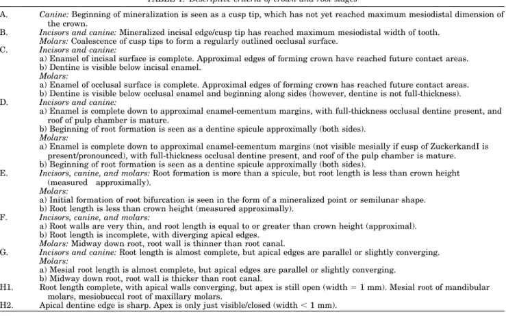
TABLE 1. Descriptive criteria of crown and root stages
A. Canine:Beginning of mineralization is seen as a cusp tip, which has not yet reached maximum mesiodistal dimension of the crown.
B. Incisors and canine:Mineralized incisal edge/cusp tip has reached maximum mesiodistal width of tooth. Molars:Coalescence of cusp tips to form a regularly outlined occlusal surface.
C. Incisors and canine:
a) Enamel of incisal surface is complete. Approximal edges of forming crown have reached future contact areas. b) Dentine is visible below incisal enamel.
Molars:
a) Enamel of occlusal surface is complete. Approximal edges of forming crown has reached future contact areas. b) Dentine is visible below occlusal enamel and beginning along sides (however, dentine is not full-thickness). D. Incisors and canine:
a) Enamel is complete down to approximal enamel-cementum margins, with full-thickness occlusal dentine present, and roof of pulp chamber is mature.
b) Beginning of root formation is seen as a dentine spicule approximally (both sides). Molars:
a) Enamel is complete down to approximal enamel-cementum margins (not visible mesially if cusp of ZuckerkandI is present/pronounced), with full-thickness occlusal dentine present, and roof of the pulp chamber is mature. b) Beginning of root formation is seen as a dentine spicule approximally (both sides).
E. Incisors, canine, and molars:Root formation is more than a spicule, but root length is less than crown height (measured approximally).
Molars:
a) Initial formation of root bifurcation is seen in the form of a mineralized point or semilunar shape. b) Root length is less than crown height (measured approximally).
F. Incisors, canine, and molars:
a) Root walls are very thin, and root length is equal to or greater than crown height (approximal). b) Root length is incomplete, with diverging apical edges.
Molars:Midway down root, root wall is thinner than root canal.
G. Incisors and canine:Root length is almost complete, but apical edges are parallel or slightly converging. Molars:
a) Mesial root length is almost complete, but apical edges are parallel or slightly converging. b) Midway down root, root wall is thicker than root canal.
H1. Root length complete, with apical walls converging, but apex is still open (width⫽1 mm). Mesial root of mandibular molars, mesiobuccal root of maxillary molars.
Radiographically, this is visible as very thin root walls that diverge, with time becoming parallel as the dentine attains full thickness, narrowing the root canal, and the apical edges converge with mat-uration of the apex. Despite the additional criteria of relative thickness of pulp canal and root dentine for root stage G in this study, this stage was still not easy to assess, and the subjective judgment of com-plete root length from cross-sectional radiographs remains a difficulty. Further dentine formation con-tinues very slowly on the approximal walls of the pulp cavity in molars; the pulp chamber size of de-ciduous molar teeth in older children becomes only marginally smaller over several years, but the root canals are noticeably narrower. The apex of the
im-mature maxillary central incisor root is considerably wider than other single-rooted deciduous teeth. An-other difference between deciduous and permanent molars is the root used to assess formation. For permanent molars, the apex of the distal root (the last forming) is assessed by the method of Demirjian et al. (1973). However, in deciduous molars the me-sial root (mesio-buccal root in maxillary molars) is longer and matures later, and therefore should be assessed.
The mean ages of crown completion (for c, m1, and
m2) from this study are similar to the published data
based on one longitudinal study (Fanning, 1961; Moorrees et al., 1963; Fanning and Brown, 1971) and from a cross-sectional study (for m2; Gilster et
al., 1964). The mean age of mandibular canine crown formation from the present study (0.81 year) is a little earlier than that calculated from detailed histological investigation of four teeth from prehis-toric Rome (FitzGerald et al., 1999). This assumes that initial mineralization timing is similar (Sun-derland et al., 1987), and also assumes a similar rate of enamel formation and proportion of prenatal/post-natal enamel estimated at about 40%/60% (H. Thomas, personal communication). Mean ages of apex closure from the present study are a little later than the published data for the mandibular canine and second molar. The mean age of apex closure of m1 is just prior to the second year (Moorrees et al.,
1963), yet Fanning and Brown (1971) reported the mean age in boys to be more than a year later. The mean age from the present study of combined sexes is just prior to the third year. Comparison with other radiographic studies of deciduous tooth formation is difficult, as some do not use cumulative distribution analysis or fail to give sufficient details of sampling or analysis (Fass, 1969; Nanda and Chawla, 1966; Nystro¨m, 1982; Nystro¨m et al., 1977).
Eruption is defined as the movement of the devel-oping tooth from within the alveolar bone, through the gingivae into the oral cavity, until it reaches occlusal contact with the opposing tooth. This pro-cess usually begins sometime after crown comple-tion, when some root is present. Resorption of alve-olar bone, making space for the crown, and
breakdown of the soft-tissue ligament over the oc-clusal bone cavity in the mandible, allow the cusp tip to reach and pass the alveolar crest. On skeletal material, a trough or groove may be observed around the erupting crown as it erupts through the alveolar bone. Sometime later, the tooth becomes palpable in the mouth and will penetrate the soft tissue. The first erupting maxillary incisors pene-trate the tightly attached gingival tissue buccal to the alveolar crest (illustrated in Hulland et al., 2000). The position of the cusp tips at the time of clinical emergence of other teeth is not described in the literature. Do nonsuccessional (or primary) teeth emerge through the gingivae around the midpoint between the alveolar and occlusal levels? Clinical observations of first permanent molars suggest that these teeth are nearer to the occlusal level when they first appear in the mouth. Results from the present study of the stage midpoint between alveo-lar and occlusal levels are simialveo-lar to the average ages of clinical emergence in British children (Leigh-ton, 1977). A recent longitudinal study of emergence of some deciduous teeth in individual children re-ported several months elapsing between being pal-pable to all cusps being visible (Hulland et al., 2000). The only mention in the literature regarding the emergence level of deciduous teeth is a footnote in a table in Kronfeld and Schour (1939); “full eruption”-the age when teeth are in occlusion. The sequence of deciduous eruption and formation is similar (i1, i2,
Fig. 3. Radiograph and line drawings of eruption (see text for criteria). Top row, alveolar bone level; lower row, midway between alveolar and occlusal levels. From left to right: molar, canine, and incisor (arrows).
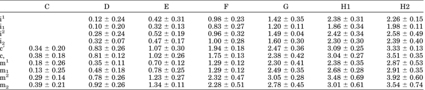
TABLE 2. Age of attainment of crown and root stages (mean⫾SD in years)
C D E F G H1 H2 i1 0.12⫾0.24 0.42⫾0.31 0.98⫾0.23 1.42⫾0.35 2.38⫾0.31 2.26⫾0.15 i1 0.10⫾0.20 0.32⫾0.13 0.83⫾0.27 1.20⫾0.11 1.86⫾0.34 1.98⫾0.11 i2 0.28⫾0.24 0.52⫾0.19 0.96⫾0.32 1.49⫾0.04 2.42⫾0.34 2.58⫾0.49 i2 0.32⫾0.07 0.47⫾0.17 1.00⫾0.28 1.60⫾0.30 2.30⫾0.30 2.39⫾0.40 c⬘ 0.34⫾0.20 0.83⫾0.26 1.07⫾0.30 1.94⫾0.18 2.47⫾0.36 3.09⫾0.25 3.33⫾0.13 c, 0.38⫾0.18 0.81⫾0.12 1.02⫾0.26 1.75⫾0.13 2.38⫾0.42 3.04⫾0.27 3.51⫾0.35 m1 0.18⫾0.26 0.35⫾0.11 0.70⫾0.12 1.29⫾0.12 2.30⫾0.41 2.38⫾0.35 2.87⫾0.53 m1 0.13⫾0.25 0.48⫾0.18 0.78⫾0.25 1.29⫾0.12 2.49⫾0.35 2.68⫾0.28 2.91⫾0.35 m2 0.29⫾0.14 0.78⫾0.26 1.23⫾0.27 2.32⫾0.47 3.05⫾0.28 3.48⫾0.69 3.92⫾0.60 m2 0.39⫾0.21 0.92⫾0.26 1.34⫾0.11 2.28⫾0.51 2.78⫾0.45 3.01⫾0.61 3.54⫾0.74
m1, c, and m2), with no clear pattern in sexual dimorphism (or between jaw differences). However, some population differences have been reported (re-viewed by Holman and Jones, 1998). Measurements of eruption prior to the clinical emergence of
decid-uous teeth have not been reported. In contrast, sev-eral aspects apart from the clinical emergence of permanent tooth eruption have been studied: timing of stages (Bengston, 1935; Garn et al., 1958; Haa-vikko, 1970; Demirjian and Levesque, 1980), rate of eruption and stage (Carlson, 1944; Shumaker and El Hadaray, 1960; Kuno, 1980; Feasby, 1981; Tsai, 2000), and root stage at clinical emergence Grøn
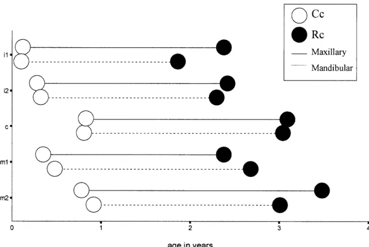
Fig. 4. Timing of root growth, from average crown complete stage to root complete stage. Solid lines, maxillary teeth; dotted lines, mandibular teeth; open circles, Cc, crown complete; solid circles, Rc, root complete.
TABLE 3. Predicting age from crown and root stages
D E F G H1 i1 0.27 0.70 1.20 1.90 i1 0.21 0.58 1.02 1.53 i2 0.40 0.74 1.22 2.00 i2 0.40 0.74 1.30 1.95 c⬘ 0.58 0.95 1.50 2.20 2.78 c, 0.60 0.92 1.38 2.06 2.71 m1 0.26 0.52 1.00 1.80 2.34 m1 0.30 0.63 1.04 1.89 2.58 m2 0.54 1.00 1.78 2.68 3.26 m2 0.65 1.13 1.81 2.53 2.90
TABLE 4. Distribution of tooth formation stages relative to deciduous first molar at stage D
Tooth Stage n B C D E F G H i1 26 21 47 i2 1 8 29 38 c 5 34 4 43 m2 6 38 3 47
TABLE 5. Distribution of tooth formation stages relative to deciduous first molar at stage F
Tooth Stage n B C D E F G H i1 12 17 14 43 i2 1 16 25 2 44 c 6 17 30 53 m2 4 39 7 50
TABLE 6. Distribution of tooth formation stages relative to deciduous second molar at stage D
Tooth Stage n B C D E F G H i1 1 4 24 13 42 i2 2 13 21 6 42 c 3 19 19 1 42 m1 6 14 11 1 32
(1962). A better understanding of patterns within and between teeth might be to view the entire tooth formation continuum as a whole rather than the traditional division of deciduous and permanent cat-egories (Schwartz and Langdon, 1991). What is clear is that deciduous teeth grow faster and are likely to
predict age more accurately than permanent teeth in early childhood. Results from the present study provide some idea of variation in crown, root, and eruption times of children from recent times, and fill several gaps in the literature. They are more com-plete than previously reported studies, providing data for maxillary teeth, and they use clearly de-scribed stages of formation and eruption. The simi-larity in development between the historical and living samples suggests little evidence of a secular trend, and highlights the value of research using juvenile individuals from skeletal collections.
CONCLUSIONS
New combined sex data on deciduous maxillary and mandibular tooth development include:
Age of attainment of crown and root growth (also adapted for prediction);
Distribution of stages relative to reference stages (m1 and m2stages D and F);
Age of attainment of eruption levels (also adapted for prediction); and
Distribution of root stages at each eruption level.
ACKNOWLEDGMENTS
We are grateful to Marie Watt and Dorothy Lunt and the trustees of the Whithorn Trust for allowing
Fig. 5. Timing of eruption stages, and mean ages of alveolar eruption, midway and occlusal levels. Solid lines, maxillary teeth; dotted lines, mandibular teeth; open bars, AE, alveolar bone level, cross Mid, midway; solid bars, Occ, occlusal level.
TABLE 7. Distribution of tooth formation stages relative to deciduous second molar at stage F
Tooth Stage n B C D E F G H i1 1 33 34 i2 11 18 29 c 1 19 18 38 m1 9 27 19 55
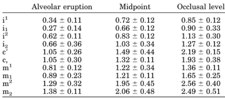
TABLE 8. Age of attainment for eruption levels (mean⫾SD in years)
Alveolar eruption Midpoint Occlusal level i1 0.34⫾0.11 0.72⫾0.12 0.85⫾0.12 i1 0.27⫾0.14 0.66⫾0.12 0.90⫾0.33 i2 0.62⫾0.11 0.83⫾0.12 1.13⫾0.30 i2 0.66⫾0.36 1.03⫾0.34 1.27⫾0.12 c⬘ 1.05⫾0.26 1.49⫾0.44 2.19⫾0.15 c, 1.05⫾0.30 1.32⫾0.11 1.93⫾0.38 m1 0.81⫾0.12 1.22⫾0.34 1.36⫾0.11 m1 0.89⫾0.23 1.21⫾0.11 1.65⫾0.25 m2 1.29⫾0.32 1.95⫾0.45 2.56⫾0.40 m2 1.38⫾0.11 2.06⫾0.48 2.49⫾0.51
us to examine material. We also thank Jennifer Thompson, Gail Krovitz, and Andrew Nelson for inviting us to take part in the symposium “Patterns of Growth and Development in the GenusHomo” at the American Association of Physical Anthropolo-gists Meeting in 2001.
LITERATURE CITED
Bengston RG. 1935. A study of the time of eruption and root development of the permanent teeth between six and thirteen years. Northwest Univ Bull 35:3–9.
Boller RJ. 1964. Fetal morphogenesis of the human dentition. J Dent Child 31:67–95.
Boyde A. 1964. The structure and development of mammalian enamel. Ph.D. thesis, University of London.
Brothwell D. 1977. On a mycoform stone structure in Orkney, and its relevance to possible further interpretation of so called souterrains. Bull Inst Archaeol 14:179 –198.
Carlson H. 1944. Studies on the rate and amount of eruption of certain human teeth. Am J Orthod Oral Surg 30:575–588. Dean MC. 1985. Variation in the developing root cone angle of the
permanent mandibular teeth of modern man and certain fossil hominids. Am J Phys Anthropol 68:233–238
Demirjian A, Levesque GY. 1980. Sexual differences in dental development and prediction of emergence. J Dent Res 59:1110 – 1122.
Demirjian A, Goldstein H, Tanner JM. 1973. A new system of dental age assessment. Hum Biol 45:211–227.
Eveleth PB, Tanner JM. 1990. Worldwide variation in human growth, 2nd ed. Cambridge: Cambridge University Press. Fanning EA. 1961. A longitudinal study of tooth formation and
root resorption. N Z Dent J 57:202–217.
Fanning EA, Brown T. 1971. Primary and permanent tooth de-velopment. Aust Dent J 16:41– 43.
Fass EN. 1969. A chronology of growth of the human dentition. J Dent Child 36:391– 401.
Feasby WH. 1981. A radiographic study of dental eruption. Am J Orthod 80:554 –560.
Finney DJ. 1952. Probit analysis. Cambridge: Cambridge Univer-sity Press.
FitzGerald C, Saunders SR, Macchiarelli R, Bondioli L. 1999. Large scale histological assessment of deciduous crown forma-tion. In: Mayhall JP, Heikkinen T, editors. Dental Morphology ’98. Oulu: Oulu University Press. p 92–101.
Garn SM, Lewis AB, Koski K, Polacheck DL. 1958. The sex difference in tooth calcification. J Dent Res 37:561–567. Gilster JE, Smith FH, Wallace GK. 1964. Calcification of
man-dibular second primary molars in relation to age. J Dent Child 31:284 –288.
Grøn A. 1962. Prediction of tooth emergence. J Dent Res 41:573– 585.
Haavikko K. 1970. The formation and the alveolar and clinical eruption of the permanent teeth. Proc Finn Dent Soc 66:103– 170.
Healy MJR. 1986. Statistics of growth standards. In: Falkner F, Tanner JM, editors. Human growth: a comprehensive treatise, volume 3. New York: Plenum Press. p 47–58.
Holman DJ, Jones RE. 1998. Longitudinal analysis of deciduous teeth eruption. II. Parametric survival analysis in Bangladeshi, Guatemalan, Japanese, and Javanese children. Am J Phys Anthopol 105:209 –230.
Hulland SA, Lucas JO, Wake MA, Hesketh KD. 2000. Eruption of the primary dentition in human infants: a prospective descrip-tive study. Pediatr Dent 22:415– 421.
Keene HJ. 1982. The morphogenetic triangle: a new conceptual tool for application to problems in dental morphogenesis. Am J Phys Anthropol 59:281–287.
Keene HJ. 1991. On heterochrony in heterodonty: a review of some problems in tooth morphogenesis and evolution. Yrbk Phys Anthropol 35:251–282.
Kronfeld R, Schour I. 1939. Neonatal dental hypoplasia. J Am Dent Assoc 26:18 –32.
Kuno T. 1980. On the eruptive process of the mandibular second molars—with particular reference to 45° oblique cephalometric analysis. J Nihon Univ Sch Dent 22:108 –114.
Leighton BC. 1977. Early recognition of normal occlusion. In: McNamara JA, editor. Contributions to the biology of occlusal development. Craniofacial growth series. Ann Arbor: Univer-sity of Michigan. p 147–167.
Liversidge HM, Molleson TI. 1999. Developing permanent tooth length as an estimate of age. J Forensic Sci 44:917–920. Liversidge HM, Dean MC, Molleson TI. 1993. Increasing human
tooth length between birth and 5.4 years. Am J Phys Anthropol 90:307–313.
Liversidge HM, Herdeg B, Ro¨sing FW. 1998. Dental age estima-tion of non-adults. In: Alt KW, Ro¨sing FW, Teschler-Nicola M, editors. Dental anthropology—fundamentals, limits and pros-pects. Vienna: Springer. p 419 – 442.
Molleson T, Cox M, Waldron AH, Whittaker DK. 1993. The Spi-talfields project. Volume 2: the anthropology: the middling sort. York: Council for British Archaeology.
Moorrees CFA, Fanning EA, Hunt EE. 1963. Formation and resorption of three deciduous teeth in children. Am J Phys Anthropol 21:205–213.
Nanda RS, Chawla TN. 1966. Growth and development of denti-tion in Indian children. I. Development of permanent teeth. Am J Orthod 52:837– 853.
Nystro¨m M. 1982. Development of deciduous dentition in a series of Finnish children. Proc Finn Dent Soc 78:1– 48.
TABLE 9. Prediction of age from eruption (in years) Alveolar level Midpoint Occlusal
i1 0.23 0.53 0.78 i1 0.18 0.46 0.78 i2 0.45 0.72 0.98 i2 0.49 0.84 1.15 c⬘ 0.94 1.27 1.84 c, 0.93 1.18 1.62 m1 0.58 1.02 1.29 m1 0.68 1.05 1.43 m2 1.04 1.62 2.25 m2 1.15 1.72 2.28
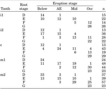
TABLE 10. Distribution of formation stage at each eruption level1 Tooth Root stage Eruption stage n Below AE Mid Occ
i1 D 14 1 15 E 10 12 10 22 F 5 12 14 G 26 26 i2 D 12 1 13 E 17 15 4 36 F 1 3 13 11 28 G 22 22 c D 12 1 13 E 4 24 11 4 43 F 6 13 19 G 29 29 m1 D 24 24 E 11 17 19 1 48 F 2 12 30 44 G 23 23 m2 D 33 3 1 37 E 13 15 10 1 39 F 3 29 25 57 G 23 23
1“Below” indicates cusp tip below alveolar bone level, AE at alveolar bone level, Mid midway between, and Occ at occlusal level.
Nystro¨m M, Kilpinen E, Kleemola-Kujala EA. 1977. A radio-graphic study of the formation of some teeth from 0.5 to 3.00 years of age. Proc Finn Dent Soc 73:167–172.
Scheuer L, Black S. 2000. Developmental juvenile osteology. Lon-don: Academic Press.
Schwartz JH, Langdon HL. 1991. Innervation of the human up-per primary dentition: implications for understanding tooth initiation and rethinking growth and eruption patterns. Am J Phys Anthropol 86:273–286.
Shellis RP. 1984. Variations in growth of the enamel crown in human teeth and a possible relationship between growth and enamel structure. Arch Oral Biol 29:697–705.
Shumaker DB, El Hadary MS. 1960. Roentgenographic study of eruption. J Am Dent Assoc 18:535–541.
Smith BH. 1991 Standards of human tooth formation and dental age assessment. In: Kelley M, Larsen CS, editors. Advances in dental anthropology. New York: Alan R. Liss. p 143–168.
Sunderland EP, Smith CJ, Sunderland R. 1987. A histological study of the chronology of initial mineralization in the human deciduous dentition. Arch Oral Biol 32:167–174.
Tsai H. 2000. Eruption process of the second molar. J Dent Child 67:275–281.
Watt ME, Lunt DA. 1999. Stages of tooth development relative to the first permanent molar in a Mediaeval population from the south west of Scotland. In: Mayhall MY, Heikkinen JT, editors. Dental morphology ’98. Oulu: Oulu University Press. p 120 – 127.