Original Article
Increased circulating macrophage-colony stimulating
factor and monocyte chemoattractant protein-1 are
predictors of in-hospital events in Chinese
patients with unstable angina pectoris
Xiao-Hong Kan1, Xue-Zhen Zhong2, Wen-Dong Zhang3, Cheng-Yao Shi3
1Key Laboratory of Cardiovascular Proteomics of Shandong Province, Department of Geriatrics, Qilu Hospital of
Shandong University, Jinan, China; 2Central Hospital of Jinan Affiliated to Shandong University, Jinan, Shandong,
China; 3Department of Pharmacy, Qilu Hospital Affiliated to Shandong University, Jinan, China
Received October 19, 2015; Accepted December 22, 2015;Epub February 1, 2016; Published February 15, 2016
Abstract: Background: Macrophage colony stimulating factor (MCSF) and monocyte chemoattractant protein-1 (MCP-1) play an important role in the activation of monocyte resulting in the onset and progression of atherosclero-sis. However, it remains unclear whether MCSF and MCP-1 have a predictive value to in-hospital events in Chinese patients with unstable angina pectoris. Methods: 110 subjects were divided into unstable angina pectoris (UAP) group (n = 60), stable angina pectoris (SAP) group (n = 30), and controls (n = 20). Blood samples were collected to measure the levels of MCSF and MCP-1 by ELISA. The severity of coronary stenosis was evaluated using coronary angiography and Gensini score method. The cardiac events of patients with UAP were recorded during the hospital. Results: The levels of MCSF and MCP-1 in UAP group were significantly higher than SAP and control group. MCSF and MCP-1 of patients with type II lesions were significantly higher than type І and type III, but MCSF and MCP-1 were not correlated with Gensini score. The incidence of cardiac events in UAP patients with high levels of MCSF and MCP-1 were significantly increased than that with non-high levels. The levels of MCSF and MCP-1 in UAP patients with car-diac events were significantly higher than that without carcar-diac events. Furthermore, multivariable logistic regression analysis revealed that MCSF and MCP-1 were all risk factors for in-hospital cardiac events. Conclusion: MCSF and MCP-1 are increased in unstable angina pectoris and may be biomarkers to evaluate the stability of plaque and prognosis of in-hospital cardiac events in Chinese patients with unstable angina pectoris.
Keywords: Macrophage colony stimulating factor, monocyte chemoattractant protein-1, unstable angina pectoris, short-term prognosis
Introduction
Increasing evidences suggest that inflam- mation plays a pivotal role in atherosclero- tic plaque instability and rupture resulting in acute coronary syndrome [1, 2]. Activated mo- nocytes/macrophages is implicated in this process through migrating to the fibrous cap of atherosclerotic plaque and producing proin-flammatory cytokines, metalloproteinases cy- tokines, chemokines and tissue factor, thus leads to a progression of plaque destabiliza- tion [3, 4]. The monocyte chemotactic protein (MCP-1) and monocyte colony-stimulating fac-tor (MCSF) are two important cytokines invol-
ved in monocytes activation and atherogene- sis [5-7].
events in Chinese patients with unstable an- gina.
Materials and methods
Patients
Sixty patients with UAP were recruited from the Department of Cardiology, Qilu Hospital, Shandong University from December 2012 to June 2014. All patients with UAP had anginal episodes at rest or angina during a mild degree of effort within the preceding 48 hours with no evidence of myocardial necrosis by enzymatic criteria. For comparison, 30 sex- and age-ma- tched patients were randomly selected as the SAP group who had typical effort angina or pos-itive treadmill exercise testing with no episode of angina at rest. 20 healthy subjects without a history of cardiovascular disease and having normal findings on physical examination, che- st roentgenography and electrocardiography echocardiography served as control group. The exclusion criteria in the study included a myo-cardial infarction within the previous month, the presence of any ECG abnormalities invali-dating ST-segment analyses, elevated serum levels of cardiac-related enzymes, thromboly- tic therapy, or body temperature >38.0°C. Fu- rthermore, all patients with inflammatory dis-eases (e.g., infections, autoimmune disdis-eases), malignancies, pulmonary disease, liver or kid-ney disease were also excluded. Informed
con-patient according to coronary angiography results as previously described [11, 12].
Laboratory measurements
A fasting morning blood sample was drawn from each patient at inclusion in the study. Aliquots of plasma were stored at -80°C and analysis was performed within a year. Plasma MCSF and MCP-1 were determined by enzyme-linked immunoassay (R&D system, USA) accor- ding to the manufacturer’s instructions. The intra-assay coefficients of variation were 5% for both tests.
In-hospital follow-up
All patients were given standard therapy acco- rding to physician preference. During intensive hospitalization, we noted any occurrence of in-hospital events, defined as cardiac death, non-fatal acute myocardial infarction, and recurre- nce of angina pectoris after conventional tre- atment. In-hospital events were evaluated by cardiologists who treated the patients without knowing the circulating MCSF and MCP-1 le- vels.
Statistics
[image:2.612.91.364.83.256.2]Statistical analysis was performed with SP- SS13.0 for Windows. Continuous data were pr- esented as mean ± SD and were compared by
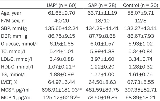
Table 1. Patient characteristics in three groups of subjects UAPa (n = 60) SAP (n = 28) Control (n = 20)
Age, year 61.65±9.70 63.71±11.19 58.07±9.71
F/M sex, n 40/20 18/10 12/8
SBP, mmHg 135.65±12.24 134.29±11.41 132.27±13.11 DBP, mmHg 86.75±9.15 87.79±8.68 86.67±7.93 Glucose, mmol/l 6.15±1.68 6.01±1.57 5.93±1.02 TC, mmol/l 5.44±1.01 5.99±1.88 5.34±0.84 LDL-C, mmol/l 3.49±0.88 3.97±1.60 3.34±0.74 HDL-C, mmol/l 1.07±0.21b,c 1.22±0.20 1.28±0.32
TG, mmol/l 1.88±0.99 1.77±1.00 1.61±0.75 LVEF, % 64.97±5.44 64.50±8.63 67.73±5.55 MCSF, pg/ml 698.91±181.93b,c 481.59±89.75 397.35±82.71
MCP-1, pg/ml 125.12±62.92b,c 78.50±19.89 68.89±18.21 aUAP, unstable angina pectoris; SAP, stable angina pectoris; SBP, systolic blood pressure; DBP, diastolic blood pressure; TC, total cholesterol; LDL-C, high density lipoprotein cholesterol; HDL-C, high density lipoprotein cholesterol; TG, triglyceride; LVEF, left ventricular ejection fraction; MCSF, macrophage colony stimulating factor; MCP-1, monocyte chemoattractant protein-1. bP<0.05 com-pared with control. cP<0.05 compared with SAP.
sent was obtained from all sub-jects based on a protocol ap- proved by the Ethics Committee of Qilu Hospital, Shandong Uni- versity.
Coronary angiography
means of one-way analysis of variance with Scheffe posteriori comparisons and student’s t test as appropriate. Categorical data were pre-sented as proportion and Chi square test was used for comparison. Multivariat analysis with multiple logistic regression method was used to further analyze covariables associated with in-hospital events. P<0.05 was considered sta-tistically significant.
Results
Patient characteristics
The three groups were well matched in terms of age and gender. Compared with SAP and con-trols, high-density lipoprotein-cholesterol (HDL-C) was lower in UAP groups, with no difference between SAP groups and controls (Table 1). With respect to other cardiovascular risk fac-tors, there was no difference between UAP and SAP groups.
Comparison of MCSF and MCP-1 and in-hospi-tal events
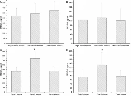
The levels of MCSF and MCP-1 in UAP group were significantly higher than SAP group and control group (P<0.05, Table 1). MCSF and MCP-1 of patients with type II lesions were sig-nificantly higher than that with type I and type III (P<0.05 or P<0.01, Figure 1), but the levels of MCSF and MCP-1 were not different among the patients with single vessel disease, two vessel disease, multiple vessel disease (P> 0.05, Figure 1). The levels of MCSF and MCP-1 were not correlated with Gensini score (P>0.05). Linear correlation analysis revealed that MCP-1 was positively associated with age, TC and GLu (r = 0.330, P<0.01; r = 0.406, P<0.001; r = 0.434, P<0.001), but MCSF was not associat-ed with risk factors of CHD (P>0.05). And MCP-1 was positively correlated with MCSF (r = 0.630,
[image:3.612.93.522.73.386.2]P<0.01).
We defined the 95% credibility interval of the MCSF (228.04-543.36 pg/ml) and MCP-1 (29.73-96.65 pg/ml) concentrations in control group as the normal reference range. The levels of MCSF and MCP-1 were considered high beyond the upper limit of normal reference range. The incidence of cardiac events in UAP patients with high levels of MCSF and MCP-1 were significantly higher than the non-high level ones (36.84% vs. 9.09%, 36.11% vs. 12.50%, respectively; all P<0.05). According to the in-hospital cardiac events, UAP patients were divided into the groups with cardiac events and without cardiac events. The levels of MCSF and MCP-1 in the former group were significantly higher than the latter (P<0.05 or P<0.01, Figure 2). Multivariable logistic regression analysis revealed that MCSF and MCP-1 were all risk factors for cardiac events (Partial regression coefficient were 1.021 and 0.674 respectively; OR were 2.776 and 1.962 respectively; P = 0.004 and 0.045 respectively; Table 2).
patients with type II lesions were significantly higher than that with type I and type III. These data suggest that high levels of MCSF and MCP-1 may reflect the unstable plaque leading to acute coronary syndrome.
[image:4.612.90.343.73.256.2]Previous studies also found that increased MCSF beyond the acute phase were strongly predictive of long term outcome in patients wi- th severe unstable angina [8] and circulating MCP-1 played an important role in the patho-genesis development of ACS [9]. However it is unclear whether circulating MCP-1 and MCSF have a predictive value to in-hospital events in Chinese patients with unstable angina. Here, we found that MCP-1 was positively associated with age, TC and GLu, but MCSF was not asso-ciated with risk factors of CHD. According to the in-hospital cardiac events, UAP patients were divided into the groups with cardiac events and without cardiac events. The levels of MCSF and MCP-1 in the former group were significan- tly higher than the latter. Multivariable logistic Figure 2. Comparison of MCSF and MCP-1 concentrations between
patients with and without cardiac events in UAP group. The levels of MCSF and MCP-1 in UAP patients with cardiac events were signifi-cantly higher than that without cardiac events. *P<0.01 compared with patients without cardiac events.
Table 2. Logistic regression analysis of cardiac events in patients with unstable angina pectoris
Partial regression
coefficient regression coefficientStandardized partial P OR
MCSF 1.021 185.751 0.004 2.776
MCP-1 0.674 42.408 0.045 1.962
MCSF, Macrophage colony stimulating factor; MCP-1, monocyte chemoat-tractant protein-1; OR, odds ratio.
Discussion
The present study demonstrated that circulating MCSF and MCP-1 were increased in Chinese patients with unstable angina pectoris. Increased MCSF and MCP-1 were correlated with atherosclerotic plaque type and were all risk factors for in-hospital cardiac events.
[image:4.612.90.343.362.413.2]regression analysis revealed that MCSF and MCP-1 were all risk factors for in-hospital car-diac events. The mechanism may be that MCSF causes monocyte/macrophage activation and MCP-1 serves as a chemoattractant for mono-cytes, resulting in monocyte migrating to fi- brous cap of an atherosclerotic plaque, produc-ing metalloproteinases and causproduc-ing plaque destabilization[3, 4].
The major limitation of the study is the relative-ly small size of the sample. Larger and long-term studies are necessary to investigate wh- ether the prognostic value of MCSF and MCP-1 is independent of or additive to other inflamma-tory marker.
Conclusion
MCSF and MCP-1 are increased in unstable angina pectoris and may be biomarkers to eval-uate the stability of plaque and prognosis of in-hospital cardiac events in Chinese patients with unstable angina pectoris.
Acknowledgements
This study was supported by the Science and Technology Research Program of Shandong Province (2009GG10002034 and 2014GSF- 118143), Chinese medicine science and tech-nology Research Program of Shandong Prov- ince (2011-206), and the National Natural Sc- ience Foundation of China (30800468 and 81470558).
Disclosure of conflict of interest
None.
Address correspondence to: Dr. Wen-Dong Zhang, Department of Pharmacy, Qilu Hospital Affiliated toShandong University, 107 West Wenhua Road, Ji- nan 250012, China. Tel: +086-15053177788; Fax: +086-531-86927544; E-mail: zhangwendong_ql@ 163.com
References
[1] Naghavi M, Libby P, Falk E, Casscells SW, Litovsky S, Rumberger J, Badimon JJ, Stefana-dis C, Moreno P, Pasterkamp G, Fayad Z, Stone PH, Waxman S, Raggi P, Madjid M, Zarrabi A, Burke A, Yuan C, Fitzgerald PJ, Siscovick DS, de Korte CL, Aikawa M, Juhani Airaksinen KE, Ass-mann G, Becker CR, Chesebro JH, Farb A, Galis
ZS, Jackson C, Jang IK, Koenig W, Lodder RA, March K, Demirovic J, Navab M, Priori SG, Rekhter MD, Bahr R, Grundy SM, Mehran R, Colombo A, Boerwinkle E, Ballantyne C, Insull W Jr, Schwartz RS, Vogel R, Serruys PW, Hans-son GK, Faxon DP, Kaul S, Drexler H, Green-land P, Muller JE, Virmani R, Ridker PM, Zipes DP, Shah PK, Willerson JT. From vulnerable plaque to vulnerable patient: a call for new definitions and risk assessment strategies: Part I. Circulation 2003; 108: 1664-1672. [2] Libby P, Ridker PM, Maseri A. Inflammation
and atherosclerosis. Circulation 2002; 105: 1135-1143.
[3] Shantsila E, Lip GY. Monocytes in acute coro-nary syndromes. Arterioscler Thromb Vasc Biol 2009; 29: 1433-1438.
[4] Uzui H, Harpf A, Liu M, Doherty TM, Shukla A, Chai NN, Tripathi PV, Jovinge S, Wilkin DJ, Aso-tra K, Shah PK, Rajavashisth TB. Increased expression of membrane type 3-matrix metal-loproteinase in human atherosclerotic plaque: role of activated macrophages and inflamma-tory cytokines. Circulation 2002; 106: 3024-3030.
[5] Shyy YJ, Wickham LL, Hagan JP, Hsieh HJ, Hu YL, Telian SH, Valente AJ, Sung KL, Chien S. Human monocyte colony-stimulating factor stimulates the gene expression of monocyte chemotactic protein-1 and increases the adhe-sion of monocytes to endothelial monolayers. J Clin Invest 1993; 92: 1745-1751.
[6] Lin J, Kakkar V, Lu X. Impact of MCP-1 in ath-erosclerosis. Curr Pharm Des 2014; 20: 4580-4588.
[7] Saitoh T, Kishida H, Tsukada Y, Fukuma Y, Sano J, Yasutake M, Fukuma N, Kusama Y, Hayakawa H. Clinical significance of increased plasma concentration of macrophage colony-stimulating factor in patients with angina pec-toris. J Am Coll Cardiol 2000; 35: 655-665. [8] Rallidis LS, Zolindaki MG, Pentzeridis PC,
Pou-lopoulos KP, Velissaridou AH, Apostolou TS. Raised concentrations of macrophage colony stimulating factor in severe unstable angina beyond the acute phase are strongly predictive of long term outcome. Heart 2004; 90: 25-29. [9] Gonzalez-Quesada C, Frangogiannis NG. Mo-
nocyte chemoattractant protein-1/ CCL2 as a biomarker in acute coronary syndromes. Curr Atheroscler Rep 2009; 11: 131-138.
[10] Ambrose JA, Winters SL, Stern A, Eng A, Teich-holz LE, Gorlin R, Fuster V. Angiographic mor-phology and the pathogenesis of unstable angina pectoris. J Am Coll Cardiol 1985; 5: 609-616.
[12] Neeland IJ, Patel RS, Eshtehardi P, Dhawan S, McDaniel MC, Rab ST, Vaccarino V, Zafari AM, Samady H, Quyyumi AA. Coronary angiographic scoring systems: an evaluation of their equiva-lence and validity. Am Heart J 2012; 15: 547-552.
[13] Libby P. Inflammation in atherosclerosis. Arte-rioscler Thromb Vasc Biol 2012; 32: 2045-2051.
[14] Drechsler M, Duchene J, Soehnlein O. Chemo-kines control mobilization, recruitment, and fate of monocytes in atherosclerosis. Arterio-scler Thromb Vasc Biol 2015; 35: 1050-1055. [15] Gerszten RE, Garcia-Zepeda EA, Lim YC, Yoshi-da M, Ding HA, Gimbrone MA Jr, Luster AD, Lus-cinskas FW, Rosenzweig A. MCP-1 and IL-8 trigger firm adhesion of monocytes to vascular endothelium under flow conditions. Nature 1999; 398: 718-723.
[16] Döring A, Sloka S, Lau L, Mishra M, van Min-nen J, Zhang X, Kinniburgh D, Rivest S, Yong VW. Stimulation of monocytes, macrophages, and microglia by amphotericin B and macro-phage colony-stimulating factor promotes re-myelination. J Neurosci 2015; 35: 1136-1148.
[17] Clinton SK, Underwood R, Hayes L, Sherman ML, Kufe DW, Libby P. Macrophage colony-stimulating factor gene expression in vascular cells and in experimental and human athero-sclerosis. Am J Pathol 1992; 140: 301-316. [18] Niu J, Kolattukudy PE. Role of MCP-1 in
cardio-vascular disease: molecular mechanisms and clinical implications. Clin Sci (Lond) 2009; 117: 95-109.
[19] Hojo Y, Ikeda U, Takahashi M, Shimada K. In-creased levels of monocyte-related cytokines in patients with unstable angina. Atherosclero-sis 2002; 161: 403-408.