Original Article
AKT2 expression changes impact on invasive
ability of breast cancer cells
Xiaolang Tan1, Lei Guo2, Shayang Luo2, Weiming Wang1, Kejing Zhang2, Feiyu Chen2
1Department of Oncology, Changsha Central Hospital, Changsha, PR China; 2Department of Breast Surgery,
Xiangya Hospital, Central South University, Changsha, PR China
Received November 24, 2015; Accepted January 23, 2016; Epub April 1, 2016; Published April 15, 2016
Abstract: AKT2 plays an important role in PI3K signaling pathway and directly relates to cell growth. Study showed that AKT2 was associated with tumor occurrence, growth, and invasion. This research aimed to detect AKT2 expres-sion impact on breast cancer cell invasive ability, to provide support for clinical prognosis. AKE2 overexpresexpres-sion vector was constructed using pEGFP-N1 as the carrier and transfected to breast cancer cell line by liposome. RT-PCR and Western blot were applied to test AKT2 expression at 12 h, 16 h, and 24 h after transfection, respectively. Transwell assay was performed to determine cell invasion. AKT2 overexpression vector pEGFP-AKT2 and inhibitory vector AKT2-sh-RNA were successfully constructed. After pEGFP-AKT2 transfection, AKT2 expression increased
sig-nificantly, and cell invasive ability also enhanced. AKT2-sh-RNA transfection declined AKT2 level and reduced breast
cancer cell invasive ability. AKT2 expression changes had certain relationship with breast cancer cell invasive ability. AKT2 overexpression may affect breast cancer prognosis.
Keywords: AKT2, breast cancer cell, transwell assay, cell transfection
Introduction
Breast cancer is a common malignant tumor with the highest incidence among female tumors. There are more than 1 million new cases every year around the world. Our country is also one of the high incidence areas [1, 2]. For breast cancer has various complicated pathogenesis, including genetic factor, environ-mental factor, psychological factor, and work stress, etc. its occurrence presents younger trend that brought great burden to society pro-ductivity and family. At present, searching for effective treatment and prediction method of breast cancer is the hotspot [3].
Following the development of biological tech-nology, especially the progress of human genomics, people awareness of the disease gradually transferred to genetic level. A variety of genes closely related to disease have been discovered and their mechanism has been elu-cidated. Meanwhile, targeted therapy also achieved certain progress in clinical treatment [4, 5]. Research suggested that every disease had various genes expression changes. Thus,
study the relationship between gene expres-sion and the disease is one of the methods of precision medicine [6].
AKT2 expression at 12 h, 16 h, and 24 h after transfection, respectively. Transwell assay was performed to determine cell invasion.
Materials and methods
General information
Breast cancer cell line MCF-7 was purchased from Shanghai Huayan Bio-Technology co., LTD. RPMI 1640, trypsin, fetal calf serum, and PBS were got from Gibco. Flask and dish were bought from Corning. Penicillin-streptomycin and Western blot related reagents were from Beyotime. Transwell chambers and Matrigel were from BD. Liposome transfection kit was from Invitrogen. AKT2 antibody was produced by Xinlebio. RNA extraction kit (Trizol) and cDNA synthesis kit were from TAKARA. PCR amplifica-tion reagents were from Promega. Primers used in the experiment were from Invitrogen. Gene sequencing was performed by BGI. Restriction enzyme and T4DNA ligase were pur-chased from NEB. Plasmid extraction kit was from Omega.
Vector construction
AKT2 sh-RNA vector was designed and verified by Chongqing Western CO., LTD. AKT2 primer
and restriction enzyme cutting site were designed based on CDS sequence published by NCBI and pEGFP-N1 carrier frame sequence. XhoI was added to the upstream, while BamH I enzyme loci was added to downstream. The primers used were as follows: upstream (5’-3’): atgaagaccgagaggccgcgac, downstream (5’-3’): gctcgcggatgctggccgagtaggag. The prod-uct length was 1260 bp. PCR amplification con-dition contained 94°C for 3 min, followed by 30 cycles of 94°C for 35 s, 58.7°C for 35 s, and 68°C for 1.5 min, and 68°C for 10 min at last. Agarose gel electrophoresis was performed to recycle the target band. The band together with pEGFP-N1 carrier was double enzyme digested overnight by XhoI and BamH I, and then the enzyme-digested products at 1260 bp and 4700 bp were collected. Next, the products were connected by T4 DNA ligase at 4°C over-night. The connection system included: 1 μl enzyme, 1 μl 10×Buffer, 2 μl carrier recycled product, 4 μl AKT2 PCR recycled product, and 2 μl ddH2O. Then it was converted to Dh5α com-petent cells in medium containing kanamycin overnight. Monoclonal bacteria was picked to perform PCR verification and sequenced to exclude mutation.
Cell culture and transfection
MCF-7 in store was water bathed at 37°C for 5 min and centrifuged at 1000 rpm/min for 3 min. Then the cells were cultured in RPMI 1640 medium containing 10% FBS and passaged after 24 h. Liposome transfection was per-formed at 16 h after cell adherence according to the kit introduction. Each group had three repeats.
RNA and protein extraction and detection
Total RNA and protein were extracted after cell transfection
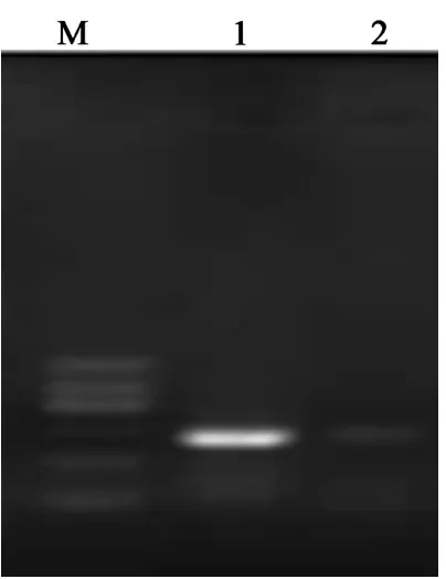
[image:2.629.98.300.76.338.2]RNA extraction: The cells were washed by pre-cooled PBS for 2-3 times and treated by 1 ml Trizol for 5 min. Then the cells were moved to an EP tube and added with 0.2 ml chloroform. After centrifuged at 15000 g and 4°C for 15 min, the upper layer was moved to a new EP tube and added with equal amount of isopropa-nol for 10 min. After centrifuged at 15000 g and 4°C for 15 min, the supernatant was removed and the RNA was treated washed by 75% ethanol twice. After centrifuged at 15000 Figure 1. M, DNA marker; 1 and 2, AKT2 CDS PCR
Table 1. RT-PCR detection gray level
Group Normal Co-inhibition pEGFP-AKT2
AKT-2 890 332 2664
β-actin 324 348 340
g and 4°C for 5 min, the RNA was solved in 20 μl DEPC water after dried for 10 min. RNA con-centration was measured, and RNA was stored at -80°C.
Protein extraction: The cells were washed by PBS after transfection and added with 50 μl RIPA. Then the cells were collected to an EP tube and vortexed for 3-4 times. After
centri-supernatant was moved to a new EP tube. The protein was quantified by BCA and stored at -80°C.
1 μg RNA was used for RT-PCR, while 40 μg protein was used for Western blot detection. β-actin was chose as internal reference.
Transwell assay
The upper chamber of Transwell chamber (bore diameter 8 μm) was coated by Matrigel (BD). 70 μl Matrigel was added to the chamber and incu-bated at 37°C for 60 min. Cell suspension in FBS-free medium containing BSA was added to the upper chamber at 1×10⁵/ml, whereas 1000 Figure 2. Cell culture and transfection (10×). A. Normal MCF-7; B. 12 h after pEGFP-AKT2 transfection; C. 12 h after
[image:3.629.333.531.253.339.2]AKT2-sh-RNA and pEGFP-N1 co-transfection.
Figure 3. PCR detection of AKT-2 after transfection. 1, normal AKT2; 2, AKT2 expression after pEGFP-AKT2 transfection; 3, pEGFP-AKT2 expression after pEGFP- AKT2-sh-RNA and pEGFP-N1 co-transfection.
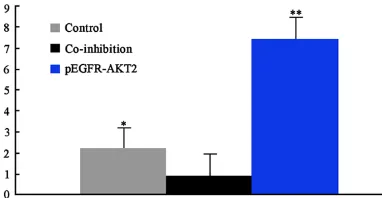
[image:3.629.98.299.262.405.2]Figure 4. RT-PCR gray level analysis. *P<0.05, **P<0.01, compared with co-inhibition group.
Figure 5. Western blot detection of AKT2 expression in transfected cells.
[image:3.629.334.529.391.495.2] [image:3.629.99.290.478.577.2]chamber. After transfection, the chamber was stained by 0.1% crystal violet for 15 min and photographed under inverted microscope. Three different fields of view were randomly selected under 100× light microscope to calcu-late the cell number.
Results
Vector construction
AKT2 CDS sequence was successfully obtained by PCR amplification. Agarose gel electrophore-sis results were showed in Figure 1. After dou-ble enzyme digestion and connected to pEGFP-N1 carrier, the PCR fragment was sequenced by BGI and compared in NCBI blast. The results showed that two fragments sequence appeared mutation, which was in accordance with NCBI published sequence. The plasmid was stored at -80°C.
Cell culture and transfection
MCF-7 cells were unfrezen and cultured in incu-bator. As shown in Figure 2, the cells appeared adherence phenomenon after 4-5 h. 5 μg pEG-FP-AKT2 plasmid was transfected to MCF-7 cells, while AKT2-sh-RNA and pEGFP-N1 vector were co-transfected to MCF-7 cells after mix-ing. Green fluorescence confirmed transfection success (Figure 2).
RT-PCR detection of AKT2 expression
Total RNA was extracted from cells and reverse transcripted to cDNA for PCR detection. Image J software was used for data analysis and
fected cells. As shown in Figures 5 and 6, AKT2 protein expression was similar to RT-PCR re- sults. AKT2 expression significantly increased to 3.36 times after pEGFP-AKT2 transfection compared with normal control (P<0.01), while it reduced to 0.42 time after AKT2-sh-RNA and pEGFP-N1 co-transfection (P<0.05). It indicat-ed that vector plays a proper role after trans-fection to regulate AKT2 expression.
Transwell Boyden assay detection of cell inva-sive ability
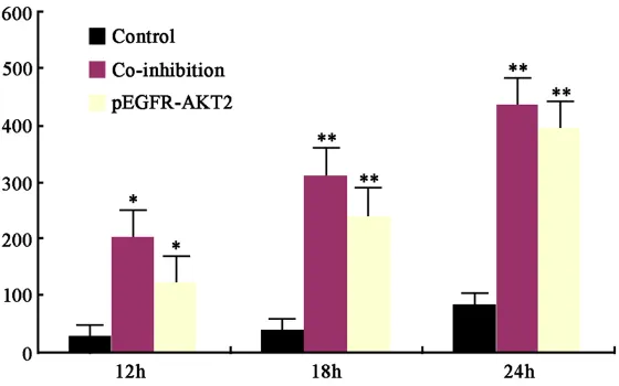
The chamber was incubated in 37°C and 5% CO2 for 12, 18, or 24 h, respectively. Then the chamber was stained by 0.1% crystal violet for 15 min and photographed under inverted microscope. Three fields of view were randomly selected to count the transmembraned cell number. pEGFR-AKT2 group presented signifi-cant difference with normal control and co-inhi-bition group (Figure 7).
[image:4.629.102.382.82.257.2]In conclusion, tumor makes the body gradually losing its normal regulation function especially in gene level, leading to local cell abnormal pro-liferation or differentiation. It forms neoplasm under in vivo and in vitro factors [12-14]. Once formed, neoplasm will not stop growth because of carcinogenic factors elimination. It is fea-tured as rapid growth and out of control, further destroying adjacent normal tissues and organs, namely the cancer cells invasion [15, 16]. Breast cancer also complies with the above laws. Abnormal gene regulation may result in Figure 7. Comparison of transmembraned cell numbers. *P<0.05, **P<0.01,
compared with co-inhibition group.
β-actin was treated as in- ternal reference. As shown in Table 1; Figures 3 and
4, AKT2 expression signifi-cantly increased to 2.78 times after pEGFP-AKT2 transfec-tion compared with normal control, while it reduced to 0.36 time after AKT2-sh-RNA and pEGFP-N1 co-transfec-tion. It suggested that vector plays proper role after trans- fection.
Western blot detection of AKT2 expression
trans-cancer cell proliferation and metastasis that lead to serious consequences [17, 18]. Therefore, study invasion related gene expres-sion and clarify its relationship with cancer cell invasion has great significance [19, 20]. Our study selected AKT2 gene that has been con-firmed as cell survival promoting factor in the body. It plays an important role in cell apopto-sis, proliferation, differentiation, physiological metabolism, senescence, and cancer. It also has certain impact on oncogenesis, invasion, and metastasis. As the main subtype of protein kinase B, AKT2 is validated to relate to tumori-genesis. Thus, explore the relationship between AKT2 expression and breast cancer cell inva-sive ability, and further clarify the association between AKT2 and tumorigenesis can provide new target for breast cancer gene therapy. Our results revealed that AKT2 expression was suc-cessfully controlled in MCF-7 cells in vitro. Further investigation showed that AKT2 overex-pression can improve MCF-7 invasive ability, while its level inhibition may weaken MCF-7 invasion. It suggested that AKT2 has a role in regulating MCF-7 cell invasion.
Acknowledgements
Key research and development program of Hunan province (Basic Research), 2015JC3016.
Disclosure of conflict of interest
None.
Address correspondence to: Dr. Lei Guo, Depart- ment of Breast Surgery, Xiangya Hospital, Central South University, 87 Xiangya Road, Changsha 410- 008, PR China. Tel: +86-731-89753015; E-mail: lei- [email protected]
References
[1] Berardi DE, Flumian C, Rodriguez CE, Diaz Bessone MI, Cirigliano SM, Bal de Kier Joffe ED, Fiszman GL, Urtreger AJ, Todaro LB. PKCdelta Inhibition Impairs Mammary Cancer Proliferative Capacity But Selects Cancer Stem Cells, Involving Autophagy. J Cell Biochem 2016; 117: 730-40.
[2] Daswani VP, Ayesa U, Venegas B, Chong PL. Concentration-induced j-aggregate formation causes a biphasic change in the release of trans-combretastatin a4 disodium phosphate from archaeosomes and the subsequent cyto-toxicity on mammary cancer cells. Mol Pharm 2015; 12: 3724-3734.
[3] Gruppi F, Hejazi L, Christov PP, Krishnamachari S, Turesky RJ, Rizzo CJ. Characterization of ni-trogen mustard formamidopyrimidine adduct formation of bis(2-chloroethyl)ethylamine with calf thymus DNA and a human mammary can-cer cell Line. Chem Res Toxicol 2015; 28: 1850-1860.
[4] Song H, Jung JI, Cho HJ, Her S, Kwon SH, Yu R, Kang YH, Lee KW, Park JH. Inhibition of tumor progression by oral piceatannol in mouse 4T1 mammary cancer is associated with decreased
angiogenesis and macrophage infiltration. J
Nutr Biochem 2015; 26: 1368-78.
[5] Accornero P, Miretti S, Bersani F, Quaglino E, Martignani E, Baratta M. Met receptor acts uniquely for survival and morphogenesis of EGFR-dependent normal mammary epithelial and cancer cells. PLoS One 2012; 7: e44982. [6] Arumugam A, Parada J, Rajkumar L. Mammary
cancer promotion by ovarian hormones in-volves IGFR/AKT/mTOR signaling. Steroids 2012; 77: 791-797.
[7] Ayoub NM, Akl MR, Sylvester PW. Combined gamma-tocotrienol and Met inhibitor treat-ment suppresses mammary cancer cell prolif-eration, epithelial-to-mesenchymal transition and migration. Cell Prolif 2013; 46: 538-553. [8] Cotrim CZ, Fabris V, Doria ML, Lindberg K,
Gustafsson JA, Amado F, Lanari C, Helguero LA. Estrogen receptor beta growth-inhibitory effects are repressed through activation of MAPK and PI3K signalling in mammary epithe-lial and breast cancer cells. Oncogene 2013; 32: 2390-2402.
[9] Fan H, Guan JL. Compensatory function of Pyk2 protein in the promotion of focal adhe-sion kinase (FAK)-null mammary cancer stem cell tumorigenicity and metastatic activity. J Biol Chem 2011; 286: 18573-18582.
[10] Feigin ME, Akshinthala SD, Araki K, Rosenberg AZ, Muthuswamy LB, Martin B, Lehmann BD, Berman HK, Pietenpol JA, Cardiff RD, Muthuswamy SK. Mislocalization of the cell polarity protein scribble promotes mammary tumorigenesis and is associated with basal breast cancer. Cancer Res 2014; 74: 3180-3194.
[11] Furth PA, Nakles RE, Millman S, Diaz-Cruz ES, Cabrera MC. Signal transducer and activator of transcription 5 as a key signaling pathway in normal mammary gland developmental biolo-gy and breast cancer. Breast Cancer Res 2011; 13: 220.
[13] Lamartiniere CA, Jenkins S, Betancourt AM, Wang J, Russo J. Exposure to the endocrine disruptor bisphenol A alters susceptibility for mammary cancer. Horm Mol Biol Clin Investig 2011; 5: 45-52.
[14] Li J, Cho YY, Langfald A, Carper A, Lubet RA, Grubbs CJ, Ericson ME, Bode AM. Lapatinib, a preventive/therapeutic agent against mam-mary cancer, suppresses RTK-mediated sig-naling through multiple sigsig-naling pathways. Cancer Prev Res (Phila) 2011; 4: 1190-1197. [15] Lyu H, Huang J, Edgerton SM, Thor AD, He Z,
Liu B. Increased erbB3 promotes erbB2/neu-driven mammary tumor proliferation and co-targeting of erbB2/erbB3 receptors exhibits potent inhibitory effects on breast cancer cells. Int J Clin Exp Pathol 2015; 8: 6143-6156. [16] Miwa HE, Koba WR, Fine EJ, Giricz O, Kenny PA,
Stanley P. Bisected, complex N-glycans and ga-lectins in mouse mammary tumor progression and human breast cancer. Glycobiology 2013; 23: 1477-1490.
[17] Nakles RE, Kallakury BV, Furth PA. The PPARgamma agonist efatutazone increases the spectrum of well-differentiated mammary cancer subtypes initiated by loss of full-length
BRCA1 in association with TP53 haploinsuffi -ciency. Am J Pathol 2013; 182: 1976-1985.
[18] Shibata MA, Iinuma M, Morimoto J, Kurose H, Akamatsu K, Okuno Y, Akao Y, Otsuki Y. alpha-mangostin extracted from the pericarp of the mangosteen (garcinia mangostana linn) reduc-es tumor growth and lymph node metastasis in an immunocompetent xenograft model of met-astatic mammary cancer carrying a p53 muta-tion. BMC Med 2011; 9: 69.
[19] Thompson MD, Mensack MM, Jiang W, Zhu Z, Lewis MR, McGinley JN, Brick MA, Thompson HJ. Cell signaling pathways associated with a reduction in mammary cancer burden by di-etary common bean (phaseolus vulgaris L.). Carcinogenesis 2012; 33: 226-232.
[20] Tiwari RV, Parajuli P, Sylvester PW.