Original Article
Evaluation of peripheral lymphadenopathy with
excisional biopsy: six-year experience
Esra Akyüz Özkan1, Ceren Canbey Göret2, Zeynep Tuba Özdemir3, Serdar Yanık4, Nuri Emrah Göret5, Meryem
Doğan6, Fatma Gökşin Cihan7, Ayşe Neslin Akkoca8
1Department of Pediatrics, Faculty of Medical, Bozok University, Yozgat, Turkey; 2Department of Pathology,
Haydarpasa Numune Training and Research Hospital, Istanbul; 3Department of Internal Medicine, Faculty of
Medical, Bozok UniversityYozgat, Turkey; 4Department of Pathology, Iskenderun State Hospital, Hatay, Turkey; 5Department of General Surgery, Kartal Dr Lütfü Kırdar Training and Research Hospital, Istanbul, Turkey; 6Department of Pathology, Faculty of Medical, Atatürk University, Erzurum, Turkey; 7Department of Family
Medicine, Faculty of Medical, Necmettin Erbakan University, Konya, Turkey; 8Department of Family Medicine,
Iskenderun State Hospital, Hatay, Turkey
Received September 18, 2015; Accepted October 24, 2015; Epub November 1, 2015; Published November 15, 2015
Abstract: Background: Lymphadenopathy (LAP) may be the first symptom of many diseases. We aimed to inves
-tigate peripheral LAP results taken by excisional biopsy. Methods: Histopathological examination of 185 lymph
node biopsy specimens were evaluated between January 2009 and April 2015 in Istanbul Private Ekin Pathology
Laboratory, retrospectively. Results: The average age of patients ranged from 1 to 86 was 41.01 ± 20.62 years. 87 of were female, 98 of male. 62 (33.5%) of excisional biopsy materials were benign lesions and 123 (66.5%) of ma
-lignant. Benign lesions were consisted of reactive hyperplasia, cat-scratch disease, toxoplasmosis, necrotizing/non-necrotizing granulomatous. Of these patients, 40 had nodes with reactive hyperplasia (15 female/25 male, mean age: 27.35 y), 14 had necrotizing granulomatous disease (9 female/5 male, mean age: 39.86 y), 2 non-necrotizing granulomatous disease (1 female/1 male mean age: 43 y), 4 had cat-scratch disease (1 female/3 male mean age: 54.25 y), 1 toxoplasmosis (26 y, female), 1 Kikuchi disease (25 y, female). In the evaluation of malign lesions; 38 were Hodgkin lymphoma (HL) (20.5%, 17 female/21 male, mean age: 34.89 y) 77 had non-Hodgkin lymphoma (NHL) (41.6%, 37 female/40 male, mean age: 52.26 y), 8 metastasis (4.3%, 5 female/3 male, mean age: 53.5 y).
Reactive LAP observed most common in cervical region, NHL in axillary-abdomen-inguinal-mediastinum and HL in
the supraclavicular region. Conclusion: Excisional biopsy can be applied safely with minimal morbidity and mortal
-ity and a gold standard diagnostic method for LAP. Although LAP is mostly related with benign lesions, malignancy should be kept in mind in differential diagnosis.
Keywords: Benign, excisional biopsy, lymphadenopathy, malignant
Introduction
Lymphadenopathy (LAP) describes the condi-tions in which lymph nodes become abnormal
in size, consistency, and it may be one of the symptoms of many diseases. The history of the patient should be considered carefully because
it may provide clues to the underlying disease.
Usually points self-limited infection in younger
adults although a malignancy in elderly [1].
No further investigation is required in most
cases, as the cause is obvious on the patient’s
first evaluation (such as infection). The cause of
peripheral LAP often cannot be ascertained on
clinical grounds alone. Laboratory tests, imag-ing studies, and tissue biopsy are recommend-ed when there are unexplainrecommend-ed conditions. The
size and distribution of the node can be identi
-fied more accurately with imaging than physical
examination. Ultrasound is a non invasive
method to assess lymph nodes in superficial
regions like the neck [2]. Ultrasound can assess the number, size, site, shape, margins, and
pat-tern of vascularity and the inpat-ternal structure of
a lymph node. Computed tomography (CT) is
helpful for LAP in the thorax or abdominopelvic
the first step in the diagnostic approach to lym
-phomas, excisional biopsy of enlarged lymph
nodes is still gold standard procedure. Excision
biopsy of the most accessible peripheral lymph node is with the possibility of early diagnosis and is a vital part of the management [5].
In general, if diameter of the lymph nodes
exceeds one cm or more it is considered to be
abnormal. Of course, there are exceptions in different regions and lymph nodes have differ
-ent sizes at differ-ent ages. The cervical region
is most commonly involved area among periph-eral LAP at any age. Genperiph-eralized LAP is usually
an indicative of an underlying disease such as
the Epstein-Barr virus, HIV, lymphoma, and autoimmune disorders [1]. It is considered that palpable supraclavicular, iliac, popliteal, epi-trochlear nodes greater than 0.5 cm, and ingui-nal nodes larger than 1.5 cm are abnormal [6]. 40 years and over, LAP in multiple region, supraclavicular lymph nodes, nodal diameter
greater than 2 cm, firm or hard texture, fixed nodes, lack of tenderness, and abnormal chest X-ray is some factors that referring the physi
-cians for tissue sampling. If there are no predic
-tive risks for malignancy, patients with periph
-eral LAP can be followed for 3 or 4 weeks before
lymph node biopsy [1].
In current study we aimed to evaluate the patients with peripheral LAP with uncertain eti-ology, retrospectively.
Material and methods
A total of 185 lymph node biopsy specimens
were examined, between January 2009 and
April 2015 in Istanbul Private Ekin Pathology
Laboratory, retrospectively. After taking in-formed consent from patients, excision biopsy of the most accessible peripheral lymph node was performed as an outpatient procedure in all of.
Fixed tissues with neutral buffered formalin, half of the fresh tissues with again neutral buff
-ered formalin, and the other half were fixed with Hollande solution and routinely processed for
light microscopy and stained with hematoxylin-eosin. Selected cases (lymphoma cases) were additionally stained with
immunohistochemis-try using a panel of antibodies or markers
through avidin-biotin peroxidase method. The statistical analyses were carried out by
Statistical Package for Social Sciences (SPSS). Variables were expressed as mean ± SD. Comparisons of variables were performed
using unpaired Student t test and p value <0.05
was considered indicate statistical
signifi-cance.
Results
The excisional lymph node biopsy materials of
185 patients were analyzed according to age,
location and gender. 87 of the patients were female, 98 were male. The mean (SD) age of the patients was 41.01 ± 20.62 years (range 1-86 years). The pathological diagnosis of
patients is shown in Table 1.
In the pathological examination, the most
com-mon (n = 123, 66.5%) lesions were malignan
-cies. The frequency of malignancies were; non-Hodgkin lymphoma (NHL) (n = 77; 62.6%), Hodgkin lymphoma (HL) (n = 38; 30.8%) and metastasis (n = 8; 6.5%), respectively. The sec -ond most common c-ondition was reactive LAP
(n = 40, 21.6%). The average age of patients with malignancy was 46.80 ± 19.64 years (5-86 y), while the average age of patients with benign lesions 29.76 ± 17.68 years (1-70 y) and this difference was statistically significant (P = 0.000).
The excisional biopsy materials demonstrated
62 (33.5%) benign lesions and 123 (66.5%)
malignant diagnoses. Benign lesions were a
heterogeneous group of disorders including
reactive hyperplasia, cat-scratch disease, toxo-plasmosis, necrotizing granulomatozis and
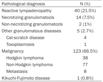
non-necrotizing granulomatozis. Of these pa-Table 1. Excisional biopsy results of patients
with lympadenopathy
Pathological diagnosis N (%)
Reactive lympadenopathy 40 (21.5%)
Necrotizing granulomatozis 14 (7.5%)
Non-necrotizing granulomatozis 2 (1%)
Other granulomatous diseases 5 (2.7%)
Cat-scratch disease 4
Toxoplasmosis 1
Malignancy 123 (66.5%)
Hodgkin lymphoma 38
Non-Hodgkin lymphoma 77
Metastasis 8
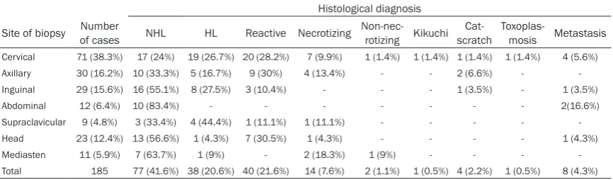
[image:2.629.98.299.105.265.2]Table 2. Site distribution and histopathological diagnosis of patients with lymphadenopathy
Histological diagnosis
Site of biopsy of casesNumber NHL HL Reactive Necrotizing Non-nec-rotizing Kikuchi scratchCat- Toxoplas-mosis Metastasis
Cervical 71 (38.3%) 17 (24%) 19 (26.7%) 20 (28.2%) 7 (9.9%) 1 (1.4%) 1 (1.4%) 1 (1.4%) 1 (1.4%) 4 (5.6%)
Axillary 30 (16.2%) 10 (33.3%) 5 (16.7%) 9 (30%) 4 (13.4%) - - 2 (6.6%) -
-Inguinal 29 (15.6%) 16 (55.1%) 8 (27.5%) 3 (10.4%) - - - 1 (3.5%) - 1 (3.5%)
Abdominal 12 (6.4%) 10 (83.4%) - - - 2(16.6%)
Supraclavicular 9 (4.8%) 3 (33.4%) 4 (44.4%) 1 (11.1%) 1 (11.1%) - - - -
-Head 23 (12.4%) 13 (56.6%) 1 (4.3%) 7 (30.5%) 1 (4.3%) - - - - 1 (4.3%)
Mediasten 11 (5.9%) 7 (63.7%) 1 (9%) - 2 (18.3%) 1 (9%) - - -
-Total 185 77 (41.6%) 38 (20.6%) 40 (21.6%) 14 (7.6%) 2 (1.1%) 1 (0.5%) 4 (2.2%) 1 (0.5%) 8 (4.3%)
HL: Hodgkin lymphoma, NHL: Non-Hodgkin lymphoma.
tients, 40 had nodes with reactive hyperplasia
(15 female, 25 male) (1-70 y, mean age: 27.35
y), 14 had necrotizing granulomatous disease
(9 female, 5 male) (16-63 y, mean age: 39.86 y), 2 of non-necrotizing granulomatous disease (1 female, 1 male, mean age: 43 y), 4 of had cat-scratch disease (1 female, 3 male, mean age: 54.25 y), 1 of toxoplasmosis (26 y, female), 1 of Kikuchi disease (25 y, female). Malign lesions included NHL, HL and metastasis; 38 of were HL (20.5%, 17 female, 21 male) (5-81 y,
mean age: 34.89 y) (18 nodular sclerozane type, 12 mixed cellular type, 8 lymphocyte
pre-dominant), 77 of NHL (41.6%, 37 female, 40 male) (6-84 y, mean age: 52.26 y) (54 diffuse large B-cell, 10 follicular lymphoma, 4 periph -eral T-cell, 3 mantle cell, 3 B cell rich T cell lym-phoma, 2 small cell lymphocytic lymlym-phoma, 1
marginal zone lymphoma), 8 of metastasis (4.3%, 5 female,3 male) (20-86 y, mean age:
53.5 y).
71 lesions were in cervical region, 30 in axillary, 11 in mediasten, 29 in inguinal, 23 in head-neck, 12 in abdomen, and 9 in supraclavicular regions.
The most frequently encountered in the servi -cal region was reactive LAP while NHL in axiller-inguinal-mediastinal-abdomen and HL in supra-clavicular region. Site distribution and
histo-pathological diagnosis of patients with LAP are
shown in Table 2.
In childhood (ages 1-18 y, mean age: 12:31 ±
4:48 y); 17 reactive LAP, 4 HL, 3 NHL, 1 necro-tizing LAP, 3 cat scratch disease was observed.
Discussion
There are 600 lymph nodes in different regions of our body [6]. Spleen, tonsils, adenoids, and
Peyer’s patches are the parts of the lymphoid tissue and clean antigens from the extracellu
-lar fluid. Peripheral lymph nodes locate deep in
the subcutaneous tissue and can be palpable when any process causes them to enlarge.
If LAP grows rapidly within 2 weeks, and does
not shrink within 4-6 weeks and not completely regressed in 8-12 weeks, biopsy should be planned [7, 8]. Usually less than one cm in diameter is accepted as normal sized lymph
node. In general, when the size of lymph node
larges more than 2 cm it suggests malignancy or a granulomatous disease [9].
Fine-needle aspiration biopsy may be useful in the differentiation of benign or malignant, but are often faced with failure diagnosis. Also it needs excisional biopsy for the definitive diag
-nosis of lymphoma. Therefore excisional biopsy in the diagnosis of LAP stated as “gold stan -dard” [10]. An excisional biopsy is a diagnostic
method that can be applied safely with minimal
morbidity and mortality.
In our cases, in the pathological examination;
the most common (n = 123; 66.5%) condition was malignancies. The frequency of diseases
were; NHL, HL, and metastasis respectively.
This is 30-40% higher than reported in previous studies of Nigerian [11-14] and western series
[15, 16]. Lymphoma incidence was reported as
28.8% by Obafunwa et al [11] and 19.1% by Pindig et al [13]. Most of these cases (17.4%)
were NHL as similar to our study. Mohan et al [10] in their study; the malignancy rate was
25.8%; and it came after non-specific lymphad -enitis and tuberculosis lymphad-enitis. Darnal et al [17] reported that malignant cases ranked
ratio was 1.08, while 1:23 in HL. These rates
were lower than the rates of Darnal [17] and Desforges’s [18] reported. In this study, non specific reactive LAP constituted 40 (21.6%)
cases. Reactive LAP has been documented as
a common cause of lymph node enlargement in the tropics and ranged from 15-22% in adults in previous Nigerian and other African series
[11-14, 19]. Moore et al [20] reported that,
even though the first biopsy results were as reactive hyperplasia in children; after 6 months of follow-up, they reported that 3 of were tuber
-culosis lymphadenitis and 15 of diagnosed as lymphoma. Therefore, even though peripheral LAP biopsy of patients is benign; the patient should not be removed from the clinical follow-up.
Hussain Gadelkarim Ahmed et al assessed
100 patients biopsy outcomes with fine needle
aspiration and benign lesions were reactive lymphoid hyperplasia (n = 64), benign
granulo-matous disease (n = 26) respectively. 10 of these cases diagnosed with malignancy, 7 (7%) were cases of NHL and the remaining 3 (3%)
were HL [21].
Mohan et al evaluated 1724 lymph node biopsy
specimens of adult patients and they found that non-specific lymphadenitis were most
seen pathology. Tuberculosis lymphadenitis and malignancies were the other common causes [10].
Adesuwa N. Olu-Eddo et al evaluated 427 lymph node biopsy specimens. Cervical LAP
was predominate region including 250 (58.5%)
cases. Tuberculosis lymphadenitis and
meta-static disease were the major causes of lymph node enlargement constituting 114 (26.7%) and 113 (26.5%) cases respectively [22].
In our study, we detected one case of Kikuchi disease. Kikuchi disease also known as
Histiocytic necrotizing lymphadenitis (HNL); is a
self-limiting, benign disease characterized by fever, cervical LAP and transient leucopenia
[23].
The most common malignant disease was NHL
in this study (n = 77). Types of NHL, the most common was fastest growing type as diffuse large B-cell lymphoma (54 cases, 70.1%) and
the second most common was slow-growing
type as follicular lymphoma (10 cases, 12.9%).
NHLs are a heterogeneous group of lymphopro
-liferative disorders originating from B-, T-, or natural killer (NK) lymphocytes. In the United States, B-cell lymphomas constitute 80% to 85% of all cases, 15-20% of is T-cell lym-phomas; but NK lymphomas are very rare [24].
In our study, we observed reactive LAP most
frequently in children. This result agrees with
Lake AM et al whom evaluated 75 children
exci-sional biopsies; 41 (55%) of were non-diagnos
-tic hyperplasia, 16 (21%) of non-caseating gran
-ulomatous lymphadenitis, 5 (7%) of caseating lesion of tuberculosis and 13 (17%) of lympho -reticular neoplasm [25] in children. Mbise RL et
al analyzed excisional lymph node biopsy of
257 children and reported tuberculosis
lymph-adenitis frequency as 67.3% of, non-specific reactive lymphadenitis as 20.6% of, malignancy as 11.3% of and histiocytosis-X as 0.8% and
Hodgkin disease was the most common
neo-plasm (34.5%) [26].
Cervical region is the most common involved regional area when compared to other
lymphat-ic regions, infections are usually exist in the front row and when posterior cervical chain
lymph nodes involved, malignancy should be
thought. In our study we found that cervical
region was the most common area and reactive LAP and HL were the most common causes in this area. Enlarged mediastinal lymph nodes are usually with pathological conditions such as lymphoma, ALL, neurogenic tumors and
tuberculosis. In our study, 11 patients (5.9%)
had mediastinal LAP and the most common
cause was NHL. During the determination of
supraclavicular LAP, malignancy should be
ruled out at first. Left supraclavicular LAP
points usually abdominal malignancies such as neuroblastoma, whereas right supraclavicular LAP intrathoracic diseases. NHL was again the
most common causes of supraclavicular LAP of
our study. Axillary LAP may be associated with
local infections or cat scratch disease or lym
-phoma. The most common cause of axillary
LAP was NHL in this study. Inguinal LAP usually
caused by infection. The most common cause of inguinal LAP was again NHL in this study.
Popliteal palpable lymph node is always consid-ered to be pathological, but we didn’t come across popliteal lymph node [27].
In general practice, less than one percent of
disease [29]. These LAPs are often due to leu -kemia in children and Hodgkin disease in ado-lescents [30]. It has been reported that the
prevalence of malignancy is 0.4% in patients under 40 years and increases to 4% over 40 years of age in the primary care setting [3].
The most frequent neoplastic diseases with LAP are leukemia, HL, NHL, solid tumor metas-tases, neuroblastoma, nasopharynx carcino-ma, rhabdomyosarcocarcino-ma, thyroid cancer, histio-cytosis and hemophagocytic syndromes. 70% of acute lymphoblastic leukemia and 30% of
acute myeloblastic leukemia patients go along with LAP. Besides neoplastic diseases;
Kawasaki disease, Rosai-Dorfman, angiofollic -ular lymphoid hyperplasia (Castleman’s dis-ease), autoimmune diseases, metabolic stor-age diseases, and some anticonvulsant drug intake can be the causes of regional or general-ized LAP [1]. In our study, from lymph node biopsy materials, NHL was the most common malignancy.
HLs are seen rare less than 10 years. The
Epstein-Barr virus infection in combination with immune deficiency is a risk factor for HL, espe -cially in less-developed countries and in low
socioeconomic conditions. NHL is the fourth
common malignancy worldwide in males with a
frequency of 6.1% [30].
In conclusion LAP is a common problem and
necessitates a careful physical examination and follow-up of the patient. Laboratory and imaging methods should be used in the differ -ential diagnosis when necessary. Excisional biopsy can be applied with minimal morbidity
and mortality in a safe manner and is a diag -nostic method that is used as the gold
stan-dard in the diagnosis of LAP. Although LAP is
mostly related with benign lesions, care should
be taken in terms of malignancy and malignan -cy should be eliminated. In this study, malig-nancies were more common than benign lesions and most common seen in the cervical region.
Disclosure of conflict of interest
None.
Address correspondence to: Dr. Esra Akyüz Özkan,
Department of Pediatrics, Faculty of Medical, Bozok
University, Yozgat, Turkey. Tel: 05067026694; Fax: 0354 2140612; E-mail: [email protected]
References
[1] Mohseni S, Shojaiefard A, Khorgami Z, Alinejad S, Ghorbani A, Ghafouri A. Peripheral lymph -adenopathy: approach and diagnostic tools. Iran J Med Sci 2014; 39 Suppl 2: 158-170. [2] Ahuja AT, Ying M. Sonographic evaluation of
cervical lymph nodes. AJR Am J Roentgenol 2005; 184: 1691-1699.
[3] Richner S, Laifer G. Peripheral lymphadenopa -thy in immunocompetent Adults. Swiss Med
Wkly 2010; 140: 98-104.
[4] Chau I, Kelleher MT, Cunningham D, Norman AR, Wotherspoon A, Trott P Rhys-Evans P,
Querci Della Rovere G, Brown G, Allen M,
Waters JS, Haque S, Murray T, Bishop L. Rapid
access multidisciplinary lymph node
diagnos-tic clinic: analysis of 550 patients. Br J Cancer
2003; 88: 354-361.
[5] Bazemore AW, Smucker DR. Lymphadenopathy
and malignancy. Am Fam Physician 2002; 66: 2103-2110.
[6] Ferrer R. Lymphadenopathy: differential diag -nosis and evaluation. Am Fam Physician 1998; 58: 1313-1320.
[7] Knight PJ, Mulne AF, Vassy LE. When is lymph
node biopsy indicated in children with en-larged peripheral nodes? Pediatrics 1982; 69: 391-396.
[8] Twist CJ, Link MP. Assessment of lymphade -nopathy in children. Pediatr Clin North Am 2002; 49: 1009-1025.
[9] Slap GB, Brooks JS, Schwartz JS. When to per
-form biopsies of enlarged peripheral lymph
nodes in young patients. JAMA 1984; 252: 1321-1326.
[10] Mohan A, Reddy MK, Phaneendra BV, Chandra A. Aetiology of peripheral lymphadenopathy in adults: Analysis of 1724 cases seen at a ter -tiary care teaching hospital in southern India. Natl Med J India 2007; 20: 78-80.
[11] Obafunwa JO, Olomu LN, Onyia NJ. Primary pe
-ripheral lymphadenopathy in Jos, Nigeria. West Afr J Med 1992; 11: 25-28.
[12] Mandong BM, Nwana EJC, Igun G, Dakum AN. Surgical lymph node biopsies in Jos University Teaching Hospital, Jos, Nigeria. Nig J Surg Res 1999; 1: 63-67.
[13] Pindiga UH, Dogo D, Yawe T. Histopathology of
primary peripheral lymphadenopathy in North Eastern Nigeria. Nig J Surg Res 1999; 1: 68-71.
[14] Thomas JO, Ladipo JK, Yawe T. Histopathology of lymphadenopathy in a tropical country. East Afr Med J 1995; 72: 703-705.
[15] Freidig EE, McClure SP, Wilson WR, Banks PM, Washington JA. Clinical histologic- microbiolo-gic analysis of 419 lymph node biopsy speci
[16] Sriwatanawongsa V, Cardoso R, Chang P.
Incidence of malignancy in peripheral lymph
node biopsy. Am Surg 1985; 51: 587-590. [17] Darnal HK, Karim N, Kamini K, Angela K. The
profile of lymphadenopathy in adults and chil -dren. Med J Malaysia 2005; 60: 590-598. [18] Desforges F, Rutherford C, Piro A. Hodgkin’s
Disease. N Engl J Med 1979; 301: 1212-1222. [19] Abba AA, Bamgboye AE, Afzal M, Rahmatullah
RA. Lymphadenopathy in adults. A clinicopath-ological analysis. Saudi Med J 2002; 23: 282-286.
[20] Moore SW, Schneider JW, Schaaf HS. Diag-nostic aspects of cervical lymphadenopathy in children in the developing world: A study of
1,877 surgical specimens. Pediatr Surg Int 2003; 19: 240-244.
[21] Ahmed HG, Elmubasher MB, Salih RA, Elhussein GE, Ashankyty IM. Fine needle
aspi-ration cytopathology of pediatric lymphade -nopathy among Sudanese children. Asian Pac J Cancer Prev 2013; 14: 4359-4363.
[22] Olu-Eddo AN, Ohanaka CE. Peripheral lymph-adenopathy in Nigerian adults. J Pak Med Assoc 2006; 56: 405-408.
[23] Kikuchi M. Lymphadenitis showing focal reticu -lum cell hyperplasia with nuclear debris and
phagocytosis. Nippon Ketsueki Gakkai Zasshi
1972; 35: 379-380.
[24] Jemal A, Siegel R, Ward E, Hao Y, Xu J, Thun
MJ. Cancer statistics, 2009. CA Cancer J Clin 2009; 59: 225-249.
[25] Lake AM, Oski FA. Peripheral lymphadenopa-thy in childhood. Ten-year experience with exci-sional biopsy. Am J Dis Child 1978; 132: 357-359.
[26] Mbise RL. Peripheral lymphadenopathy in
chil-dren in Dar es Salaam, Tanzania. A study from
biopsy material. Ann Trop Paediatr 1984; 4: 83-85.
[27] Çeçen E. Peripheral Lymphadenopathy in
Childhood. ADÜ Tıp Fakültesi Dergisi 2009; 10:
45-52.
[28] Fijten GH, Blijham GH. Unexplained
lymphade-nopathy in family practice. An evaluation of the probability of malignant causes and the effec
-tiveness of physicians’ workup. J Fam Pract
1988; 27: 373-376.
[29] Kliegman RM, Nieder ML, Super DM.
Lym-phadenopathy. In: Fletcher J, Bralow L, editors. Practical strategies in pediatric diagnosis and
therapy. St. Louis: WB Saunders Co; 1996. pp.
791-803.
[30] Naz E, Mirza T, Aziz S, Danish F, Siddiqui ST, Ali A. Frequency and clinicopathologic correlation of different types of non Hodgkin’s lymphoma according to WHO classification. J Pak Med