© 2015 IJSRST | Volume 1 | Issue 4 | Print ISSN: 2395-6011 | Online ISSN: 2395-602X Themed Section: Science and Technology
Validation of Cholinesterase (Acetyl and Butyryl) Activity
Estimation in the Blood and Brain of Wistar Rats
Arabinda Adak, Rajesh Eswarappa, Sriram Nagarajan, S. K. Mukhopadhayay
Department of Toxicology,Jai Research Foundation, Vapi, Gujarat, India
ABSTRACT
The present study was carried out with an objective to validate the estimation method of both acetylcholinesterase (AChE) and butyrylcholinesterase (BChE) activities in blood and brain tissue of Wistar rats as well as to generate historical control data, based on sizable sample number, for our laboratory. In the present study, the butyrylcholinesterase activity was estimated in plasma and acetylcholinesterase in erythrocytes, plasma and brain tissue in 14 weeks old Wistar rats (male-15, female-15). In brain tissue, the activity of AChE was 1.71±0.25 U/g in males and 1.98±0.27 U/g in females. In erythrocytes, the activity was 1285.65±379.14 U/L in males and 1684.71±218.75 U/L in females. The AChE activity in plasma was 440.86±71.39 U/L and 495.29 ± 205.80 U/L in males and females, respectively while the plasma butyrylcholinesterase activity was 59.47 ± 23.65 U/L in males and 831.35 ± 174.95 U/L in females. In conclusion, both acetylcholinesterase and butyrylcholinesterase activities seemed to be higher in females than males, in general and the methods used were based on cholinesterase assay kits obtained from M/S Abnova Corporation, Taiwan and M/S Agappe Diagnostics, Kerala, India to estimate AChE and BChE, respectively. These kits were found to be convenient, accurate and sensitive with high throughput for the estimation of AChE and BChE activities in Wistar rats.
Keywords: Blood, Brain, Cholinesterase (Acetyl And Butyryl), Wistar Rat
I.
INTRODUCTION
Acetylcholine (ACh), a neurotransmitter, plays an important role in the functioning of nervous system by enabling chemical communication between a nerve cell and a target cell. Ever since its discovery as a neurotransmitter by Sir Henry Dale and Otto Loewi for which they were awarded the Nobel Prize in 1936, its function in health and dysfunction in disease has been increasingly recognized. In the recent past, its role in learning and memory too has been demonstrated indubitably. Further, pharmacological manipulation of cholinergic function has been found useful in the treatment of CNS disorders like Alzheimer’s and Parkinson’s disease. ACh per se has a very short half-life, as such its direct estimation is a little difficult in brain tissue. There are several approaches to evaluate cholinergic functions indirectly, like estimating the activities of choline acetyltransferase (ChAT) and acetylcholinesterase (AChE) by immunochemical and
histochemical techniques but these are tedious and time consuming. However, estimation of AChE activity provides relatively an easy and valuable assessment of cholinergic functions (Srikumar et al., 2004).
There are two types of cholinesterase enzymes: Acetylcholinesterase (AChE) or true cholinesterase (primarily present in erythrocytes and synaptic junction of nervous tissue) and Butyrylcholinesterase (BChE) or pseudo cholinesterase (primarily present in plasma and liver). Invariably pseudocholinesterase / butyrylcholinesterase is estimated from plasma or serum sample while true/acetyl cholinesterase enzyme is measured from the anticoagulated / heparinized whole blood or specifically from RBC and brain tissue or specifically from its different parts. The method of AChE activity estimation is popularly known as Ellman’s method named after George Ellman and associates who developed this method in 1961 (Ellman et al., 1961). Methods to estimate acetyl and butyryl cholinesterase differ. The automated method of cholinesterase activity estimation is easy and desirable for routine work (Wilson et. al., 1996).
The present study was planned with the sole objective to validate the method of estimating the normal activity of butyrylcholinesterase in plasma and acetylcholinesterase in erythrocytes (RBC), plasma and brain tissue of Wistar rats of either sex to generate the historical control data for this laboratory.
II.
METHODS AND MATERIAL
The present study was carried out using 14 weeks old Wistar rats (male-15, female-15) obtained from Animal Breeding Facility, Jai Research Foundation. The approval of the Institute Animal Ethic Committee (IAEC) was obtained prior to the commencement of the study. All the animals were housed in stainless steel cages in air conditioned rooms at 20±30C having 30 to 70% relative humidity with 12 h artificial light and 12 h dark cycle. The rate of air change in experiment rooms was minimum of 15 times per hour. Standard feed pellets of rats/mice and clean filtered water were provided ad libitum. These animals appeared apparently normal without any abnormal clinical expression during life-phase. The terminal body weight of males and females used was 400-450g and 250-275g, respectively.
Collection and preparation of blood/ plasma/ erythrocyte samples:
Approximately 1.0 ml blood per animal was collected in heparinized (@100 IU//ml of blood) vials by puncturing
orbital plexus of Wistar rats under light anaesthesia by isoflurane inhalation. The collected blood was centrifuged at 3000 rpm for 10 minutes. The supernatant (plasma) and pellet (erythrocytes/RBC) were collected separately in labeled centrifuge tubes for further analysis. Prior to sample analysis, the erythrocyte pellets were diluted 40 folds in the assay buffer by accurately pipetting 5 µL erythrocyte pellet into 195 µL assay buffer and mixed thoroughly to estimate the activity of AChE (Magnotti, 1987) while the plasma sample was analysed undiluted for both acetylcholinesterase and butyrylcholinesterase.
Collection and preparation of brain tissue:
Post euthanasia by carbon dioxide asphyxiation, the rat carcasses were positioned on ventral recumbency (i.e. face downward) on necropsy table. After deflecting the skin, sub-cutis and muscles, the skull was cut opened using bone-cutters and scissors. The whole brain (including cerebrum and cerebellum) was dissected out quickly and carefully from the cranial cavity and put in the petridish chilled on crushed ice until weighing. Post weighing the brain was cut longitudinally into two halves so that each half contained cerebrum, hippocampus and cerebellum. The right half which was supposed to be used for acetyl cholinesterase assay was kept in a labeled ependorf tube placed on ice and was again weighed while the left half was fixed in 10% neutral buffered formalin for histopathology. Tissue or cell lysates of right half of brain was prepared from each sample by brief homogenization in 0.1 M phosphate buffer (pH 7.5) at the ratio of 1:10 (w/v), followed by refrigerated centrifugation at 14,000 rpm for 5 minutes. The supernatant was collected and stored in ependorf tubes until further analysis. Samples were assayed fresh.
Reagents/Equipment Required:
1. 0.1 M Phosphate buffer (pH 7.5)
Solution A: 2.76g of monobasic sodium phosphate, monohydrate was dissolved in 100 ml of distilled water.
Solution B: 2.84g of dibasic sodium phosphate was dissolved in 100 ml of distilled water.
adding distilled water to have 0.1M phosphate buffer.
2. Acetylcholinesterase assay kit (Code KA 1607) manufactured by M/S Abnova Corporation, Taiwan and supplied by M/S Thermo Fisher Scientific, Mumbai. The kit contained assay buffer, calibrator and reagent.
3. Butyrylcholinesterase assay kit (Code 11205001) manufactured and supplied by M/S Agappe Diagnostics, Kerala, India. The kit contained cholinesterase R1 (buffer solution) and cholinesterase R2 (tablets containing 5, 5 DTNB and butyrylthiocholine) and calibrator.
4. Refrigerated centrifuge 5. Tissue homogenizer 6. Clear-bottom 96-well plate 7. Multi-channel pipette
8. Micro-plate reader (EL808IUTM, BioTek Instruments, USA)
9. Clinical chemistry analyzer (BT 2000 Plus, Biotecnica, Italy)
10. Dissecting instruments: knives, saw, scissors, forceps and bone-cutter
Assay Principle
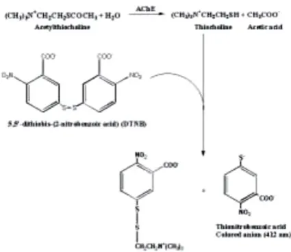
The AChE activity is measured by providing an artificial substrate (acetylthiocholine, ATC). AChE cleavage/breaks acetylthiocholine and release thiocholine. Thiocholine so released is allowed to react with the -SH reagent 5, 5’ - dithiobis - (2-nitrobenzoic acid) (DTNB). This DTNB in turn is reduced to thionitrobenzoic acid, a yellow coloured anion which is measured at 412 nm. The colour intensity of thionitrobenzoic acid is taken as a direct estimation of AChE activity. The intensity of the produced colour is directly proportionate to the AChE enzyme activity in the sample (Srikumar et al., 2004).
Figure 1: The steps involved in estimation of AChE activity using Ellman’s reaction
Assay procedure for acetylcholinesterase:
1. Working Reagent preparation: 2 mg test reagent (supplied with kit) was required per sample. Accordingly keeping in view the number of samples to be analysed, the amount of test reagent was calculated and weighed. Per 2 mg of test reagent 200 µL assay buffer (supplied with kit) was added and vortexed to dissolve the test reagent to prepare working reagent. Only freshly prepared working reagent was to be used and that too within 30 minutes.
2. Calibrator: 200 µL distilled water and calibrator (provided with kit) each was added separately into two adjacent wells of the clear bottom 96-well plate.
3. Samples: In another two adjacent separate wells 10 µL sample per well was added as sample was to be analysed in duplicate.
4. Reaction: 190 µL freshly prepared working reagent (sl.no.1 above) was added to each sample well and the plate was taped briefly to mix. OD412nm at 2
nd
and 10th minute in a micro-plate reader (ELx808IUTM, BioTek Instruments, USA) was recorded.
5. Calculataion: Acetylcholinesterase activity (U/L) was calculated as follows:
OD10 – OD2
AChE Activity = --- x n x 200 (U/L)
OD10 and OD2 were the OD412nm values of the sample at
10th and 2nd minute, respectively. ODCAL and ODH2O
were the OD412nm values of the calibrator and distilled
water at 10th minute. n was the dilution factor (n = 1 in case of brain and plasma while n = 40 in case of blood and RBC/erythrocytes. The number “200” was the equivalent activity of the calibrator under the assay conditions.
Note: If the AChE activity without considering the dilution factor (n) was greater than 600 U/L or showed any abnormal values, the sample was further diluted in the assay buffer and the assay was repeated. The results were multiplied by the dilution factor.
Final calculation in case of brain: The concentration of brain homogenate was calculated according to its dilution with phosphate buffer and finally, AChE activity was expressed as U/g. For example, if the weight of brain sample (taken for AChE activity) was 1 g and 10 ml phosphate buffer was added to the brain tissue, the concentration of the homogenate would be 100 g/L. If the calculated value of AChE activity was 200 U/L, the final AChE activity of the brain tissue would be 2 U/g.
Assay procedure for butyrylcholinesterase:
1. Reagent preparation: One tablet (R2) was dissolved in one vial of buffer solution (R1) and left for 10 minutes for complete dissolution. The stability of this working reagent is 2 hours at 2-8 0C.
2. Samples: 10 µL plasma was diluted in 10 µL saline solution to get 20 µL diluted sample.
3. Reaction: 3000 µL freshly prepared working reagent was added to 20 µL diluted sample and mixed. The mixed sample was loaded in the clinical chemistry analyzer and the change in absorbance per minute (▲OD/60 sec) during 2 minutes was measured.
4. Calculation: Butyrylcholinesterase activity (U/L) was calculated as follows:
BChE Activity = (▲OD/60 sec) x 22710
III.
RESULT AND DISCUSSION
The results of AChE in the brain, RBC and plasma and BChE activity in plasma collected from Wistar rats of either sex have been presented in Table 1.
Historically, there have been technical difficulties with the measurement of the plasma and RBC cholinesterase(s), particularly for the latter (Wilson, et al., 1996). Although in recent years there have been improvements in assay methods of blood cholinesterase activity. It is important to consider carefully the methodological issues that might affect the accuracy and variability of the data when assessing the effects of certain pesticides on cholinesterase activity in blood. A number of methods are available for measuring blood cholinesterase activity and the methods vary from laboratory to laboratory. The colorimetric method, based on the Ellman reaction, is considered a reliable method when performed properly and is commonly used for measuring plasma and RBC cholinesterase activity (Ellman, et al., 1961; US EPA, 1992; ASCP, 1994).
The inhibition of acetylcholinesterase activity is a key step in the toxicity of certain organophosphorous and carbamate pesticides (Mileson, et al, 1998; US EPA, 1999) as it plays an important role in developing nervous system disorders owing to their potential pharmacological and toxicological actions. Monitoring the AChE activities in preclinical toxicity studies of organophosphorous and carbamate compounds is being increasingly demanded. Therefore, measurements of cholinesterase inhibition rate represent a critical biochemical biomarker of potential adverse effects. Inhibition of acetylcholinesterase in the central nervous system is considered to be an indicator of an adverse effect. Information from blood cholinesterase inhibition data is also considered to provide important insights into potential hazard. The measurement of erythrocyte (RBC) acetylcholinesterase (AChE) activity is generally preferred over plasma cholinesterase activity because it may provide a better representation of the inhibition of the neural target enzyme, acetylcholinesterase. Although effects of pesticide both on RBC and plasma cholinesterase activities provide adverse reflection of anti-AChE action on nervous system, the RBC AChE activity provides better reflection of neuronal AChE inhibition than that that of plasma, which is a variable
acetylcholinesterase depending upon the species. In the present investigation, it has been found that:
a) Acetylcholinesterase and butyrylcholinesterase activities seem to be higher in females than males. b) Acetylcholinesterase activity in RBC seems to be
2-3 times higher than plasma.
c) The distribution of acetyl and butyryl cholinesterase in plasma is 7: 1 in males and 1:2 in females.
d) AChE activity measured using Acetylcholinesterase assay kit (M/S Abnova, Taiwan) based on improved Ellman method, is convenient, accurate and sensitive with high throughput.
IV. ACKNOWLEDGEMENTS
The authors are thankful to Dr. M. C. Prasad for his valuable guidance and to the Management, Jai Research Foundation, Vapi, Gujarat for providing the necessary facilities to carry out the present work and granting permission to publish the findings.
V.
REFERENCES
[1] Appleyard, M. E. (1992). Secreted
acetylcholinesterase: non-classical aspects of a classical enzyme. Trends Neurosci. 15: 485-490. [2] Ellman, G.L., Courtney, K.D., Andres, V. Jr. and
Feather-Stone, R. M. (1961). A new and rapid colormetric determination of acetylcholinesterase activity. Biochem. Pharmaco. 7:88-95.
[3] Hammond, P. and Brimijoin, S. (1988). Acetylcholinesterase in Huntington’s and Alzheimer’s diseases: Simultaneous enzyme essay and immunoassay of multiple brain regions. J. Neurochem. 50: 1111-1116.
[4] Magnotti, R. A. (1987). Measurement of acetylcholinesterase in erythrocytes in the field. Clin. Chem. 33/10, 1731-1735.
[5] Mileson, B.E., Chambers, J.E., Chen, W.L., Dettbarn, W., Ehrich, M., Eldefrawi, A.T., Gaylo, D.W., Hamernik, K., Hodgson, E.H., Karczmar, A.G., Padilla, S., Pope, C.N., Rihardson, R.J., Saunders, D. R., Sheets, L.P., Sultatos, L.G. and Wallace, K.B. (1998). Common mechanism of toxicity: a case study of organophosphorous pesticides. Toxicol. Sciences. 41: 8-20.
[6] Mileson, B. E. (1999a). Common mechanism of toxicity: evaluation of carbamate pesticides. unpublished report of the international life sciences institute/risk science institute, Washington, DC). March, 2012. Pp.19.
[7] Ravi, S. M., Prabhu, B.M., Raju, T.R. and Bindu, P.N. (2000). Long-term effects of postnatal aluminium exposure on acetylcholinesterase activity and biogenic amine neurotransmitters in rat brain. Indian J Physiol Pharmacol. 44: 473-478.
[8] Srikumar, B. N., Ramkumar, K., Raju, T.R., and Shanakranarayana Rao, B.S. (2004). Assay of acetylcholinesterase activity in the brain. Brain and Behaviour. 25:142-144.