research communications
Acta Cryst.(2019). E75, 565–570 https://doi.org/10.1107/S2056989019004389
565
Received 25 March 2019Accepted 31 March 2019
Edited by W. T. A. Harrison, University of Aberdeen, Scotland
‡ Additional correspondence author, e-mail: [email protected].
Keywords:crystal structure; pyrazolone; pyra-zole; tautomer; Hirshfeld surface analysis.
CCDC reference:1906841
Supporting information:this article has supporting information at journals.iucr.org/e
Co-crystallization of a neutral molecule and its
zwitterionic tautomer: structure and Hirshfeld
surface analysis of 5-methyl-4-(5-methyl-1
H
-pyrazol-3-yl)-2-phenyl-2,3-dihydro-1
H
-pyrazol-3-one 5-methyl-4-(5-methyl-1
H
-pyrazol-2-ium-3-yl)-3-oxo-2-phenyl-2,3-dihydro-1
H
-pyrazol-1-ide
monohydrate
Abdullah M. Asiri,a,b‡ Khalid A. H. Alzahrani,b Hassan M. Faidallah,b Khalid A. Alamry,bMukesh M. Jotanicand Edward R. T. Tiekinkd*
a
Center of Excellence for Advanced Materials Research, King Abdulaziz University, PO Box 80203, Jeddah 21589, Saudi Arabia,bChemistry Department, Faculty of Science, King Abdulaziz University, PO Box 80203, Jeddah 21589, Saudi Arabia,cDepartment of Physics, Bhavan’s Sheth R. A. College of Science, Ahmedabad, Gujarat 380001, India, and
d
Research Centre for Crystalline Materials, School of Science and Technology, Sunway University, 47500 Bandar Sunway, Selangor Darul Ehsan, Malaysia. *Correspondence e-mail: [email protected]
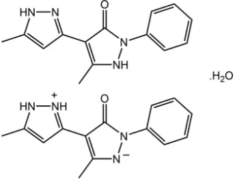
The title compound, 2C14H14N4OH2O, comprises a neutral molecule containing
a central pyrazol-3-one ring flanked by an N-bound phenyl group and a C-bound 5-methyl-1H-pyrazol-3-yl group (at positions adjacent to the carbonyl substituent), its zwitterionic tautomer, whereby the N-bound proton of the central ring is now resident on the pendant ring, and a water molecule of crystallization. Besides systematic variations in geometric parameters, the two independent organic molecules have broadly similar conformations, as seen in the dihedral angle between the five-membered rings [9.72 (9) for the neutral molecule and 3.32 (9)for the zwitterionic tautomer] and in the dihedral angles between the central and pendant five-membered rings [28.19 (8) and 20.96 (8) (neutral molecule); 11.33 (9) and 11.81 (9)]. In the crystal, pyrazolyl-N— H O(carbonyl) and pyrazolium-N—H N(pyrazolyl) hydrogen bonds between the independent organic molecules give rise to non-symmetric nine-membered { HNNH NC3O} and { HNN HNC3O} synthons, which differ
in the positions of the N-bound H atoms. These aggregates are connected into a supramolecular layer in the bc plane by water-O—H N(pyrazolide), water-O—H O(carbonyl) and pyrazolyl-N—H O(water) hydrogen bonding. The layers are linked into a three-dimensional architecture by methyl-C— H (phenyl) interactions. The different interactions, in particular the weaker contacts, formed by the organic molecules are clearly evident in the calculated Hirshfeld surfaces, and the calculated electrostatic potentials differentiate the tautomers.
1. Chemical context
Molecules related to the title compound, i.e. containing a pyrazolone ring, are of particular interest owing to their pharmaceutical potential. Applications in this context include their possible utilization as cardiovascular drugs (Higashiet al., 2006), as hypoglycemic agents (Daset al., 2008) and as anti-inflammatory and analgesic agents (Badawey & El-Ashmawey, 1998). This class of compound has also been evaluated as anti-microbials (Sahu et al., 2007) and display fungicidal activities (Singh & Singh, 1991). In the course of
studies in this area, the title compound, which has been synthesized previously (Kumaret al., 1995), was characterized crystallographically on a crystal isolated from an ethanol solution and found to contain neutral and zwitterionic tauto-mers.
Tautomerism relates to a phenomenon whereby isomeric structures undergo inter-conversion by the migration of, typically, an atom, often a proton, or small group within the molecule. While different tautomers can co-exist in solution, in crystals usually only one form is found (Rubcˇic´et al., 2012). Notable examples of tautomers crystallizing in the same crystal begin with biologically relevant isocytosine (Sharma & McConnell, 1965) and the histidine residue in the structure of l-His-Gly hemihydrate (Steiner & Koellner, 1997). Such
behaviour has also been observed, for example, in a synthetic compound, namely, N-(3-hydroxysalicylidene)-4-methoxy-aniline (Pizzalaet al., 2000). Herein, the crystal and molecular structures of the title compound, (I), are described along with an analysis of the calculated Hirshfeld surfaces.
2. Structural commentary
The crystal of (I), Fig. 1, comprises a neutral molecule of
methyl-1H-pyrazol-3-yl)-2-phenyl-2,3-dihydro-1H-pyrazol-3-one, its zwitterionic tautomer 5-methyl-4-(5- methyl-1H-pyrazol-2-ium-3-yl)-3-oxo-2-phenyl-2,3-dihydro-1H-pyrazol-1-ide and a molecule of water. Evidence of tautomerism is twofold. Firstly, in the nature of the hydrogen-bonding interactions operating in the crystal; see Supra-molecular features. Secondly, in small but systematic variations in key geometric parameters, Table 1. Thus, the N5—N6 bond in the zwitterion is longer than the equivalent N1—N2 bond in the neutral species with a concomitant shortening of the C16— N6 and lengthening of the C16—C17 bonds with respect to the bonds in the neutral species. The most notable changes in angles relate to protonation/deprotonation. Thus, the angle at the protonated N2 atom is greater than the angle at the deprotonated N6, and the same is true for the angles subtended at the N7 and N3 atoms. Molecules in (I) may also exhibit rotational isomerism about the C3—C5 and C17—C19 bonds. In the present case, the carbonyl group issynwith the pendent ring imine-N atom in the neutral molecule and is designated the NH-Z form (Kumaret al., 1995); the zwitter-ionic tautomer has a similar conformation.
The differences in geometric parameters characterizing the two independent organic molecules in (I) notwithstanding, the molecules present very similar conformations as highlighted in the overlay diagram, Fig. 2. Thus, the r.m.s. deviations of the
566
Asiriet al. 2C14H14N4OH2O Acta Cryst.(2019). E75, 565–570
[image:2.610.313.566.92.266.2] [image:2.610.85.257.137.268.2]research communications
Table 1
Selected geometric parameters (A˚ ,) for (I).
Atoms Parameter Atoms Parameter
C1—O1 1.266 (2) C15—O2 1.263 (2)
N1—N2 1.376 (2) N5—N6 1.401 (2)
C1—N1 1.389 (2) C15—N5 1.396 (2)
C2—N2 1.338 (2) C17—N6 1.329 (2)
C1—C3 1.429 (2) C15—C17 1.416 (2)
C2—C3 1.391 (2) C16—C17 1.405 (2)
N3—N4 1.364 (2) N7—N8 1.349 (2)
C5—N3 1.346 (2) C19—N7 1.349 (2)
C7—N4 1.338 (2) C21—N8 1.337 (2)
C5—C6 1.414 (2) C19—C20 1.404 (2)
C6—C7 1.375 (2) C20—C21 1.389 (2)
N2—N1—C1 109.07 (13) C15—N5—N6 111.51 (13)
N2—N1—C9 121.16 (14) N6—N5—C23 119.25 (14)
C1—N1—C9 129.62 (14) C15—N5—C23 128.95 (15)
N1—N2—C2 108.78 (14) N5—N6—C16 105.37 (13)
N4—N3—C5 103.94 (14) N8—N7—C19 110.58 (14)
[image:2.610.48.297.528.671.2]N3—N4—C7 113.60 (14) N7—N8—C21 108.36 (14)
Figure 1
[image:2.610.315.566.589.709.2]The molecular structures of the constituents of (I), showing displacement ellipsoids at the 70% probability level. The pyrazolyl-N4, N8—H O2, O1(carbonyl), pyrazolium-N7—H N3(pyrazolyl) and water-O— H N6(pyrazolium) hydrogen bonds are shown as dashed lines. Note the non-symmetric nine-membered { HNNH NC3O} and { HNN HNC3O} synthons formed between the neutral and tauto-meric molecules.
Figure 2
fitted atoms of the N1, N3, N5 and N7 rings are 0.019, 0.003, 0.006 and 0.006, respectively. The dihedral angle between the N1 and N3 rings is 9.72 (9), and that between each of these and the appended phenyl ring are 28.19 (8) and 20.96 (8). The comparable values for the zwitterionic tautomer are 3.32 (9), 11.33 (9) and 11.81 (9), respectively, indicating that this molecule is closer to planar, as vindicated by the difference in the N2—N1—C9—C14 and N6—N5—C23—C28 torsion angles of 26.2 (2) and7.4 (2), respectively.
3. Supramolecular features
The molecular packing of (I) features substantial conventional hydrogen-bonding interactions and the description of these can be conveniently divided by discussing interactions without
the involvement of the water molecule of crystallization and then considering the role of the water molecule. Table 2 lists the geometric parameters of the specific intermolecular interactions in the crystal of (I). There are three hydrogen bonds formed between the two organic molecules comprising the asymmetric unit and these involve the three outer-ring amine-H atoms as donors and a ring-N and the two carbonyl-O atoms as acceptors. The resulting pyrazolyl-N4, N8— H O1, O2(carbonyl) and pyrazolium-N7—H N3(pyrazol-yrazolyl) hydrogen bonds give rise to non-symmetric nine-membered { HNNH NC3O} and { HNN HNC3O}
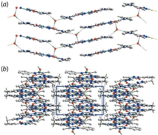
synthons, differing only in the relative placement of one of the ring-N-bound H atoms, Fig. 1. The two-molecule aggregates are assembled into a supramolecular layer in thebcplane by hydrogen bonding involving the water molecule, which func-tions as a donor to the pyrazolide-N6 and carbonyl-O1 atoms and as an acceptor from a pyrazolyl-N2 atom, Fig. 3a. Further consolidation of the layers is provided by – interactions with the shortest of these involving the N1-pyrazolyl and phenyl(C9–C14)i rings [inter-centroid separation = 3.5810 (9) A˚ , inter-planar angle = 6.29 (8) for symmetry operation: (i) 1x, 12+ y,
3
2z]. As shown in Fig. 3b, the
points of contact between layers involve methyl-C8—H
interactions with the symmetry-related phenyl (C23–C28) ring, Table 2, resulting in a three-dimensional architecture.
4. Hirshfeld surface analysis
The Hirshfeld surface calculations for (I) were calculated employing Crystal Explorer (Turner et al., 2017) and were conducted in accord with recent studies (Tanet al., 2019) to investigate the influence of intermolecular interactions between the neutral and zwitterionic tautomers, along with the water molecule of crystallization, on the molecular packing.
The donors and acceptors of the hydrogen bonds summar-ized in Table 2 and discussed in the previous section are clearly evident as the broad and bright-red spots on the Hirshfeld surface mapped overdnormfor the neutral tautomer in Fig. 4a
andband for the zwitterionic tautomer in Fig. 4c. In addition to these, a short intramolecular H H contact between symmetry-related pyrazolyl-H4Nand H7Natoms (Table 3) is viewed as faint-red spots near these atoms on the dnorm
-mapped Hirshfeld surfaces of the tautomers in Fig. 4aandc. It is clear from the views of the Hirshfeld surfaces mapped over the calculated electrostatic potential for neutral molecule in
research communications
Acta Cryst.(2019). E75, 565–570 Asiriet al. 2C
[image:3.610.44.297.437.654.2]14H14N4OH2O
567
Figure 3Supramolecular association in the crystal of (I): (a) a view of the supramolecular layer in thebc plane whereby the dimeric aggregates shown in Fig. 1 are connected by water-O1W O1(carbonyl), water-O1W N6(pyrazolide) (pink dashed lines) and pyrazolyl-N2— H O1W(water) hydrogen bonds (blue dashed lines) and (b) a view of the unit-cell contents shown in projection down thecaxis. The C—H interactions are shown as purple dashed lines. In both images, the non-participating and non-acidic H atoms are omitted.
Table 2
Hydrogen-bond geometry (A˚ ,).
Cg1 is the centroid of the (C23–C28) ring.
D—H A D—H H A D A D—H A
N2—H2N O1Wi 0.90 (1) 1.79 (1) 2.673 (2) 168 (2) N4—H4N O2 0.88 (1) 1.90 (1) 2.764 (2) 168 (2) N7—H7N N3 0.89 (1) 2.28 (2) 2.970 (2) 134 (1) N8—H8N O1 0.90 (1) 1.79 (1) 2.664 (2) 166 (2) O1W—H1W O1ii
0.85 (2) 1.92 (2) 2.7641 (17) 172 (2) O1W—H2W N6 0.86 (2) 1.94 (2) 2.7979 (19) 178 (2)
C8—H8A Cg1iii 0.98 2.71 3.492 (2) 137
C8—H8B Cg1iv 0.98 2.89 3.755 (2) 148
Symmetry codes: (i) x;yþ1
2;zþ12; (ii) xþ1;yþ1;zþ1; (iii) xþ2;yþ1;zþ1; (iv)xþ2;y;zþ1.
Figure 4
Views of the Hirshfeld surface for (I) mapped overdnormin the range
Fig. 5aandb, and for the zwitterionic tautomer in Fig. 5cand d, that they have quite distinct charge distributions on their surfaces. The presence of non-protonated pyrazolyl-N3 and N6 atoms results in a pronounced electronegative regions adjacent to carbonyl-O1 atom in the neutral form and oppo-site to carbonyl-O2 atom in the zwitterion as shown by the intense-red regions in Fig. 5a–d; hence facilitating the charge-assisted hydrogen bonds with pyrazolyl-N7 and water-O1W atoms, respectively. The donors and acceptors of inter-molecular water-O1W–-H O1(carbonyl) and pyrazolyl-N2—H O1W(water) are also viewed as blue and red regions in Fig. 5eandf.
The short interatomic contacts characterizing weak inter-molecular interactions in the crystal of (I) are viewed as characteristic red spots near the involved atoms on the Hirshfeld surface of the overall structure by modifying (making more sensitive) thednormrange, see Fig. 6 and Table 3.
[image:4.610.312.566.102.197.2]The short intra- and inter-layer C C contacts formed by the methyl-C22 atom with methyl-C8 and pyrazolyl-C15 atoms (Table 3) are viewed as small red spots near these atoms in Fig. 6a. The presence of faint-red spots near pyrazole-N2, C2, C17, C21 and phenyl-C13, C14 atoms in Fig. 6 represent their participation in short interatomic C C and C N contacts (Table 3) arising from–contacts between the N1-pyrazolyl and phenyl (C9–C14), and pyrazolyl-N5 and pyrazolyl-N7 rings (Table 5). In addition to this, the influence of short interatomic H H and C H/H C contacts (Table 3) on the packing is also evident as the faint-red spots near methyl-H8B and phenyl-C28 atoms, and the spots near the methyl-H18A and phenyl-C14, H14 atoms in Fig. 6. The involvement of the methyl-H8A and H8B atoms as the donors and the phenyl (C23–C28) ring as the acceptor in the C—H contacts are also confirmed from the Hirshfeld surface mapped with the shape-index property through blue and red regions, respec-tively, in Fig. 7.
568
Asiriet al. 2C14H14N4OH2O Acta Cryst.(2019). E75, 565–570
research communications
Table 4
Percentage contributions of interatomic contacts to the Hirshfeld surface for (I).
Contact Percentage contribution
neutral tautomer zwitterion overall
H H 49.8 52.4 52.4
O H/H O 11.1 9.4 8.8
N H/H N 8.2 14.0 9.1
C H/H C 21.5 15.1 19.9
C C 5.2 5.9 5.9
C N/N C 4.9 3.1 3.7
C O/O C 0.2 0.1 0.2
Table 3
Summary of short interatomic contacts (A˚ ) in (I).
Contact Distance Symmetry operation
H4N H7N 2.05 x,y,z
H13 H18A 2.28 x,1
2y, 1 2+z
H14 H18A 2.05 x,1
2y,12+z
C1 H1W 2.60 1x, 1y, 1z
C10 H1W 2.81 1x, 1y, 1z
C14 H18A 2.75 x,1
2y, 1 2+z
C28 H8B 2.75 2x,y, 1z
N2 C14 3.238 (2) 1x,1
2+y, 3 2z
C2 C13 3.382 (3) 1x,1
2+y, 3 2z
C8 C22 3.317 (2) 1 +x,y,z
C15 C22 3.352 (3) 1x, 1y, 1z
[image:4.610.314.565.238.287.2]C17 C21 3.391 (3) 1x, 1y, 1z
Figure 5
Views of Hirshfeld surface mapped over the calculated electrostatic potential for (a) and (b) the neutral tautomer in the range0.157 to +0.225 atomic units (a.u.), (c) and (d) zwitterionic tautomer in the range
0.152 to +0.259 a.u., and (e) and (f) for the overall structure in the range
0.166 to +0.250 a.u.. The red and blue regions represent negative and positive electrostatic potentials, respectively.
Figure 6
Views of Hirshfeld surfaces mapped overdnormfor the overall structure in the range0.031 to +1.343 arbitrary units.
Table 5
Geometric data (A˚ ) for additional–interactions in the crystal of (I).
First ring Second ring Separation Symmetry operation
Cg(N1,N2,C1–C3) Cg(C9–C14) 3.5810 (9) 1x,1 2+y,
3 2z
[image:4.610.45.296.409.526.2]Cg(N5,N6,C15–C17) Cg(N7,N8,C19–C21) 3.8064 (9) 1x, 1y, 1z Cg(N7,N8,C19–C21) Cg(N7,N8,C19–C21) 3.6886 (10) 1x,y, 1z
Figure 7
[image:4.610.314.566.601.699.2] [image:4.610.46.296.621.717.2]The overall two dimensional fingerprint plot in Fig. 8aand those delineated into H H, O H/H O, N H/H N and C H/H C, C C and C N/N C contacts for the overall structure are illustrated in Fig. 8b–g and the percentage contributions from the different interatomic contacts to the Hirshfeld surfaces of the neutral tautomer, zwitterion and the overall structure are summarized in Table 4.
In the fingerprint plot delineated into H H contacts, Fig. 8b, the short contact involving the methyl-H18A and phenyl-H14 atoms, Table 3, are viewed as the pair of two adjacent short peaks at de + di 2.1 A˚ while the points
corresponding to the H13 H18Acontact are merged within the plot. The involvement of the water molecule in the N— H O hydrogen bond results in a pair of long spikes atde+di 1.8 A˚ in the fingerprint plot delineated into O H/H O contacts, Fig. 8c; these encompass the pair of spikes
corre-sponding to the O—H O hydrogen bond involving
carbonyl-O1 atom. The percentage contribution from these contacts to the Hirshfeld surface of the overall structure is less than the individual tautomers (Table 4) as the atoms of the organic components comprising the asymmetric unit are self-associated by hydrogen bonds as well as participating in hydrogen bonding with the water molecule.
The fingerprint plot delineated into N H/H N contacts in Fig. 8dshows interatomic distances are at van der Waals separations or longer in the crystal. The significant 19.9% contribution from C H/H C contacts to the Hirshfeld surface of (I), Table 4, arises from a significant number of methyl-C—H (phenyl) interactions (Tables 2 and 3) and short phenyl-, pyrazolyl-C H(water, methyl) contacts (Table 3). The presence of C—H interactions are viewed as the pair of characteristic wings in Fig. 8ewith the shortest C H contact represented as the pair of peaks at de + di 2.7 A˚ , Table 3. It is evident from the fingerprint plots delineated into C C and C N/N C contacts in Fig. 8fand g, arise from the presence of inter-layer–contacts between pyrazolyl and phenyl rings whereas the other short C C contacts summarized in Table 3 are intra-layer, i.e. methyl-C8 C22(methyl). The small contribution from C O/O C
contacts appears to have a negligible effect on the molecular packing.
5. Database survey
There are no direct precedents for the neutral molecule found in (I) in the crystallographic literature. Arguably, the most closely related species is the compound whereby the nitrogen-bound proton in the pendant five-membered ring has been substituted by a phenyl ring to give (II) – this structure has been reported three times [Bertolasi et al., 1995 (ZILJIN); Kumar et al., 1995 (ZILJIN01); Ghandour et al., 2017 (ZILJIN02)]. Here, owing to the presence of the phenyl ring, there is a significant twist between the five-membered rings as seen in the Ccarbonyl—C—C—Nexternal ring torsion angle of
57.1 (3). In two other derivatives, a similar situation pertains. In the derivative where the original phenyl ring of the neutral molecule in (I) is substituted with a benzenesulfonamide group (EXIJEB; Asiri et al., 2011), the equivalent torsion angle is 132.9 (2). Finally, when both phenyl groups of (II) are substituted with 4-chlorobenzene rings (KUZPIF; Rabnawaz et al., 2010), Ccarbonyl—C—C—Nexternal ring torsion angles of 57 (1) and 56 (1)are found for the two crystallographically independent molecules comprising the asymmetric unit.
research communications
Acta Cryst.(2019). E75, 565–570 Asiriet al. 2C
[image:5.610.312.558.92.412.2]14H14N4OH2O
569
Figure 8(a) The full two-dimensional fingerprint plot for (I) and (b)–(f) those delineated into H H, O H/H O, N H/H N, C H/H C, C C and C N/N C contacts, respectively.
Table 6
Experimental details.
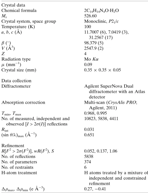
Crystal data
Chemical formula 2C14H14N4OH2O
Mr 526.60
Crystal system, space group Monoclinic,P21/c
Temperature (K) 100
a,b,c(A˚ ) 11.7007 (6), 7.0419 (3), 31.2567 (17) (
) 98.379 (5)
V(A˚3) 2547.9 (2)
Z 4
Radiation type MoK
(mm1) 0.09
Crystal size (mm) 0.350.350.05
Data collection
Diffractometer Agilent SuperNova Dual
diffractometer with an Atlas detector
Absorption correction Multi-scan (CrysAlis PRO; Agilent, 2011)
Tmin,Tmax 0.968, 0.995
No. of measured, independent and observed [I> 2(I)] reflections
10823, 5838, 4411
Rint 0.031
(sin/)max(A˚
1
) 0.651
Refinement
R[F2> 2(F2)],wR(F2),S 0.052, 0.137, 1.06
No. of reflections 5838
No. of parameters 374
No. of restraints 6
H-atom treatment H atoms treated by a mixture of independent and constrained refinement
max, min(e A˚
3) 0.27,0.41
Computer programs:CrysAlis PRO(Agilent, 2011),SHELXS97 (Sheldrick, 2015a),
[image:5.610.46.299.558.696.2]6. Synthesis and crystallization
4-Acetoacetyl-3-methyl-1-phenyl-2-pyrazolin-5-one 1 (2.5 g, 10 mmol) and hydrazin hydrazine (1 ml) were refluxed in a mixture of ethanol (50 ml) and acetic acid (50 ml) for 2 h. The reaction mixture was allowed to stand at room temperature. The precipitate was filtered and recrystallized from ethanol solution as fine needles, M.p. 409–410 K. Yield: 70%.
7. Refinement details
Crystal data, data collection and structure refinement details are summarized in Table 6. The carbon-bound H atoms were placed in calculated positions (C—H = 0.95–0.98 A˚ ) and were included in the refinement in the riding-model approximation, with Uiso(H) set to 1.2–1.5Ueq(C). The O- and N-bound H
atoms were refined with distance restraints of 0.840.01 and 0.860.01 A˚ , respectively, and withUiso(H) = 1.5Ueq(O) and
1.2Ueq(N).
Acknowledgements
The University of Malaya’s X-ray crystallography laboratory is thanked for the intensity data.
Funding information
This research was funded by the Deanship of Scientific Research (DSR) at King Abdulaziz University, Jeddah, under grant No. RG-03–102-428.
References
Agilent (2011). CrysAlis PRO. Agilent Technologies, Yarnton, England.
Asiri, A. M., Al-Youbi, A. O., Faidallah, H. M., Alamry, K. A. & Ng, S. W. (2011).Acta Cryst.E67, o2474.
Badawey, E. A. M. & El-Ashmawey, I. M. (1998).Eur. J. Med. Chem. 33, 349–361.
Bertolasi, V., Gilli, P., Ferretti, V. & Gilli, G. (1995).Acta Cryst.B51, 1004–1015.
Brandenburg, K. (2006).DIAMOND. Crystal Impact GbR, Bonn, Germany.
Das, N., Verma, A., Shrivastava, P. K. & Shrivastava, S. K. (2008).
Indian J. Chem. Sect. B,47, 1555–1558.
Farrugia, L. J. (2012).J. Appl. Cryst.45, 849–854.
Gans, J. & Shalloway, D. (2001). J. Mol. Graph. Model. 19, 557– 559.
Ghandour, I., Mague, J. T., Bouayad, A., Chakroune, S., Essassi, E. M. & Kandri Rodi, Y. (2017).IUCrData,2, x170853.
Higashi, Y., Jitsuiki, D., Chayama, K. & Yoshizumi, M. (2006).Recent.
Pat. Cardiovasc. Drug. Discov.1, 85–93.
Kumar, D., Singh, S. P., Martı´nez, A., Fruchier, A., Elguero, J., Martı´nez-Ripoll, M., Carrio´, J. S. & Virgili, A. (1995).Tetrahedron,
51, 4891–4906.
Pizzala, H., Carles, M., Stone, W. E. E. & Thevand, A. (2000).J. Mol.
Struct.526, 261–268.
Rabnawaz, M., Raza Shah, M. & Ng, S. W. (2010).Acta Cryst.E66, o2569.
Rubcˇic´, M., Uzˇarevic´, K., Halasz, I., Bregovic´, N., Malisˇ, M., Ðilovic´, I., Kokan, Z., Stein, R. S., Dinnebier, R. E. & Tomisˇic´, V. (2012).
Chem. Eur. J.18, 5620–5631.
Sahu, S. K., Azam, A. M., Banerjee, M., Choudhary, P., Sutradhar, S., Panda, P. K. & Misra, P. K. (2007).J. Indian Chem. Soc.84, 1011– 1015.
Sharma, B. D. & McConnell, J. F. (1965).Acta Cryst.19, 797–806. Sheldrick, G. M. (2015a).Acta Cryst.A71, 3–8.
Sheldrick, G. M. (2015b).Acta Cryst.C71, 3–8.
Singh, D. & Singh, D. (1991).J. Indian Chem. Soc.68, 165–167. Steiner, T. & Koellner, G. (1997).Chem. Commun.pp. 1207–1208. Tan, S. L., Jotani, M. M. & Tiekink, E. R. T. (2019).Acta Cryst.E75,
308–318.
Turner, M. J., Mckinnon, J. J., Wolff, S. K., Grimwood, D. J., Spackman, P. R., Jayatilaka, D. & Spackman, M. A. (2017). Crystal Explorer v17. The University of Western Australia.
Westrip, S. P. (2010).J. Appl. Cryst.43, 920–925.
570
Asiriet al. 2C14H14N4OH2O Acta Cryst.(2019). E75, 565–570
supporting information
sup-1
Acta Cryst. (2019). E75, 565-570
supporting information
Acta Cryst. (2019). E75, 565-570 [https://doi.org/10.1107/S2056989019004389]
Co-crystallization of a neutral molecule and its zwitterionic tautomer: structure
and Hirshfeld surface analysis of 5-methyl-4-(5-methyl-1
H
-pyrazol-3-yl)-2-phenyl-2,3-dihydro-1
H
-pyrazol-3-one 5-methyl-4-(5-methyl-1
H
-pyrazol-2-ium-3-yl)-3-oxo-2-phenyl-2,3-dihydro-1
H
-pyrazol-1-ide monohydrate
Abdullah M. Asiri, Khalid A. H. Alzahrani, Hassan M. Faidallah, Khalid A. Alamry, Mukesh M.
Jotani and Edward R. T. Tiekink
Computing details
Data collection: CrysAlis PRO (Agilent, 2011); cell refinement: CrysAlis PRO (Agilent, 2011); data reduction: CrysAlis
PRO (Agilent, 2011); program(s) used to solve structure: SHELXS97 (Sheldrick, 2015a); program(s) used to refine
structure: SHELXL2018 (Sheldrick, 2015b); molecular graphics: ORTEP-3 for Windows (Farrugia, 2012), QMolGans &
Shalloway (2001) and DIAMOND (Brandenburg, 2006); software used to prepare material for publication: publCIF
(Westrip, 2010).
5-Methyl-4-(5-methyl-1H-pyrazol-3-yl)-2-phenyl-2,3-dihydro-1H-pyrazol-3-one 5-methyl-4-(5-methyl-1H
-pyrazol-2-ium-3-yl)-3-oxo-2-phenyl-2,3-dihydro-1H-pyrazol-1-ide monohydrate
Crystal data
2C14H14N4O·H2O Mr = 526.60 Monoclinic, P21/c a = 11.7007 (6) Å b = 7.0419 (3) Å c = 31.2567 (17) Å β = 98.379 (5)° V = 2547.9 (2) Å3
Z = 4
F(000) = 1112 Dx = 1.373 Mg m−3
Mo Kα radiation, λ = 0.71073 Å Cell parameters from 3750 reflections θ = 2.3–27.5°
µ = 0.09 mm−1 T = 100 K Plate, colourless 0.35 × 0.35 × 0.05 mm
Data collection
Agilent SuperNova Dual
diffractometer with an Atlas detector Radiation source: SuperNova (Mo) X-ray
Source
Mirror monochromator
Detector resolution: 10.4041 pixels mm-1 ω scan
Absorption correction: multi-scan (CrysAlis PRO; Agilent, 2011)
Tmin = 0.968, Tmax = 0.995 10823 measured reflections 5838 independent reflections 4411 reflections with I > 2σ(I) Rint = 0.031
θmax = 27.6°, θmin = 2.4° h = −15→9
supporting information
sup-2
Acta Cryst. (2019). E75, 565-570
Refinement
Refinement on F2 Least-squares matrix: full R[F2 > 2σ(F2)] = 0.052 wR(F2) = 0.137 S = 1.06 5838 reflections 374 parameters 6 restraints
Primary atom site location: structure-invariant direct methods
Hydrogen site location: mixed
H atoms treated by a mixture of independent and constrained refinement
w = 1/[σ2(F
o2) + (0.0593P)2 + 0.7451P] where P = (Fo2 + 2Fc2)/3
(Δ/σ)max = 0.001 Δρmax = 0.27 e Å−3 Δρmin = −0.40 e Å−3
Special details
Geometry. All esds (except the esd in the dihedral angle between two l.s. planes) are estimated using the full covariance matrix. The cell esds are taken into account individually in the estimation of esds in distances, angles and torsion angles; correlations between esds in cell parameters are only used when they are defined by crystal symmetry. An approximate (isotropic) treatment of cell esds is used for estimating esds involving l.s. planes.
Fractional atomic coordinates and isotropic or equivalent isotropic displacement parameters (Å2)
x y z Uiso*/Ueq
supporting information
sup-3
Acta Cryst. (2019). E75, 565-570
C13 0.29632 (14) 0.0537 (3) 0.78551 (6) 0.0193 (4) H13 0.284374 0.077360 0.814448 0.023* C14 0.40700 (14) 0.0643 (2) 0.77450 (6) 0.0178 (4) H14 0.470414 0.098477 0.795649 0.021* O2 0.77522 (10) 0.25890 (19) 0.51120 (4) 0.0248 (3) N5 0.74784 (12) 0.3174 (2) 0.43598 (5) 0.0155 (3) N6 0.65481 (12) 0.3330 (2) 0.40247 (5) 0.0173 (3) N7 0.56152 (12) 0.2282 (2) 0.53983 (5) 0.0178 (3) H7N 0.6363 (9) 0.222 (3) 0.5506 (6) 0.021* N8 0.47846 (12) 0.1938 (2) 0.56454 (5) 0.0177 (3) H8N 0.4983 (16) 0.166 (3) 0.5926 (3) 0.021* C15 0.71023 (14) 0.2837 (2) 0.47567 (6) 0.0165 (4) C16 0.56071 (14) 0.3123 (2) 0.42123 (6) 0.0163 (4) C17 0.58821 (14) 0.2820 (2) 0.46598 (5) 0.0153 (3) C18 0.44402 (15) 0.3236 (3) 0.39471 (6) 0.0237 (4) H18A 0.452353 0.350197 0.364543 0.036* H18B 0.399461 0.425608 0.405806 0.036* H18C 0.403635 0.202539 0.396371 0.036* C19 0.51492 (14) 0.2495 (2) 0.49802 (5) 0.0151 (3) C20 0.39512 (14) 0.2301 (2) 0.49695 (6) 0.0169 (4) H20 0.337894 0.239186 0.472140 0.020* C21 0.37622 (14) 0.1949 (2) 0.53908 (6) 0.0166 (4) C22 0.26704 (15) 0.1657 (3) 0.55739 (6) 0.0220 (4) H22A 0.282458 0.086869 0.583433 0.033* H22B 0.210356 0.102077 0.535950 0.033* H22C 0.236420 0.288951 0.564862 0.033* C23 0.86164 (14) 0.3190 (2) 0.42595 (6) 0.0158 (4) C24 0.95656 (15) 0.3283 (3) 0.45857 (6) 0.0241 (4) H24 0.945472 0.331769 0.488071 0.029* C25 1.06731 (16) 0.3324 (3) 0.44760 (7) 0.0287 (5) H25 1.131685 0.340211 0.469881 0.034* C26 1.08599 (15) 0.3254 (3) 0.40489 (6) 0.0248 (4) H26 1.162274 0.326228 0.397846 0.030* C27 0.99162 (15) 0.3172 (3) 0.37267 (6) 0.0207 (4) H27 1.003385 0.313770 0.343246 0.025* C28 0.87980 (14) 0.3139 (2) 0.38275 (6) 0.0180 (4) H28 0.815760 0.308261 0.360297 0.022* O1W 0.65340 (10) 0.59378 (18) 0.33486 (4) 0.0195 (3) H1W 0.6112 (15) 0.686 (2) 0.3408 (7) 0.029* H2W 0.6537 (17) 0.512 (2) 0.3552 (5) 0.029*
Atomic displacement parameters (Å2)
U11 U22 U33 U12 U13 U23
supporting information
sup-4
Acta Cryst. (2019). E75, 565-570
N4 0.0173 (7) 0.0344 (9) 0.0117 (8) 0.0005 (6) 0.0052 (6) 0.0054 (7) C1 0.0185 (8) 0.0150 (8) 0.0116 (8) −0.0001 (6) 0.0042 (7) −0.0001 (7) C2 0.0183 (8) 0.0167 (8) 0.0136 (9) −0.0010 (6) 0.0047 (7) 0.0004 (7) C3 0.0164 (8) 0.0174 (8) 0.0114 (8) 0.0005 (6) 0.0031 (6) 0.0009 (7) C4 0.0182 (8) 0.0248 (9) 0.0150 (9) −0.0011 (7) 0.0016 (7) 0.0021 (7) C5 0.0168 (8) 0.0161 (8) 0.0124 (8) −0.0002 (6) 0.0035 (7) −0.0008 (7) C6 0.0158 (8) 0.0238 (9) 0.0122 (9) 0.0001 (7) 0.0023 (7) 0.0012 (7) C7 0.0180 (8) 0.0185 (8) 0.0158 (9) 0.0023 (7) 0.0056 (7) 0.0025 (7) C8 0.0186 (9) 0.0255 (9) 0.0215 (10) 0.0017 (7) 0.0092 (7) 0.0046 (8) C9 0.0162 (8) 0.0160 (8) 0.0141 (9) 0.0008 (6) 0.0054 (7) 0.0038 (7) C10 0.0213 (8) 0.0199 (8) 0.0124 (9) −0.0004 (7) 0.0038 (7) 0.0014 (7) C11 0.0188 (8) 0.0212 (9) 0.0180 (9) −0.0016 (7) 0.0000 (7) 0.0011 (7) C12 0.0170 (8) 0.0224 (9) 0.0206 (10) −0.0006 (7) 0.0068 (7) 0.0012 (8) C13 0.0211 (9) 0.0255 (9) 0.0126 (9) 0.0021 (7) 0.0065 (7) 0.0005 (7) C14 0.0177 (8) 0.0234 (9) 0.0123 (9) 0.0007 (7) 0.0023 (7) 0.0011 (7) O2 0.0178 (6) 0.0448 (8) 0.0114 (6) −0.0039 (6) 0.0005 (5) 0.0048 (6) N5 0.0149 (7) 0.0214 (7) 0.0103 (7) 0.0001 (6) 0.0025 (5) 0.0021 (6) N6 0.0153 (7) 0.0249 (8) 0.0111 (7) 0.0002 (6) 0.0003 (6) 0.0011 (6) N7 0.0153 (7) 0.0268 (8) 0.0118 (7) −0.0001 (6) 0.0038 (6) 0.0005 (6) N8 0.0192 (7) 0.0247 (8) 0.0101 (7) −0.0011 (6) 0.0050 (6) 0.0012 (6) C15 0.0185 (8) 0.0197 (8) 0.0116 (8) −0.0021 (7) 0.0031 (7) −0.0008 (7) C16 0.0176 (8) 0.0197 (8) 0.0122 (9) 0.0002 (7) 0.0043 (7) 0.0011 (7) C17 0.0167 (8) 0.0182 (8) 0.0115 (8) −0.0014 (6) 0.0036 (6) −0.0008 (7) C18 0.0165 (8) 0.0415 (11) 0.0132 (9) −0.0004 (8) 0.0023 (7) 0.0054 (8) C19 0.0182 (8) 0.0166 (8) 0.0109 (8) −0.0002 (6) 0.0032 (6) −0.0012 (7) C20 0.0161 (8) 0.0222 (9) 0.0124 (9) −0.0011 (7) 0.0027 (6) −0.0001 (7) C21 0.0181 (8) 0.0169 (8) 0.0155 (9) −0.0002 (6) 0.0046 (7) 0.0006 (7) C22 0.0220 (9) 0.0267 (9) 0.0190 (10) −0.0002 (7) 0.0094 (7) 0.0006 (8) C23 0.0156 (8) 0.0176 (8) 0.0151 (9) 0.0000 (6) 0.0053 (7) 0.0016 (7) C24 0.0199 (9) 0.0377 (11) 0.0148 (9) −0.0012 (8) 0.0033 (7) 0.0013 (8) C25 0.0189 (9) 0.0462 (12) 0.0206 (10) −0.0001 (8) 0.0014 (8) 0.0044 (9) C26 0.0174 (9) 0.0330 (10) 0.0257 (11) 0.0013 (8) 0.0085 (8) 0.0033 (8) C27 0.0214 (9) 0.0252 (9) 0.0172 (9) 0.0017 (7) 0.0090 (7) 0.0000 (7) C28 0.0180 (8) 0.0224 (9) 0.0136 (9) 0.0002 (7) 0.0027 (7) 0.0000 (7) O1W 0.0239 (7) 0.0224 (6) 0.0131 (7) 0.0050 (5) 0.0058 (5) 0.0022 (5)
Geometric parameters (Å, º)
supporting information
sup-5
Acta Cryst. (2019). E75, 565-570
C1—C3 1.429 (2) C16—C17 1.405 (2) C2—C3 1.391 (2) C16—C18 1.493 (2) C2—C4 1.490 (2) C17—C19 1.429 (2) C3—C5 1.461 (2) C18—H18A 0.9800 C4—H4A 0.9800 C18—H18B 0.9800 C4—H4B 0.9800 C18—H18C 0.9800 C4—H4C 0.9800 C19—C20 1.404 (2) C5—C6 1.414 (2) C20—C21 1.389 (2) C6—C7 1.375 (2) C20—H20 0.9500 C6—H6 0.9500 C21—C22 1.488 (2) C7—C8 1.493 (2) C22—H22A 0.9800 C8—H8A 0.9800 C22—H22B 0.9800 C8—H8B 0.9800 C22—H22C 0.9800 C8—H8C 0.9800 C23—C24 1.396 (2) C9—C14 1.394 (2) C23—C28 1.397 (2) C9—C10 1.395 (2) C24—C25 1.388 (2) C10—C11 1.390 (2) C24—H24 0.9500 C10—H10 0.9500 C25—C26 1.385 (3) C11—C12 1.389 (2) C25—H25 0.9500 C11—H11 0.9500 C26—C27 1.383 (3) C12—C13 1.384 (2) C26—H26 0.9500 C12—H12 0.9500 C27—C28 1.390 (2) C13—C14 1.390 (2) C27—H27 0.9500 C13—H13 0.9500 C28—H28 0.9500 C14—H14 0.9500 O1W—H1W 0.851 (9) O2—C15 1.263 (2) O1W—H2W 0.858 (9)
supporting information
sup-6
Acta Cryst. (2019). E75, 565-570
H4A—C4—H4B 109.5 C16—C18—H18C 109.5 C2—C4—H4C 109.5 H18A—C18—H18C 109.5 H4A—C4—H4C 109.5 H18B—C18—H18C 109.5 H4B—C4—H4C 109.5 N7—C19—C20 105.87 (15) N3—C5—C6 110.48 (15) N7—C19—C17 119.85 (15) N3—C5—C3 121.32 (15) C20—C19—C17 134.28 (16) C6—C5—C3 128.19 (16) C21—C20—C19 106.93 (15) C7—C6—C5 105.80 (15) C21—C20—H20 126.5 C7—C6—H6 127.1 C19—C20—H20 126.5 C5—C6—H6 127.1 N8—C21—C20 108.26 (14) N4—C7—C6 106.17 (15) N8—C21—C22 120.92 (16) N4—C7—C8 121.10 (16) C20—C21—C22 130.81 (16) C6—C7—C8 132.67 (16) C21—C22—H22A 109.5 C7—C8—H8A 109.5 C21—C22—H22B 109.5 C7—C8—H8B 109.5 H22A—C22—H22B 109.5 H8A—C8—H8B 109.5 C21—C22—H22C 109.5 C7—C8—H8C 109.5 H22A—C22—H22C 109.5 H8A—C8—H8C 109.5 H22B—C22—H22C 109.5 H8B—C8—H8C 109.5 C24—C23—C28 119.36 (15) C14—C9—C10 120.62 (15) C24—C23—N5 120.93 (16) C14—C9—N1 119.57 (15) C28—C23—N5 119.70 (15) C10—C9—N1 119.81 (15) C25—C24—C23 119.51 (18) C11—C10—C9 118.77 (16) C25—C24—H24 120.2 C11—C10—H10 120.6 C23—C24—H24 120.2 C9—C10—H10 120.6 C26—C25—C24 121.44 (18) C12—C11—C10 120.89 (16) C26—C25—H25 119.3 C12—C11—H11 119.6 C24—C25—H25 119.3 C10—C11—H11 119.6 C27—C26—C25 118.82 (16) C13—C12—C11 119.79 (16) C27—C26—H26 120.6 C13—C12—H12 120.1 C25—C26—H26 120.6 C11—C12—H12 120.1 C26—C27—C28 120.91 (17) C12—C13—C14 120.26 (16) C26—C27—H27 119.5 C12—C13—H13 119.9 C28—C27—H27 119.5 C14—C13—H13 119.9 C27—C28—C23 119.95 (16) C13—C14—C9 119.61 (16) C27—C28—H28 120.0 C13—C14—H14 120.2 C23—C28—H28 120.0 C9—C14—H14 120.2 H1W—O1W—H2W 107.0 (19) C15—N5—N6 111.51 (13)
supporting information
sup-7
Acta Cryst. (2019). E75, 565-570
N2—C2—C3—C1 −0.27 (19) N6—C16—C17—C15 −0.1 (2) C4—C2—C3—C1 179.35 (17) C18—C16—C17—C15 −179.85 (17) N2—C2—C3—C5 177.71 (15) N6—C16—C17—C19 −178.60 (17) C4—C2—C3—C5 −2.7 (3) C18—C16—C17—C19 1.7 (3) O1—C1—C3—C2 176.04 (17) O2—C15—C17—C16 −178.44 (18) N1—C1—C3—C2 −1.59 (18) N5—C15—C17—C16 0.70 (18) O1—C1—C3—C5 −1.9 (3) O2—C15—C17—C19 0.2 (3) N1—C1—C3—C5 −179.55 (15) N5—C15—C17—C19 179.32 (15) N4—N3—C5—C6 0.35 (19) N8—N7—C19—C20 1.01 (19) N4—N3—C5—C3 −179.20 (15) N8—N7—C19—C17 −178.27 (15) C2—C3—C5—N3 170.80 (16) C16—C17—C19—N7 −178.44 (17) C1—C3—C5—N3 −11.6 (3) C15—C17—C19—N7 3.3 (3) C2—C3—C5—C6 −8.7 (3) C16—C17—C19—C20 2.5 (3) C1—C3—C5—C6 168.93 (17) C15—C17—C19—C20 −175.72 (18) N3—C5—C6—C7 −0.34 (19) N7—C19—C20—C21 −0.76 (19) C3—C5—C6—C7 179.16 (16) C17—C19—C20—C21 178.35 (18) N3—N4—C7—C6 0.0 (2) N7—N8—C21—C20 0.35 (19) N3—N4—C7—C8 177.54 (15) N7—N8—C21—C22 −178.43 (15) C5—C6—C7—N4 0.18 (19) C19—C20—C21—N8 0.26 (19) C5—C6—C7—C8 −176.92 (18) C19—C20—C21—C22 178.88 (18) N2—N1—C9—C14 26.2 (2) C15—N5—C23—C24 −15.1 (3) C1—N1—C9—C14 −148.83 (17) N6—N5—C23—C24 171.73 (15) N2—N1—C9—C10 −153.49 (15) C15—N5—C23—C28 165.82 (16) C1—N1—C9—C10 31.5 (3) N6—N5—C23—C28 −7.4 (2) C14—C9—C10—C11 2.1 (3) C28—C23—C24—C25 0.0 (3) N1—C9—C10—C11 −178.19 (15) N5—C23—C24—C25 −179.08 (17) C9—C10—C11—C12 −2.8 (3) C23—C24—C25—C26 −0.7 (3) C10—C11—C12—C13 1.2 (3) C24—C25—C26—C27 1.0 (3) C11—C12—C13—C14 1.1 (3) C25—C26—C27—C28 −0.7 (3) C12—C13—C14—C9 −1.7 (3) C26—C27—C28—C23 0.0 (3) C10—C9—C14—C13 0.1 (3) C24—C23—C28—C27 0.3 (3) N1—C9—C14—C13 −179.61 (16) N5—C23—C28—C27 179.44 (16)
Hydrogen-bond geometry (Å, º)
Cg1 is the centroid of the (C23–C28) ring.
D—H···A D—H H···A D···A D—H···A
N2—H2N···O1Wi 0.90 (1) 1.79 (1) 2.673 (2) 168 (2) N4—H4N···O2 0.88 (1) 1.90 (1) 2.764 (2) 168 (2) N7—H7N···N3 0.89 (1) 2.28 (2) 2.970 (2) 134 (1) N8—H8N···O1 0.90 (1) 1.79 (1) 2.664 (2) 166 (2) O1W—H1W···O1ii 0.85 (2) 1.92 (2) 2.7641 (17) 172 (2) O1W—H2W···N6 0.86 (2) 1.94 (2) 2.7979 (19) 178 (2) C8—H8A···Cg1iii 0.98 2.71 3.492 (2) 137 C8—H8B···Cg1iv 0.98 2.89 3.755 (2) 148