492
https://doi.org/10.1107/S2056989018003791 Acta Cryst.(2018). E74, 492–496research communications
Received 12 February 2018 Accepted 5 March 2018
Edited by W. T. A. Harrison, University of Aberdeen, Scotland
Keywords:crystal structure; chalcone; absorp-tion spectra; HOMO–LUMO; Hirshfeld surface.
CCDC reference:1817217
Supporting information:this article has supporting information at journals.iucr.org/e
(
E
)-1,3-Bis(anthracen-9-yl)prop-2-en-1-one: crystal
structure and DFT study
Dian Alwani Zainuri, Ibrahim Abdul Razak and Suhana Arshad*
X-ray Crystallography Unit, School of Physics, Universiti Sains Malaysia, 11800 USM, Penang, Malaysia. *Correspondence e-mail: [email protected]
The title compound, C31H20O, was synthesized using a Claisen–Schmidt
condensation. The enone group adopts an s-trans conformation and the anthracene ring systems are twisted at angles of 85.21 (19) and 83.98 (19)from
the enone plane. In the crystal, molecules are connected into chains along [100] via weak C—H interactions. The observed band gap of 3.03 eV is in excellent agreement with that (3.07 eV) calculated using density functional theory (DFT) at the B3LYP/6–311++G(d,p) level. The Hirshfeld surface analysis indicates a high percentage of C H/H C (41.2%) contacts in the crystal.
1. Chemical context
Anthrancene and its derivatives constitute a very well-known class with interesting photophysical properties and they are used extensively in the design of luminescent chemosensors and switches (Montaltiet al., 2000). A chalcone molecule with a -conjugated system provides a large charge-transfer axis with appropriate substituent groups on the terminal aromatic rings. Strong intermolecular charge transfer (ICT) will give rise to second harmonic generation (SHG) efficiency and this may enhance the non-linear optical (NLO) properties (D’silva et al., 2011). Furthermore, -conjugated molecular materials with fused rings are the focus of considerable interest in the emerging area of organic electronics, since the combination of good charge-carrier mobility and high stability may lead to potential optoelectronic applications (Wuet al., 2010). As part of our work in this area, we now report the synthesis and combined experimental and theoretical studies of the title compound, (I).
2. Structural commentary
The molecular structure of (I) is shown in Fig. 1 (for the optimized structure, see Fig. S1 in the Supporting informa-tion). The structure consists of two anthracene rings (AnthA and AnthB) . AnthAis formed by the aromatic rings labeled as Cg1(C1–C6), Cg2(C1/C6–C8/C13/C14) and Cg3(C8–C13). Anth B consists of Cg4(C18/C19/C24–C26/C31, Cg5(C19– C24) andCg6(C26–C31).
The C—C distances in the central ring of the anthracene units show little variation compared to the other rings (Anth A: C20—C21, C22—C23, C27—C28 and C29—C30; AnthB: C2—C3, C4—C5, C9—C10 and C11—C12), which are much shorter. These observations are consistent with an electronic structure for the anthracene units where a central ring displaying aromatic delocalization is flanked by two isolated diene units (Glidewell & Lloyd, 1984). Both theoretical and experimental structures exist in an E configuration with respect to the C16 C17 double bond [experimental = 1.291 (2) A˚ and DFT (see below) = 1.34 A˚].
The enone moiety (O1/C15–C17) shows ans-trans config-uration with the O1—C15—C16—C17 torsion angle being 179.19 (19) and 179.64 in the experimental and calculated structures, respectively. Additionally, the enone moiety [O1/ C15–C17, maximum deviation of 0.0039 (18) A˚ at C16] forms dihedral angles of 85.21 (19) and 83.98 (19)with the AnthA
[C1–C14, maximum deviation of 0.103 (2) A˚ at C11] and Anth B [C18–C31, maximum deviation of 0.016 (3) A˚ at C27] groups, respectively. The large dihedral-angle deviation indi-cates that the possibility for electronic effects between the anthracene units through the enone moiety has decreased (Jung et al., 2008). This is in contrast with the molecular structure of (E )-1-(anthracen-9-yl)-3-(2-chloro-6-fluorophen-yl)prop-2-en-1-one (Abdullah et al. 2016), which shows the enone moiety locked in an s-cisconfiguration because of the intramolecular hydrogen bond. Furthermore, the bulkiness of the anthracene ring gives rise to a highly twisted structure at
both terminal rings. Compound (I) is twisted at the C17—C18 and C14—C15 bonds with C16—C17—C18—C19 and C1— C14—C15—C16 torsion angles of 84.0 (2) and 93.65 (19),
respectively (see Fig. S2 in the Supporting information). The corresponding torsion angles for the DFT study are 48.01 and 94.05, respectively. We propose that the torsion-angle
difference of about 35.9between the experimental and DFT studies are the result of the formation of intermolecular C— H interactions involving the anthracene units. The observed intermolecular interactions in the crystal packing are the main cause of the angle difference when this interaction is not taken into consideration during the optimization process.
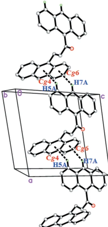
3. Supramolecular features
In the crystal of (I), C—H interactions are mainly responsible for the packing. Two C—H interactions (Fig. 2 and Table 1) occur between anthracene rings (Anth A and AnthB), connecting the molecules into infinite zigzag chains propagating along the [100] direction.
research communications
Acta Cryst.(2018). E74, 492–496 Zainuriet al. C
[image:2.610.45.295.119.158.2]31H20O
493
Figure 2 [image:2.610.348.529.347.726.2]The weak C—H interactions in the crystal of (I).
Table 1
Hydrogen-bond geometry (A˚ ,).
Cg4 andCg6 are the centroids of the C18/C19/C24–C26/C31 and C26–C31 rings, respectively.
D—H A D—H H A D A D—H A
C5—H5A Cg4i 0.93 2.75 3.511 (2) 140
C7—H7A Cg6i 0.93 2.91 3.672 (2) 140
[image:2.610.47.294.555.727.2]Symmetry code: (i)x 1;y;z.
Figure 1
4. Theoretical chemistry study
The optimization of the molecular geometries leading to energy minima was achieved using DFT [with Becke’s non-local three parameter exchange and the Lee–Yang–Parr correlation functional (B3LYP)] with the 6-311++G (d,p) basis set as implemented inGaussian09program package (Frischet al., 2009). The selected bond lengths and angles of the opti-mized structure in comparison to the experimental values are presented in Table S2 in the Supporting information and the optimized structure is presented in Figure S1. Agreement between experimental and calculated geometrical data is generally good and any deviations may be ascribed to the fact that the optimization is performed in an isolated condition, whereas the crystal environment affects the molecular geometry (Ramyaet al., 2015).
5. Absorption spectrum and frontier molecular orbitals
The longest wavelength absorption maxima for (I) is observed in the UV region at 383 nm as shown in Fig. 3. The TD–DFT calculation at the B3LYP/6-311G++(d,p) level shows that this feature is due to an electronic transition from the highest occupied molecular orbital (HOMO) to the lowest unoccupied molecular orbital (LUMO). In the ground state (HOMO), the charge densities are mainly delocalized over the anthracene rings and the enone moiety, while in the LUMO state, the charge densities are accumulated on the Anth Aand enone moiety (see Fig. S3 in the Supporting information). The calculated max of 390 nm is shifted from the experimental
value, which may be attributed to solvent effects, compared to the gas-phase calculation.
The HOMO–LUMO energy gap (Fig. S3) relates to the chemical activity of the molecule (Kosar & Albayrak, 2011). The predicted energy gap of 3.07 eV shows excellent agree-ment with the estimated experiagree-mental energy gap of 3.03 eV. These optical band-gap values indicate the potential suitability of this compound for optoelectronic applications, as
previously reported by Prabhu et al. (2016). Additionally, Nietfeldet al.(2011) compared the structural, electrochemical and optical properties of fused-ring and non-fused ring compounds, indicating that fused rings have lower band gaps than other structures.
6. Hirshfeld Surface analysis
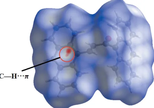
Fig. 4 shows the Hirshfeld surface mapped over dnorm. As
expected, the dnorm surfaces reveal the C—H
inter-molecular interaction as a large depression (bright-red spot). The presence of this C—H interaction is also indicated through the combination of pale-orange and bright-red spots that are present on the Hirshfeld surfaces mapped over de
(Fig. 5a) and shape-index (Fig. 5b).
The two-dimensional fingerprint plots shown in Fig. 6 illustrate the difference between the intermolecular inter-action patterns and the major intermolecular contacts asso-ciated with the title compound. The H H contacts (Fig. 6b) appear to be the major contributor to the Hirshfeld surface and are seen as one distinct spike with a minimum value forde
+dithat is less than the sum of the van der Waals radii (2.4 A˚ ).
The intermolecular C—H interactions are characterized by the short interatomic C H/H C (41.2%) contacts and
494
Zainuriet al. C31H20O Acta Cryst.(2018). E74, 492–496
[image:3.610.45.291.68.256.2]research communications
Figure 3
[image:3.610.317.563.75.248.2]UV–Vis absorption spectra of (I).
Figure 4
[image:3.610.316.565.592.709.2]View of the Hirshfeld surfaces mapped overdnormfor (I).
Figure 5
View of the Hirshfeld surfaces for (I) mapped over (a)deand (b)
their presence is indicated by the distribution of points around a pair of wings atde+di2.6 A˚ (Fig. 6c).
7. Database survey
A survey of the Cambridge Structural Database (CSD, Version 5.38, last update Nov 2016; Groom et al., 2016) revealed fused-ring substituted chalcones similar to the title compound. There are four compounds which have an anthracene-ketone subtituent on the chalcone: 9-anthryl styryl ketone and 9,10-anthryl bis(styryl ketone) (Harlow et al., 1975), (2E )-1-(anthracen-9-yl)-3-[4-(propan-2-yl)phenyl]-prop-2-en-1-one (Girishaet al., 2016) and (E )-1-(anthracen-9-yl)-3-(2-chloro-6-fluorophenyl) prop-2-en-1-one (Abdullah et al., 2016). Junget al.(2008) reported two ferrocenyl chalcones containing an anthracenyl subtituent, 9-(2-ferrocenylethenyl-carbonyl)anthracene and 1-(9-anthracenyl)-3-ferrocenyl-2-propen-1-one. Other related compounds include, 1-(anthrac-en-9-yl)-2-methylprop-2-en-1-one (Agrahari et al., 2015) and 9-anthroylacetone (Cicognaet al., 2004).
8. Synthesis and crystallization
A mixture of 9-acetylanthracene (0.5 mmol) and 9-anthrace-necarboxaldehyde (0.5 mmol) was dissolved in methanol (20 ml). A catalytic amount of NaOH (5 ml, 20%) was added to the solution dropwise with vigorous stirring. The reaction mixture was stirred for about 5-6 h at room temperature. After stirring, the contents of the flask were poured into ice-cold water (50 ml). The resultant crude products were filtered, washed successively with distilled water and recrystallized from acetone solution as yellow blocks. The single crystal (Fig. S4) used for data collection was obtained by the slow-evaporation technique using acetone as the solvent.
9. Refinement
Crystal data collection and structure refinement details are summarized in Table 2. All H atoms were positioned geome-trically (C—H =0.93 A˚ ) and refined using riding model with Uiso(H)=1.2Ueq(C).
Funding information
The authors thank the Malaysian Government and Universiti Sains Malaysia (USM) for the research facilities and the Fundamental Research Grant Scheme (FRGS) No. 203/ PFIZIK/6711572 and for Short Term Grant Scheme (304/ PFIZIK/6313336) to conduct this work. DAZ thanks the Malaysian Government for the My Brain15 scholarship.
References
Abdullah, A. A., Hassan, N. H. H., Arshad, S., Khalib, N. C. & Razak, I. A. (2016).Acta Cryst.E72, 648–651.
Agrahari, A., Wagers, P. O., Schildcrout, S. M., Masnovi, J. & Youngs, W. J. (2015).Acta Cryst.E71, 357–359.
Bruker (2009). APEX2, SAINT and SADABS. Bruker AXS Inc., Madison, Wisconsin, USA.
Cicogna, F., Ingrosso, G., Lodato, F., Marchetti, F. & Zandomeneghi, M. (2004).Tetrahedron,60, 11959–11968.
D’silva, E. D., Podagatlapalli, G. K., Rao, S. V., Rao, D. N. & Dharmaprakash, S. M. (2011). Cryst. Growth Des. 11, 5362– 5369.
Frisch, M. J.,et al.(2009).Gaussian 09.Gaussian, Inc., Wallingford CT, USA.
Girisha, M., Yathirajan, H. S., Jasinski, J. P. & Glidewell, C. (2016). Acta Cryst.E72, 1153–1158.
Glidewell, C. & Lloyd, D. (1984).Tetrahedron,40, 4455–4472. Groom, C. R., Bruno, I. J., Lightfoot, M. P. & Ward, S. C. (2016).Acta
Cryst. B72, 171–179.
Harlow, R. L., Loghry, R. A., Williams, H. J. & Simonsen, S. H. (1975). Acta Cryst.B31, 1344–1350.
research communications
Acta Cryst.(2018). E74, 492–496 Zainuriet al. C
[image:4.610.46.299.68.161.2]31H20O
495
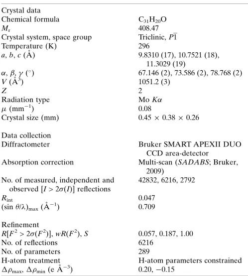
Table 2Experimental details.
Crystal data
Chemical formula C31H20O
Mr 408.47
Crystal system, space group Triclinic,P1
Temperature (K) 296
a,b,c(A˚ ) 9.8310 (17), 10.7521 (18), 11.3029 (19)
,,(
) 67.146 (2), 73.586 (2), 78.768 (2)
V(A˚3) 1051.2 (3)
Z 2
Radiation type MoK
(mm 1) 0.08
Crystal size (mm) 0.450.380.26
Data collection
Diffractometer Bruker SMART APEXII DUO CCD area-detector Absorption correction Multi-scan (SADABS; Bruker,
2009) No. of measured, independent and
observed [I> 2(I)] reflections
42832, 6216, 2792
Rint 0.047
(sin /)max(A˚ 1) 0.709
Refinement
R[F2> 2(F2)],wR(F2),S 0.057, 0.187, 1.00
No. of reflections 6216
No. of parameters 289
H-atom treatment H-atom parameters constrained max,min(e A˚ 3) 0.20, 0.15
[image:4.610.313.562.90.367.2]Computer programs:APEX2andSAINT(Bruker, 2009),SHELXL2013(Sheldrick, 2015),SHELXTL(Sheldrick, 2008) andPLATON(Spek, 2009).
Figure 6
Jung, Y., Son, K., Oh, Y. E. & Noh, D. (2008).Polyhedron,27, 861– 867.
Kosar, B. & Albayrak, C. (2011).Spectrochim. Acta A Mol. Biomol. Spectrosc.78, 160–167.
Montalti, M., Prodi, L. & Zaccheroni, N. (2000).J. Fluoresence,10, 71–76.
Nietfeld, J. P., Schwiderski, R. L., Gonnella, T. P. & Rasmussen, S. C. (2011).J. Org. Chem.76, 6383–6388.
Prabhu, A. N., Upadhyaya, V., Jayarama, A. & Bhat, K. B. (2016). Mol. Cryst. Liq. Cryst.637, 76–86.
Ramya, T., Gunasekaran, S. & Ramkumaar, G. R. (2015). Spectro-chim. Acta A Mol. Biomol. Spectrosc.149, 132–142.
Sheldrick, G. M. (2008).Acta Cryst. A64, 112–122. Sheldrick, G. M. (2015).Acta Cryst. C71, 3–8. Spek, A. L. (2009).Acta Cryst. D65, 148–155.
Wu, W., Liu, Y. & Zhu, D. (2010).Chem. Soc. Rev.39, 1489–1502.
496
Zainuriet al. C31H20O Acta Cryst.(2018). E74, 492–496
supporting information
sup-1 Acta Cryst. (2018). E74, 492-496
supporting information
Acta Cryst. (2018). E74, 492-496 [https://doi.org/10.1107/S2056989018003791]
(
E
)-1,3-Bis(anthracen-9-yl)prop-2-en-1-one: crystal structure and DFT study
Dian Alwani Zainuri, Ibrahim Abdul Razak and Suhana Arshad
Computing details
Data collection: APEX2 (Bruker, 2009); cell refinement: SAINT (Bruker, 2009); data reduction: SAINT (Bruker, 2009);
program(s) used to solve structure: SHELXTL (Sheldrick, 2008); program(s) used to refine structure: SHELXL2013
(Sheldrick, 2015); molecular graphics: SHELXTL (Sheldrick, 2008); software used to prepare material for publication:
SHELXTL (Sheldrick, 2008) and PLATON (Spek, 2009).
(E)-1,3-Bis(anthracen-9-yl)prop-2-en-1-one
Crystal data
C31H20O
Mr = 408.47
Triclinic, P1
a = 9.8310 (17) Å
b = 10.7521 (18) Å
c = 11.3029 (19) Å
α = 67.146 (2)°
β = 73.586 (2)°
γ = 78.768 (2)°
V = 1051.2 (3) Å3
Z = 2
F(000) = 428
Dx = 1.290 Mg m−3
Mo Kα radiation, λ = 0.71073 Å Cell parameters from 3766 reflections
θ = 2.3–22.1°
µ = 0.08 mm−1
T = 296 K Block, yellow
0.45 × 0.38 × 0.26 mm
Data collection
Bruker SMART APEXII DUO CCD area-detector
diffractometer
Radiation source: fine-focus sealed tube
φ and ω scans
Absorption correction: multi-scan (SADABS; Bruker, 2009)
42832 measured reflections 6216 independent reflections 2792 reflections with I > 2σ(I)
Rint = 0.047
θmax = 30.3°, θmin = 2.0°
h = −13→13
k = −15→15
l = −15→15
Refinement
Refinement on F2
Least-squares matrix: full
R[F2 > 2σ(F2)] = 0.057
wR(F2) = 0.187
S = 1.00 6216 reflections 289 parameters 0 restraints
Hydrogen site location: inferred from neighbouring sites
H-atom parameters constrained
w = 1/[σ2(F
o2) + (0.0746P)2 + 0.1037P]
where P = (Fo2 + 2Fc2)/3
(Δ/σ)max < 0.001
Δρmax = 0.20 e Å−3
supporting information
sup-2 Acta Cryst. (2018). E74, 492-496
Special details
Geometry. All esds (except the esd in the dihedral angle between two l.s. planes) are estimated using the full covariance matrix. The cell esds are taken into account individually in the estimation of esds in distances, angles and torsion angles; correlations between esds in cell parameters are only used when they are defined by crystal symmetry. An approximate (isotropic) treatment of cell esds is used for estimating esds involving l.s. planes.
Fractional atomic coordinates and isotropic or equivalent isotropic displacement parameters (Å2)
x y z Uiso*/Ueq
O1 0.44003 (14) 0.39564 (13) 0.83904 (17) 0.0974 (5) C1 0.18125 (17) 0.58896 (16) 0.72383 (17) 0.0557 (4)
C2 0.2456 (2) 0.55577 (19) 0.6096 (2) 0.0745 (5)
H2A 0.3398 0.5184 0.5977 0.089*
C3 0.1725 (3) 0.5774 (2) 0.5176 (2) 0.0895 (6)
H3A 0.2174 0.5560 0.4428 0.107*
C4 0.0287 (3) 0.6321 (2) 0.5335 (2) 0.0848 (6)
H4A −0.0202 0.6472 0.4690 0.102*
C5 −0.0372 (2) 0.66204 (17) 0.6411 (2) 0.0702 (5)
H5A −0.1325 0.6962 0.6513 0.084*
C6 0.03472 (17) 0.64307 (16) 0.74020 (18) 0.0564 (4) C7 −0.03088 (16) 0.67773 (16) 0.84986 (18) 0.0589 (4)
H7A −0.1270 0.7093 0.8621 0.071*
C8 0.04251 (16) 0.66686 (15) 0.94246 (17) 0.0551 (4) C9 −0.0227 (2) 0.70868 (18) 1.05166 (19) 0.0707 (5)
H9A −0.1189 0.7397 1.0651 0.085*
C10 0.0525 (3) 0.7042 (2) 1.1362 (2) 0.0834 (6)
H10A 0.0083 0.7335 1.2065 0.100*
C11 0.1973 (2) 0.6556 (2) 1.1191 (2) 0.0776 (5)
H11A 0.2487 0.6544 1.1772 0.093*
C12 0.26246 (19) 0.61070 (18) 1.01937 (18) 0.0659 (5)
H12A 0.3577 0.5762 1.0115 0.079*
C13 0.18947 (16) 0.61474 (15) 0.92571 (16) 0.0529 (4) C14 0.25475 (16) 0.57424 (15) 0.81840 (16) 0.0528 (4) C15 0.40796 (17) 0.51694 (18) 0.80046 (18) 0.0633 (5) C16 0.51726 (17) 0.61184 (17) 0.73382 (18) 0.0666 (5)
H16A 0.6119 0.5756 0.7213 0.080*
C17 0.49221 (16) 0.74227 (16) 0.69108 (16) 0.0564 (4)
H17A 0.3972 0.7775 0.7032 0.068*
C18 0.59990 (15) 0.84051 (15) 0.62495 (16) 0.0510 (4) C19 0.65765 (16) 0.87661 (16) 0.48951 (17) 0.0547 (4) C20 0.6174 (2) 0.82323 (19) 0.40902 (19) 0.0701 (5)
H20A 0.5499 0.7606 0.4473 0.084*
C21 0.6744 (2) 0.8611 (2) 0.2788 (2) 0.0881 (6)
H21A 0.6459 0.8247 0.2282 0.106*
C22 0.7768 (3) 0.9552 (2) 0.2183 (2) 0.0955 (7)
H22A 0.8155 0.9807 0.1280 0.115*
C23 0.8190 (2) 1.0082 (2) 0.2896 (2) 0.0830 (6)
supporting information
sup-3 Acta Cryst. (2018). E74, 492-496
C24 0.76171 (18) 0.97267 (17) 0.42761 (18) 0.0634 (5) C25 0.80165 (19) 1.02850 (18) 0.5019 (2) 0.0720 (5)
H25A 0.8687 1.0914 0.4605 0.086*
C26 0.74538 (19) 0.99421 (17) 0.6362 (2) 0.0658 (5)
C27 0.7864 (3) 1.0506 (2) 0.7140 (3) 0.0899 (7)
H27A 0.8524 1.1146 0.6740 0.108*
C28 0.7316 (3) 1.0133 (2) 0.8443 (3) 0.0980 (7)
H28A 0.7605 1.0513 0.8933 0.118*
C29 0.6313 (2) 0.9178 (2) 0.9075 (2) 0.0841 (6)
H29A 0.5946 0.8925 0.9981 0.101*
C30 0.58805 (19) 0.86234 (18) 0.83782 (19) 0.0676 (5)
H30A 0.5211 0.7994 0.8812 0.081*
C31 0.64201 (16) 0.89751 (15) 0.70003 (17) 0.0555 (4)
Atomic displacement parameters (Å2)
U11 U22 U33 U12 U13 U23
supporting information
sup-4 Acta Cryst. (2018). E74, 492-496
C31 0.0509 (9) 0.0493 (9) 0.0624 (11) 0.0010 (7) −0.0161 (8) −0.0162 (8)
Geometric parameters (Å, º)
O1—C15 1.2109 (19) C16—H16A 0.9300
C1—C14 1.397 (2) C17—C18 1.473 (2)
C1—C2 1.416 (3) C17—H17A 0.9300
C1—C6 1.433 (2) C18—C19 1.396 (2)
C2—C3 1.349 (3) C18—C31 1.403 (2)
C2—H2A 0.9300 C19—C20 1.418 (3)
C3—C4 1.411 (3) C19—C24 1.431 (2)
C3—H3A 0.9300 C20—C21 1.343 (3)
C4—C5 1.333 (3) C20—H20A 0.9300
C4—H4A 0.9300 C21—C22 1.406 (3)
C5—C6 1.418 (2) C21—H21A 0.9300
C5—H5A 0.9300 C22—C23 1.335 (3)
C6—C7 1.380 (2) C22—H22A 0.9300
C7—C8 1.389 (2) C23—C24 1.422 (3)
C7—H7A 0.9300 C23—H23A 0.9300
C8—C9 1.417 (2) C24—C25 1.374 (3)
C8—C13 1.432 (2) C25—C26 1.385 (3)
C9—C10 1.346 (3) C25—H25A 0.9300
C9—H9A 0.9300 C26—C27 1.419 (3)
C10—C11 1.404 (3) C26—C31 1.431 (2)
C10—H10A 0.9300 C27—C28 1.340 (3)
C11—C12 1.344 (3) C27—H27A 0.9300
C11—H11A 0.9300 C28—C29 1.401 (3)
C12—C13 1.421 (2) C28—H28A 0.9300
C12—H12A 0.9300 C29—C30 1.345 (3)
C13—C14 1.391 (2) C29—H29A 0.9300
C14—C15 1.501 (2) C30—C31 1.416 (2)
C15—C16 1.461 (2) C30—H30A 0.9300
C16—C17 1.291 (2)
C14—C1—C2 123.06 (16) C15—C16—H16A 117.6
C14—C1—C6 119.19 (16) C16—C17—C18 126.16 (14)
C2—C1—C6 117.74 (16) C16—C17—H17A 116.9
C3—C2—C1 121.13 (19) C18—C17—H17A 116.9
C3—C2—H2A 119.4 C19—C18—C31 120.92 (15)
C1—C2—H2A 119.4 C19—C18—C17 120.16 (15)
C2—C3—C4 120.9 (2) C31—C18—C17 118.92 (15)
C2—C3—H3A 119.6 C18—C19—C20 123.09 (16)
C4—C3—H3A 119.6 C18—C19—C24 119.04 (16)
C5—C4—C3 120.04 (19) C20—C19—C24 117.87 (16)
C5—C4—H4A 120.0 C21—C20—C19 121.5 (2)
C3—C4—H4A 120.0 C21—C20—H20A 119.3
C4—C5—C6 121.60 (19) C19—C20—H20A 119.3
supporting information
sup-5 Acta Cryst. (2018). E74, 492-496
C6—C5—H5A 119.2 C20—C21—H21A 119.7
C7—C6—C5 122.16 (16) C22—C21—H21A 119.7
C7—C6—C1 119.23 (15) C23—C22—C21 120.5 (2)
C5—C6—C1 118.59 (18) C23—C22—H22A 119.8
C6—C7—C8 121.90 (15) C21—C22—H22A 119.8
C6—C7—H7A 119.0 C22—C23—C24 121.5 (2)
C8—C7—H7A 119.0 C22—C23—H23A 119.2
C7—C8—C9 122.02 (16) C24—C23—H23A 119.2
C7—C8—C13 119.12 (16) C25—C24—C23 122.26 (19)
C9—C8—C13 118.85 (16) C25—C24—C19 119.60 (17)
C10—C9—C8 121.02 (18) C23—C24—C19 118.14 (19)
C10—C9—H9A 119.5 C24—C25—C26 122.21 (17)
C8—C9—H9A 119.5 C24—C25—H25A 118.9
C9—C10—C11 120.5 (2) C26—C25—H25A 118.9
C9—C10—H10A 119.8 C25—C26—C27 122.71 (19)
C11—C10—H10A 119.8 C25—C26—C31 118.98 (17)
C12—C11—C10 120.56 (19) C27—C26—C31 118.31 (19)
C12—C11—H11A 119.7 C28—C27—C26 121.1 (2)
C10—C11—H11A 119.7 C28—C27—H27A 119.4
C11—C12—C13 121.64 (18) C26—C27—H27A 119.4
C11—C12—H12A 119.2 C27—C28—C29 120.8 (2)
C13—C12—H12A 119.2 C27—C28—H28A 119.6
C14—C13—C12 123.25 (15) C29—C28—H28A 119.6
C14—C13—C8 119.31 (15) C30—C29—C28 120.3 (2)
C12—C13—C8 117.42 (16) C30—C29—H29A 119.9
C13—C14—C1 121.16 (15) C28—C29—H29A 119.9
C13—C14—C15 120.13 (15) C29—C30—C31 121.57 (19)
C1—C14—C15 118.69 (15) C29—C30—H30A 119.2
O1—C15—C16 120.98 (16) C31—C30—H30A 119.2
O1—C15—C14 120.98 (15) C18—C31—C30 122.86 (15)
C16—C15—C14 118.03 (14) C18—C31—C26 119.26 (16)
C17—C16—C15 124.87 (15) C30—C31—C26 117.88 (16)
C17—C16—H16A 117.6
C14—C1—C2—C3 −176.92 (17) C14—C15—C16—C17 1.4 (3)
C6—C1—C2—C3 1.6 (3) C15—C16—C17—C18 179.28 (17)
C1—C2—C3—C4 −0.9 (3) C16—C17—C18—C19 84.0 (2)
C2—C3—C4—C5 −0.6 (3) C16—C17—C18—C31 −96.6 (2)
C3—C4—C5—C6 1.4 (3) C31—C18—C19—C20 −179.19 (14)
C4—C5—C6—C7 177.88 (16) C17—C18—C19—C20 0.3 (2)
C4—C5—C6—C1 −0.7 (3) C31—C18—C19—C24 0.3 (2)
C14—C1—C6—C7 −0.8 (2) C17—C18—C19—C24 179.77 (14)
C2—C1—C6—C7 −179.38 (14) C18—C19—C20—C21 179.48 (16)
C14—C1—C6—C5 177.76 (14) C24—C19—C20—C21 0.0 (3)
C2—C1—C6—C5 −0.8 (2) C19—C20—C21—C22 0.2 (3)
C5—C6—C7—C8 −175.90 (14) C20—C21—C22—C23 0.1 (3)
C1—C6—C7—C8 2.6 (2) C21—C22—C23—C24 −0.5 (3)
supporting information
sup-6 Acta Cryst. (2018). E74, 492-496
C6—C7—C8—C13 −1.7 (2) C22—C23—C24—C19 0.7 (3)
C7—C8—C9—C10 −176.32 (16) C18—C19—C24—C25 −0.7 (2)
C13—C8—C9—C10 2.2 (3) C20—C19—C24—C25 178.85 (15)
C8—C9—C10—C11 −1.1 (3) C18—C19—C24—C23 −179.92 (15)
C9—C10—C11—C12 −1.1 (3) C20—C19—C24—C23 −0.4 (2)
C10—C11—C12—C13 2.1 (3) C23—C24—C25—C26 179.77 (16)
C11—C12—C13—C14 177.40 (16) C19—C24—C25—C26 0.6 (3)
C11—C12—C13—C8 −0.9 (2) C24—C25—C26—C27 179.65 (17)
C7—C8—C13—C14 −1.0 (2) C24—C25—C26—C31 −0.1 (3)
C9—C8—C13—C14 −179.59 (14) C25—C26—C27—C28 −178.81 (19)
C7—C8—C13—C12 177.34 (14) C31—C26—C27—C28 0.9 (3)
C9—C8—C13—C12 −1.2 (2) C26—C27—C28—C29 −0.3 (4)
C12—C13—C14—C1 −175.44 (14) C27—C28—C29—C30 −0.3 (3)
C8—C13—C14—C1 2.8 (2) C28—C29—C30—C31 0.4 (3)
C12—C13—C14—C15 2.9 (2) C19—C18—C31—C30 −178.97 (14)
C8—C13—C14—C15 −178.87 (14) C17—C18—C31—C30 1.6 (2)
C2—C1—C14—C13 176.59 (14) C19—C18—C31—C26 0.2 (2)
C6—C1—C14—C13 −1.9 (2) C17—C18—C31—C26 −179.29 (14)
C2—C1—C14—C15 −1.8 (2) C29—C30—C31—C18 179.39 (16)
C6—C1—C14—C15 179.75 (14) C29—C30—C31—C26 0.2 (3)
C13—C14—C15—O1 95.9 (2) C25—C26—C31—C18 −0.3 (2)
C1—C14—C15—O1 −85.7 (2) C27—C26—C31—C18 179.96 (15)
C13—C14—C15—C16 −84.7 (2) C25—C26—C31—C30 178.88 (15)
C1—C14—C15—C16 93.65 (19) C27—C26—C31—C30 −0.9 (2)
O1—C15—C16—C17 −179.19 (19)
Hydrogen-bond geometry (Å, º)
Cg4 and Cg6 are the centroids of the C18/C19/C24–C26/C31 and C26–C31 rings, respectively.
D—H···A D—H H···A D···A D—H···A
C5—H5A···Cg4i 0.93 2.75 3.511 (2) 140
C7—H7A···Cg6i 0.93 2.91 3.672 (2) 140
Symmetry code: (i) x−1, y, z.
Comparison of experimental and calculated molecular geometry parameters (Å, °)
Parameters Exp DFT
C15—O1 1.21 (19) 1.22
C1—C14 1.40 (2) 1.40
C1—C2 1.42 (3) 1.42
C2—C3 1.35 (3) 1.35
C3—C4 1.41 (3) 1.41
C4—C5 1.33 (3) 1.33
C5—C6 1.42 (2) 1.42
C6—C7 1.380 (2) 1.38
C7—C8 1.39 (2) 1.39
C8—C9 1.42 (2) 1.42
supporting information
sup-7 Acta Cryst. (2018). E74, 492-496
C10—C11 1.40 (3) 1.40
C11—C12 1.34 (3) 1.34
C12—C13 1.42 (2) 1.42
C13—C14 1.39 (2) 1.39
C14—C15 1.50 (2) 1.52
C15—C16 1.46 (2) 1.48
C16—C17 1.29 (2) 1.35
C17—C18 1.47 (2) 1.47
C18—C19 1.40 (2) 1.40
C19—C20 1.42 (3) 1.42
C20—C21 1.34 (3) 1.34
C21—C22 1.41 (3) 1.41
C22—C23 1.34 (3) 1.34
C23—C24 1.42 (3) 1.42
C24—C25 1.37 (3) 1.37
C25—C26 1.39 (3) 1.38
C26—C27 1.42 (3) 1.42
C27—C28 1.34 (3) 1.34
C28—C29 1.40 (3) 1.40
C29—C30 1.35 (3) 1.35
C30—C31 1.42 (2) 1.42
C31—C18 1.40 (2) 1.40
C14—C15—C16 118.03 (14) 119.35
O1—C15—C14 120.98 (15) 120.25
O1—C15—C16 120.98 (16) 120.40
C15—C16—C17 124.87 (15) 123.93