Received 9 November 2017 Accepted 12 November 2017
Edited by W. T. A. Harrison, University of Aberdeen, Scotland
‡ Additional correspondence author, e-mail: [email protected].
Keywords:crystal structure; carbothioamide; hydrogen bonding; Hirshfeld surface analysis.
CCDC reference:1585129
Supporting information:this article has supporting information at journals.iucr.org/e
A new monoclinic polymorph of
N
-(3-methylphen-yl)ethoxycarbothioamide: crystal structure and
Hirshfeld surface analysis
Mukesh M. Jotani,a‡ Chien Ing Yeoband Edward R. T. Tiekinkb*
a
Department of Physics, Bhavan’s Sheth R. A. College of Science, Ahmedabad, Gujarat 380 001, India, andbResearch Centre for Crystalline Materials, School of Science and Technology, Sunway University, 47500 Bandar Sunway, Selangor Darul Ehsan, Malaysia. *Correspondence e-mail: [email protected]
The title compound, C10H13NOS, is a second monoclinic polymorph (space
group P21/c, Z0 = 2) of the previously reported C2/c (Z = 1) polymorph
[Tadbuppa & Tiekink (2005). Z. Kristallogr. New Cryst. Struct.220, 395–396]. Two independent molecules comprise the asymmetric unit of the new polymorph and each of these exists as a thioamide–thione tautomer. In each molecule, the central CNOS chromophore is strictly planar [r.m.s. deviations = 0.0003 and 0.0015 A˚ ] and forms dihedral angles of 6.17 (5) and 20.78 (5)with
the N-bound 3-tolyl rings, thereby representing the major difference between the molecules. The thione-S and thioamide-N—H atoms are syn in each molecule and this facilitates the formation of an eight-membered thioamide { SCNH}2synthon between them; the dimeric aggregates are consolidated by
pairwise 3-tolyl-C—H S interactions. In the extended structure, supra-molecular layers parallel to (102) are formedviaa combination of 3-tolyl-C— H (3-tolyl) and weak –interactions [inter-centroid distance between 3-tolyl rings = 3.8535 (12) A˚ ]. An analysis of the Hirshfeld surfaces calculated for both polymorphs reveals the near equivalence of one of the independent molecules of theP21/cform to that in theC2/cform.
1. Chemical context
Molecules of the general formula ROC( S)N(H)R0 [R =
alkyl, aryl], O-thiocarbamates, are readily prepared from the reaction of an alcohol, ROH, with an isothiocyanide deriva-tive, R0N C S. Since the first report of the structure of
discovered. Thus, (I) was synthesized afresh for complexation to phosphanegold(I) and during characterization exhibited distinctive crystallographic properties from a previously described material, i.e. a C2/c form (Tadbuppa & Tiekink, 2005), hereafter (Ic). In the present report, the crystal and molecule structures of a new monoclinic polymorph of (I),i.e. (Ip), are described along with a Hirshfeld surface analysis of both polymorphs, conducted in order to discover distinctive packing patterns.
2. Structural commentary
The crystallographic asymmetric unit of (Ip), Fig. 1, comprises two independent molecules which are chemically indis-tinguishable, Table 1. The thione-S and thioamide-N—H atoms aresynin each molecule and each exists as a thioamide– thione tautomer. The central OC( S)N chromophores are strictly planar with the r.m.s. deviation of the four fitted atoms being 0.0003 A˚ [0.0015 A˚ for the S11-molecule]. The bond lengths follow the expected trends with the C1—O1, N1 bonds being significantly shorter than the C9—O1 and C2—N1 bonds, respectively. The angles about the quaternary atom vary systematically, with those involving the thione-S1 atom being greater than the O1—C1—N1 bond angle. Of the bond angles involving the thione-S1 atom, the angle involving the O1 atom is greater by 2–3 than that formed by the sterically
less encumbered N1 atom. The major difference between the key geometric parameters listed in Table 1 is found in the angles subtended at the N1 atom with the angle for the S1-molecule being nearly 3wider than that for the S1-molecule. There is also a conformational difference between the two
molecules, readily quantified in terms of the dihedral angles formed between the central chromophore and 3-tolyl rings of 6.17 (5) and 20.78 (5)for the S1- and S11-molecules,
respec-Figure 2
[image:2.610.332.535.69.391.2] [image:2.610.113.229.181.310.2]Overlay diagram of the two independent molecules of (Ip) (S1-molecule, red image; S11-molecule, green) and that of the originalC2/cpolymorph (blue image), (Ic). The molecules have been superimposed so that the central S, O and N atoms are coincident.
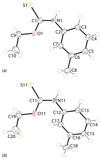
Figure 1
[image:2.610.313.563.509.698.2]The molecular structures of the two independent molecules comprising the asymmetric unit of (Ip) showing the atom-labelling scheme and displacement ellipsoids at the 70% probability level.
Table 1
Selected geometric parameters (A˚ ,) in (Ip) and (Ic).
Parameter (Ip), S1-molecule (Ip), S11-moleculea
(Ic)
C1—S1 1.6768 (19) 1.6752 (19) 1.6720 (18) C1—O1 1.321 (2) 1.319 (2) 1.325 (2) C1—N1 1.338 (2) 1.339 (2) 1.337 (2) C9—O1 1.457 (2) 1.454 (2) 1.451 (2) C2—N1 1.421 (2) 1.423 (2) 1.426 (2) S1—C1—O1 124.23 (14) 125.00 (15) 124.53 (12) S1—C1—N1 122.06 (14) 121.61 (15) 122.11 (13) O1—C1—N1 113.71 (16) 113.39 (16) 113.37 (15) C1—O1—C9 118.72 (15) 119.01 (15) 119.29 (15) C1—N1—C2 132.48 (16) 129.60 (16) 130.17 (15)
[image:2.610.44.297.621.734.2]tively. As seen from the overlay diagram, Fig. 2, the ethyl groups have an open conformation and overlap closely with the C1—O1—C9—C10 and C11—O11—C19—C20 torsion angles being178.76 (17) and 177.42 (18), respectively.
Geometric parameters for the original polymorph of (I),i.e. (Ic), are also included in Table 1. A comparison of these show the values in (Ip) and (Ic) to be equal within experimental error and those of (Ic) often lying between the two indepen-dent values found for (Ip). As evidenced from Fig. 2, there is a greater twist in the molecule as indicated by the dihedral angle of 30.44 (6) formed between the central chromophore and
the 3-tolyl ring. The orientation of the O-bound ethyl group is as for both molecules of (Ip) with the C1—O1—C9—C10 torsion angle being176.96 (17).
3. Supramolecular features
The most notable feature of the molecular packing of (I) is the
presence of an eight-membered thioamide synthon,
{ SCNH}2, formed via thioamide-N—H S(thione)
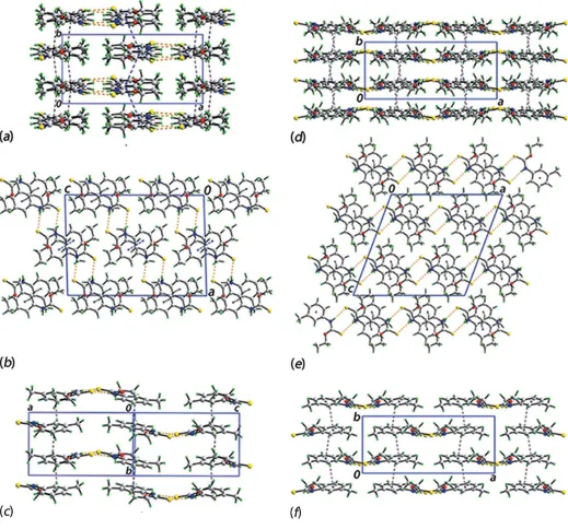
hydrogen bonds, between the two independent molecules comprising the asymmetric unit, Fig. 3 and Table 2. As shown in Fig. 3, the N—H S hydrogen bonds are supported by 3-tolyl-C—H S interactions, Table 2, with that involving the S1 atom being slightly beyond the standard distance criteria in PLATON(Spek, 2009). Globally, like molecules stack along theb-axis direction. The S1-molecules are connectedviaweak
– interactions between the 3-tolyl rings with the inter-centroid distance being 3.8535 (12) A˚ for the symmetry operation 1x, 2y, 1z. The connections between the S11-molecules are of the type 3-tolyl-C—H (3-tolyl), Table 2. The columns pack into alternating layers of S1- and S11-molecules parallel to [001], Fig. 4a, and connections
between them are made through the thioamide-N—
H S(thione) hydrogen bonds mentioned above, resulting in supramolecular layers parallel to (102), Fig. 4b. The layers, Fig. 4c, stack with no directional interactions between them.
The molecular packing in (Ic) has not been discussed in any detail (Tadbuppa & Tiekink, 2005) and hence, is now
described. The eight-membered thioamide synthon,
{ SCNH}2, seen in the packing of (Ip) is also found in the
packing of (Ic), Table 3, with an important difference, that being the synthon has crystallographic twofold symmetry; the putative 3-tolyl-C—H S interaction is long at 2.92 A˚ .
Globally, molecules pack into columns parallel to thebaxis and are sustained by 3-tolyl-C—H (3-tolyl) interactions, Fig. 4d and Table 3. Connections between columns are made by the aforementioned thioamide-N—H S(thione) hydrogen bonds. The result is supramolecular layers that stack along thecaxis, Fig. 4e. A view of the layer is shown in Fig. 4f. From the images of Fig. 4, it is obvious that despite some similarities, the molecular packing in polymorphs (Ip) and (Ic) are distinct. This point is highlighted in the analysis of the Hirshfeld surfaces of (Ip) and (Ic) discussed in the next section.
4. Analysis of the Hirshfeld surfaces of (Ip) and (Ic)
[image:3.610.45.296.109.175.2]The Hirshfeld surfaces for the individual molecules in (Ip), overall (Ip) and for (Ic) were calculated in accord with a recent report on a pair of polymorphs (Kuanet al., 2017). The calculations clearly reveal the similarities and differences in
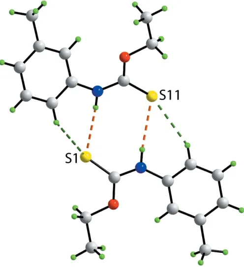
Figure 3
A view of the supramolecular dimer in (Ip) sustained by thioamide-N—
H S(thione) hydrogen bonds and supported by 3-tolyl-C—
H S(thione) interactions, shown as orange and green dashed lines, respectively.
Table 2
Hydrogen-bond geometry (A˚ ,).
Cg1 is the centroid of the (C12–C17) ring.
D—H A D—H H A D A D—H A
N1—H1N S11 0.87 (1) 2.62 (1) 3.4859 (16) 174 (2) N11—H11N S1 0.87 (1) 2.54 (1) 3.3985 (15) 171 (2) C3—H3 S11 0.95 2.86 3.708 (2) 150 C13—H13 S1 0.95 2.94 3.7090 (19) 139 C17—H17 Cg1i 0.95 2.82 3.471 (2) 127
Symmetry code: (i)xþ2;y1 2;zþ
1 2.
Table 3
Hydrogen-bond geometry (A˚ ,) for (Ic).
Cg1 is the centroid of the (C2–C7) ring.
D—H A D—H H A D A D—H A
N1—H1n S1i 0.87 2.58 3.4142 (16) 160 C7—H7 Cg1ii 0.94 2.91 3.4973 (17) 122
Symmetry code: (i)x,y,1 2z; (ii)
1
2x,
1 2+y,
[image:3.610.316.564.426.698.2]the intermolecular interactions instrumental in the crystals of the polymorphs.
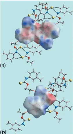
The appearance of bright-red spots near the thioamide-H and thione-S atoms, diminutive red spots near the 3-tolyl-H, ethoxy-H atoms and thione-S atoms on the Hirshfeld surfaces mapped over dnorm shown in Fig. 5 for both independent
molecules of (Ip) as well as for polymorph (Ic) are indicative of comparable thioamide-N—H S(thione) and 3-tolyl-C— H S(thione) interactions, and short interatomic H H contacts in their respective crystals, Table 4; values in Table 4
[image:4.610.47.566.72.556.2]were obtained from an analysis employing theCrystalExplorer package (Wolff et al. 2012). As there are two independent molecules in monoclinic polymorph (Ip), it exhibits a pair of the above-mentioned intermolecular interactions shown with labels 1 to 4 in Fig. 5a–c, whereas in form (Ic) they are labelled as 1 and 2 in Fig. 5dande. In addition to the above, the faint-red spots viewed near 3-tolyl-C14 in Fig. 5band ethoxy-H20B in Fig. 5cindicate the significance of short interatomic C H/ H C contacts, Table 4, in the packing of (Ip). The donors and acceptors of intermolecular interactions are also represented Figure 4
Molecular packing in (Ip): (a) a view of the unit-cell contents shown in projection down thecaxis, (b) a view of the unit-cell contents shown in projection down thebaxis and (c) a view of the supramolecular layer. Molecular packing in (Ic): (d) a view of the unit-cell contents shown in projection down thec
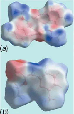
with blue and red regions, respectively, on the Hirshfeld surfaces mapped over electrostatic potential in Fig. 6. The new monoclinic polymorph (Ip) has distinct and a greater number of short interatomic contacts than for (Ic) owing, in part, to the presence of two distinct molecules per asymmetric unit, Table 4. The short interatomic H H contacts together with intermolecular N—H S and C—H S interactions formed with the atoms of reference molecules within Hirshfeld
surfaces mapped over electrostatic potential for (Ip) and (Ic) are highlighted in Fig. 7.
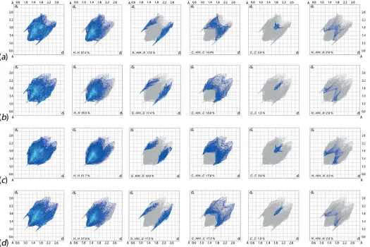
[image:5.610.44.299.93.225.2]The overall two-dimensional fingerprint plots for the S1 and S11-containing molecules of (Ip), the whole asymmetric unit of (Ip) and for the polymorph (Ic) are illustrated in Fig. 8a–d, Table 4
Summary of short interatomic contacts (A˚ ) in (Ip) and (Ic)a.
Contact Distance Symmetry operation
(Ip)
H3 H14 2.35 x,3
2y, 1 2+z
H5 H9B 2.28 x,3
2y,12+z
H15 H19A 2.31 x,3
2y, 1 2+z H19B H19B 2.08 2x, 2y, 1z
C10 H20C 2.87 1 +x,y,z
C14 H20B 2.77 2x,1
2+y, 1 2z
N1 H18B 2.73 2x,1
2+y, 1 2z (Ic)
H7 H9b 2.37 1
2+x, 1 2y,
1 2+z
H9a H9a 2.11 x,y,z
H10b H10b 2.32 1
2x,12y,z
[image:5.610.314.566.103.206.2]Note: (a) the atom numbering for the molecule in (Ic) follows that for the S1-molecule in (Ip).
Table 5
Percentage contributions of interatomic contacts to the Hirshfeld surfaces for the individual molecules in (Ip), overall (Ip) and (Ic).
Contact Percentage contribution
(Ip), S1-molecule (Ip), S11-molecule overall (Ip) (Ic)
H H 57.4 55.5 61.7 57.0
S H/H S 17.6 17.4 10.0 17.3
C H/H C 14.0 18.8 17.8 17.3
C C 3.9 1.5 3.0 1.9
N H/H N 2.6 2.9 3.0 2.6
C O/O C 2.5 2.5 2.7 2.4
O H/H O 1.0 1.2 1.2 1.1
[image:5.610.314.566.301.689.2]C N/N C 0.9 0.3 0.6 0.4
Figure 5
Views of the Hirshfeld surfaces mapped over dnorm for the (a)
[image:5.610.46.295.385.694.2]S1-containing molecule of (Ip) in the range0.147 to +1.345 au, (b) and (c) S11-containing molecule in (Ip) in the range0.149 to +1.274 au and (d) and (e) molecule of polymorph (Ic) in the range0.109 to 1.397 au.
Figure 6
respectively. In addition, the fingerprint plots delineated into H H, S H/H S, C H/H C, C C and N H/H N contacts (McKinnon et al., 2007) are included in Fig. 8; the relative contributions from different interatomic contacts to the Hirshfeld surfaces are summarized in Table 5. The nearly similar distribution of points in the fingerprint plots for S11-containing molecule of (Ip) and that of (Ic) indicate similarity in their molecular environments although some of the equivalent interatomic distances differ, Tables 2–4.
The fingerprint plots delineated into H H contacts, Fig. 8b andd, have needle-like tips pointing atde+di2.1 A˚
indi-cating short interatomic H H contacts, Table 4, for the
S11-containing molecule of (Ip) and for (Ic), both involving ethoxy-H atoms. The other short interatomic contacts in both forms are characterized from the points located within the pair of short peaks in (Ip) and a single short peak in (Ic), respec-tively, atde+di< 2.4 A˚ ,i.e. at the sum of their van der Waals
radii.
The involvement of ethoxy-H atoms in short interatomic C H/H C contacts decreases the percentage contribution from H H contacts to the Hirshfeld surface of the S11-containing molecule whereas the contribution from equivalent contacts to the surface of the S1-containing molecule of (Ip) and that of (Ic) are almost same, Table 5. The increase in percentage contribution from these contacts to the Hirshfeld surface of overall asymmetric unit of (Ip) is due to the inter-molecular N—H S and C—H S interactions between the respective atoms of S1- and S11-containing molecules thereby decreasing the contribution from S H/H S contacts to the overall surface, Table 5. This fact is confirmed from the nearly same percentage contribution from S H/H S contacts to the Hirshfeld surfaces of the individual S1- and S11-containing molecules of (Ip) and of the molecule of the (Ic) form, Table 5, and also from pair of forceps-like tips atde+di2.6 A˚ with
the nearly same distribution of points in their respective fingerprint plots in Fig. 8.
The similar distribution of points in the fingerprint plot delineated into C H/H C contacts for the S11-containing molecule of (Ip), Fig. 8b, and of (Ic), Fig. 8d, indicate their involvement in the intermolecular C—H contacts showing pairs of tips atde +di2.8 and 2.9 A˚ , respectively. This is
confirmed by the slight increase in the percentage contribution from these contacts to the Hirshfeld surface of the S11-containing molecule of (Ip) cf. the S1-containing molecule, Table 5. In other words, the contribution from C H/H C contacts to the surface of the S1-containing molecule of (Ip), Table 5, is decreased due to the absence of C—H contacts involving this molecule whereas the greater percentage contribution from C C contacts to the Hirshfeld surface of this molecule results from the presence of – stacking interactions between the symmetry-related 3-tolyl rings. This is also evident from the arrow-like distribution of points aroundde=di= 1.8 A˚ in the C C delineated fingerprint plot
shown in Fig. 8a.
The contribution of 3.0% from N H/H N contacts to the Hirshfeld surface of whole asymmetric unit of polymorph (Ip) indicate the presence of short interatomic N H/H N contacts between the thioamide-N1 and tolyl-H18B atoms, Table 4, although all of the delineated fingerprint plots have a similar distributions of points, Fig. 8, at least to a first approximation. The other interatomic contacts summarized in Table 4 make only small contributions to the Hirshfeld surfaces and have negligible contributions on the respective molecular packings.
5. Database survey
[image:6.610.44.296.71.535.2]According to a search of the Cambridge Structural Database (Version 5.38, May update; Groomet al., 2016), there are 22 Figure 7
Figure 8
[image:7.610.47.572.362.712.2]The full two-dimensional fingerprint plot and those delineated into H H, S H/H S, C H/H C, C C and N H/H N (left to right) contacts for (a) S1-molecule of (Ip), (b) S11-molecule of (Ip), (c) overall (Ip) and (d) (Ic).
Table 6
Hydrogen-bonding patterns inROC( S)N(H)R0.
Number R R0
Z0
Hydrogen bonds Motif Refcode Ref.
(II) Me phenyl 1 N—H S A OJIHAQ Hoet al.(2003)
(III) Me 4-NO2-phenyl 1 N—H S A CAZFUF Hoet al.(2005)
(IV) Me 4-C( O)OMe-phenyl 1 N—H S A CAZGAM Hoet al.(2005)
(V) Me 4-Cl-phenyl 2 N—H S A000
CAZCEQ Hoet al.(2005)
(VI) Me 4-C( O)Me-phenyl 1 N—H O B CAZGIU Hoet al.(2005)
(VII) Me 2-tolyl 1 N—H S A TAZSIX Kuanet al.(2005)
(VIII) Me 4-tolyl 2 N—H S A000
TIBYUZ Hoet al.(2007)
(IX) Et phenyl 3 N—H S A000000 PINPIL Taylor & Tiekink (1994)
(Ip) Et 3-tolyl 2 N—H S A000
– This work
(Ip) Et 3-tolyl 1 N—H S A000000000 TAZTUK Tadbuppa & Tiekink (2005)
(X) Et 4-tolyl 1 N—H S A TIBYOT Tadbuppa & Tiekink (2007a)
(XI) Et 3-OMe-phenyl 1 N—H S A UDUPAL Hanifet al.(2007)
(XII) Et 4-NO2-phenyl 1 N—H S A NENLAU Bensonet al.(2006)
(XIII) Et 4-Cl-phenyl 1 N—H S A DEYQEE Tadbuppa & Tiekink (2007b)
(XIV) n-Pr phenyl 2 N—H S A000
PAWKAB Sudkaowet al.(2012)
(XV) i-Pr Ph 1 N—H S A ADOGUW Kuanet al.(2007)
(XVI) i-Pr 4-tolyl 1 N—H S A ADOGOQ Kuanet al.(2007)
(XVII) i-Pr 4-Cl-phenyl 1 N—H S A ADOHAD Kuanet al.(2007)
(XVIII) i-Pr 4-NO2-phenyl 1 N—H S A MISDEY Elliset al.(2008) (XIX) 4-pyridylmethyl phenyl 2 N—H N C IFACOI Xiaoet al.(2006)
(XX) i-Bu phenyl 1 N—H S A000000000 KEQJAS Jianet al.(2006)
(XXI) 2,4-Me2-phenyl phenyl 1 N—H S A POVVOL Abrahamet al.(1995) (XXII) 2,4-(OMe)2-phenyl R1
a
1 N—H N C OSIZOG Zhouet al.(2010)
(XXIII) Cy phenyl 2 N—H S A000
VEFKUO Sahooet al.(2012)
monofunctional carbothioamide molecules related to the title compound, with (Ip) and (Ic) being the only pair of poly-morphs characterized thus far. Referring to Table 6, the overwhelming majority of the 24 crystallographically char-acterized thioamides feature an eight-membered thioamide, { SCNH}2, synthon. Thus, in 13 of the characterized
struc-tures, the synthon is formed about a centre of inversion, motif A. In five structures, two independent molecules (Z0 = 2)
comprise the asymmetric unit, as in (Ip), and these associated via the { SCNH}2 synthon but with no crystallographically
imposed symmetry, motif A000. There is a single example of a
structure withZ0= 3 (Taylor & Tiekink, 1994). Here, one of
the independent molecules self-associates about a centre of inversion, as in motif A, whereas the two remaining inde-pendent molecules are connected by the { SCNH}2synthon,
as found in motifA000. This is motifA000000. Two structures feature the { SCNH}2 synthon located about a twofold axis of
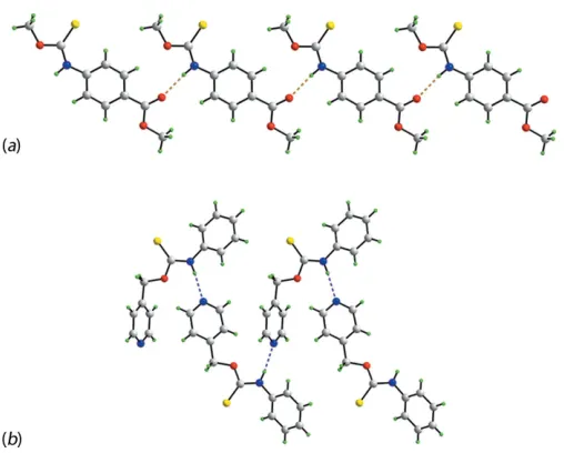
symmetry, as in (Ic), i.e. motif A000000000. The remaining three structures do not feature thioamide-N—H S(thione) hydrogen bonding. In the structure of MeOC( S)N(H)(4-C( O)Me-phenyl) (VI) (Hoet al., 2005), motifB, thioamide-N—H O(carboxy) hydrogen bonding is observed, leading to a linear supramolecular chain as shown in Fig. 9a. This structure is noteworthy as being the only example where the conformation of the thioamide moiety isantirather than the normally observedsyn. The final variation, motifC, is found in two structures, Table 6. The structure of (4-pyridyl)-CH2OC( S)N(H)phenyl (XIX) (Xiaoet al., 2006) serves as
an exemplar. Thus, in the crystal of (XIX), thioamide-N— H N(pyridyl) hydrogen bonds lead to a zigzag chain as shown in Fig. 9b. In summary, an inspection of the data in Table 6 indicates the predominance of thioamide-N—
H S(thione) hydrogen bonding in these carbothioamides, at least in the absence of competing synthons, as seen in motifsB andC
.
6. Synthesis and crystallization
[image:8.610.45.299.66.270.2]All chemicals and solvents were used as purchased without purification. To prepare (Ip), 3-tolyl isothiocyanate (Merck; 2.5 mmol, 0.34 ml) was added to NaOH (Merck; 2.5 mmol, 0.10 g) in EtOH (Merck; 3 ml) and the mixture was stirred at room temperature for 2 h, followed by the addition of excess 5M HCl solution. The resulting mixture was stirred for another 1.5 h. The final product was extracted with chloroform (Merck; 10 ml) and left for evaporation at room temperature, Figure 9
Supramolecular aggregation in related carbothioamide structures: (a)
linear supramolecular chain in the crystal of MeOC(
S)N(H)(4-C( O)Me-phenyl) (VI) mediated by thioamide-N—H O(carboxy)
hydrogen bonding shown as orange dashed lines and (b) zigzag chain in the crystal of (4-pyridyl)CH2OC( S)N(H)phenyl mediated by
thio-amide-N—H N(pyridyl) hydrogen bonding shown as blue dashed lines.
Table 7
Experimental details.
Crystal data
Chemical formula C10H13NOS
Mr 195.27
Crystal system, space group Monoclinic,P21/c
Temperature (K) 100
a,b,c(A˚ ) 14.3999 (5), 7.0388 (3), 19.9725 (7)
() 91.727 (3)
V(A˚3) 2023.45 (13)
Z 8
Radiation type MoK
(mm1) 0.28
Crystal size (mm) 0.200.200.05
Data collection
Diffractometer Agilent SuperNova, Dual, Mo at zero, Atlas
Absorption correction Multi-scan (CrysAlis PRO; Agilent, 2011)
Tmin,Tmax 0.662, 1.000 No. of measured, independent and
observed [I> 2(I)] reflections
15588, 4577, 3514
Rint 0.040
(sin/)max(A˚ 1
) 0.651
Refinement
R[F2> 2(F2)],wR(F2),S 0.047, 0.125, 1.03 No. of reflections 4577
No. of parameters 245
No. of restraints 2
H-atom treatment H atoms treated by a mixture of independent and constrained refinement
max, min(e A˚
3) 0.72,0.24
[image:8.610.312.553.90.394.2]yielding brown crystals after 1 week. M.p. (Kru¨ss KSP1N melting point meter): 339–340 K. IR (Perkin Elmer Spectrum 400 FT Mid-IR/Far-IR spectrophotometer; cm1): 3211 (s) (N—H), 1451 (s) (C—N), 1209 (s) (C S), 1064 (s) (C—O).
7. Refinement
Crystal data, data collection and structure refinement details are summarized in Table 7. The carbon-bound H atoms were placed in calculated positions (C—H = 0.95–0.99 A˚ ) and were included in the refinement in the riding-model approximation, with Uiso(H) set to 1.2–1.5Ueq(C). The nitrogen-bound H
atoms were located in a difference Fourier map but were refined with a distance restraint of N—H = 0.880.01 A˚ , and withUiso(H) set to 1.2Ueq(N). Owing to poor agreement, one
reflection, i.e. (15 1 4), was omitted from the final cycles of refinement.
Acknowledgements
The authors thank the staff of the University of Malaya’s X-ray diffraction laboratory for the data collection. Sunway University is thanked for support of biological and crystal engineering studies of carbothioamides and their coinage metal complexes.
References
Abraham, S. P., Samuelson, A. G. & Nethaji, M. (1995).Proc. Indian Acad. Sci. Chem. Sci.107, 255–271.
Agilent (2011). CrysAlis PRO. Agilent Technologies, Yarnton, England.
Benson, R. E., Broker, G. A., Daniels, L. M., Tiekink, E. R. T., Wardell, J. L. & Young, D. J. (2006).Acta Cryst.E62, o4106–o4108. Brandenburg, K. (2006).DIAMOND. Crystal Impact GbR, Bonn,
Germany.
Ellis, C. A., Miller, M. A., Spencer, J., Zukerman-Schpector, J. & Tiekink, E. R. T. (2009).CrystEngComm,11, 1352–1361.
Ellis, C. A., Tiekink, E. R. T. & Zukerman-Schpector, J. (2008).Acta Cryst.E64, o345.
Farrugia, L. J. (2012).J. Appl. Cryst.45, 849–854.
Gans, J. & Shalloway, D. (2001).J. Mol. Graph. Model.19, 557–609. Groom, C. R., Bruno, I. J., Lightfoot, M. P. & Ward, S. C. (2016).Acta
Cryst.B72, 171–179.
Hanif, M., Qadeer, G., Shi, L. & Rama, N. H. (2007).Acta Cryst.E63, o3062.
Ho, S. Y., Bettens, R. P. A., Dakternieks, D., Duthie, A. & Tiekink, E. R. T. (2005).CrystEngComm,7, 682–689.
Ho, S. Y., Cheng, E. C.-C., Tiekink, E. R. T. & Yam, V. W.-W. (2006). Inorg. Chem.45, 8165–8174.
Ho, S. Y., Kuan, F. S. & Tiekink, E. R. T. (2007).Acta Cryst.E63, o1723–o1724.
Ho, S. Y., Lai, C. S. & Tiekink, E. R. T. (2003).Acta Cryst.E59, o1155– o1156.
Jian, F.-F., Yu, H.-Q., Qiao, Y.-B. & Liang, T.-L. (2006).Acta Cryst. E62, o3416–o3417.
Kuan, F. S., Jotani, M. M. & Tiekink, E. R. T. (2017).Acta Cryst.E73, 1465–1471.
Kuan, F. S., Mohr, F., Tadbuppa, P. P. & Tiekink, E. R. T. (2007). CrystEngComm,9, 574–581.
Kuan, F. S., Tadbuppa, P. P. & Tiekink, E. R. T. (2005).Z. Kristallogr. New Cryst. Struct.220, 393–394.
McKinnon, J. J., Jayatilaka, D. & Spackman, M. A. (2007).Chem. Commun.pp. 3814–3816.
Ooi, K. K., Yeo, C. I., Mahandaran, T., Ang, K. P., Akim, A. M., Cheah, Y.-K., Seng, H.-L. & Tiekink, E. R. T. (2017). J. Inorg. Biochem.166, 173–181.
Sahoo, S. K., Chakraborty, S. & Patel, B. K. (2012).J. Sulfur Chem.33, 143–153.
Sheldrick, G. M. (2008).Acta Cryst.A64, 112–122. Sheldrick, G. M. (2015).Acta Cryst.C71, 3–8.
Slater, N. H., Buckley, B. R., Elsegood, M. R. J., Teat, S. J. & Kimber, M. C. (2016).Cryst. Growth Des.16, 3846–3852.
Spek, A. L. (2009).Acta Cryst.D65, 148–155.
Sudkaow, P., Yeo, C. I., Ng, S. W. & Tiekink, E. R. T. (2012).Acta Cryst.E68, o1774.
Tadbuppa, P. P. & Tiekink, E. R. T. (2005).Z. Kristallogr. New Cryst. Struct.220, 395–396.
Tadbuppa, P. P. & Tiekink, E. R. T. (2007a).Acta Cryst.E63, o1779– o1780.
Tadbuppa, P. P. & Tiekink, E. R. T. (2007b).Acta Cryst.E63, o1885– o1886.
Taylor, R. L. & Tiekink, E. R. T. (1994).Z. Kristallogr.209, 64–67. Westrip, S. P. (2010).J. Appl. Cryst.43, 920–925.
Wolff, S. K., Grimwood, D. J., McKinnon, J. J., Turner, M. J., Jayatilaka, D. & Spackman, M. A. (2012). University of Western Australia.
Xiao, H.-L., Wang, K.-F. & Jian, F.-F. (2006).Acta Cryst.E62, o2852– o2853.
Yeo, C. I., Halim, S. N. A., Ng, S. W., Tan, S. L., Zukerman-Schpector, J., Ferreira, M. A. B. & Tiekink, E. R. T. (2014).Chem. Commun. 50, 5984–5986.
Yeo, C. I., Sim, J.-H., Khoo, C.-H., Goh, Z.-J., Ang, K.-P., Cheah, Y.-K., Fairuz, Z. A., Halim, S. N. B. A., Ng, S. W., Seng, H.-L. & Tiekink, E. R. T. (2013).Gold Bull.46, 145–152.
Zhou, L., Tan, C. K., Jiang, X., Chen, F. & Yeung, Y.-Y. (2010).J. Am. Chem. Soc.132, 15474–15476.
sup-1 Acta Cryst. (2017). E73, 1889-1897
supporting information
Acta Cryst. (2017). E73, 1889-1897 [https://doi.org/10.1107/S2056989017016280]
A new monoclinic polymorph of
N
-(3-methylphenyl)ethoxycarbothioamide:
crystal structure and Hirshfeld surface analysis
Mukesh M. Jotani, Chien Ing Yeo and Edward R. T. Tiekink
Computing details
Data collection: CrysAlis PRO (Agilent, 2011); cell refinement: CrysAlis PRO (Agilent, 2011); data reduction: CrysAlis
PRO (Agilent, 2011); program(s) used to solve structure: SHELXS97 (Sheldrick, 2008); program(s) used to refine
structure: SHELXL2014 (Sheldrick, 2015); molecular graphics: ORTEP-3 for Windows (Farrugia, 2012), QMol (Gans &
Shalloway, 2001) and DIAMOND (Brandenburg, 2006); software used to prepare material for publication: publCIF
(Westrip, 2010).
N-(3-Methylphenyl)ethoxycarbothioamide
Crystal data
C10H13NOS
Mr = 195.27
Monoclinic, P21/c
a = 14.3999 (5) Å b = 7.0388 (3) Å c = 19.9725 (7) Å β = 91.727 (3)° V = 2023.45 (13) Å3
Z = 8
F(000) = 832 Dx = 1.282 Mg m−3
Mo Kα radiation, λ = 0.71073 Å Cell parameters from 6144 reflections θ = 2.4–27.5°
µ = 0.28 mm−1
T = 100 K Slab, colourless 0.20 × 0.20 × 0.05 mm
Data collection
Agilent SuperNova, Dual, Mo at zero, Atlas diffractometer
Radiation source: SuperNova (Mo) X-ray Source
Mirror monochromator
Detector resolution: 10.4041 pixels mm-1
ω scan
Absorption correction: multi-scan (CrysAlis PRO; Agilent, 2011)
Tmin = 0.662, Tmax = 1.000
15588 measured reflections 4577 independent reflections 3514 reflections with I > 2σ(I) Rint = 0.040
θmax = 27.6°, θmin = 2.5°
h = −18→18 k = −9→9 l = −18→25
Refinement
Refinement on F2
Least-squares matrix: full R[F2 > 2σ(F2)] = 0.047
wR(F2) = 0.125
S = 1.03 4577 reflections 245 parameters 2 restraints
H atoms treated by a mixture of independent and constrained refinement
w = 1/[σ2(F
o2) + (0.0629P)2 + 0.8006P]
where P = (Fo2 + 2Fc2)/3
(Δ/σ)max = 0.001
Δρmax = 0.72 e Å−3
sup-2 Acta Cryst. (2017). E73, 1889-1897
Special details
Geometry. All esds (except the esd in the dihedral angle between two l.s. planes) are estimated using the full covariance
matrix. The cell esds are taken into account individually in the estimation of esds in distances, angles and torsion angles; correlations between esds in cell parameters are only used when they are defined by crystal symmetry. An approximate (isotropic) treatment of cell esds is used for estimating esds involving l.s. planes.
Fractional atomic coordinates and isotropic or equivalent isotropic displacement parameters (Å2)
x y z Uiso*/Ueq
sup-3 Acta Cryst. (2017). E73, 1889-1897
C17 0.99808 (13) 0.6749 (3) 0.23923 (9) 0.0176 (4) H17 1.0404 0.6265 0.2724 0.021* C18 1.12821 (13) 0.6769 (3) 0.15808 (10) 0.0245 (5) H18A 1.1566 0.5877 0.1904 0.037* H18B 1.1622 0.7975 0.1599 0.037* H18C 1.1308 0.6234 0.1129 0.037* C19 1.05091 (14) 0.7744 (3) 0.44091 (10) 0.0256 (5) H19A 1.0493 0.6584 0.4689 0.031* H19B 1.0238 0.8806 0.4663 0.031* C20 1.14753 (15) 0.8193 (4) 0.42346 (12) 0.0349 (5) H20A 1.1840 0.8490 0.4643 0.052* H20B 1.1477 0.9291 0.3933 0.052* H20C 1.1750 0.7096 0.4012 0.052*
Atomic displacement parameters (Å2)
U11 U22 U33 U12 U13 U23
S1 0.0230 (3) 0.0401 (3) 0.0140 (3) −0.0017 (2) 0.00422 (18) −0.0020 (2) O1 0.0190 (7) 0.0284 (8) 0.0141 (7) 0.0011 (6) −0.0009 (5) −0.0020 (6) N1 0.0155 (7) 0.0236 (9) 0.0139 (8) 0.0005 (7) 0.0022 (6) −0.0005 (7) C1 0.0200 (9) 0.0174 (10) 0.0176 (10) −0.0011 (8) 0.0017 (7) 0.0017 (8) C2 0.0211 (9) 0.0129 (9) 0.0132 (9) 0.0008 (7) 0.0039 (7) 0.0011 (7) C3 0.0182 (9) 0.0233 (11) 0.0178 (10) 0.0014 (8) 0.0019 (7) 0.0004 (8) C4 0.0251 (10) 0.0262 (11) 0.0157 (10) −0.0002 (9) −0.0014 (8) −0.0010 (8) C5 0.0279 (10) 0.0234 (11) 0.0133 (9) 0.0026 (8) 0.0052 (7) −0.0009 (8) C6 0.0205 (9) 0.0181 (10) 0.0184 (10) −0.0003 (8) 0.0052 (7) 0.0035 (8) C7 0.0199 (9) 0.0203 (10) 0.0154 (10) −0.0016 (8) 0.0014 (7) 0.0020 (8) C8 0.0226 (10) 0.0331 (12) 0.0221 (11) 0.0014 (9) 0.0077 (8) 0.0024 (9) C9 0.0254 (10) 0.0337 (12) 0.0155 (10) −0.0020 (9) −0.0057 (8) −0.0002 (9) C10 0.0267 (11) 0.0404 (14) 0.0329 (13) −0.0041 (10) −0.0037 (9) −0.0003 (11) S11 0.0225 (3) 0.0423 (3) 0.0127 (3) 0.0072 (2) 0.00474 (18) 0.0035 (2) O11 0.0203 (7) 0.0236 (8) 0.0137 (7) 0.0012 (6) −0.0003 (5) −0.0007 (6) N11 0.0147 (7) 0.0238 (9) 0.0141 (8) 0.0025 (7) 0.0027 (6) 0.0016 (7) C11 0.0200 (9) 0.0200 (10) 0.0155 (9) 0.0075 (8) 0.0027 (7) 0.0009 (8) C12 0.0196 (9) 0.0152 (9) 0.0121 (9) −0.0005 (7) 0.0031 (7) 0.0003 (7) C13 0.0154 (9) 0.0225 (10) 0.0158 (10) −0.0017 (8) 0.0006 (7) −0.0010 (8) C14 0.0219 (9) 0.0217 (10) 0.0141 (9) −0.0031 (8) −0.0032 (7) 0.0010 (8) C15 0.0242 (10) 0.0225 (10) 0.0110 (9) −0.0033 (8) 0.0030 (7) 0.0001 (8) C16 0.0198 (9) 0.0161 (9) 0.0175 (10) −0.0014 (8) 0.0042 (7) −0.0007 (8) C17 0.0201 (9) 0.0165 (9) 0.0162 (10) 0.0029 (8) 0.0010 (7) 0.0010 (8) C18 0.0236 (10) 0.0286 (12) 0.0217 (11) 0.0038 (9) 0.0090 (8) 0.0029 (9) C19 0.0265 (10) 0.0323 (12) 0.0176 (10) 0.0042 (9) −0.0044 (8) −0.0055 (9) C20 0.0302 (12) 0.0359 (14) 0.0382 (14) −0.0012 (10) −0.0047 (10) −0.0008 (11)
Geometric parameters (Å, º)
sup-4 Acta Cryst. (2017). E73, 1889-1897
O1—C9 1.457 (2) O11—C19 1.454 (2) N1—C1 1.338 (2) N11—C11 1.339 (2) N1—C2 1.421 (2) N11—C12 1.423 (2) N1—H1N 0.872 (9) N11—H11N 0.872 (9) C2—C3 1.394 (3) C12—C13 1.393 (3) C2—C7 1.397 (2) C12—C17 1.393 (2) C3—C4 1.378 (3) C13—C14 1.380 (3)
C3—H3 0.9500 C13—H13 0.9500
C4—C5 1.388 (3) C14—C15 1.389 (3)
C4—H4 0.9500 C14—H14 0.9500
C5—C6 1.391 (3) C15—C16 1.389 (3)
C5—H5 0.9500 C15—H15 0.9500
C6—C7 1.390 (3) C16—C17 1.383 (3) C6—C8 1.510 (3) C16—C18 1.513 (2)
C7—H7 0.9500 C17—H17 0.9500
C8—H8A 0.9800 C18—H18A 0.9800 C8—H8B 0.9800 C18—H18B 0.9800 C8—H8C 0.9800 C18—H18C 0.9800 C9—C10 1.480 (3) C19—C20 1.479 (3) C9—H9A 0.9900 C19—H19A 0.9900 C9—H9B 0.9900 C19—H19B 0.9900 C10—H10A 0.9800 C20—H20A 0.9800 C10—H10B 0.9800 C20—H20B 0.9800 C10—H10C 0.9800 C20—H20C 0.9800
sup-5 Acta Cryst. (2017). E73, 1889-1897
C6—C7—H7 119.9 C16—C17—H17 119.9 C2—C7—H7 119.9 C12—C17—H17 119.9 C6—C8—H8A 109.5 C16—C18—H18A 109.5 C6—C8—H8B 109.5 C16—C18—H18B 109.5 H8A—C8—H8B 109.5 H18A—C18—H18B 109.5 C6—C8—H8C 109.5 C16—C18—H18C 109.5 H8A—C8—H8C 109.5 H18A—C18—H18C 109.5 H8B—C8—H8C 109.5 H18B—C18—H18C 109.5 O1—C9—C10 106.11 (17) O11—C19—C20 107.04 (17) O1—C9—H9A 110.5 O11—C19—H19A 110.3 C10—C9—H9A 110.5 C20—C19—H19A 110.3 O1—C9—H9B 110.5 O11—C19—H19B 110.3 C10—C9—H9B 110.5 C20—C19—H19B 110.3 H9A—C9—H9B 108.7 H19A—C19—H19B 108.6 C9—C10—H10A 109.5 C19—C20—H20A 109.5 C9—C10—H10B 109.5 C19—C20—H20B 109.5 H10A—C10—H10B 109.5 H20A—C20—H20B 109.5 C9—C10—H10C 109.5 C19—C20—H20C 109.5 H10A—C10—H10C 109.5 H20A—C20—H20C 109.5 H10B—C10—H10C 109.5 H20B—C20—H20C 109.5
C9—O1—C1—N1 178.80 (17) C19—O11—C11—N11 178.92 (17) C9—O1—C1—S1 −1.1 (3) C19—O11—C11—S11 −0.6 (3) C2—N1—C1—O1 −3.2 (3) C12—N11—C11—O11 −3.8 (3) C2—N1—C1—S1 176.68 (16) C12—N11—C11—S11 175.77 (16) C1—N1—C2—C3 −172.1 (2) C11—N11—C12—C13 −147.0 (2) C1—N1—C2—C7 7.3 (3) C11—N11—C12—C17 35.4 (3) C7—C2—C3—C4 0.2 (3) C17—C12—C13—C14 −1.1 (3) N1—C2—C3—C4 179.66 (18) N11—C12—C13—C14 −178.82 (17) C2—C3—C4—C5 −0.7 (3) C12—C13—C14—C15 −0.3 (3) C3—C4—C5—C6 0.7 (3) C13—C14—C15—C16 0.7 (3) C4—C5—C6—C7 −0.2 (3) C14—C15—C16—C17 0.1 (3) C4—C5—C6—C8 −178.83 (19) C14—C15—C16—C18 179.80 (18) C5—C6—C7—C2 −0.3 (3) C15—C16—C17—C12 −1.5 (3) C8—C6—C7—C2 178.33 (18) C18—C16—C17—C12 178.84 (18) C3—C2—C7—C6 0.3 (3) C13—C12—C17—C16 2.0 (3) N1—C2—C7—C6 −179.08 (18) N11—C12—C17—C16 179.53 (17) C1—O1—C9—C10 −178.76 (17) C11—O11—C19—C20 177.42 (18)
Hydrogen-bond geometry (Å, º)
Cg1 is the centroid of the (C12–C17) ring.
D—H···A D—H H···A D···A D—H···A
sup-6 Acta Cryst. (2017). E73, 1889-1897
C13—H13···S1 0.95 2.94 3.7090 (19) 139 C17—H17···Cg1i 0.95 2.82 3.471 (2) 127