Comparative Structural and Vibrational Study of
8-Hydroxyquinoline and 8-Hydroxyquinoline
Succinate Compounds: A DFT Study
S. N. Saravanamoorthy1, G. Sathiyapriya2, M. Sivasakthi3
1, 2, 3
Department of Physics, Devanga Arts College, Aruppukottai, TamilNadu, India.
Abstract: Quinoline based compounds efficiently form molecular complexes with various organic acids through hydrogen bonding. These compounds are classified as electron-deficient heterocyclic in nature and exhibit acceptor-donor properties with intramolecular charge transfer. 8-Hydroxyquinolinesuccinate is a π donor-acceptor molecular compound in which 8-HQ acts as donor and succinic acid as electron acceptor. The present work deals with the interaction of title compounds using Density Functional Theory (DFT) implemented in Gaussian-09 software package. All geometry optimizations, the vibrational analysis, electronic transition study of UV and NMR spectra of title compounds were interpreted with aid of normal coordinate analysis based on DFT/B3LYP using 6-311G basis set. Dihedral angles of 8-HQ varies between 0 ͦ and 180 ͦ. This shows that the molecule
of 8-HQ is practically planar. 8-HQSC crystal is stabilized by the hydrogen bonds N2-H13….O22 and O22-H12….O1 formed by the existence of complementary functional groups. The negative chemical shift values of -145.5869 ppm do exist for nitrogen atom in 8-HQ. Energy gap of 8-HQ is obtained theoretically by DFT method is 4.52 eV and from HOMO-LUMO diagram is 4.53 eV. From the Mulliken atomic charges confirms the extensive intermolecular charge transfer between adsorbate and adsorbent of the title compound during the interaction. The calculated results are found to be in good agreement with the previous investigations.
Keywords: 8-Hydroxyquinoline, 8-Hydroxyquinoline Succinate, Vibrational spectra, Density Functional Theory (DFT), HOMO, LUMO.
I. INTRODUCTION
8-Hydroxyquinoline is a bicycle compound and has typical phenolic properties. It consists of carbocyclic ring and pyridine ring with hydroxyl group substituted at position-8. Due to the unique chemical properties of 8-HQ, these compound derivatives have application in agricultural and medical fields.
The compound shows a variety of applications ranging from pharmacological and pharmaceutical agents to electron carriers in organic light emitting diodes and fluorescent chemo sensors for metal ions. In medical field, its derivatives can be used as insecticides, antibacterial, fungicidal, neuroprotective, and anti-HIV agents [1]. Organic functionalized NLO materials attracted a great deal of attention due to its extensive applications in harmonic generation, amplitude, phase modulation, switching and other signal processing devices [2-4].
A possible reason for the significant increase in the effective nonlinear optical response observed in organic molecules is due to the
existence of π-conjugation between the appropriate electron donor and acceptor groups, chirality and hydrogen bonding. Functional modification in electron-donor systems affects the optical and electrical properties of the quinolone derivatives.
8-Hydroxyquinolinesuccinate is a π donor-acceptor molecular compound in which 8-HQ acts as donor and succinic acid as electron acceptor. Theoretical studies are valuable approaches to explore the mechanism of reactions in the molecules and their electronic structures levels in addition to electronic parameters that acquired by means of theoretical calculations employ the computational methods of quantum chemistry. The improvement in theoretical studies and applications have accomplish a point where predicted features of logical accuracy can be obtained from DFT (density functional theory) studies [5,6].
II. DFT CALCULATIONS
Molecular geometries of the singlet ground state of compounds 8-HQ and 8-HQSC were fully optimized at B3LYP/6-311G level using Gaussian09 program. In geometry optimizations every bond length, bond angle and dihedral angle was permitted to relax, free of constraints. For each compound frequency calculations were carried out, verifying that the optimized structure obtained corresponded to energy minimum, thus only positive frequencies were expected. Frontier molecular orbitals were used to identify the adsorption centers of the inhibitor molecule. For the simplest transfer of electrons, adsorption should occur at the part of the molecule where the softness (σ) has the highest value. Electronegativity, hardness and softness have conformed to be very useful quantities in quantum molecular description calculations [7]. Moreover, we have computed UV, Nuclear Magnetic Resonance (NMR), highest occupied molecular orbitals (HOMO) and the lowest unoccupied molecular orbitals (LUMO) energies in order to understand the properties of the molecules.
III. RESULTS AND DISCUSSION
We have computed the structures of 8-HQ and 8-HQSC compounds using DFT calculations and quantities of the spectroscopic interest for the two compounds and compared the results with previous studies.
A. Molecular Geometries
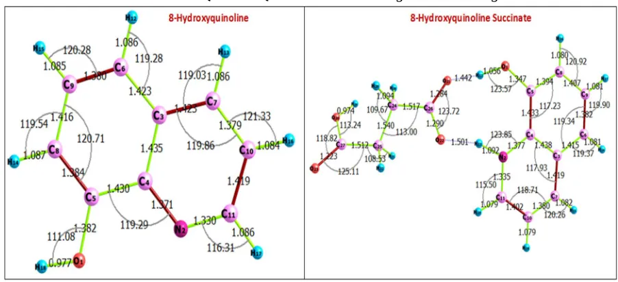
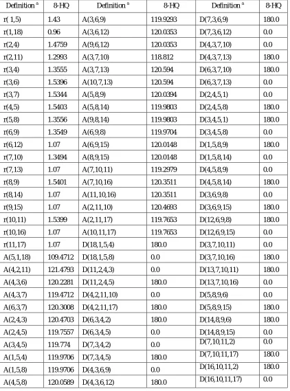
[image:2.612.79.530.292.498.2]The optimized molecular structures of the 8-HQ and 8-HQSC with atomic labeling are shown in Fig. 1.
Fig. 1 optimized molecular structures of the 8-HQ and 8-HQSC with atomic labeling
The interatomic bond lengths and bond angles of 8-HQ and 8-HQSC derived from DFT study are listed in Table 1 and 2. 8-HQ consists of two rings, one phenol and other pyridine type ring. The angle between the phenolic and pyridine ring is only 1.382 ͦ [8].
The average C-C bond length in the rings is 1.456 Å whereas the average C-N bond is 1.387 Å. The observed C-C bond length in the two rings vary from 1.349 to 1.540 Å. Nearly identical C-C bonds indicate the complete delocalization of π -electrons in the individual hetero atom rings. The obtained C-O bond length is 1.43 Å. It is found that the C-C bonds and hetero atom C-N bond are
nearly the same as that found in experimental result. The average value of the bond angles in the two benzene rings is 120 ͦ. The deviation from this ideal value is quite significant. Dihedral angles are useful to identify the molecular conformation. Dihedral angles of 8-HQ varies between 0 ͦ and 180 ͦ. This shows that the molecule of 8-HQ is practically planar. 8-HQSC single crystal growth and its characterization were reported by R.Thirumurugan et.al (2014). The N-site of the 8-HQ leads to the formation of cation and it is confirmed by the enhancement of the internal angle at N2 (N2-C11-C10 bond angle of 121.0427) compared with
122.12(2) ͦ observed in 8-HQSC single crystal study [9]. The deprotonation occurs due to the succinic acid molecule and it leads to an anion. The variations of the carboxyl bond distances C26-O21 and C26-O22 are 1.3013 Å and 1.2584 Å respectively. The dihedral angle between 8-hydroxyquinolinium cation and succinate anion are 8.8499 ͦ coincide with the already existing report. It
TABLE I
Optimized Geometrical Parametersb of 8-Hydroxyquinoline at b3lyp/6-311g level.
a
For atomic numbering scheme, see Fig. 1.
b
Bond lengths (r) in Angstrom, bond angles (A) and dihedral angles (D) in degree.
Definition a 8-HQ Definition a 8-HQ Definition a 8-HQ
r( 1,5) 1.43 A(3,6,9) 119.9293 D(7,3,6,9) 180.0
r(1,18) 0.96 A(3,6,12) 120.0353 D(7,3,6,12) 0.0
r(2,4) 1.4759 A(9,6,12) 120.0353 D(4,3,7,10) 0.0
r(2,11) 1.2993 A(3,7,10) 118.812 D(4,3,7,13) 180.0
r(3,4) 1.3555 A(3,7,13) 120.594 D(6,3,7,10) 180.0
r(3,6) 1.5396 A(10,7,13) 120.594 D(6,3,7,13) 0.0
r(3,7) 1.5344 A(5,8,9) 120.0394 D(2,4,5,1) 0.0
r(4,5) 1.5403 A(5,8,14) 119.9803 D(2,4,5,8) 180.0
r(5,8) 1.3556 A(9,8,14) 119.9803 D(3,4,5,1) 180.0
r(6,9) 1.3549 A(6,9,8) 119.9704 D(3,4,5,8) 0.0
r(6,12) 1.07 A(6,9,15) 120.0148 D(1,5,8,9) 180.0
r(7,10) 1.3494 A(8,9,15) 120.0148 D(1,5,8,14) 0.0
r(7,13) 1.07 A(7,10,11) 119.2979 D(4,5,8,9) 0.0
r(8,9) 1.5401 A(7,10,16) 120.3511 D(4,5,8,14) 180.0
r(8,14) 1.07 A(11,10,16) 120.3511 D(3,6,9,8) 0.0
r(9,15) 1.07 A(2,11,10) 120.4693 D(3,6,9,15) 180.0
r(10,11) 1.5399 A(2,11,17) 119.7653 D(12,6,9,8) 180.0
r(10,16) 1.07 A(10,11,17) 119.7653 D(12,6,9,15) 0.0
r(11,17) 1.07 D(18,1,5,4) 180.0 D(3,7,10,11) 0.0
A(5,1,18) 109.4712 D(18,1,5,8) 0.0 D(3,7,10,16) 180.0
A(4,2,11) 121.4793 D(11,2,4,3) 0.0 D(13,7,10,11) 180.0
A(4,3,6) 120.2281 D(11,2,4,5) 180.0 D(13,7,10,16) 0.0
A(4,3,7) 119.4712 D(4,2,11,10) 0.0 D(5,8,9,6) 0.0
A(6,3,7) 120.3008 D(4,2,11,17) 180.0 D(5,8,9,15) 180.0
A(2,4,3) 120.4703 D(6,3,4,2) 180.0 D(14,8,9,6) 180.0
A(2,4,5) 119.7557 D(6,3,4,5) 0.0 D(14,8,9,15) 0.0
A(3,4,5) 119.774 D(7,3,4,2) 0.0 D(7,10,11,2) 0.0
A(1,5,4) 119.9706 D(7,3,4,5) 180.0 D(7,10,11,17) 180.0
A(1,5,8) 119.9706 D(4,3,6,9) 0.0 D(16,10,11,2) 180.0
Table II
Optimized Geometrical Parametersb of 8-Hydroxyquinoline Succinate At B3lyp/6-311g Level
a
For atomic numbering scheme, see Fig. 1.
b
Bond lengths (r) in Angstrom, bond angles (A) and dihedral angles (D) in degree.
Definition a 8-HQSC Definition a 8-HQSC Definition a 8-HQSC
B. Vibrational Spectra
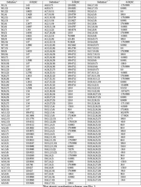
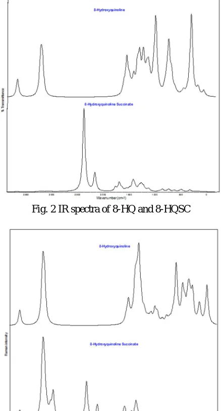
[image:5.612.196.415.291.697.2]The 8-hydroxyquinoline has 18 atoms with 48 fundamental modes of vibration. These vibrations are divided into 33 in-plane and 15 out-of-plane vibrations. The detailed vibrational assignment of fundamental modes of frequencies of 8-HQ reported by Krishnakumar et al. [10] and calculated frequency using B3LYP/6-311G method are reported in Table 3. IR and Raman spectra of 8-HQ and 8-HQSC are shown in Fig. 2 and 3. The accurate position of the OH bond is based on the strength of the hydrogen bond. In 8-HQ, OH-N hydrogen bond absorption is found at 3688 cm-1. The OH group in-plane and out-of-plane modes are found at 1518 cm-1 (δ OH) and 896 cm-1 (γ OH) respectively in agreement with the literature data [10,11]. In addition, a certain number of diatomic molecular fragments such as C2 and CN provide useful indications for identification of organic materials [12,13] and also the mixing of several bands are possible in this region. νC11N2 and ν C9N1 are appeared at 1354 cm-1 and 1306 cm-1 respectively. In the IR spectra of 8-HQ, the non-linearity of hydrogen bond have an impact over the carbonyl group frequency. The IR band is observed at 1231 cm−1 and assigned to νC5O1 vibration. The frequencies at 591 cm−1 and 491 cm−1are active due to δC5O and γC5O respectively. Generally, The presence of skeletal modes of semiunsaturated CC bonds vibrations are appeared in the 1650 -1450 cm−1 frequency region [14]. Due to stretching modes, a change of dipole moment occurs in mono-substituted benzene. As a result of this, strong to medium intensity bands are expected for aromatic C-C modes. The medium intensity IR bands have been assigned to the ring stretching modes at 1657 cm−1 and 1551 cm−1. The rest of bands are observed at 1475, 1441, 1394, 1294 and 1251 cm−1. Table 3 shows that the results of observed and calculated IR and Raman frequencies yield good agreement.
Fig. 2 IR spectra of 8-HQ and 8-HQSC
Table III
Detailed Assignment Of Fundamental Vibrations Of 8-Hydroxyquinoline
No Symmetry
Species CS
Observed frequency
[Ref 10] Calculated using B3LYP/6-311G method
Characterization of normal modes
Infrared Raman Frequency
(cm-1) IR intensity
Raman activity
1 A' 3418 -- 3688 39.4493 174.2442 OH Stretch
2 A' 3097 -- 3197 31.5145 307.2668 C10H Stretch
3 A' 3084 -- 3192 36.7406 184.4129 C9H Stretch
4 A' -- 3066 3170 25.6162 143.3291 C7H, C8H Stretch
5 A' 3048 -- 3167 10.3337 19.5749 C11H, C10H Stretch
6 A' -- 3019 3160 15.594 119.7586 C5H Stretch
7 A' -- 3003 3157 15.4611 91.6807 C8H, C11H Stretch
8 A' -- 1585 1657 10.6069 2.417 C11C10, C11N2 Stretch
9 A' 1577 -- 1632 7.9544 10.349 C9C8, C10C7 Stretch
10 A' 1508 -- 1595 54.174 38.5904 C5C3, C11C10 Stretch
11 A' 1472 1475 1551 17.919 3.2791 C5C9, C5C4 Stretch
12 A' 1454 -- 1518 4.028 6.1479 OH In plane bending
13 A' 1434 1432 1475 24.5718 48.8213 C10H In plane bending
14 A' 1410 1404 1441 2.4819 32.7303 C11H In plane bending
15 A' 1381 1382 1394 43.874 116.1304 C8C5 Stretch
16 A' 1286 -- 1354 57.3398 20.6337 C7H In plane bending
17 A' 1273 1276 1306 5.8492 11.8936 C9N1 Stretch
18 A' -- 1229 1294 63.3758 3.1707 C5C4, C7C3 Stretch
19 A' 1221 -- 1251 9.4239 3.2718 C8H In plane bending
20 A' 1206 -- 1231 19.0607 3.1143 C5O1, C4N2 Stretch
21 A' 1169 -- 1198 32.0379 1.1901 C10H, C9H In plane bending
22 A' -- 1161 1176 6.3397 5.1521 C8H, C11H In plane bending
23 A' 1147 1141 1105 31.3946 16.7081 C5H In plane bending
24 A' 1093 1100 1065 112.562 2.4727 C11H, C8H In plane bending
25 A' 1059 1061 1054 11.922 7.2649 C7H In plane bending
26 A" 1034 -- 1021 0.776 0.4964 C9H, C5H, C10H In plane bending
27 A" 974 -- 997 0.4162 0.2262 C11H, C8H out of plane bending
28 A" 958 -- 969 0.2897 1.3472 C5H, C10H out of plane bending
29 A' -- 951 897 11.6846 2.123 C9H, C7H out of plane bending
30 A" 896 891 896 0.9874 0.8258 OH out of plane bending
31 A" 867 866 849 23.1273 0.5872 C10H, C11H out of plane bending
32 A' 818 -- 832 11.2632 0.9562 In plane bending
33 A" -- 808 827 79.5941 0.5774 C7H, C5H out of plane bending
34 A" 781 -- 775 31.62 1.0173 C8H, C10H out of plane bending
35 A' 741 -- 711 7.8229 30.3592 In plane bending
36 A" 710 707 665 0.7412 0.3332 Ring torsion out of plane
37 A" 636 -- 603 0.6398 1.5936 Ring torsion out of plane
38 A' 575 579 591 1.4922 10.9747 C5O In plane bending
39 A' 545 548 551 7.1895 10.9747 In plane bending
40 A' 493 496 497 0.2951 5.2661 Ring torsion out of plane
41 A" 465 464 491 0.0504 0.1473 C5O out of plane bending
42 A' 440 -- 472 0.6737 6.1355 C5O In plane bending
43 A" 422 423 444 2.7541 2.5516 In plane bending
44 A" 355 -- 394 135.057 4.8773 In plane bending
45 A' 266 268 284 10.2579 1.6713 Butterfly, C5O out of plane bending
46 A" 194 195 268 4.7871 0.9137 Ring torsion out of plane
47 A" 137 -- 180 9.7229 0.1523 Ring torsion out of plane
The detailed frequencies of 8-HQSC with their assignments using B3LYP/6-311G method are reported in Table 4. Absorption bands are appeared at 3671.19 cm-1 for O-H stretching vibration and the peaks at 3179 cm-1 to 3234 cm-1 for H stretching. The C-N stretching vibration is observed at 1685 cm-1. C-O-H plane bending vibration peak is assigned at 1476 cm-1. The peaks at 1265, 1243 and 1109 cm-1 are found due to the absorption band for C-H in plane bending. The band near 788 cm-1 represents the C-H out of plane bending. These bands give significant information about the type of aromatic substitution. The carbonyl anti symmetric stretching vibration band of COO- group is reported at 1550 cm-1. The ring C-C stretching vibration takes place in the region of 1685 cm-1 [15]. The band observed at 901 cm-1 is coincide with out of plane bending of C-O deformation reported by Dhanya et.al.[16] and Thirumurugan et. al.[9]. The ring breathing mode is assigned for frequencies vary from 1135 cm-1 to 1221 cm-1 which is in good agreement with the quinolone and isoquinoline. The vibrations 573 cm-1, 588 cm-1 and 596 cm-1 are assigned to C-O in plane bending vibration which is similar to the quinoline vibrations [17]. The C-C in plane and out of plane bending was observed at 1094 cm-1 and 811 cm-1. The ring stretching vibration of quinoline is appeared at 1388 cm-1 due to CH2 deformation. The in plane
[image:7.612.107.505.282.731.2]bending vibrations of quinoline was reported between 500 and 750 cm-1 [9]. 763, 726, 712, and 517 cm-1 are the observed vibrational frequencies and it represents the position of in plane bending vibrations of quinoline [18].
Table IV
Detailed Assignment Of Fundamental Vibrations Of 8-Hydroxyquinoline Succinate
No Symmetry
Species CS
Calculated using B3LYP/6-311G method
Characterization of normal modes Frequency (cm
-1) IR intensity
Raman activity
1 A 17.704 4.7703 2.1324
2 A 20.1561 0.0849 4.2526
3 A 43.3615 1.2506 2.1156
4 A 53.0698 1.5074 2.3815
5 A 60.2886 6.3626 0.6632
6 A 78.7225 4.4232 0.9685
7 A 98.4331 1.7687 0.9418
8 A 149.7372 14.0647 1.7833 Ring torsion out of plane
9 A 158.376 2.042 1.4286
10 A 168.6823 9.1345 1.2512
11 A 189.3775 0.4449 0.4868 Ring torsion out of plane
12 A 211.9531 17.2501 1.3824
13 A 261.482 1.2386 1.9407 Ring torsion out of plane
14 A 284.4859 0.4151 2.337 Butterfly, C-O out of plane bending
15 A 315.4841 124.4251 0.8878
16 A 419.5327 5.2182 5.2244
17 A 444.755 9.0316 3.0373 In plane bending
18 A 473.7569 71.8554 1.8973 C-O In plane bending
19 A 483.7636 27.8823 8.0535
20 A 489.0768 2.3909 0.5339
21 A 493.2749 59.1196 4.4129 Ring torsion out of plane
22 A 517.1057 17.804 2.9363
23 A 557.5937 0.9324 6.168
24 A 573.2466 8.77 3.3844 C-O In plane bending
25 A 588.9931 4.213 14.1451 C-O In plane bending
26 A 596.047 6.7392 2.5574 C-O In plane bending
27 A 618.7497 82.0213 0.493
28 A 648.4574 3.2074 0.184
29 A 670.6983 0.808 0.2296 Ring torsion out of plane
30 A 712.0108 28.5188 12.0959 In plane ring bending
31 A 726.3639 135.3893 30.7011 In plane ring bending
32 A 763.458 22.4179 0.8622 In plane ring bending
33 A 788.6721 6.0965 0.4678 C-H out of plane bending
34 A 811.1474 3.2219 0.6847 C-C out of plane bending
35 A 829.7912 4.6196 0.4663 C-H In plane bending
36 A 845.3951 78.4139 0.06 C-H out of plane bending
38 A 901.049 44.5341 0.391 C-O deformation
39 A 921.5914 1.8191 0.74 OH out of plane
40 A 931.972 4.2592 14.9542 C-C Stretch
41 A 982.1853 1.4416 0.5616 C-H out of plane bending
42 A 1012.1852 0.0239 0.7336 C-H twisting
43 A 1032.50 0.8316 0.3784 C-H twisting
44 A 1055.3228 2.883 1.2055
45 A 1061.79 0.2874 1.848
46 A 1064.718 5.5046 5.2345
47 A 1094.3708 2.2443 20.628 C-C in plane bending
48 A 1109.5658 137.8005 19.6538 C-H In plane bending
49 A 1135.8563 62.4353 1.6332 In plane bending ring
50 A 1165.4514 35.669 2.2518 In plane bending ring
51 A 1186.2482 4.5663 6.451 In plane bending ring
52 A 1212.8627 256.9344 7.4529 In plane bending ring
53 A 1221.7421 1.8444 1.7833 In plane bending ring
54 A 1243.6844 128.5237 20.8728 C-C Stretch;C-H in plane bending
55 A 1259.9714 67.7117 3.2453 C-C Stretch;C-H in plane bending
56 A 1261.1842 151.9825 1.6521 C-H In plane bending
57 A 1265.0347 93.747 6.1412 C-H In plane bending
58 A 1297.9659 253.547 3.031 C-C Stretch
59 A 1315.1723 5.4672 0.9914 CH2 Wagging
60 A 1342.1056 20.0742 13.3958 CH2 Twisting
61 A 1347.2734 19.1216 4.0157 C-C Stretch
62 A 1373.6268 80.3438 12.0537 Asymmetry COO- Stretching
63 A 1388.1458 156.8427 8.1857 Ring stretch
64 A 1399.9736 73.0545 58.3957 C-C Stretch
65 A 1409.7217 73.2905 61.9312 In plane bending
66 A 1419.2441 542.7768 0.8832 Wagging
67 A 1476.6181 76.4168 13.015 C-O-H plane bending
68 A 1487.4443 78.0765 18.6307 In plane bending ring
69 A 1508.5421 0.738 16.2588 CH2 symmetry stretch
70 A 1520.6674 30.8446 9.3289 In plane bending ring
71 A 1524.4133 20.6308 1.199 CH2 symmetry stretch
72 A 1550.4135 121.8452 2.5442 Anti-symmetry COO- group
73 A 1612.6288 42.7094 36.8778 C-C Stretch
74 A 1631.0969 13.9091 56.5387 C-C Stretch
75 A 1638.6294 136.6119 9.6169 C-C Stretch
76 A 1685.2279 575.2508 3.9248 C-C, C-N Stretch
77 A 1759.0941 257.989 18.2197 COO Stretch
78 A 2157.0276 1235.975 106.9134 COO Stretch; OH Stretch
79 A 2369.0515 5683.2459 277.3536 OH Stretch
80 A 3017.2281 5.2265 150.2304 CH2 rocking
81 A 3027.6895 17.1555 39.0364 CH2 Symmetrical Stretching
82 A 3073.2668 19.3408 64.8093 CH2 Symmetrical Stretching and
rocking
83 A 3141.1116 2.4908 26.9589 CH2 Asymmetrical Stretching
84 A 3179.1 3.8786 55.3858 CH Stretch
85 A 3184.2364 3.9848 85.8094 CH Stretch
86 A 3197.6576 20.2462 177.0382 CH Stretch
87 A 3217.8745 9.8881 60.2354 CH Stretch
88 A 3218.2467 0.594 236.2681 CH Stretch
89 A 3234.2359 1.1029 164.5032 CH Stretch
C. NMR spectral analysis
NMR chemical shifts calculations of 8-HQ have been carried out by using B3LYP/6-311G GIAO (Gauge Including Atomic Orbital) method. GIAO method is somewhat superior since it exhibits a faster convergence of the calculated properties upon extension of the basis set used [19]. GIAO method is one of the most common approaches for calculating isotropic nuclear magnetic shielding tensors [20]. The chemical shifts are used to identify the organic compounds and ionic species. It is helpful to recognize the accurate predictions of optimized molecular geometrics for the reliable calculations of magnetic properties [21]. The NMR spectrum of 8-HQ and 8-HQSC is presented in Fig. 4.
In 8-HQ, the observed chemical shift of the carbon atoms was identified from 17.8577 ppm to 70.1716 ppm. The chemical shift value of hydrogen atoms was reported from 23.4832 ppm to 28.9341 ppm. The chemical shift of oxygen atom is 185.9363 ppm. The negative chemical shift values of -145.5869 ppm do exist for nitrogen atom. This is due to the strong shielding effect of the macrocyclic aromatic ring current on protons inside the macrocycle oppose the protons outside the macrocycle.
[image:9.612.81.533.291.376.2]In the case of 8-HQSC, the observed chemical shift of the carbon atoms was identified from 0.0939 ppm to 154.041 ppm. The peak appeared in 154.041 ppm clearly indicates the presence of imine carbon. An imine is a functional group of C-N double bond. The chemical shift value of hydrogen atoms was reported from 25.686 ppm to 31.802 ppm. The chemical shift of oxygen atom varies from -198.067 to 127.993ppm. The chemical shift value nitrogen atom appears at 82.095 ppm. It is interesting to note that the NMR signals for 8-HQSC which was found in the experimental spectrum showed its presence in the computed NMR spectrum.
Fig. 4 NMR spectrum of 8-HQ and 8-HQSC
D. UV Spectral Analysis
Computation of UV spectra using B3LYP/6-311G is able to detect accurate absorption wavelengths at a relatively small computing time which is correspond to vertical electronic transitions computed on the ground state geometry. The UV-VIS spectrum of 8-HQ and 8-HQSC is shown in Fig. 5. The experimental and theoretical excitation energies, absorption wavelength and oscillator strength are noted in Table 5. These transitions are on the basis of major contribution of molecular orbitals. The orbitals contributions ≤ 10%
are neglected [22]. Experimentally measured absorption wavelengths of 8-HQ are good in agreement with the theoretical wavelengths 274, 315, 340, and 408 nm. Energy gap of 8-HQ is obtained theoretically by DFT method is 4.52 eV and from HOMO-LUMO diagram is 4.53 eV.
Optical transmission range and the cutoff wavelength are very important factor for optical application. The energy gap of the 8-HQSC corresponding to the cutoff wavelength 434.39 nm, 241.7 nm, 192.87 nm is 2.85 eV, 5.13 eV and 6.43 eV respectively. The group contributions to the molecular orbital and the density of state (DOS) are calculated using Gauss-sum 2.2 program [23]. The calculated TDOS diagram of 8-HQ and 8-HQSC is shown in Fig. 6. The DOS spectra were produced by convoluting the molecular orbital information with Gaussian cures of unit height [24].
[image:9.612.114.496.575.706.2]Table V
The Experimental and Theoretical Excitation Energies, Absorption Wavelength and Oscillator Strength.
λmax (nm) Band gap
(eV)
Energy
(cm-1) f
8-Hydroxyquinoline
274.03 4.52 36492 0.0024
315.43 3.93 31701 0.0747
340.35 3.64 29381 0.0024
408.84 3.03 24458 0.0014
8-Hydroxyquinoline Succinate
192.87 6.43 50830.26 0.0226
241.7 5.13 6297.554 0.0028
434.39 2.85 78269.6 0.0348
Fig. 6 The calculated TDOS diagram of 8-HQ and 8-HQSC
E. Chemical Reactivity Studies
The highest occupied molecular orbitals and the lowest unoccupied molecular orbitals are the main orbital taking part in chemical reactions of the molecules called as Frontier molecular orbitals (FMOs) [25]. The HOMO behaves as an electron donor and LUMO acts an electron acceptor. The molecular orbital analysis provides a platform for understanding the phenomenon of charge transfer through optical molecular excitations [26]. The chemical hardness and reactivity of Hydroxyquinoline (HQ) and 8-Hydroxyquinolinium succinate (8-HQSC) can be predicted from HOMO-LUMO energy gap. Calculated quantum molecular descriptors of 8-HQ and 8-HQSC compounds are presented in Table 6.
The HOMO and LUMO energies of 8-Hydroxyquinoline are -6.08 eV and -1.55 eV respectively. The energy gap between the HOMO and LUMO shows the molecular chemical stability [27]. The energy gap of 8-HQ is 4.53 eV. The ionization energy (I) and electron affinity (A) can be expressed through HOMO and LUMO orbital energies as I = - EHOMO = 6.08 eV and A = - ELUMO = 1.55
eV. The global hardness (η) is predicted by the relation η = (I-A)/2 = 2.265 eV. The electron affinity can be used in combination with ionization energy to give electronic chemical potential µ = -(EHOMO + ELUMO)/2 = 3.815 eV. Considering the chemical hardness,
if one molecule has large HOMO-LUMO gap, it is a hard molecule or small HOMO-LUMO gap it is a soft molecule. The global
electrophilicity index (ω) is calculated in terms of chemical potential and the hardness as ω = -(µ2/2η) = -3.212 eV and assess the lowering of energy due to maximal electron flow between donor and acceptor. The inverse of the hardness is expressed as the global
softness S=(1/ η) = 0.441. ∆Nmax =-µ/η=-1.68 isthe maximum amount of electronic charge that the electrophile system may accept. In the 8-HQSC compound, the HOMO and LUMO energies are -6.56 eV and -3.1 eV respectively. The energy gap of 8-HQSC is 3.46 eV. The ionization energy (I) and electron affinity (A) of the 8-HQSC is I = - EHOMO = 6.56 eV and A = - ELUMO = 3.1 eV. The
global hardness (η) is η = (I-A)/2 = 1.73 eV. The electronic chemical potential is found to be µ = 4.83 eV. The global
Table VI
Calculated Quantum Molecular Descriptors Of 8-HQ and 8-HQSC Compounds
S.No. Parameters Values
8-HQ 8-HQSC
1 EHOMO (a.u) -6.08 -6.56
2 ELUMO (a.u) -1.55 -3.1
3 IA: Ionization energy = - EHOMO (eV) 6.08 6.56
4 EA: Electron affinity = - ELUMO (eV) 1.55 3.1
5 Eg: Energy gap = EHOMO - ELUMO (eV) 4.53 3.46
6 η: Hardness =Energy gap/2 (eV) 2.265 1.73
7 µ: Electronic chemical potential = (EHOMO + ELUMO)/2 (eV)
3.815 4.83
8 ω: Electrophilicity index=µ2/2η -3.212 -6.74
9 S: softness =(1/ η) 0.441 0.578
10 χ: Electro negativity = -µ -3.815 -4.83
11 ΔNmax=-µ/η 1.68 -2.79
F. Mulliken Atomic Charges
The binding capacity and the molecular conformation are greatly depending on the electric charges of the atoms [28-30]. The bar diagram of Mulliken charge distribution is shown in Fig. 7. In the case of 8-HQ, the atom O1 and N2 presents a strong electronegativity. All hydrogen atoms have a net positive charge. In particular, the hydrogen atom H18 has large net positive charge. In the 8-HQSC, Totally 13 carbon atoms are present. Among these atoms, six atoms have positive charge value and seven have negative charge value. The atoms C3 (-0.135) and C10 (-0.263) enforce a small positive charge on C7 (0.004). All the hydrogen atoms present less positive charge except H13 which confirms the electron movement through the nitrogen atom to the quinoline ring. The atom N2 shows the largest electronegativity and the atom C27 shows the largest electro positivity. Negative charge is observed for oxygen atoms and highest negative value of oxygen (-0.628) is observed in quinoline ring. Thus analysis of Mulliken atomic charges reveals the extensive intermolecular charge transfer in the molecule.
Fig. 7 The bar diagram of Mulliken charge distribution
IV. CONCLUSIONS
The optimized stable molecular structure of 8-HQ and 8-HQSC were calculated using B3LYP/6-311G basis set. Nearly identical C-C bonds indicate the complete delocalization of π -electrons in the individual hetero atom rings. The dihedral angle between
[image:11.612.49.566.397.523.2]REFERENCES
[1] Saleh N Al-Busafi, Fakhr Eldin O Suliman, and Zaid R Al-Alawi, “8-Hydroxyquinoline and its derivatives: Synthesis and Applications”, Research & Reviews: Journal of Chemistry, vol. 3(1), pp. 1-10, 2014.
[2] E. W. Van Stryland, H. Vanherzeele, M. A. Woodall, M. J. Soileau, A. L. Smirl, S. Guha, and T. F. Bogess, “Two-photon absorption, nonlinear refraction, and optical limiting in semiconductors”, Optical Engineering, vol. 24, pp. 613-623, 1985.
[3] Sheik-Bahae, A. A. Said, T. H. Wei, D. J. Hagan, and E. W. Van Stryland, “Sensitive measurement of optical nonlinearities using a single beam”, IEEE Journal of quantum electronics, vol. 26, pp. 760-769, 1990.
[4] J. J. Rodrigues, L. Misoguti, F. D. C. R. Nunes Mendonca, and S. C. Zilio, “Optical properties of L-threonine crystals”, Optical Materials, vol. 22, pp. 235-240, 2003.
[5] E. Güneş, and C. Parlak, “DFT, FT-Raman and FT-IR investigations of 5-methoxysalicylic acid”, Spectrochimica Acta A: Molecular and Biomolecular Spectroscopy, vol. 82, pp. 504-514, 2011.
[6] Chengteh Lee, Weitao Yang, and Robert G. Parr., “Development of the Colle-Salvetti correlation-energy formula into a functional of the electron density”, Physical Review B, vol. 37, pp. 785–789, 1988.
[7] Nuha Ahmed Wazzan, and Fatma Mohamed Mahgoub, “DFT calculations for corrosion inhibition of Ferrous Alloys by Pyrazolopyrimidine derivatives”, Open Journal of Physical Chemistry, vol. 4, pp. 6-14, 2014.
[8] J. E. Fleming, and H. Lynton, “Crystal and molecular structure of the pyridine solvate of bis(8-hydroxyquinoline)silver(I)”, Canadian Journal of Chemistry, vol. 46, pp. 471-477, 1968.
[9] R. Thirumurugan, B. Babu, K. Anitha, and J. Chandrasekaran, “Investigation on growth, structure and characterization of succinate salt of 8-hydroxyquinoline: an organic NLO crystal”, Spectrochimica Acta A, Molecular and biomolecular spectroscopy, vol. 140, pp. 44-53, 2014.
[10] V. Krishnakumar, and R. Ramasamy, “DFT studies and vibrational spectra of isoquinoline and 8-hydroxyquinoline”, Spectrochim. Acta A, vol. 61, pp. 673-683, 2005.
[11] Kami Arici, Murat Yurdakul, and Senay Yrdakul. “HF and DFT studies of the structure and vibrational spectra of 8-hydroxyquinoline and its mercury (II) halide complex”, Spectrochim. Acta A, vol. 61, pp. 37-43, 2005.
[12] S. Grégoire V. Motto-Ros, Q, L. Ma, W, Q. Lei, X, C. Wang, F. Pelascini, F. Surma, V. Detalle, and J. Yu, “Correlation between native bonds in a polymeric material and molecular emissions from the laser-induced plasma observed with space and time resolved imaging”, Spectrochim. Acta Part B, vol. 74, pp. 31– 37, 2012.
[13] Á. Fernández-Bravo, T. Delgado, P. Lucena, and J. J. Laserna, “Vibrational emission analysis of the CN molecules in laser-induced breakdown spectroscopy of organic compounds”, Spectrochim. Acta Part B, vol. 89, pp. 77–83, 2013.
[14] J. Bellamy, The Infrared Spectra of Complex Molecules. Wiley, New York, 1975.
[15] G. Socrates, Infrared Characteristic Group Frequencies, John Wiley, New York, 2000.
[16] V. S. Dhanya, M. R. Sudarsanakumaran, S. Suma, S. Prasanna, K. Rajendra Babu, B. Suresh Kumar, and J. Sunalya M Roy, “Growth and characterization of a new polymorph of lead succinate: A promising NLO material”, Journal of Crystal Growth, vol. 319, pp. 96-101, 2011.
[17] V. Krishnakumar, R. Nagalakshmi, and P. Janaki, “Growth and spectroscopic characterization of a new organic nonlinear optical crystal: 8-Hydroxyquinoline”, Spectrochim Acta A Molecular and biomolecular spectroscopy, vol. 61(6), pp. 1097-1103, 2005.
[18] J. Balaji, and P. Ramnivasmirtha, “Growth, Spectroscopic, Hyperpolarizability and Dielectric studies on 8-Hydroxy Quinolinium Benzoate crystal”, International Journal of Modern Science and Technology, vol. 3(1), pp. 17-26, 2018.
[19] S. Muthu, G. Ramachandran, and J. Uma Maheswari, “Vibrational spectroscopic investigation on the structure of 2-ethylpyridine-4-carbothioamide”, Spectrochimica Acta A, vol. 93, pp. 214-222, 2012
[20] T. Schlick, Molecular Modeling and Simulation: An Interdisciplinary Guide, vol.21, second ed., Springer, New York, 2010.
[21] N. Subramanian, N. Sundaraganesan, and J. Jayabharathi, “Molecular structure, spectroscopic (FT-IR, FT-Raman, NMR, UV) studies and first-order molecular hyperpolarizabilities of 1,2-bis(3-methoxy-4-hydroxybenzylidene)hydrazine by density functional method” Spectrochim Acta A, vol. 76, pp. 259-269, 2010. [22] H. Bougharraf, R. Benallal, M. Elfaydy, D. Mondieig, Ph. Negrier, and S. Massip, “Experimental and theoretical investigation of molecular structure and
charge transfer within some 8-hydroxyquinoline derivatives”, International journal of engineering sciences and research technology, vol. 5(6), pp. 209-222, 2016.
[23] N, M. O’ Boyle, A, L. Tenderholt, K.M. Langer, “cclib: a library for package-independent computational chemistry algorithms”, Journal of computational chemistry, vol. 29, pp. 839– 845, 2008.
[24] S. Renuga, M. Karthikesan, S. Muthu, “FTIR and Raman spectra, electronic spectra and normal coordinate analysis of N,N-dimethyl-3-pyridin-2-yl-propan-1-amine by DFT method”, Spectrochimica Acta Part A: Molecular and Biomolecular Spectroscopy, vol. 127, pp. 439-453, 2014.
[25] K. Fukui, “Role of Frontier Orbitals in Chemical Reactions”, Science, vol. 218, pp. 747-754, 1982.
[26] T. Yakhanthip, N. Kungwan, J. Jitonnom, P. Anuragudom, S. Jungsuttiwong, and S. Hannongbua, “Theoretical investigation on the electronic and optical properties of poly(fluorenevinylene) derivatives as light emitting materials”, International journal of photoenergy, vol. 2011, pp. 1-9, 2011.
[27] R. G. Pearson, “Absolute electronegativity and hardness correlated with molecular orbital theory”, Proc. Natl. Acad. Sci. USA, vol. 83, pp. 8440-8441, 1986. [28] H. Bougharraf, R. Benallal, M. El faydy, D. Mondieg, Ph. Negrier, T. Sahdane, B. Lakhrissi, B. Kabouchi and A. Zawadzka, “Synthesis, spectroscopic
characterization, X-Ray analysis, and DFT-HF calculations of 5-ethoxymethyl-8-hydroxyquinoline”, Optical and Quantum Electronics, Springer Verlag., vol. 48, pp. 141, 2016.
[29] R. S. Mulliken, “Electronic Population Analysis on LCAO-MO Molecular Wave Functions”, The Journal of chemical physics, vol. 23(10), pp. 1833-1840, 1955.