STRUCTURAL ANALYSIS OF ELECTRICAL PROPERTIES OF CELLS
AND TISSUES
Author: R. S. Eisenberg R. T. Mathias
Department of Physiology Rush University
Chicago, lllinois
lamc" l Rae
D~'p~Hlnh.·nf ut ('h\qotog\
~'hh Un1<C"'1I
<-'l11cago. JBinoj.,
I. INTRODUCTION
The functions of electricity in living organisms are as diverse as they are imporrant. The conduction of information in the nervous system is probably the best known
func-lion 01 electricity in biology; but the transduction of environmental informal ion into nervous signals, rhe regulation of cellular volume, and the transport of metabolites arc equally important biological functions, all intimately linked to the flow of electric current. Bioelectricity has special characteristics because biological currents are appar-ently always carried by ions rather than electrons. Biological potentials arise from the movement of ,>pecific ions driven by concentration gradients across membranes ~pec
iali~ed to conlrol the movemen! of those ions. Biological membranes are formed by a very thin (8 nm) high~resistance lipid matrix containing macromolecules embedded like i"lands in an oily sea. Conduction of ions across membranes occurs almost exclusively through these specialized macromolecules and not through the lipid phase; however, the thin lipid matrix contributes a relatively large capacitance and so it is an important factor in the time course of potential changes across membranes.
The concentration gradients which drive bioelecrric currents are quite limited sources of electrochemical potential. Biological current flmvs themselves are also quile small, both because of the small electrochemical potentials and because of the high resistance of the protein-lipid membrane. Despite these limitations, bioelectricity allows animals many functions which would otherwise be awkward or impossible. Undoubtedly, that is why the use of electricity is so widespread among animals.
The widespread use of electricity in biological function deserves further specifica-tion, if only because that use may not be well known to physical scientists. Almost all signaling in the nervous system is electrical. The signals which move long distances in the nervous system are all-or-none action potentials, produced by the interplay of volt-age-dependent membrane conductances, specific to particular ions (Hodgkin and Hux-leyH). These propagating waves would be called solitary waves or solitons in the ap-plied mathematics literature (Whitham'l). Local signals in the nervous system are cieci rical and it seems dear that information processing on a msec time scale is a/ways electrical. Too little is known about slower processes, like memory and learning, to be SLUe of their electrical content. But even if these slow processes are in some sense biochemical, they must acquire and transmit data through an electrical interface.
204 (RC Crilical Rc~ic\\s in BiocIlllinccrinll
fiber and radially into the fiber through the ~et of tubular invagination,> called the t-"y~tem. It is the propagation pattern of the cardiac action potential which allows the healthy heart to function as a pump. The failure of the propagation pattern is the immediate cause of deal h in many patients. Other musdes, including some amphibian skeletal muscle, smooth muscle, and many invertebrate muscles, do not need and do not use a propagating action potential to coordinate contraction. Even in these case'>, however, it appears that a change in voltage across a IllU,>cle membrane i~ an essential step in the sequence of event'> which link a nerve action potential to muscular contrac-tioll.
Electricity is also used for functiom other than signaling. Epithelial tissue~, which function by transporting solutes from one part of the animal to another, almost always have current flow associated with their activity. Epithelia often lise a potential gradient to a.'>siq ill transport. Surprisingly enough, the active transport of a nonelectrolyte, .'>ll\.:h as a sugar, i" usually found to be coupled to the movement of an electrolyte. even though nonelectrolytes, being uncharged, need not have any particular interaction with electrical phenomena. The membrane macromolecules which form the active transport syslern--called a "pump" in the biological literature-'>eem to link the transmembrane movement of charged and uncharged molecules.
The widespread use of electricity in biology is probably a result of the necessity of a steady resting potential across cell membranes. The resting potential found in almost all cells is some 50 to 100 mY, inside negative. In order for a cell to maintain constant volume and to trap within the cytoplasm the expensive biological macromolecules which form the metabolic apparatu'> and genetic code, the membrane of the cell must be impermeable to macromolecules. A necessary consequence of membrane imperme-ability (to charged macromolecules) is a charge imbalance in the cell interior which, although 100 small to be measured by chemical techniques, is large enough to produce the resting potential. Cells can be expected therefore to have had resting potentials from the earliest phases of evolution. Since evolution is the supreme pragmatist, mod-ifying swim bladders into lungs, and fins into feet, it is not surprising that the voltage across the cell membrane is used so widely for signaling and transport. It is also not surprising that bioelectric phenomena have a central role in the function of most cells and tissues.
The analysis of electrical behavior and properties is then a central topic in biology, both the analysis of electrical function itself and the analysis of the mechanisms which produce that function. This review discusses one part of that analysis: the parsing of the overall properties of a tissue or cell into the properties of the structures which comprise it. We consider it obvious that neither function nor mechanism can be under-stood until the structures producing that function, and containing that mechanism, are identified and separated from structures doing other things. As obvious as this may be, the identification and isolation of the electrical properties of the components of cells and tissues has often been the rate-limiting step in the understanding of func-tion in complex tissues. This review is devoted, therefore, to a procedure which system-atically parses the electrical properties of cells and tissues into the properties of their constitutent structures. Most of this procedure is modern, but reasonably well tested. We also introduce some new ideas which, if they survive practical test, will make the procedure easier.
II.
NECESSITY OF STRUCTURAL LOCALIZATION
biologist, the role of structure is always in view and should always be apparent. Each function of an animal is produced by a separate structure, an organ system, which in turn is made of cells with specialized structure, different specializations being apparent at different levels of magnification. The naked eye examines organ systems, the light microscope examines the cells and tissues of the organ, the electron microscope exam-ines the cells and subcellular components, and X-ray diffraction can examine individ-ual molecules. So far, as each organ is investigated, each structure is found to have a definite role in its function; and conversely, as each structure, at each level of magni-fication, is investigated it is found to have an identifiable function.
Structure, like all other biological phenomena, is the result of evolution and so the significance of a structure must be viewed in the context of evolutionary, not human design. Gould, lJ following in the tradition of Simpson,·4 has written eloquently, and
with convincing examples, of the processes of evolution. Evolution does not proceed in what seems, to the human mind, to be a logical manner. Rather it produces well-adapted systems by selecting among the variations in natural characteristics provided by the mechanisms of heredity. * I t is easier for selection to produce adaptation by making a succession of small changes in existing systems, each of which involves changes in only a few genes, than by inventing a new system which would require simultaneous and usefully correlated change in a multiplicity of genes. The range of variations provided by heredity is therefore limited and selection produces well adapted, not optimally adapted, systems: optimal adaptations would often require mechanisms beyond the possibilities provided by heredity. So we see that evolution does not build the way a human being does.
In our context then, we expect and find that evolution proceeds by adding structural complexities one on another. Each structural complexity contributes to the electrical properties one measures, and each structural complexity is expected to contribute spe-cifically to the electrical function of the tissue.
The electrical properties of primary concern are those which the tissue or cell uses in its natural role. These are usually produced in a rather intricate manner by the different structures of the preparation. In complex tissues the analysis of structure, the analysis of function, and the analysis of mechanism all depend on the structural localization of electrical properties.
There is a separate, less profound but highly practical reason to study the role of structure in electrical function. There is usually much structure interposed between the place (or "port" in the language of electrical circuit theory) at which electrical mea-surements are made and the place where the electrical properties originate. Only a limited number of ports are accessible to experimentation. It is certainly true that great, perhaps the greatest, advances in physiology occur when a new port is made accessible, e.g., by penetrating cells with microelectrodes to record intracellular voltages. Each method of access to a biological tissue, however, has its own artifacts as well as advan-tages. Usually one measures properties which depend on all the membranes and inter-nal structures, but with variable weighting. For example, measurements from nerve and muscle fibers rarely give the properties of the excitable membranes directly. The measurements give" input" properties, which depend on the geometry of the prepa-ration fully as much as on the excitable membrane. Even when measurements are made directly from a membrane which has no obvious structural complexity, such as a large unmyelinated axon, there are hidden structural complications of great physiological
206 ( R ( ' ('riliL'JI Rniew, in Bioengineering
importance. The axon membrane which is producing the phenomena of interest is in series with a resistance, at the minimum the resistance of the bathing solution, and this resistance significantly alters the potential which is recorded inside the axon.
We can see from the previous discussion that structural analysis is an essential step in interpreting the electrical properties of cells and tissues and of the experiments done to measure these properties. Our procedure for structural analysis of electrical prop-erties is essentially the procedure for creating, solving, and testing the electrical state-ment of the structure of the preparation. We now turn to those topics, providing an outline of the procedure and then a fuller description of each part.
III. PROCEDURE FOR STRUCTURAL LOCALIZATION
I. Structural description
Qualitative: a diagram of the topology Quantitative: the morphometric parameters 2. Theory: the electrical structure
Physical laws and structural description
Partial differential equations boundary conditions 3. Theoretical predictions
Solution of equations, with known error
Physical meaning and circuit representation of solution Linear properties, then nonlinear properties
4. Electrical measurements
Curve fitting to transients or frequency response Determination of circuit parameters
5. Integrals of transients
An alternative, promising, but untested method Lumped circuits with the minimal number of elements Redundant lumped circuits
Distributed circuits
Experimental utility and verification 6. Experimental verification
Interventions to modify parameters predictably
Measurement of morphological and electrical parameters Comparision of predictions and measurements
A. Structural Description: Qualitative
to determining the connections between cells. Even in less complicated and better stud-ied tissues, there is much "knowledge" that is still changing.
I'or example. the pattern of striations in vertebrate skeletal mmde has been examined for hundreds of years. but only recently has that pattern, and therefore the pattern of the internal membrane systems and contractile filament,. been understood (Peachey and Eisenberg !H). II not uncommon for profound changes 10 occur in the pen:eption of a structure as morphological techniques, particularly tissue preservation, improve. The analy'il is then confronted with the frustrating necessity to reanalvze a tissue, just when the first analysis is complete. At times a .<,!fllcture is successfully analyzed. by theoreticians, just when it is revised by the mor-phologists. Indeed. that happened recently with the H,ystem of frog skeletal muscle. The week that Mathias ct al. q first derived their description of the expected properties of a branching planar network of t-tubules wa, the same week that Peachey and Eisenberg'H showed the t-system to be helicoidal, ~ planar. Fortu-nately. in this case the consequence, of the third dimension were not profound. 11
The structural description of a tissue requires then the identification of the mem-brane systems and compartments which make up the tissue. Some of the aspects of the necessary morphology are not so straightforward, however. It is often necessary to use stains, with ill-defined chemical properties, to identify subcellular structures. Electron dense extracellular markers are usually needed to identify components of ex-tracellular space not obviously connected to the exterior of the cell. Such components often are tubules which pass out of the thin sections of tissue used in electron micros-copy. Identification of the connection between different compartments of tissue is also difficult with purely structural techniques, since the specialized junctions which con-nect these components may not always be apparent. Finally, as good as present tech-niques of tissue preservation are, morphologists, like the rest of experimental scientists, work up to and just beyond the limit of resolution of their methods. It is characteristic of the most significant, but unresolved questions concerning small structures that quite different images are seen when the tissue containing the structures is prepared in dif-ferent ways.
The topological description of a tissue, as important as it is, is still only the first step in the analysis of structure in electrical tissues. The morphology of a tissue iden-tifies the possible paths for current flow, but it does not tell us how much current is likely to flow into each structure. Morphometries are needed fully as much as mor-phology to answer this question.
B. Structural Analysis: Quantitative
V·ie now consider methods of measuring the amounts of the various structures de-scribed in the qualitative diagram of the tissue or cell. The first difficulties in measuring the amounts of various structures in a tissue arise from two incompatible requirements: on the one hand, the components of the tissue must be manipUlated to be observable in the light or electron microscope; on the other hand, the tissue must be preserved as close to the natural state as possible. It is beyond the scope of this review, and probably of current knowledge. to discuss this subject definitively. A great deal of work is un-derway to understand and improve currently used techniques, and some workers are enthusiastic that rapid freezing may make tissue preservation and detailed tissue obser-vation compatible. Suffice it to say here that the folklore of morphology provides a variety of poorly understood techniques which preserve structure surprisingly well, at least in those cases where independent measurements of structure are available. We proceed now to the discussion of morphometric analysis, leaving the subject of tissue preservation to other workers.
208 CRe Critical Reviews in Bioengineering
implementation. At the moment the human visual and nervous systems are required to recognize and identify structures having any degree of ambiguity or complexity. Computer algorithms have serious difficulty recognizing even contrasty objects of sim-ple structure on noiseless backgrounds. The presence of noise, low-contrast images, irregular, even broken, structures tend to confuse the computer and reduce the utility of this approach.
Curve tracing is a compromise system for planimetry which avoids the digitization
of the entire micrograph. Here a cursor is traced by human hand over the structure of interest, various technologies being used to continually record the location of the de-vice. The digital record of the cursor location is a sequence of numbers which define the boundary of the structure of interest. From this sequence, the area enclosed, the perimeter, and other parameters of interest (shape factors, center of "mass", and so on) can be determined. In the case of two-dimensional objects these parameters can completely specify the structure. Of course, most biological objects exist in three di-mensions and so cannot be reconstructed from a single two-dimensional image. A de-terministic analysis of the full three-dimensional structure requires serial sectioning, a technique which is as impractical (because of crumbling of material at the face of the sections) as it is tedious (because of the difficulties of cutting, handling, and observing so many sections without losing even one). A sampling of structures in different ori-entations, however, may be used to reconstruct the three-dimensional structure. If nor-mal morphological procedures provide a fair representation of all the different aspects of the tissue (or in statistical language, if they provide an unbiased uniformly distrib-uted random sample of the structure), then random sections can be used to measure structural parameters. Such a statistical approach to the reconstruction of the third dimension suggests the possibility of using a statistical approach in the analysis of even two-dimensional images. This statistical approach is called stereology.
The method of stereology, as it is usually if somewhat imprecisely calJed, allows the efficient measurement of many micrographs by giving up the attempt to determine all the information in a single micrograph. No attempt is made to measure the area or perimeter of the piece of a structure seen in a single picture. Rather stereological pro-cedures measure an unbiased sample of each micrograph. The average of many sam-ples from many micrographs can then be considered to be a good representation of the mean structure. In other words, stereology reconstructs a two-dimensional image, and then a three-dimensional structure, statistically, using unbiased samples of the structure. The mean of the samples becomes the representation of the mean structure. The methods used in stereology are beyond the scope of this review (see Weibel,4,) for a pleasant and useful introduction; see Eisenberg, et al. 14 for an application to a tissue
of complex structure).
The statistical aspects of stereology are in their infancy. Little work has been done to determine the best estimators of the biological parameters of interest. The word "best" implies most practical as well as the usual statistical properties of most effi-cient, unbiased (or with known bias) and so on. And there are undoubtedly parameters of interest for which no estimators are known at all. The reader may wish to consult Solomon 45 for an admirable discussion of the statistical properties of a classical ster-eological problem: the determination of the number IT using a statistical process intro-duced by Buffon.8 Unfortunately, Solomon does not discuss the stereo logical
cir-cumstances. Finally it should be added that little is known about the application of stereological methods to partially or fully oriented structures, particularly the aniso-tropic structures so characteristic of the complex tissues we are interested in here. It seems likely that the anisotropic electrical parameters necessary to describe these tissues (Eisenberg et al. 16) must be accompanied by estimates of anisotropic morphometric parameters. These in turn probably must be determined from measurements of specif-ically oriented sections (see Appendix of Mathias et aI.15). On the other hand, the average properties of such preparations may well require measurements from uno-riented, random sections. It seems unlikely that a general prescription can be con-structed.
In our opinion, the methods of stereology are the methods of choice for the meas-urement of biological structures as much because of their accuracy as their efficiency. But the method of curve tracing is so appealing that we feel it important to justify our opinion. Human errors are more commonplace in curve tracing than might be realized. Operators will tend to measure the outside or the inside of curves; often they will miss the curve altogether; operators tend to miss corners; they will follow dangerous pro-cedures when the curve being traced is difficult to follow, as is so often the case when the curve is a membrane caught in a grazing section. These errors are both random and systematic. The former produce much unnecessary variance; the latter can produce errors.
The tedium involved in curve tracing, and its effects, should not be minimized. Al-though the human operator can trace curves of structures the computer cannot iden-tify, he cannot trace them without a great deal of effort. Tedium produces mistakes, discourages massive investigations, and encourages dangerous short cuts. These
em-barrassingly practical matters often turn out to determine the course of scientific re-search and so deserve mention here.
Another set of difficulties concerning the method of curve tracing arises from a combination of the cost (i.e., human time and effort) of the technique and certain statistical realities. The piece of a tissue seen in most micrographs, certainly in most electron micrographs, is an exceedingly small sample of a rather variable structure. Variation is present at many different levels. The micrograph is usually a small sample of a single cell, and there is considerable variation to be expected from place to place within the cell. The cell itself is a small sample of the tissue; the tissue is a small sample of the preparation; and the preparation itself has usually been insulted by an experi-mental procedure which is not entirely reproducible. For all these reasons, the meas-urement of many micrographs, from many cells, from many tissues, of many prepa-rations, is required to produce reliable results. The expense of the curve tracing method makes it impractical to acquire so much data; and the use of small amounts of data may produce serious errors. Stereological procedures are much less expensive and re-quire large amounts of data. It is therefore much easier to make stereological measure-ments independent of sampling errors.
We conclude that stereological methods are preferable where possible but, as is usu-ally the case in an experimental science, one must tailor the methods to the questions and use statistical sampling where appropriate, oriented sectioning where appropriate, and resort to curve tracing if appropriate statistical estimators are not known.
C. Theory: The Electrical Structure
210 CRC Critical Reviews in Bioengineering
not usually correspond to individual structural components. The elements of a canon-ical circuit are composites of the properties of many structural components, and so the canonical elements will vary in a complex manner with experimental conditions even if the structural components vary in a simple manner. A circuit model which corresponds directly to the structure of the preparation would not have this property; its parameters are the properties of individual structural components. We feel that the theory or circuit used to interpret experimental data must be based directly and con-vincingly on the morphology of the preparation, with each component of the circuit corresponding to a structure. Otherwise the resulting estimates of electrical parameters are as arbitrary as the canonical circuits themselves.
The theoretical approach we advocate starts with as fundamental a set of physical laws as possible. The partial differential equations and boundary conditions of electro-statics are the correct starting point if they can in fact be solved. These equations specify the microscopic electrical potential at every point within the preparation. If
the preparation consists of cells of simple geometry, the equations can be easily solved. Descriptions of the derivation and solution of one dimensional problems can be found in Jack et
aL;27
three-dimensional problems are described in Eisenberg and Johnson; 17 subsequent work is reviewed in Peskoff and Eisenberg,40 and Eisenberg et aL 18 In thecase of invaginated cells or syncytial tissues, made of many electrically connected cells, the application of the physical laws specifying the microscopic potential yields un-wieldy expressions which appear awkward to solve in the general case. Barcilon et aL 7
have coped with these microscopic expressions in an interesting special case and derived much simpler but approximate equations for an average or macroscopic potential. Their derivation is for the particular situation of straight unbranched tubules with random orientation. The appropriate description of other more complicated situations, the more common situations biologically, remains an open question mathematically until a derivation is performed which includes branched and wiggling tubules and clefts between cells.
Fortunately, a heuristic description of such preparations is available (see Eisenberg et aI., 16 which includes references and discussion of other papers on this subject). Equations are derived there for the average potential in a small but macroscopic piece of tissue, a piece large enough to contain a representative sample of the entire tissue, but small enough to be meaningfully described by a single potential, or pair of poten-tials if an intracellular and extracellular medium are present. These equations can be derived for anisotropic situations and appear to have some validity, judging from the microscopic analysis of Barcilon el al. 1 The boundary condition used by Eisenberg et
al. 16 appears to be in error, however. Or perhaps, to put it both more kindly and more precisely, it appears to be a special case of the boundary condition appropriate for complex tissues. The appropriate boundary condition for tissues containing an extracellular space other than straight unbranched tubules is not known. One can an-ticipate, however, that it is likely to be of the general form derived by Barcilon et aV with a different relationship between the measurable effective electrical parameters and the specific electrical and morphometric parameters of the components of the tissue.
It is even possible that the original membrane boundary condition, as used by Eisen-berg et al.,16 may apply to some tissues with branched and wiggling extracellular spaces, if the definition of the effective parameters is modified.
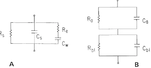
extra-cellular spaces, can be described as resistive. While this description must be (and fortunately can be) tested experimentally, it is not the most likely source of error. In the linear case, one can begin in the same spirit by describing a small patch of mem-brane as a resistor in parallel with a capacitor. The resistor describes the macromole-cules which span the membrane and allow ionic current flow, perhaps through an aqueous channel in the center of a protein. The capacitor represents the dielectric be-havior of the oily lipid matrix which forms the bulk of the membrane. Note that by restricting ourselves to the strictly linear situation, we mean to exclude the nonlinear ionic conductances as described in, e.g., Hodgkin and Huxley.24 It is certainly reason-able, and probably correct, to describe membrane permeability as a fixed conductance, although errors may be introduced in a secondary manner by nonlinear phenomena inside or outside of membranes, e.g., accumulation and/or depletion of ions in small regions of extra- or intracellular space, near membranes. It is also reasonable, but not so certainly correct, to describe displacement current in the membrane as that through a fixed-voltage and time independent-capacitance. That is certainly a good descrip-tion of artificial membranes made of lipids, but perhaps is not such a good descripdescrip-tion of biomembranes.
The structural analysis of nonlinear properties is of greater biological significance than analysis of the linear properties we have been discussing, for the simple reason that most physiological functions are highly nonlinear. The current voltage relation-ship for even the simplest ionic channel through a membrane will not be a straight line and only by keeping the voltage excursions small can one justify the assumption of linearity. Indeed the voltage dependence of many ionic channels is very steep and usu-ally involves time dependence as well. The sodium selective channel which initiates the nerve action potential is one well-known example of a voltage and time-dependent system, as quantitatively described in Hodgkin and Huxley.26 The analytical descrip-tion of nonlinear, time-varying conductances has usually been a kinetic scheme where the rate of a chemical reaction (e.g., the opening of a gate in a channel) depends on transmembrane potential. In the case of the sodium channel, it is widely thought that three such reactions must occur in succession before the channel can conduct, since the conductance depends on a probability function cubed. Other ionic channels have different kinetic behavior and in fact it may not be possible to represent some channels by a chemical kinetic scheme.
The detailed description of an ionic channel is relevant to our goal of structural analysis, since the equations describing a tissue will depend on the properties of its membranes. If the nonlinear case is to be treated, a nonlinear representation of the membranes must be used. Since a general description of a nonlinear ionic channel is not available, and may not be possible, one cannot write equations that are appropriate for the general nonlinear situation. A further complication is that nonlinear time-vary-ing conductances often give rise to current flows which can mimic delayed (i.e., reac-tive) currents produced by structural complexities. *
For these reasons, it is necessary, in our opinion, to perform a structural analysis of linear electrical properties before one can hope to determine the nonlinear properties of the individual components of the cell. The linear properties are themselves of con-siderable interest, so it is not necessary to apologize for their study. Nonetheless, the
212 CRC Critical Reviews in Bioengineering
essential justification for the study of the linear properties of tissues which function nonlinearly is that we must study the more definable Linear case first, if we are to proceed without piling ambiguity upon assumption.
D. Theoretical Predictions
The differential equations and boundary conditions describing the electrical prop-erties of tissues and cells range in difficulty from trivial to intractable. The equations describing the steady-state potential in a small spherical cell need not be even differ-ential equations. The equations describing current flow in a thin axon are relatively simple: one-dimensional current flow in such a preparation is described by the "telegrapher's equation" of 19th century fame. The equations describing current flow
in multidimensional tissues or near the tip of a microelectrode are more difficult; fi-nally, the equations describing nonlinear phenomena are essentially intractable, requir-ing numerical analysis on a digital computer.
The fundamental goals of analysis of biological preparations are rather different from those in analagous physical situations, and this difterence colors the entire ap-proach to the problem. Often the biologist is more interested in qualitative, parametric results than in quantitative predictions of the field in space and time. Thus the precise distribution of potential in a cell is rarely important. Much more significant is the variation of the properties of the cell or tissue with changes in the properties of its components, with changes in size, shape, membrane resistance, and so on. Peskoff and Eisenberg4
() argue this case in some detail and conclude that the techniques of singular perturbation theory are particularly well suited to biological needs.
In a perturbation analysis, the solution of a differential equation is represented by an asymptotic series in powers of some small parameter. The differential equation is then broken into a series of problems, the solution of each problem giving a coefficient of the power series expansion. Note that each coefficient is in fact a function, describ-ing the spatial and temporal distribution of a component of the solution. The proper-ties of singular perturbation theory which are most useful are quite specific: first, the breaking of a problem into subproblems, each of some mathematical complexity but each with a simple physical interpretation; and second, the isolation and relative sizing of the important dimensionless parameters of a problem. Other methods may be used to give similar results, but it should be emphasized that the limiting factor in the valid-ity of perturbation results lies in the properties of the resulting expansion, no matter how that expansion is generated. These expansions are usually sufficiently complex, involving a significant number of physically (but not mathematically) interrelated pa-rameters, that a discussion of uniformity is rarely undertaken. This is an important enough point to warrant further discussion.
Approximate solutions of field problems are usually leading terms in an expansion of the solution of that problem. Since the solutions and their expansions involve many dimensionless parameters, the expansion cannot be expected to be valid for all possible values of the parameters. Consider an expansion of the form V(f';E) =
vii!)
+ EVJi>
~ + ~
+
Ely 2(r)+ ...
+ E"V.(r). The coefficients V n(fJ, as well as £, will usually depend onthe morphometric properties of the tissue. We assume E
«
1, but the functions Vn(t)may contain a morphometric parameter (J such that V,,(;) a: {In. The expansion intro-duced may thus contain terms of the form ({J£)n. Since we must expect that under some conditions {JE ~ 1, our solution will diverge under those conditions and may not be a useful approximation. The use of the leading terms of the expansion as an approximate solution requires the additional assumption that the parameter {J must be about equal to one. In more formal language, the expansion is nonuniform in the parameter {J,
extra-cellular spaces, can be described as resistive. While this description must be (and fortunately can be) tested experimentally, it is not the most likely source of error. In the linear case, one can begin in the same spirit by describing a small patch of mem-brane as a resistor in parallel with a capacitor. The resistor describes the macromole-cules which span the membrane and allow ionic current flow, perhaps through an aqueous channel in the center of a protein. The capacitor represents the dielectric be-havior of the oily lipid matrix which forms the bulk of the membrane. Note that by restricting ourselves to the strictly linear situation, we mean to exclude the nonlinear ionic conductances as described in, e.g., Hodgkin and Huxley.24 It is certainly reason-able, and probably correct, to describe membrane permeability as a fixed conductance, although errors may be introduced in a secondary manner by nonlinear phenomena inside or outside of membranes, e.g., accumulation and/or depletion of ions in small regions of extra- or intracellular space, near membranes. It is also reasonable, but not so certainly correct, to describe displacement current in the membrane as that through a fixed-voltage and time independent-capacitance. That is certainly a good descrip-tion of artificial membranes made of lipids, but perhaps is not such a good descripdescrip-tion of biomembranes.
The structural analysis of nonlinear properties is of greater biological significance than analysis of the linear properties we have been discussing, for the simple reason that most physiological functions are highly nonlinear. The current voltage relation-ship for even the simplest ionic channel through a membrane will not be a straight line and only by keeping the voltage excursions small can one justify the assumption of linearity. Indeed the voltage dependence of many ionic channels is very steep and usu-ally involves time dependence as well. The sodium selective channel which initiates the nerve action potential is one well-known example of a voltage and time-dependent system, as quantitatively described in Hodgkin and Huxley. 26 The analytical descrip-tion of nonlinear, time-varying conductances has usually been a kinetic scheme where the rate of a chemical reaction (e.g., the opening of a gate in a channel) depends on transmembrane potential. In the case of the sodium channel, it is widely thought that three such reactions must occur in succession before the channel can conduct, since the conductance depends on a probability function cubed. Other ionic channels have different kinetic behavior and in fact it may not be possible to represent some channels
by a chemical kinetic scheme.
The detailed description of an ionic channel is relevant to our goal of structural analysis, since the equations describing a tissue will depend on the properties of its membranes. If the nonlinear case is to be treated, a nonlinear representation of the membranes must be used. Since a general description of a nonlinear ionic channel is not available, and may not be possible, one cannot write equations that are appropriate for the general nonlinear situation. A further complication is that nonlinear time-vary-ing conductances often give rise to current flows which can mimic delayed (i.e., reac-tive) currents produced by structural complexities.
*
For these reasons, it is necessary, in our opinion, to perform a structural analysis of linear electrical properties before one can hope to determine the nonlinear properties of the individual components of the cell. The linear properties are themselves of con-siderable interest, so it is not necessary to apologize for their study. Nonetheless, the
212 CRe Critical Reviews in Bioengineering
essential justification for the study of the linear properties of tissues which function nonlinearly is that we must study the more definable linear case first, if we are to
proceed without piling ambiguity upon assumption.
D. Theoretical Predictions
The differential equations and boundary conditions describing the electrical prop-erties of tissues and cells range in difficulty from trivial to intractable. The equations describing the steady-state potential in a small spherical cell need not be even differ-ential equations. The equations describing current flow in a thin axon are relatively simple: one-dimensional current flow in such a preparation is described by the "telegrapher's equation" of 19th century fame. The equations describing current flow
in multidimensional tissues or near the tip of a microelectrode are more difficult; fi-nally, the equations describing nonlinear phenomena are essentially intractable, requir-ing numerical analysis on a digital computer.
The fundamental goals of analysis of biological preparations are rather different from those in analagous physical situations, and this diHerence colors the entire ap-proach to the problem. Often the biologist is more interested in qualitative, parametric results than in quantitative predictions of the field in space and time. Thus the precise distribution of potential in a cell is rarely important. Much more significant is the variation of the properties of the cell or tissue with changes in the properties of its components, with changes in size, shape, membrane resistance, and so on. Peskoff and Eisenberg40 argue this case in some detail and conclude that the techniques of singular perturbation theory are particularly well suited to biological needs.
in a perturbation analysis, the solution of a differential equation is represented by an asymptotic series in powers of some small parameter. The differential equation is then broken into a series of problems, the solution of each problem giving a coefficient of the power series expansion. Note that each coefficient is in fact a function, describ-ing the spatial and temporal distribution of a component of the solution. The proper-ties of singular perturbation theory which are most useful are quite specific: first, the breaking of a problem into subproblems, each of some mathematical complexity but each with a simple physical interpretation; and second, the isolation and relative sizing of the important dimensionless parameters of a problem. Other methods may be used to give similar results, but it should be emphasized that the limiting factor in the valid-ity of perturbation results lies in the properties of the resulting expansion, no matter how that expansion is generated. These expansions are usually sufficiently complex, involving a significant number of physically (but not mathematically) interrelated pa-rameters, that a discussion of uniformity is rarely undertaken. This is an important enough point to warrant further discussion.
Approximate solutions of field problems are usually leading terms in an expansion of the solution of that problem. Since the solutions and their expansions involve many dimensionless parameters, the expansion cannot be expected to be valid for all possible values of the parameters. Consider an expansion of the form V(t,E} = V
o<"i1
+
EV 1(0~ + ~
+
E'V2(r)+ ... +
£nV,,(r). The coefficients V,,(n, as well as £, will usually depend onthe morphometric properties of the tissue. We assume £
< <
I, but the functions Vit)
" ~
may con tam a morphometrIC parameter (3 such that V n(r) ex:
f3".
The expansionintro-duced may thus contain terms of the form ({3£)n. Since we must expect that under some conditions {JE. ~ I, our solution will diverge under those conditions and may not be a useful approximation. The use of the leading terms of the expansion as an approximate solution requires the additional assumption that the parameter {3 must be about equal to one. In more formal language, the expansion is nonuniform in the parameter {3,
The form of the expansion will depend on the relative size of the different parameters in the problem. Since the form of the expansion determines the form of an equivalent circuit, different relative sizes of parameters can produce quite distinct images of the tissue. These different images correspond to different physical and physiological situ-ations, which often have not been recognized. In this manner the mathematical inves-tigation of nonuniformity can produce important physiological insights.
Perturbation analysis has been widely used in problems specifying the electrical properties of cells and tissues of complex structure. The solutions generated by pertur-bation methods have so far always reduced to simple circuits with known error terms. These simple equivalent circuits are useful because they summarize a wide range of properties in a neat form understood by most electrophysiologists. They also can be studied with the methods of circuit theory, which are often more easy to apply than the equivalent techniques of applied mathematics. Finally, these circuits have in them-selves an obvious relation to the structure of the preparation: individual circuit com-ponents represent the effective properties of individual structural systems, whether membranes or compartments of intra- or extracellular space. The equivalent circuit therefore has a life of its own and can be used to explain phenomena that are beyond the conditions under which the original partial differential equations were derived and solved.
Equivalent circuits can be, and often have been, introduced into the scientific liter-ature without the use of field theory because they are simply a listing of the significant pathways for current flow in a preparation. Such pathways can often be determined without much formal theory. And so the reader may wonder why the complexity of field theory is necessary. The mathematical analysis is obviously necessary to provide estimates of error, and so to avoid controversy concerning the appropriateness and range of validity of the equivalent circuits. Furthermore mathematical analysis is needed to determine the relationship between the measurable effective parameters and the underlying specific parameters which describe the properties of the cellular com-ponents. Finally, it is not always possible to identify a priori the significant pathways for current flow-this was certainly the case in the analysis of syncytial tissues (Eisen-berg et al. 16)-and then the analytical approach is essential.
The theoretical analysis of complex structures must be done in a certain manner if it is to serve its proper role as a tool in the measurement of the electrical properties of biological structures. The theory should be
1. Reductionist and rigorous, reducing physiological phenomena to fundamental physical laws with as few interposed approximations as possible
2. Realistic, involving as precise a description of the tissue as possible 3. Accurate, giving error bounds on all approximate expressions
4. Usable, giving expressions which can be directly compared to commonly used heuristic results and to experimental data
Theoretical analyses which satisfy many of these criteria are now available for a number of preparations as summarized in the reviews already cited. But the reader must not be lulled into thinking this a closed field. The following is an incomplete list of significant open problems, all apparently solvable with known perturbation meth-ods, all awaiting solution:
1. The electric field expected in an anisotropic cylindrical, thin plane, or thick plane syncytial tissue
214 CRC Critical Reviews in Bioengineering
3. The electric field expected outside a cylindrical preparation
4. The electrical interaction expected between neighboring cells, with the common biological shapes, assuming no specialized connections between cells
S. The meaning of "tortuosity" for branched and wiggling defts or tubules in prep-arations of several different geometries
6. The formulation, solution, and testing of the appropriate differential equations to describe the accumulation and depletion of ions in small extracellular com-partments within cells and tissues
7. The formulation. solution, and testing of the approriate differential equations to describe the spread of potential in dendritic trees, recognizing the analogy with syncytial tissues and satisfying the criteria just described
E. Electrical Measurements
The practical use of electrical measurements to specify properties of the components of complex tissues has been the subject of many papers lValdiosera et aI., 4(;47 Mathias
et aJ., q Eisenberg e( a1., 16.1" Schneider and Chandler;1l Chandler and Schneider,lo
Schoenberg et aI., 41 Schoenberg and Fozzard,42 see the many earlier papers cited in
those articles) and the recent paper of Mathias et a1.,'5 presents a review of current knowledge. For that reason another long discussion is neither needed nor appropriate. Here we will concentrate on a few general issues and develop a new method which may make the structural interpretation of electrical measurements easier.
The assignment of particular electrical properties to the individual structures of a cell or tissue requires the comparison of at least one experimental response to a theo-retical prediction. It is better, of course, to compare many predictions and responses.
It is better yet to compare a number of predictions and responses measured and com-puted under different conditons, with different patterns of current flow. Living tissue
The assignment of particular electrical properties to the individual structures of a cell or tissue requires the comparison of at least one experimental response to a theo-retical prediction. [t is better, of course, to compare many predictions and responses.
It is better yet to compare a number of predictions and responses measured and com-puted under different conditons, with different patterns of current flow. Living tissue is too delicate to allow multiple experimental manipUlations which change the pattern of current flow, For example, pharmacological agents and physiological interventions will change the conductive properties of membranes, but most such experiments are prolonged and difficult to perform without damage to the tissue. However, the pattern of current can be changed without detrimental manipulations because of the capacitive propenies of membranes. The capacitive properties of a biomembrane guarantee that different frequencies of applied current will induce different patterns of induced cur-rent flow (and of induced potential). It is possible then to compare experimental results and theoretical predictions under many conditions of current flow simply by compar-ing the temporal variation of potential with that predicted by theory.
F. Analysis in the Frequency Domain
The confrontation between theory and experiment can be made either in the fre-quency domain or the time domain. The phrase "in the frefre-quency domain" originally
meant that the input signal and resulting output were sinusoids. Here "frequency do-main" refers to the sinusoidal components of a wide class of inputs and outputs, as determined from their Fourier transforms.
There are several different ways to perform a frequency domain analysis:
ensures, for a linear time invariant system, that the response will also be a sinu-soid at just that one frequency, but perhaps with a different amplitude and phase. 2. The system may be perturbed by the sum of sinusoids of different frequencies with prescribed energy at each frequency. A stochastic version of such a signal, with equal energy (on the average) at each frequency, is called "white noise". A deterministic approximation to such a signal can be produced by periodic repeti-tion at the rate F (in Hz) of a waveform that appears to be (but is not) random. Such a periodic waveform is easy to make by analog filtering of the binary output of a shift register oscillator. The resulting waveform is often called pseudo-ran-dom noise, but it should be clearly realized that the waveform is in fact a strictly deterministic periodic signal, containing a rich spectrum of those sinusoids with frequencies greater than F.
3. The system might be perturbed by a typical transient excitation, a step function, or an impulse, and then appropriate mathematical steps taken to numerically convert the waveform, and the response to that waveform, into the frequency domain.
In an ideal system any of these methods might be expected to work reasonably weIl, but in the real world the third approach, using step functions or impulses, is quite difficult. For example. if a step function input were used, one would in effect be con-centrating all the energy of the input at frequencies close to zero, since the energy content of a step is proportional to the reciprocal of frequency. The advantages of frequency domain analysis would then be lost, since the range of frequencies examined, and therefore the distributions of potential, are limited. Furthermore if the amplifiers and electrodes add contaminating noise at many frequencies, the signal-to-noise ratio would be very high at zero frequency but very low at higher frequencies. Measurements of circuit parameters which depend only on low-frequency behavior would be possible, but measurement of parameters which depend on high-frequency behavior would be difficult. Even prolonged signal averaging does not help very much since, in the real world, the low-frequency signals, containing so much energy, limit the usable dynamic range. The analysis of a response to a step function must be expected to give less information than the analysis of a broad band signal.
The use of a signal with equal energies at all frequencies would seem to offer a way out of these difficulties and signals approximating white noise are sometimes used for this very reason. Those are the pseudo-random, periodic signals we have just discussed. The only nonperiodic transient signal with a flat frequency spectrum is, however, par-ticularly impractical to use. That signal is a so-called delta function (better "delta functional "): a very large, supposedly infinite spike, containing unity area. Even if such a signal is approximated as a triangle or rectangle of short duration and large height, it is most difficult to use. The signal is so large that it tends to excite confusing nonlinearities in the biological preparation or irrelevant nonlinearities in the recording apparatus. For these reasons, frequency domain analysis cannot easily be done with transient waveforms. Direct analysis of the time domain response to step functions can provide a great deal of useful information concerning the electrical parameters of a preparation. The analysis, however, must use a fundamentally different approach than analysis in the frequency domain.
Analysis in the frequency domain follows the procedure, detailed in many papers-particularly, Valdiosera et al. 46,47 and Mathias et al.3s-of applying a
elec-November 1980 215
ensures, for a linear time invariant system, that the response will also be a sinu-soid at just that one frequency, but perhaps with a different amplitude and phase. 2. The system may be perturbed by the sum of sinusoids of different frequencies with prescribed energy at each frequency. A stochastic version of such a signal, with equal energy (on the average) at each frequency, is called "white noise". A deterministic approximation to such a signal can be produced by periodic repeti-tion at the rate F (in Hz) of a waveform that appears to be (but is not) random. Such a periodic waveform is easy to make by analog filtering of the binary output of a shift register oscillator. The resulting waveform is often called pseudo-ran-dom noise, but it should be clearly realized that the waveform is in fact a strictly deterministic periodic signal, containing a rich spectrum of those sinusoids with frequencies greater than F.
3. The system might be perturbed by a typical transient excitation, a step function, or an impulse, and then appropriate mathematical steps taken to numerically convert the waveform, and the response to that waveform, into the frequency domain.
In an ideal system any of these methods might be expected to work reasonably well, but in the real world the third approach, using step functions or impulses, is quite difficult. For example, if a step function input were used, one would in effect be con-centrating all the energy of the input at frequencies close to zero, since the energy content of a step is proportional to the reciprocal of frequency. The advantages of frequency domain analysis would then be lost, since the range of frequencies examined, and therefore the distributions of potential, are limited. Furthermore if the amplifiers and electrodes add contaminating noise at many frequencies, the signal-to-noise ratio would be very high at zero frequency but very low at higher frequencies. Measurements of circuit parameters which depend only on low-frequency behavior would be possible, but measurement of parameters which depend on high-frequency behavior would be difficult. Even prolonged signal averaging does not help very much since, in the real world, the low-frequency signals, containing so much energy, limit the usable dynamic range. The analysis of a response to a step function must be expected to give less information than the analysis of a broad band signal.
The use of a signal with equal energies at all frequencies would seem to offer a way out of these difficulties and signals approximating white noise are sometimes used for this very reason. Those are the pseudo-random, periodic signals we have just discussed. The only nonperiodic transient signal with a flat frequency spectrum is, however, par-ticularly impractical to use. That signal is a so-called delta function (better "delta functional"): a very large, supposedly infinite spike, containing unity area. Even if such a signal is approximated as a triangle or rectangle of short duration and large height, it is most difficult to use. The signal is so large that it tends to excite confusing nonlinearities in the biological preparation or irrelevant nonlinearities in the recording apparatus. For these reasons, frequency domain analysis cannot easily be done with transient waveforms. Direct analysis of the time domain response to step functions can provide a great deal of useful information concerning the electrical parameters of a preparation. The analysis, however, must use a fundamentally different approach than analysis in the frequency domain.
Analysis in the frequency domain follows the procedure, detailed in many papers-particularly, Valdiosera et al. 4647 and Mathias et al. JS_of applying a
elec-trical parameters of the cell or tissue. When the data are in the frequency domain, this procedure is found to work-both in principle and in practice.
O. Analysis in the Time Domain
It is only natural to expect that curve fining to time domain data (e.g., the response to a step function) would be as successful as curve fitting in the frequency domain. Unfortunately, this expectation is not fulfilled: despite many attempts to use them, curve fitting procedures do not work well in the time domain.
Transient measurements have been widely used in physiology (and are extensively reviewed by Jack et al.,17) to measure the electrical parameters of preparations. But careful reading of the literature will show that such investigations have been successful in essentially two situations: one, when the system is highly nonlinear and a frequency domain analysis produces a multitude of confusions; two, when a particularly simple electrical structure is assumed for a preparation.
In the first situation, when the tissue is highly nonlinear, transient measurements are entirely appropriate and necessary. However, it has not yet been possible to deter-mine the nonlinear electrical structure of complex tissues. That is to say, it has not been possible to assign the different nonlinear properties of a preparation to the cellu-lar structures which produce them. Such a nonlinear structural analysis clearly requires a previous linear structural analysis.
Curve fitting to transient meaurements have been used in one other situation: when the electrical structure is assumed to be quite simple. In this case the entire preparation has been assumed to be a single cell, either a spherical cell (a resistor and capacitor in parallel) or a cylindrical cell (where the resistor and capacitor are distributed along the resistance of the cell interior). This assumption is usually in conflict with the known anatomy, so transient measurements are then used to determine the "effective" or "total" capacitance and conductance of the preparation, meaning the sum of all the membrane capacitors or conductors. But, as Adrian and Almers3
,4 point out, even the
determination of "total" capacitance or conductance is correct only if the preparation really is a single cell as assumed; the determination is incorrect if the electrical structure of the preparation is complex (Vaughan et al. 48).
The use of transient measurements to determine circuit parameters of more complex circuits is usually unsuccessful and has in fact rarely been used. The mathematical properties of a linear system guarantee that the response to a transient input is a sum of exponentials. Thus fitting experimental results with a theoretical model in the time domain means in practice the fitting of noisy data with a sum of exponentials. The purpose of the fitting is to determine the amplitudes, time constants (i.e., the expo-nents) and the number of exponential terms. The amplitudes and time constants in turn determine the parameters of the equivalent circuit, which are the electrical prop-erties of individual structures of the preparation.
November 1980 211
his textbook "On Numerical Methods that [Usually] Work, "where he warns of such problems in vivid terms: "One of the perennial problems that plagues [the numerical analyst] .. .is the fitting of data by a series of exponential functions .... The answer to this problem lies in the .. .laboratory [doing a different kind of experiment] ... and the sooner the hopeful innocent can be sent there and away from the computer room, the better everyone will be. For it is well known that an exponential equation of this type ... is extremely ill-conditioned. That is, there are many combinations of fparametersl ... that will fit the most exact data quite well indeed (will you believe four significant figures?) and when experimental noise is thrown into the pot, the entire operation becomes hopeless .... " These authors conclude, as may we, that curve fitting to transient data contaminated with noise is an undesirable way to determine electrical parameters. Indeed, it is this very fact which motivated the first workers (Falk and Fatt20
and Fatt21
) to use measurements in the frequency domain to determine the
prop-erties of individual cell structures.
H. Integrals of Transients
One might conclude for these reasons that transient analysis is of little use in struc-tural analysis, but Adrian et al. 5 have introduced a quite different method of treating
transient data. Their method provides good estimates of the effective (but not always the total) capacitance of a preparation. Their approach avoids curve fitting altogether; rather it is based on the numerical evaluation of the integral of the transient current following a step change in voltage. This integral directly gives an estimate of the effec-tive capacitance, since it measures the charge movement due to the voltage change. We will spend some time deriving and extending this method and will point out several applications \vhich are not discussed in the literature to our knowledge (although re-lated results have been independently derived and kindly communicated to us by Dr. Roger Tsien and Drs. Vaughan and Loo).
The fundamental idea introduced by Adrian et al., which we feel deserves the name breakthrough, is the computation of an integral of the transient response of a prepa-ration and the evaluation of that integral in terms of properties of theoretical models of the preparation, usually an equivalent circuit with a structural interpretation.
We consider a class of integrals of the following type
I n J
r
t)n ig(t,y:x)o H ( 0 , y; x)f (t, x) } d t (1)
where n 0,1,2, ... ; t is the time after the stimulus; the input f(t,x) is applied at the spatial location x. The response to the input is g(t,y; x) measured at a different spatial location y, still within the tissue. The spatial coordinates (x,y) may in general be vectors representing the locations of, e.g., a current passing microelectrode and a voltage re-cording microelectrode within a three-dimensional cell or tissue.
The Laplace transforms of time signals are defined in the usual manner (Churchill, I I
WidderSl )
t. . It. =
G(s.y:x)
=
£ {g(t.y;x) J=
So
g(t,y;x)e~stdt ( 2)where the lower limit of the integral in Equation 2 and in all subsequent integrals of this type is taken to be 0-; that is to say, the lower limit of the integral is just before any discontinuity which occurs at time zero.
The input/output relationship for the network (i.e., tissue) is defined as
t. G(s,Y;x) H(s,y;x) '"
F(s, x)
The quantity H(O,y,x), used in the above integral, defines the steady-state (time in-variant) response of the network. We consider biological tissues, working in their linear regions, without inductive elements. The function H(O,y; x) can then be determined from the resistive elements of the network with the capacitive elements treated as open circuits. The function H(O, y; x) can be considered either an input resistance or an input conductance, depending on whether the input f(') and output g(') are current and voltage, or vice versa.
We now evaluate the integrals in terms of the input/output function H(s,y; x). We later will show that, in many cases, all the circuit parameters of the preparation can be determined from the inpulioutput function and thus from the experimentally deter-mined integrals. Previous work has used only one integral to determine just one param-eter, the effective capacitance.
Several properties of the Laplace transform are used which are derived and described in most texts concerning Laplace transforms (Churchill, II Widder, 5 l 5 l give the
do-mains of validity of the following expressions):
I. The steady value of a function is given, if it reaches a steady value, by
lim g(t) lim ~G(s) (4)
2. The steady value of an integral of a function is given, if it reaches a steady value, by
lim
£t
!!(tldl cc G(O) (5)I~CN
3. A class of integrals of a function g(t) can be written in terms of the derivatives G(n,(O) of the Laplace transform of the function: In particular, the first and all higher order derivatives can be determined from integrals:
lim s-·O
dnG(s) ~ c(n)(O)
dsn
(6)
Use of these properties allows the integral l" in Equation 1 to be written as
In lim d n
H(O) .
SF(S»)
(7)s-.O ds n
The rule for repeated differentiation of a product
n
C)~[H(S)
H(O)]
d kIn lim Z; [s I, (s) J
5-,0 k~O k ds n ,- k ds k
(1\)
where
n!
(9)
k!(n -k)!
We expand H(s) about s 0; apply the definition of the derivative: