UNCORRECTED PROOF
Progress in Neurobiology 000 (2000) 000 – 000Voltage-gated proton channels in microglia
Claudia Eder
a,*, Thomas E. DeCoursey
baInstitut fu¨r Physiologie der Charite´,Humboldt Uni6ersita¨t,Tucholskystr.2,D10117Berlin,Germany bDepartment of Molecular Biophysics and Physiology,Rush Presbyterian St. Luke
’s Medical Center,1750West Harrison,Chicago,
IL60612, USA
Received 24 July 2000
Abstract
Microglia, macrophages that reside in the brain, can express at least 12 different ion channels, including voltage-gated proton channels. The properties of H+currents in microglia are similar to those in other phagocytes. Proton currents are elicited by
depolarizing the membrane potential, but activation also depends strongly on both intracellular pH (pHi) and extracellular pH (pHo). Increasing pHo or lowering pHi promotes H+ channel opening by shifting the activation threshold to more negative potentials. H+channels in microglia open only when the pH gradient is outward, so they carry only outward current in the steady
state. Time-dependent activation of H+currents is slow, with a time constant roughly 1 s at room temperature. Microglial H+
currents are inhibited by inorganic polyvalent cations, which reduce H+current amplitude and shift the voltage dependence of
activation to more positive potentials. Cytoskeletal disruptive agents modulate H+ currents in microglia. Cytochalasin D or
colchicine decrease the current density and slow the activation of H+currents. Similar changes of H+currents, possibly due to
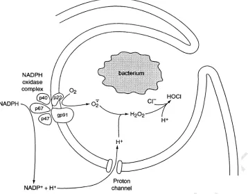
cytoskeletal reorganization, occur in microglia during the transformation from ameboid to ramified morphology. Phagocytes, including microglia, undergo a respiratory burst, in which NADPH oxidase releases bactericidal superoxide anions into the phagosome and stoichiometrically releases protons into the cell, tending to depolarize and acidify the cell. H+currents may help
regulate both the membrane potential and pHi during the respiratory burst. By compensating for the efflux of electrons and counteracting intracellular acidification, H+channels help maintain superoxide anion production. © 2001 Elsevier Science Ltd.
All rights reserved.
Contents
1. Introduction . . . 000
2. pH changes in the brain . . . 000
2.1. Activity-dependent pH changes in the brain. . . 000
2.2. Influence of extracellular pH changes on brain function. . . 000
2.3. Influence of extracellular and intracellular pH changes on microglial function . . . 000
2.3.1.Effects of pH on microglial function . . . 000
www.elsevier.com/locate/pneurobio
Abbre6iations:ACM, astrocyte conditioned media; ADP, adenosine diphosphate; ATP, adenosine triphosphate; [Ca2+]
i, intracellular free Ca2+ concentration; CGD, chronic granulomatous disease; CNS, central nervous system; CRAC, Ca2+-release-activated Ca2+[channels];E
H, Nernst potential for H+; EIPA, 5-(N-ethyl-N-isopropyl)-amiloride; fMLP,N-formyl-Met-Leu-Phe chemotactic peptide; GABA-A, gamma aminobutyric
acid;gH, H+chord conductance;gH,max, maximum or limiting value ofgH; GM-CSF, granulocyte-macrophage colony-stimulating factor; HBC, hydrogen-bonded chain; ICAM-1, intercellular adhesion molecule-1; IFN-g, gamma-interferon;IH, H+current amplitude;IH,max, maximum H+ current amplitude; InsP3, inositol triphosphate; IR, inward rectifier; [K+]o, extracellular K+concentration; LFA-1, leukocyte function-associated antigen-1; LPS, lipopolysaccharide; MHC, major histocompatibility complex; NADPH, reduced nicotinamide-adenine dinucleotide phosphate; O−
2 , superoxide anion; DpH, PH gradient (pHo– pHi); pHi, intracellular pH; pHo, extracellular pH; PKC, protein kinase C; PMA, phorbol myristate acetate;Prel, relative permeability; TEA, tetraethylammonium; TNF-h, tumor necrosis factor;Vthreshold, threshold level of depolarization required to activate H+currents;V
rev, reversal potential.
* Corresponding author. Tel.: +49-30-28026302; fax: +49-30-28026669.
E-mail address:[email protected] (C. Eder).
UNCORRECTED PROOF
2.3.2.Effects of pH on ion channels in microglia . . . 000
2.3.3.Effects of pH on NADPH oxidase function. . . 000
3. Regulation of the intracellular pH of microglia . . . 000
4. H+channels in microglia . . . . 000
4.1. Cells/species expressing H+channels . . . . 000
4.2. How are H+channels studied? . . . . 000
4.2.1.Voltage-clamp . . . 000
4.2.2.pH measurements . . . 000
4.3. Properties of H+channels . . . . 000
4.3.1.Selectivity . . . 000
4.3.2.Temperature dependence . . . 000
4.3.3.Permeation mechanism . . . 000
4.3.4.Gating kinetics . . . 000
4.3.5.pH dependence of gating. . . 000
4.3.6.Pharmacology . . . 000
4.4. Physiological modulation of H+channels . . . . 000
4.4.1.Regulation by astrocytic factors, lipopolysaccharide and cytoskeletal disruptive agents . . . 000
4.4.2.Modulation by arachidonic acid . . . 000
4.4.3.Phosphorylation . . . 000
4.5. Physiological functions of H+channels . . . . 000
4.5.1.General principles . . . 000
4.5.2.Specific functions . . . 000
4.5.3.Respiratory burst in microglia and other phagocytes . . . 000
4.6. Taxonomy of H+channels . . . . 000
4.6.1.Varieties of H+channels. . . . 000
4.6.2.Are they really channels?. . . 000
4.6.3.Comparison of H+channels with other ion channels . . . . 000
4.6.3.1.The single-channel conductance is very small . . . 000
4.6.3.2.The proton selectivity of the channels is almost perfect . . . 000
4.6.3.3.Nothing blocks H+channels . . . . 000
4.6.3.4.No multiple occupancy . . . 000
4.6.3.5.H+channels do not inactivate . . . . 000
4.6.4.Molecular identification of H+channels. . . . 000
5. Conclusions . . . 000
Acknowledgements . . . 000
References . . . 000
1. Introduction
Microglial cells are the resident macrophages of the brain. Microglia and peripheral macrophages share many phenotypic markers and are capable of perform-ing similar functions. In the normal adult brain mi-croglia appear to be in a functionally resting state, in which they are highly ramified, extending processes far into surrounding brain tissue, acting as antennae. A variety of stimuli, including neuronal injury, trauma, ischemia, inflammation, infection, and several neuro-logical diseases lead to microglial activation. Activation of microglia appears to proceed through a series of steps that include changes in morphology and expres-sion of surface antigens, release of cytokines and cyto-toxins, antigen presentation, and phagocytosis. A number of excellent reviews describing the properties and functions of microglia in the normal and
patholog-ical brain have been published recently (Raivich et al., 1999; Stoll and Jander, 1999; Streit et al., 1999), there-fore we do not describe these topics in detail here.
Electrophysiological properties of microglial cells have been studied intensively during the past decade. It has been demonstrated that microglia are capable of expressing a wide variety of ion channels. Under certain conditions, microglial cells express channels that selec-tively conduct K+, H+, Na+, Ca2+, or Cl−. Changes in the functional state of the microglial cells cause changes in the expression levels of some ion channels,
including inward rectifier and delayed rectifier K+
channels. Properties, expression, regulation and func-tional roles of the 12 different ion channels discovered so far in microglia have been reviewed recently (Eder, 1998).
chan-UNCORRECTED PROOF
nels in microglia. Because only a handful of papershave been published specifically on this narrow topic, the scope of the review will be somewhat more general.
The properties of H+ channels are similar in most
phagocytes, and are at least qualitatively similar in most respects in all cells and species. There are several
distinct varieties of H+ channels, however, and their
distinguishing features may have significant functional consequences.
2. pH changes in the brain
Within the central nervous system (CNS), neuronal activity is accompanied by substantial changes in extra-cellular pH. Changes in pH occur under pathological situations, such as ischemia and epilepsy, but also in the normally functioning brain. Because activation of
pro-ton channels depends strongly on voltage, pHi, and
pHo, activity-dependent pH changes of the external
milieu will influence the activity of microglial proton channels. We therefore first give an overview of the pH changes in the brain and their possible influence on neuronal and microglial activity.
2.1. Acti6ity-dependent pH changes in the brain
In many CNS regions, a rapid extracellular alkalin-ization is seen at the onset of neuronal activity. Such activity-dependent alkalosis of the extracellular space was detected in cortex (Urbanics et al., 1978), in the cerebellum (Kraig et al., 1983; Chesler and Chan, 1988), in some spinal cord preparations (Endres et al., 1986; Jendelova and Sykova, 1991), and in the hippocampus (Jarolimek et al., 1989; Walz, 1989; Chen and Chesler, 1992; Chen and Chesler, 1992a,b; Kaila et al., 1992). Alkaline pH shifts have a rapid onset and can sometimes be maintained for as long as the stimuli are applied (Chesler and Kaila, 1992). In many cases, alkaline pH shifts are transient and are followed by a more slowly developing extracellular acidification (see below).
Ligand-gated channels play a major role in the gener-ation of activity-induced alkaline transients. Activgener-ation of GABA-A receptors leads to the efflux of bicarbonate through anion channels resulting in a consequent rise in
pHo (Kaila and Voipio, 1987; Chen and Chesler, 1990;
Kaila et al., 1990; Chen and Chesler, 1992a). A bicar-bonate-independent alkaline shift can be elicited by activation of ionotropic glutamate receptors (Chesler and Chan, 1988; Chen and Chesler, 1992a) or by direct electrical stimulation of a neuronal population (Chen and Chesler, 1992b; Grichtchenko and Chesler, 1996; Paalasmaa and Kaila, 1996; Tong and Chesler, 1999). It has been suggested that the glutamate-induced
extracel-lular alkalinization is caused by fluxes of H+/OH−
equivalents down their electrochemical gradient
through the glutamate receptor-coupled ion channels of both neurons and glial cells (Chen and Chesler, 1992b; Deitmer and Munsch, 1992). Since the glutamate-in-duced alkalinization was suppressed upon removal of
extracellular Ca2+, it has been proposed by others
(Paalasmaa et al., 1994; Smith et al., 1994) that this alkalinization results from the activation of a cellular Ca2+/H+ATPase. Activation of the Ca2+/H+ATPase leads to a decrease in the concentration of extracellular
H+due to the outward transport of intracellular Ca2+
in exchange for extracellular H+ (Schwiening et al.,
1993).
In some CNS regions, e.g. in the optic nerve (Davis et al., 1987), in the spinal cord (Sykova and Svoboda, 1990), and in the cortex (Siesjo¨ et al., 1985; Chesler and Kraig, 1987), neuronal activity induces an early acid shift. Activity-induced alkaline transients are also often followed by a slow, persistent acid shift. Moreover, prolonged acidification of the extracellular space has been detected during repetitive stimulation, epilepti-form activity, spreading depression, and ischemia (Chesler and Kaila, 1992).
Several potential mechanisms may underlie
acidifica-tion, including lactic acid efflux, Na+/H+ exchange,
Na+/HCO
3
−/Cl−-coupled transport, or electrogenic
Na+/HCO
3
− cotransport (Chesler, 1990). In
hippocam-pal slices, the late stimulus-elicited acidification is
caused by accumulation of interstitial CO2(Voipio and
Kaila, 1993). Since the capacity of the tissue to acidify the extracellular space increased over the first postnatal weeks in correlation with the proliferation of glia, it has been proposed that acid secretion by glial cells is also an important source for the extracellular acidification (Chesler and Kaila, 1992; Deitmer and Rose, 1996).
2.2. Influence of extracellular pH changes on brain function
Physiological alterations in pHocan induce a variety
of changes in brain functions, including alterations in the neuronal excitability, changes in the resting mem-brane potential, and induction or inhibition of ionic currents in neurons and glial cells. Since many enzymes are sensitive to small shifts in the concentration of protons (e.g. NADPH oxidase, Section 2.3.3.), most
cellular processes are modulated by changes in pHi.
Changes in the steady-state pHohave a strong influence
neuroprotec-UNCORRECTED PROOF
tive due to proton inhibition of NMDA receptors.However, rapid acidification may cause the activation of proton-gated ion channels (Korkushko and Krysh-tal, 1984; Grantyn and Lux, 1988; Bevan and Yeats, 1991) and would thus contribute to membrane
depolar-ization, subsequent Ca2+ accumulation and
neurode-generation. Extracellular pH changes have also been shown to influence the bursting behavior of neurons (Church and McLennan, 1989; de Curtis et al., 1998), presumably due to an enhancement or inhibition of
voltage-activated conductances (Tombaugh and
Somjen, 1996). Dependence on extracellular pH has been reported for a wide variety of transmitter recep-tors and voltage-gated ion channels (for reviews see Green and Andersen, 1991; Chesler and Kaila, 1992). Proton-induced inhibition of voltage-gated ion channels may occur due to direct binding of protons to specific sites on the channel. Protons can induce surface charge screening effects (Hille, 1968), decrease the permeability of ion channels, and modulate their kinetics (Hille, 1992).
2.3. Influence of extracellular and intracellular pH changes on microglial function
2.3.1. Effects of pH on microglial function
Whereas intensive studies have been performed in order to investigate the influence of alkaline or acid shifts in the extracellular and intracellular pH on neu-ronal activity, only little is known about their influence on microglial activity. It has been demonstrated that
changes in pHo influence the organization of the
mi-croglial cytoskeleton and modulate mimi-croglial motility (Faff and Nolte, 2000). In an acidic environment, basal motility and C5a-induced chemotaxis of microglia were decreased. These effects were paralleled by rearrange-ment of the actin cytoskeleton. Since similar
observa-tions have been made upon decreases in pHi without
affecting pHo, it had been proposed that changes in pHi
were responsible for the observed cytoskeletal reorgani-zation and inhibition of microglial motility during ex-tracellular acidification (Faff and Nolte, 2000). As in
many other cell types, in microglia pHifollows changes
in pHo. An alkaline shift of pHiwas found to result in elevation of [Ca2+]
i (Minelli et al., 2000).
2.3.2. Effects of pH on ion channels in microglia An increase in the concentration of intracellular
pro-tons causes inhibition of microglial inward rectifier K+
channels (Eder et al., 1995a). In recordings using intra-cellular solutions with pH of less than 7.0, inward
rectifier K+ currents decreased rapidly and finally
dis-appeared within a few minutes after establishing the
whole-cell configuration. In contrast, changes in pHo
did not influence inward rectifier currents. In microglia, inward rectifier K+channels are believed to help set the
resting membrane potential (Fischer et al., 1995; Visentin et al., 1995; Eder, 1998), similar to their role in other macrophages (Gallin and Sheehy, 1985; McKin-ney and Gallin, 1990; Gallin, 1991; DeCoursey and Grinstein, 1999). It is reasonable to expect that intracel-lular acidification would lead to membrane depolariza-tion of microglial cells due to the proton-induced inhibition of inward rectifier channels.
Delayed rectifier outward K+ channels in microglia
can be modulated by both intracellular and extracellu-lar protons. Intracelluextracellu-lar acidification reduced the am-plitudes of delayed rectifier currents, while intracellular alkalinization enhanced whole-cell delayed rectifier cur-rents. Neither kinetics nor voltage sensitivity of delayed
rectifier currents were affected by changes in pHi from
5.8 to 7.8 (Eder and Heinemann, 1996). In contrast, a shift of the steady-state activation and inactivation curves of the delayed rectifier currents in depolarizing direction was observed during acidification of the exter-nal milieu, while extracellular alkalinization showed the opposite effect (Eder and Heinemann, 1996). In addi-tion to this surface charge screening effect, time-depen-dent inactivation of microglial delayed rectifier currents was slower when superfusing cells with acid solutions than with alkaline ones (Eder and Heinemann, 1996), presumably due to specific interaction of protons with the channel (Deutsch and Lee, 1989). Modulation of delayed rectifier channels by intra- and extracellular pH changes may have significant functional implications, since delayed rectifier channels in microglia and other immune cells play an important role in the maintenance of a large driving force for Ca2+ influx through Ca2+
-release-activated Ca2+ (CRAC) channels (Lewis and
Cahalan, 1995). Functional CRAC channels seem to be required for several macrophage activities (DeCoursey and Grinstein, 1999). Because untreated microglia do
not express delayed rectifier K+ channels (Eder, 1998),
it is unlikely that they play a role in maintaining the membrane potential in these cells. In activated mi-croglia that express both delayed rectifier and inward
rectifier K+ channels, delayed rectifier channels might
serve this function when there is sufficient depolarizing force to overcome the capacity of inward rectifier
chan-nels to keep the membrane potential near EK. The
depolarizing shift of delayed rectifier channel gating
produced by low pHo has been reported to depolarize
the membrane potential of microglia activated by LPS (Chung et al., 1998).
2.3.3. Effects of pH on NADPH oxidase function NADPH oxidase is of central importance in the killing of bacteria and other invaders by phagocytes. This enzyme functions best at pH 7.0 – 7.5 (Rossi, 1986; Clark, 1990). Superoxide anion release is strongly and
progressively inhibited by interventions that lower pHi
UNCORRECTED PROOF
with almost complete inhibition at pHi6.0 (Simchowitz,
1985). The oxidase is also inhibited by decreases in pHo
(Gabig et al., 1979; Liberek et al., 1993). Because pHi
tends to follow pHothe effects of an acidic environment
may be mediated by the consequent decrease in pHi
(Simchowitz, 1985; Liberek et al., 1993). One of the main functions proposed for voltage-gated proton channels in phagocytes is to ensure pH homeostasis during the respiratory burst, which represents a severe challenge to the pH regulatory mechanisms of the cell.
3. Regulation of the intracellular pH of microglia
In the present paper, voltage-gated H+ channels of
microglia are described in detail. In addition to H+
channels, several other H+ transport systems that have
been detected in microglia might be involved in pHi
regulation of the cells under physiological and patho-physiological conditions. Faff and colleagues (1996) reported that cultured microglial cells can recover from acidification due to the activity of an amiloride- and
EIPA-sensitive Na+/H+ exchanger under HCO
3
− free
conditions. In peripheral macrophages, Na+/H+
ex-change in addition to a plasmalemmal vacuolar-type
H+-ATPase (V-ATPase) participate in pH
i recovery
from intracellular acid loads (Bidani et al., 1994; McK-inney and Moran, 1995). Whether V-ATPase activity is important for pH regulation and determination of basal
pHi in microglia as reported for other macrophages
needs to be experimentally demonstrated.
A H+/K+-ATPase that is activated at increased
con-centrations of extracellular K+ and H+ has also been
detected in microglia (Shirihai et al., 1998). Since
in-creases in the concentration of extracellular K+ and
decreases in extracellular pH are hallmarks of injured brain tissue, this transporter may play an important
role in pHiregulation in microglia during pathological
conditions.
In the presence of HCO3−a more alkaline resting pHi
than under HCO3− free conditions was determined in
cultured mouse microglia. Two mechanisms, namely
Na+/HCO
3
− cotransport and Na+-dependent Cl−/
HCO3− exchange seem to be responsible for the
ob-served HCO3− induced intracellular alkalinization (Faff
et al., 1996).
Increasing pHi in microglia leads to an increase in
[Ca2+]
i(Minelli et al., 2000). Intriguingly, this response
is inhibited by removal of extracellular Ca2+ if pH
i is
increased by raising pHo but not by an NH4+ prepulse
(Minelli et al., 2000). These data provide indirect
evi-dence that a Ca2+/H+ exchanger may exist in
microglia.
So far all H+ transporters have been studied in
cultured microglial cells. Further experiments are re-quired to demonstrate the existence of these
trans-porters in microglia of the normal and pathological brain as well as the contribution of each of those
transport systems in microglial pHi regulation under
various conditions.
4. H+ channels in microglia
4.1. Cells/species expressing H+ channels
Voltage-gated proton channels were first described by Thomas and Meech in snail neurons almost two decades ago (Thomas and Meech, 1982). These studies
were elegant (Fig. 1): pHo and pHi were monitored
using pH electrodes, and membrane current was mea-sured under voltage clamp during acid injection and other manipulations (Thomas and Meech, 1982; Meech and Thomas, 1987; Thomas, 1988). Shortly afterwards,
voltage gated proton channels were observed in Am
-bystoma (axolotl or salamander) oocytes (Barish and Baud, 1984). The possibility that voltage gated proton channels might exist in mammalian cells was proposed by Henderson and colleagues (Henderson et al., 1987) on the basis of pH measurements in human neutrophils. The first direct voltage-clamp evidence for voltage gated proton channels in mammalian cells was in rat alveolar epithelial cells (DeCoursey, 1991). Henderson’s prediction that voltage gated proton channels exist in human neutrophils was confirmed by patch-clamp in 1993 (DeCoursey and Cherny, 1993). Proton currents were first described in microglia in 1995 (Eder et al.,
1995a). H+ currents have now been observed in
cul-tured murine (Eder et al., 1995a; Klee et al., 1998,
1999), rat (Visentin et al., 1995), and human
(McLarnon et al., 1997) microglia, as well as in two microglia-derived cell lines, BV-2 and MLS-9.
The cells in which voltage gated proton channels have been reported are listed in Table 1, and include a dozen species, and two dozen cells and cell lines. Sev-eral new cells that express these channels are reported each year, so the table is growing. In surveying cells as potential expression systems for voltage gated proton channels, it has been difficult to find cells that definitely do not express this conductance. Cell lines such as CHO or HEK that are commonly used as expression systems for ion channels, have been found to express native voltage gated proton channels, although at low density (Cherny et al., 1997; Table 1). Amphibian oocytes, another popular expression system, also have
robust native H+ currents (Barish and Baud, 1984;
Humez et al., 1995). The level of expression spans three orders of magnitude in different cell types. The greatest expression level in a native cell is in human eosinophils,
where the H+ current density can be \200 pA/pF
UNCORRECTED PROOF
highly buffered pipette solutions (100 mM buffer), theH+efflux during large depolarizations depletes
intracel-lular buffer and raises pHi, producing a characteristic
‘droop’ of the current (see also Section 4.3.4).
4.2. How are H+ channels studied?
Two main approaches have been used to study voltage gated proton channels: electrical recording us-ing voltage-clamp technique and measurement of pH changes using pH electrodes or pH sensitive fluorescent dyes. Both approaches are important and each has advantages and limitations that must be recognized.
4.2.1. Voltage-clamp
For the study of small cells like microglia, the patch-clamp technique (Hamill et al., 1981) is invaluable. Conventional microelectrode puncture experiments are not feasible, because the leak inevitably caused by electrode penetration of the cell membrane conducts large non-specific currents that obscure the physiologi-cal membrane conductances. The essential feature of the patch-clamp technique is the formation of an elec-trically tight seal between the patch pipette and the cell membrane. A good seal does not allow even small ions
to pass between the pipette and the membrane, and allows recording the current flowing through a single ion channel in the patch of membrane spanning the pipette tip. The whole-cell configuration allows record-ing from all the channels in the entire cell membrane. There are several limitations, however. In the whole-cell configuration, the cytoplasm is dialyzed as a result of its continuity with the pipette solution. This is at once an advantage and a limitation. The advantage of being able to control the intracellular solution by diffusion from the pipette is offset by the loss of cytoplasmic constituents, which may interrupt second messenger pathways and alter the behavior of ion channels. This particular limitation can be overcome by use of the
permeabilized-patch configuration. Including
[image:6.612.113.467.382.645.2]pore-forming molecules, such as ATP (Lindau and Fernan-dez, 1986), nystatin (Horn and Marty, 1988), or amphotericin B (Rae et al., 1991) in the pipette solution results in eventual incorporation of these molecules into the patch membrane, where they allow passage of ionic current (carried by small ions) but prevent the loss of larger cytoplasmic molecules such as proteins and en-zymes. The value of this approach is demonstrated by the fact that the response of voltage gated proton channels in neutrophils to PMA is lost in conventional
Fig. 1. Simultaneous records of membrane potential (Em), voltage-clamp current, HCl injection current, and pHi (from top to bottom) that enabled the discovery of voltage-gated proton channels in snail neurons (Helix aspersa) by Thomas and Meech (1982). Several HCl injections were made to lower pHitoB6.5, then the membrane was depolarized stepwise. At−10 mV and at more positive voltages a decaying outward current is observed. The recovery of pHi(bottom trace) from the acid load clearly is faster at more positive voltages, consistent with the outward current being carried by H+. The outward H+current decays because the driving force (V−E
H) decreases as pHiincreases (due to the continuous H+ efflux). H+currents in small cells like microglia decay two orders of magnitude faster because of the much smaller cell volume than in snail
neurons. The neuron was pretreated with CsCl to inhibit K+ currents and SITS (4-acetamido-4%-isothiocyanatostilbene-2,2%-disulfonic acid) to
UNCORRECTED PROOF
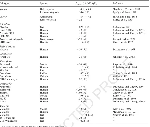
Table 1IH density in cells reported to have H+channelsc
IH,max(pA/pF) (pHi) Reference Species
Cell type
Neuron Helix aspersa 4.5 (6.8) Meech and Thomas, 1987
14.6 (5.9) Byerly and Suen, 1989 Lynmaea stagnalis
8.4 (7.2)
Oocyte Ambystoma Barish and Baud, 1984
Rana esculenta 32 Humez et al., 1995
Epithelium
27.3 (5.5)
Rat DeCoursey, 1991
Alveolar
Human
Lung A549 2 (5.5) DeCoursey and Cherny, 1994b
4 (5.5) DeCoursey and Cherny, 1994b Prostate PC-3 Human
1 (6.5)
Human b
HEK-293
Rana pipiens
Renal proximal tubule B75 (6.5) Gu and Sackin, 1995 1.6 (5.5) Cherny et al., 1997
CHO ovary Hamster
Skeletal muscle
Human
Myocyte 10 (5.5) Bernheim et al., 1993
Lymphocyte
36 (6.0) Schilling et al., 2000a Human
Jurkat E6-1
Macrophage
30 (6.0)
Peritoneal Mouse Kapus et al., 1993a
1.1 (6.0)
Human Holevinsky et al., 1994
Monocyte-derived
Rat
Alveolar 2 (7.5) b
Osteoclasts Rabbit 6.7 (6.0) Nordstro¨m et al., 1995 a? (7.3)
Chicken Weidema, 1995
Osteoclasts
Human
THP-1 monocyte 22 (5.5) DeCoursey and Cherny, 1996a
Granulocyte
17 (6)
Human DeCoursey and Cherny, 1993
Neutrophil
Human
Eosinophil 200 (6.0) Gordienko et al., 1996
100 (5.5)
Basophil Human Cherny et al., 1999
9.6 (5.5)
Mouse Kuno et al., 1997
Mast cell
Human
HL-60 133 (5.5) Demaurex et al., 1993
5 (6.0) DeCoursey and Cherny, 1994b
K-562 Human
Microglia
Microglia Mouse 42 (6.0) Eder et al., 1995a
Microglia Human a? (7.3) McLarnon et al., 1997
a66 (7.2)
Rat Visentin et al., 1995
Microglia
Rat
BV-2 microglia 20 (5.5) b
0.22 (5.5) b MLS-9 microglia Rat
aIdentity of the conductance not established with certainty.
bUnpublished data of V.V. Cherny and T.E. DeCoursey, and for MLS-9 cells P.S. Pennefather.
cThis table includes only cells in which the existence of H+channels was established by direct voltage-clamp studies. Not included are cells in
which the existence of H+channels was established by indirect measurements such as pH changes.I
H,maxis the largest H+current measured in a given cell (normalized to capacity, which reflects surface area), usually at150 mV positive toVrev;gH,maxvalues were converted to current atVrev+150 mV. In studies where typical values or cell size was not specified, estimates were made from data in figures, etc. and are preceded by a tilde (), as are values from surveys including a small number of cells. All values are at room temperature (20–25°C).
whole-cell recording, but is preserved by use of the permeabilized patch configuration (DeCoursey et al., 2000). In order to use the permeabilized-patch ap-proach to study voltage-gated proton channels, it is
necessary to take additional measures to control pHi,
because buffer molecules are too large to pass from the pipette to the cell through the nystatin or amphotericin B pores. The classical weak base gradient approach, in
which pHiis controlled by establishing a gradient of a
weak base (or acid) by virtue of the much higher permeability of the neutral form of the molecule (Ja-cobs, 1920; McLaughlin and Dilger, 1980; Roos and
Boron, 1981; Boron, 1983), has been used successfully for this purpose (Grinstein et al., 1994; DeCoursey et al., 2000).
4.2.2. pH measurements
The operation of voltage gated proton channels in intact cells can be deduced by carefully designed
mea-surements of intracellular and extracellular pH.
Thomas and Meech (1982) used pH sensitive electrodes in their pioneering study of voltage gated proton cur-rents in snail neurons (Fig. 1). Combined with
UNCORRECTED PROOF
demonstrated clearly the existence of a conductive H+
flux pathway. Many recent studies take advantage of the existence of fluorescent pH sensitive dyes to
moni-tor pHi. When combined with voltage-clamp, these
studies can provide relatively unambiguous information
relating H+ currents to cell functions. The main
draw-back of the use of pH measurements in isolation to
study H+ fluxes is that it can be difficult to establish
which transport molecule is involved in a particular response. The pH can change as a result of any of a number of different mechanisms that result in flux of acid-equivalents. Cells have a wide variety of such
mechanisms, including Na+/H+-antiport, K+/H+
ex-change, H+-ATPases, HCO
3
− transporters such as
HCO3−/Cl− exchange and NaHCO3/HCl exchange
(Roos and Boron, 1981), Cl−/OH− exchange (Sun et
al., 1996), as well as H+ channels. pH changes can also
result from the flux of the neutral form of weak acids or bases across the membrane (McLaughlin and Dilger, 1980; Roos and Boron, 1981; Boron, 1983; Cherny et al., 1990). Some of these transport mechanisms require ATP, some are electroneutral, some require counteri-ons, and most cells have a multiplicity of transporters. Unambiguous identification of the transport mecha-nism is thus difficult and requires careful elimination of all other possibilities.
A second limitation of the use of pH sensing fluores-cent dyes is that the spatial resolution is limited. Living cells may have substantial variation in local pH. In large cells, intracellular pH gradients can be detected using pH sensing microelectrodes (Vanheel et al., 1988) or pH sensing dyes (Gonda et al., 1999; Stewart et al.,
1999). For example, apical-to-basal pHi differences as
high as 0.84 Units were observed in the epithelial HT29 cell line (Maouyo et al., 2000) and stable 1 Unit gradi-ents were seen in cardiac myocytes (Spitzer et al., 2000). However, local pH in the ‘reaction layer’ adjacent to the membrane cannot be resolved using this approach. The reaction layer, a small, operationally-defined space
near the membrane during H+ flux in which
proton-buffer reactions are not at equilibrium, has a thickness
comparable with the plasma membrane (B10 nm
un-der ‘typical’ conditions) (Delahay, 1954; Neher, 1986; Kasianowicz et al., 1987; Mathias et al., 1990; De-Coursey, 1991). Local pH near the membrane is likely to be quite different from the bulk pH whenever there is membrane transport of acid equivalents. Membrane transporters that are sensitive to pH, including proton channels, can sense only the local pH.
4.3. Properties of H+ channels
The H+ channels in microglial cells exhibit many
properties in common with proton channels in other cells. Therefore, we describe the general properties that
are characteristic of all H+ channels, including their
selectivity, pH dependence of gating, and
pharmacolog-ical properties. For properties that differ among H+
channels, such as gating kinetics, we compare the prop-erties of microglial channels with those in other cells.
4.3.1. Selecti6ity
Selectivity refers to the extent to which a channel discriminates among permeating ions. This property is
studied by measuring the reversal potential, Vrev (Fig.
2), and varying the ionic composition of the solutions on either side of the membrane. Traditionally, the
relative permeability of a channel for one ion (Y)
compared with another ion (X) of like charge is
calcu-lated using the Goldman-Hodgkin-Katz (Goldman, 1943; Hodgkin and Katz, 1949; Hille, 1992) voltage equation:
Vrev=RT zF ln
[X]o+Prel[Y]o
[X]i+Prel[Y]i
(1)
where [X]o and [X]i indicate are the extracellular and
intracellular concentrations of cation X, Prel is the
Fig. 2. (A) The determination of reversal potential,Vrev, is illustrated in a murine microglial cell. A depolarizing prepulse opens many channels, and then when the potential is repolarized to various voltages (diagram), ‘tail currents’ are observed, which decay as H+
channels close. The initial tail current is outward or inward depend-ing on the relation between the potential andVrev; atVrevthere is no net current. (B) Mean values ofVrevat severalDpH are plotted, along with a line which shows a slope of 40 mV/Unit. The Nernst potential for H+, has a slope 58 mV/Unit. The discrepancy between V
UNCORRECTED PROOF
relative permeability defined asPY/PX, zis the valenceof the ion, andR,T, andFhave their usual
thermody-namic meanings. The Goldman – Hodgkin – Katz equa-tion is based on assumpequa-tions that are not generally
true, and for H+ channels this equation produces
nearly meaningless values of the absolute permeability
of H+(DeCoursey & Cherny, 1997). The main problem
is that the Goldman – Hodgkin – Katz equation assumes that each ion permeates independently of other ions, and hence predicts the conductance to be directly pro-portional to permeant ion concentration. As discussed
below (Section 4.3.3), the H+ conductance increases
anomalously gradually as pH is lowered, and thus the absolute permeability does not scale according to H+ concentration. Nevertheless, Eq. (1) still provides a useful and explicitly defined estimate ofrelati6e perme-ability. The reversal potential, Vrev, is measured in ion substitution experiments (Fig. 2), and Eq. (1) is used to
calculate Prel. By this method, the voltage-gated H+
channel is found to be at least 106– 108more permeable
to H+ than to any other ion (Demaurex et al., 1993;
Kapus et al., 1993a; DeCoursey and Cherny, 1994a,b; Cherny et al., 1995; Gordienko et al., 1996; DeCoursey
and Cherny, 1997). In fact, because no change inVrevis
detected when either cations or anions in the bathing solution are replaced, there is no evidence that any other ion can permeate at all. Ordinary ion channels typically have relative permeabilities of 100:1, soPrel\
106 is unheard of. This extreme selectivity is a strong
argument that the permeation mechanism of H+
chan-nels differs drastically from the traditional water-filled pore concept that applies to most other ion channels (see Section 4.3.3).
4.3.2. Temperature dependence
Voltage gated proton channels are extremely sensitive to temperature, with a stronger temperature depen-dence than almost any other ion channel (Kuno et al.,
1997; DeCoursey and Cherny, 1998). H+ currents are
larger and turn on faster at higher temperatures. The
Q10 of the H+ current amplitude is \2 (Byerly and
Suen, 1989; DeCoursey and Cherny, 1998). The rates of
H+ channel opening and closing have Q
10 6 – 9 for
various mammalian cells including microglia (De-Coursey and Cherny, 1998). For example, a 1°C in-crease in temperature during fever would enhance the
activation of H+ current by 34%.
4.3.3. Permeation mechanism
Most ion channels are water-filled pores that provide a low resistance hydrophilic pathway that enables ions to traverse the hydrophobic interior of the cell mem-brane. John Nagle and colleagues proposed a very different type of conduction pathway for protons. A continuous chain of molecules or chemical groups hy-drogen-bonded together and spanning a cell membrane
(a hydrogen-bonded-chain or HBC) could comprise an efficient pathway for protons to cross the membrane (Nagle and Morowitz, 1978; Nagle and Tristram-Nagle, 1983). Protons hop along a HBC much like the way they hop from one water to the next in bulk solution, by a Grotthuss mechanism (de Grotthuss, 1806). An intriguing aspect of HBC conduction is that it requires two distinct steps, the hopping of a proton across the chain, and then an obligatory structural rearrangement of the hydrogen bonds that reorients the chain to permit entry of a subsequent proton into the chain. In fact, the proton-hopping step carries only about two-thirds of the charge, with the HBC reorientation step moving the remaining one-third charge (Scheiner and Nagle, 1983). An important goal of future structure-function studies of voltage-gated proton channels will be to determine the amino acids and other elements (such as intercalated water molecules) that comprise the permeation pathway.
Several unique features of voltage-gated proton chan-nels can be explained most easily if the pathway for protons is postulated to be a hydrogen-bonded-chain
(HBC), rather than a water-filled pore. First, the H+
channel is extremely selective. How could a simple hole in a membrane protein discriminate so perfectly
be-tween a hydronium ion (H3O+) and a K+ ion, which
have identical net charge and similar radii? On the other hand, if protons permeate via a HBC mechanism, this could account for the extremely high selectivity.
Second, several properties of H+ conduction through
gramicidin channels, which are known to be water-filled pores (Myers and Haydon, 1972; Levitt et al., 1978), differ from the corresponding properties of
voltage-gated H+ channels. The deuterium isotope effect
(De-Coursey and Cherny, 1997) and the temperature dependence of the conductance (Byerly and Suen, 1989; Kuno et al., 1997; DeCoursey and Cherny, 1998) are
substantially greater for voltage-gated H+ channels
than for H+ permeation through gramicidin (Akeson
and Deamer, 1991). In light of the complex, two-step hop-turn mechanism required, HBC conduction mecha-nisms could easily be envisioned as having stronger temperature and isotope effects than a simple
water-filled pore. Furthermore, the H+ conductance is
pro-portional to H+ concentration over a wide range in
gramicidin, but the voltage-gated H+ conductance is
only weakly pH dependent (Cherny et al., 1995; Coursey and Cherny, 1995; DeCoursey, 1998; De-Coursey and Cherny, 1999a,b). Finally, the rate at which protons can permeate the water-filled gramicidin
channel is truly phenomenal, up to 2×10⁹ H+/s
(Cukierman et al. 1997), and may be limited only by the
rate at which H+ can diffuse to the channel entry
(DeCoursey and Cherny, 1999a). In contrast the esti-mated voltage gated proton channel current is several
UNCORRECTED PROOF
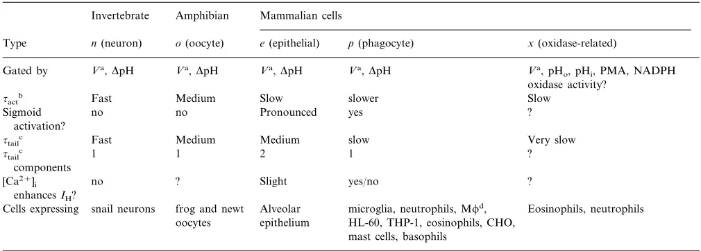
Table 2Varieties of H+Channelse
Amphibian
Invertebrate Mammalian cells
o(oocyte) e(epithelial) p(phagocyte)
n(neuron) x(oxidase-related)
Type
Va,DpH
Gated by Va,DpH Va,DpH Va,DpH Va, pH
o, pHi, PMA, NADPH oxidase activity?
Fast
~actb Medium Slow slower Slow
no no Pronounced yes
Sigmoid ?
activation?
Medium Medium
~tailc Fast slow Very slow
1 1
~tailc 2 1 ?
components
no ? Slight yes/no
[Ca2+]
i ?
enhancesIH?
frog and newt Alveolar Eosinophils, neutrophils snail neurons
Cells expressing microglia, neutrophils, Mfd, epithelium
oocytes HL-60, THP-1, eosinophils, CHO, mast cells, basophils
aV, voltage (depolarization). b~
act, activation time constant (channel opening rate). c~
tail, tail current time constant (channel closing rate). dM, macrophage.
eTable adapted and extended from (DeCoursey, 1998). It is not yet clear whether typespandxare distinct molecules or different functional modes of the same channel.
(DeCoursey and Cherny, 1993), and seems very likely to be limited by the rate at which permeation occurs
rather than by diffusion of H+ to the channel mouth
(DeCoursey and Cherny, 1994b, 1996b, 1997, 1998, 1999a,b; Cherny et al., 1995).
4.3.4. Gating kinetics
Gating kinetics refers to the rates at which ion chan-nels open and close. Practically all ion chanchan-nels have at least two fundamental conformations, called ‘open’ and ‘closed’. Open channels conduct ionic current at a virtually constant rate; closed channels do not conduct detectable current. Channels jump back and forth be-tween the open and closed states in a random, or stochastic, manner, and the time required for these gating transitions is too brief to have been resolved clearly. Thus the opening rate under specific conditions is not really the time it takes a closed channel to open, but rather a measure of the average time spent by each channel in the closed state before it randomly opens.
Strictly speaking the rate is the inverse of this time
interval. The closing rate is the inverse of the time each channel on average stays open before it closes. For an ensemble, or large population, of channels, macro-scopic currents provide the opening and closing rates in the form of the time course with which the total current increases or decreases as many channels open or close. In voltage dependent ion channels, by definition, either the opening or closing rates or both are voltage depen-dent. A depolarization-activated channel such as the voltage gated proton channel opens upon depolariza-tion because at positive membrane potentials the open-ing rate is greater than the closopen-ing rate.
Voltage gated proton currents turn on upon depolar-ization with either an exponential or sigmoid time course. In cases where the time course is sigmoid, the current waveform can be approximated by a delay followed by an exponential rise. The degree of sig-moidicity seems to vary in different cells (DeCoursey, 1998; Table 2), and thus in cells in which the time course appears to be exponential there may simply be a small delay in relation to the exponentially-rising phase of current. The sigmoidicity increases when the mem-brane is held at more negative voltages preceding a given test pulse -the ‘Cole-Moore’ effect (Cole and Moore, 1960) — indicating that there are at least two closed states with a voltage dependent transition be-tween them (DeCoursey and Cherny, 1994b). The
clos-ing time-course is generally monoexponential in
phagocytes (Kapus et al., 1994; DeCoursey and Cherny, 1996a), including microglia (Klee et al., 1999). Many ion channels exhibit ‘inactivation,’ which means that the current is not sustained indefinitely during a prolonged depolarizing voltage pulse. Typi-cally the current rises to a peak, and then decays as the channels enter a non-conducting state that differs from the normal closed state. Inactivated channels are refrac-tory to a second stimulus, and must first recover from inactivation before they can reopen. As originally defined by Hodgkin and Huxley (1952), the inactivation process is slower than the activation process. Voltage gated proton channels do not inactivate. Under most
experimental conditions, the H+ current decays during
UNCORRECTED PROOF
1991; Kapus et al., 1993a; Demaurex et al., 1993;DeCoursey and Cherny, 1993, 1994b; Humez et al., 1995; Gordienko et al., 1996; Schrenzel et al., 1996). However, this decay is the result of depletion of intra-cellular protonated buffer and the consequent increase
in pHi resulting directly from the massive H+ efflux.
This has been demonstrated by direct impalement of neurons with pH sensitive electrodes (Thomas and Meech, 1982; Meech and Thomas, 1987), by
measure-ments of shifts in Vrev (DeCoursey, 1991; DeCoursey
and Cherny, 1994b; Humez et al., 1995; Gordienko et al., 1996), and by pH sensitive fluorescent dyes (De-maurex et al., 1993; Kapus et al., 1993a; Schrenzel et al., 1996).
4.3.5. pH dependence of gating
A key property that distinguishes voltage gated pro-ton channels from other ion channels, and which also provides the basis for the functional role of these channels, is the exquisite regulation of their gating by pH. Voltage gated proton channels open preferentially at positive voltages. Unlike most other voltage-gated ion channels however, voltage gated proton channels do not have absolute voltage dependence. Instead, the position of the voltage-activation curve is highly sensi-tive to both pHoand pHi. In a systematic study, it was
found that the voltage dependence was determined by
the pH gradient, DpH (defined as pHo– pHi), rather
than by pHoor pHi alone (Cherny et al., 1995). Thus,
changingDpH by one pH Unit, whether accomplished
by changing pHoor pHior both, shifted the
voltage-ac-tivation curve by 40 mV. Lowering pHi or increasing
pHo shifts the curve toward more negative voltages,
tending to open H+channels at any given voltage. Fig.
3 illustrates the negative shift of the voltage range in
which H+ current is activated in murine microglia as
pHois increased progressively from pHo5.8 to 8.2. One
important result of theDpH dependence of H+channel
gating is that all H+channels except typex(see below)
open only when there is an outward electrochemical proton gradient. The effects of pH on other types of ion
channels are qualitatively similar to the effects of H+
channels, but generally are much less pronounced.
Lowering pHo shifts the voltage dependence of gating
in the positive direction and decreases the maximum conductance, but usually to a smaller extent than for
H+ channels (Hille, 1968; Drouin and The, 1969;
Mozhayeva and Naumov, 1970; Woodhull, 1973). The
effects of changes in pHion other channels are usually
much smaller than are effects of changes in pHoand in
some cases there is no detectable shift in voltage-depen-dent gating (Wanke et al., 1979; Deutsch and Lee, 1989; DeCoursey, 1995; Eder and Heinemann, 1996). Because the main function of voltage-gated proton channels is to extrude metabolically-produced acid, their profound sensitivity of pHiis of central importance.
Voltage gated proton channels can be opened by any
combination of lowering pHi, increasing pHo, or
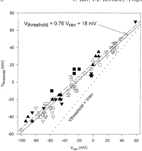
depo-larizing the membrane potential. The threshold voltage, Vthreshold, defined as the minimum level of
depolariza-tion needed to begin to activate H+channels at a given
DpH, can be predicted by a simple formula:
Vthreshold= 20 mV−40 DpH. (2)
The applicability of this relationship is shown in Fig.
4. Over a wide range of pHo and pHi, Vthreshold is
invariably positive to Vrev — the dotted line shows
equality of Vthreshold and Vrev. The slope of the lines
through the data is significantly lower, 40 mV/Unit.
This DpH sensitivity has led most investigators to
con-clude that the general function of voltage gated proton channels must be to extrude excess metabolic acid. It has been observed (DeCoursey and Cherny, 1994b) that acid extrusion via voltage gated proton channels is accomplished at no metabolic cost to the cell, other than the cost of synthesizing the channel molecules. In
contrast, H+-ATPases and antiporters such as the Na+
Fig. 3. Families of H+currents in the same microglial cell at several
UNCORRECTED PROOF
Fig. 4. The threshold voltage for activating H+currents, defined asthe voltage at which clearly time-dependent outward current was detected,Vthreshold, is plotted as a function ofVrevmeasured in the same cell and the same solution. The dotted line shows equality of the two parameters; all of the data fall above this line, indicating that
Vthresholdis always positive toVrev. Open symbols indicate measure-ments with H2O in the bath, filled symbols with D2O. Bath solutions included pHoranging 6.5 – 10.0 and pDo7.0 – 10.0. Pipette solutions (pHi) are indicated by the shape of the symbol: pD 7.0 (D), pD 8.0 (9), pD 9.0 (hexagons), pH 7.5 (2), pH 5.5 (), 50 mM NH4+() The lines show the results of linear regression of the H2O data (solid line),r=0.963, slope=0.760, y-intercept=18.1 mV. The D2O data (dashed line) were described byr=0.926, slope=0.750, intercept=
22.0 mV. Data in alveolar epithelial cells, from Fig. 11 of DeCoursey and Cherny, 1997.
Two key assumptions are required for the model to work. First, the protonation sites are accessible only to the external or internal solution at any given time, never both simultaneously. Second, the conformational change that switches the accessibility of the channel can occur only when the sites are deprotonated. This model
reproduces the main features of the regulation of H+
channel gating by pHo and pHi (Cherny et al., 1995).
Identifying the molecular correlates of these regulatory protonation sites will be an important goal of future
structure-function studies of H+ channel molecules.
4.3.6. Pharmacology
Voltage gated proton channels are unusual in having no high affinity blockers. Most ion channels are sensi-tive to specific proteins found in toxins or venoms — in fact, the toxicity of many of these substances is a direct result of their effects on ion channels. Two main classes of inhibitors exist for voltage gated proton channels: weak organic bases and polyvalent cations. The
inhibi-tion by weak organic bases (4-aminopyridine, TEA+,
D600, verapamil, amiloride, rimantadine, amantadine) (Byerly et al., 1984; Meech and Thomas, 1987; Bern-heim et al., 1993; DeCoursey and Cherny, 1994a,b) may reflect direct interaction of these molecules with the channel protein, such as occurs with other ion channels. However, because in most cases relatively high concen-trations are required, it is conceivable that some or all of the inhibition may be the result of local pH changes near the membrane due to the flux of neutral form of these molecules. Influx of neutral form of a weak base will lower the pH at the outer surface of the membrane and raise pH near the inner face of the membrane. Both
changes would reduce H+ currents by decreasing the
driving force and by shifting the voltage-activation curve in the positive direction. Further studies are needed to evaluate this hypothesis, which is discussed further elsewhere (DeCoursey and Cherny, 1994b).
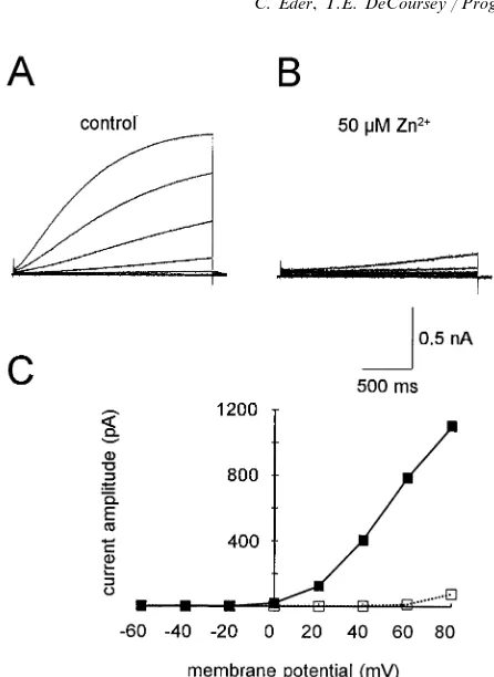
Microglial H+currents are inhibited during
extracel-lular application of 1 mM 4-aminopyridine or TEA+
and by several inorganic polyvalent cations at micro-molar concentrations with the following order of po-tency: Zn2+\La3+\Ni2+\Cd2+\Co2+\Ba2+ (Eder et al., 1995a). The effects of polyvalent cations on
H+ currents resemble qualitatively their effects on
many other ion channels. As illustrated in Fig. 6, the rate of channel activation is slowed, and the voltage dependence of channel opening is shifted in the positive direction. At first glance it appears reasonable to de-scribe these effects as voltage dependent block, because the fractional reduction of H+ current differs at differ-ent voltages. However, several types of evidence make
it clear that Zn2+ does not enter the channel and block
at a site within the membrane electrical field. There is no effect on the shape of the instantaneous
current-voltage relationship, block/unblock kinetics are
incom-/H+-antiporter consume metabolic energy in the form
of ATP either directly or indirectly by dissipating the
Na+ gradient. Furthermore, in comparison with other
acid-regulating membrane transporters, when activated maximally, voltage gated proton channels extrude acid
at 100 times higher rate than any other transporter
(DeCoursey and Cherny, 1994b).
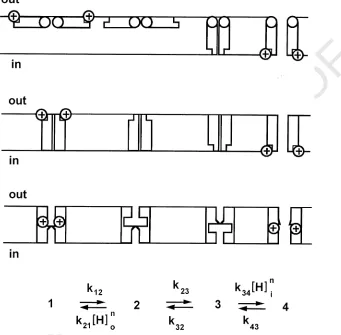
How does the regulation of gating by DpH work?
Cherny et al. (1995) proposed a simple general model to
account for the DpH dependence of the voltage gated
proton channels. The features of this model are illus-trated by three cartoons in Fig. 5, each of which represents a physical mechanism that could operate to
produce the observedDpH dependence. The channel is
UNCORRECTED PROOF
patible with the observed effects of Zn2+, and the voltagedependent effects all are consistent with the idea that
Zn2+ binds to an external site where it alters the
transmembrane voltage sensed by the voltage-sensor of
the H+channel (Cherny and DeCoursey, 1999).
Inhibi-tion by polyvalent metal caInhibi-tions is extremely sensitive to pHo: the apparent efficacy of ZnCl2was 10-fold lower at
pHo6 than at pHo7, and 100-fold lower at pHo5 than
at pHo 6 (Cherny and DeCoursey, 1999). This pHo
dependence suggests that the external Zn2+ receptor on
voltage-gated proton channels is comprised of multiple
protonatable groups which together coordinate the Zn2+
atom. The active form appears to be the divalent cation:
i.e. Zn2+ not ZnOH+ (Cherny and DeCoursey, 1999).
In practical terms, any evaluation of the potency of metal cations for inhibiting H+current is complicated by the strong pH dependence. Furthermore, many biologi-cal buffers bind Zn2+at least weakly (Table 1 of Cherny and DeCoursey, 1999). These factors must be taken into consideration in order to compare potency among stud-ies done under different conditions. Studied under
com-parable conditions, Zn2+ inhibits H+ currents more
potently than it inhibits other channels such as Ca2+
currents (Mahaut-Smith, 1989) and delayed rectifier K+
[image:13.612.138.479.238.573.2]currents (Spires and Begenisich, 1994; Eder et al., 1995b; Cherny and DeCoursey, 1999).
Fig. 5. Model of the regulation of H+channel gating by the pH gradient,DpH. The state diagram at the bottom defines the kinetic model; the
three cartoons illustrate possible physical manifestations of this model. The closed channel conformation (state 1) is stabilized by protonation of an external site, and the open conformation (state 4) is stabilized by protonation of an internal site. These hypothetical allosteric regulatory protonation sites might be the same or distinct. Only the external or internal site is accessible to protonation at any given time, not both simultaneously. A conformational change (the transition between states 2 and 3) exposes the protonation site to the internal solution. The formation of a conducting H+channel requires a conformational change in each channel protomer which can occur only when the regulatory site
is deprotonated. The open channel probability therefore increases at high pHoor low pHi. The model defined by the state diagram can be envisioned physically as (top) a ‘butterfly’ in which the protonation site on each channel protomer or ‘wing’ moves across the membrane, (middle) distinct external and internal sites which when protonated, allosterically prevent protonation at the opposite site, (lower) a protonation site in a proton well whose accessibility depends on a small conformational change, or other variants not illustrated. The voltage dependence of H+
channel gating could arise either from voltage-dependent binding/unbinding of protons to the regulatory protonation site, or from a voltage dependent conformational change, or some combination of the two. We assigned all of the voltage-dependence to proton binding, so that the regulatory sites behave like ‘proton wells’. The gating of voltage gated proton channels in rat alveolar epithelial cells was described by the following parameters:din=dout=0.71,Kw=10,m=0,n=1.5,k12=1000 s−1,k32=106s−1,k43,fast=3 s−1,k43,slow=0.05 s−1, pKin=pKout
UNCORRECTED PROOF
Fig. 6. Identical families of pulses were applied in the absence (A) orpresence (B) of 50mM ZnCl2in a microglial cell. Note the profound slowing of the turn-on of the H+current. The currents at the end of
the pulses in A and B are plotted in C. Note that theVthreshold is shifted by60 mV toward more positive voltages. Taken from Fig. 7 of Eder et al. (1995a).
scale, and could occur directly or via second messenger pathways.
Changes in H+ channel expression have been
de-tected upon deactivation or activation of microglia.
Direct modulation of H+ channels has not studied
extensively yet in microglia. Therefore, the influence of modulatory agents, such as arachidonic acid or PMA,
on microglial H+ currents can currently only be
hy-pothesized by analogy with data that have been ob-tained in other cells.
Following common parlance, we describe modes of microglial behavior as untreated, resting, activated, or deactivated. These terms are useful, but unquestionably also represent an oversimplification and they may be misleading. For example, ramified (and presumably resting) microglia in culture are not by any means inert. They display high levels of pinocytotic activity, suggest-ing that microglia may normally function to cleanse brain fluids (Booth and Thomas, 1991). Deactivated microglia, nominally having reverted from an activated to a resting state (Merrill and Zimmerman, 1991; Loughlin et al., 1993; Suzumura et al., 1993), neverthe-less differ from unstimulated resting microglia. For
example, the patterns of K+ channel expression are
different in deactivated microglia compared with either unstimulated or activated cells (Schilling et al., 2000b). Activation can be defined in many ways, because a variety of disparate stimuli result in a large array of responses, ranging from morphological to functional. The capacity to respond differentially to different stim-uli seems logical and appropriate in light of the many roles of microglia. For example, microglia clean up necrotic cellular debris, phagocytose invading bacteria and kill them by undergoing a respiratory burst, and release an armamentarium of cytokines such as chemo-tactic signals to peripheral macrophages. As in other phagocytes, the respiratory burst in microglia can be elicited by various agonists. In addition, many
sub-stances such as TNF-a, IFN-g, or GM-CSF ‘prime’
macrophages, such that the response to a given agonist is greatly enhanced by pretreatment with priming agents (Klebanoff, 1999). One of the most widely used activators of microglia and other macrophages is bacte-rial LPS. LPS elicits little or no respiratory burst in neutrophils, but greatly enhances the release of super-oxide anion induced by fMLP, PMA, or immune com-plexes (Guthrie et al., 1984; DeLeo et al., 1998). Other
important chemokines such as TNF-a or IFN-g also
prime microglia, with TNF-aitself eliciting some
super-oxide anion release, but IFN-g eliciting no response
(Chao et al., 1995). Some authors refer to priming of macrophages as ‘activation.’ In summary, although we use general terms to describe microglial behavior
pat-terns, we emphasize the arbitrariness of this
nomenclature.
The effects of ZnCl2 and CdCl2 are weaker and
qualitatively different for internal than for external
application. Intracellularly applied Zn2+ reduces the
H+ current weakly even at relatively high
concentra-tions (]0.17 mM free Zn2+) and slows deactivation
without effect on activation (Cherny and DeCoursey, 1999). Effects of both external and internal metals are qualitatively consistent with their binding to the hypo-thetical regulatory protonation sites whereby the
voltage dependence of gating is established by theDpH.
4.4. Physiological modulation of H+ channels
Physiological modulation mechanisms can be sepa-rated into two general categories: changes in the total number of active channels in the cell membrane and changes in the properties of the channels. Up- or
down-regulation of the gH presumably requires regulation of
UNCORRECTED PROOF
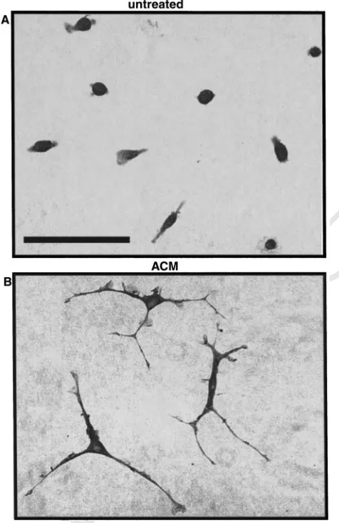
4.4.1. Regulation by astrocytic factors,lipopolysaccharide and cytoskeletal disrupti6e agents Treatment of microglial cells with astrocyte-condi-tioned medium (ACM) leads to deactivation that is characterized by a variety of changes in the im-munophenotypical, morphological and
electrophysio-logical properties. The striking morphological
transformation from amoeboid to ramified is illustrated in Fig. 7. ACM-induced deactivation of microglia also results in downregulation of surface antigens such as LFA-1, ICAM-1 and MHC class II molecules, and
upregulation of delayed rectifier K+ channels (Eder et
al., 1999).
The expression of microglial proton channels is also regulated by factors released from astrocytes. Twenty-four hours after exposure to ACM, the microglial pro-ton current density was about 50% smaller than that in untreated microglial cells. In addition, proton currents of ACM-treated microglial cells activated significantly more slowly than proton currents of untreated
mi-croglia (Klee et al., 1999). Intriguingly, proton currents changed in a similar way in microglia after exposure for 24 h to lipopolysaccharide (LPS) (Klee et al., 1999), which shifts microglia and other macrophages into an activated functional state (Hauschildt and Kleine, 1995). Moreover, after differentiation of THP-1 mono-cytes into macrophage-like cells (Auwerx, 1991),
voltage gated proton currents were decreased by 50%
and activation during depolarization was twice slower (DeCoursey and Cherny, 1996a). Thus, several physio-logical mediators produce similar changes in the
ampli-tude and properties of H+ currents. It is not clear
whether these rather subtle modifications of H+
cur-rents have functional consequences, or if they simply reflect a non-specific response to cytoarchitectural stress that is common to these cellular responses, as discussed next.
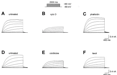
It appears that the expression and properties of voltage gated proton channels can be regulated by cytoskeletal interactions. Cytoskeletal reorganization
may be responsible for the alterations in H+ currents
observed after treatment of microglia with ACM or LPS. As illustrated in Fig. 8, exposure of microglial cells to several cytoskeletal disruptive agents produced
the same kinds of changes in H+ currents as just
discussed for ACM or LPS treatment, namely smaller
H+ current density and slower time-dependent
activa-tion (Klee et al., 1998, 1999). These effects were ob-served after 24 h treatment of murine microglia with the cytoskeletal disruptive agents cytochalasin D or colchicine. In contrast, stabilization of the cytoskeleton by phalloidin or taxol did not have any significant
effect on microglial H+currents. Since acute changes in
microglial H+currents were not observed during
short-term treatment with cytoskeletal disruptive agents (Klee et al., 1998), the incorporation of new channels in the membrane presumably was inhibited due to disruption of the cytoskeleton without direct modulation of the
channels. The slowing of H+ current activation is
evi-dently an indirect response to metabolic changes occur-ring after treatment of the cells.
It has been reported that cytochalasin D inhibits neutrophil spreading and the concomitant activation of NADPH oxidase (Demaurex et al., 1996). These obser-vations together provide intriguing hints of a relation-ship between NADPH oxidase activity and the voltage gated proton channel in phagocytes (see also below).
4.4.2. Modulation by arachidonic acid
[image:15.612.45.283.315.682.2]During the respiratory burst, phagocytes, including microglia, release arachidonic acid, which itself is a bioactive fatty acid that enhances phagocyte responses. In addition, neurons and astrocytes are capable of releasing arachidonic acid. Arachidonic acid is released by neuronal and glial cells following binding of neuro-transmitters to receptors, e.g. glutamate, acetylcholine,
UNCORRECTED PROOF
Fig. 8. Families of H+currents in microglial cells before (A, D) or after treatment for 1 day with 2mM cytochalasin D (B), 20mM phalloidin
(C), 1mM colchicine (E), or 0.5mM taxol (F). All families recorded during the same pulse sequence (inset). Taken from Klee et al. (1998).
serotonin, adrenaline, or ATP. Several pathological conditions, such as ischemia, hypoglycemia, epilepsy, or hypoxia are associated with elevated concentrations of free arachidonic acid (reviewed by Katsuki and Okuda, 1995). Henderson and colleagues have shown that arachidonic acid is a strong stimulus for both the
respiratory burst-associated H+ conductance
(Hender-son and Chappell, 1992) and for NADPH oxidase, concluding that it was necessary to trigger this response (Henderson et al., 1993). Subsequent studies have
sup-ported this role, suggesting that PKC activates the gH
indirectly, by stimulating phospholipaseA2, which leads
to release of arachidonic acid (Kapus et al., 1993b; Suszta´k et al., 1997; Dana et al., 1998; Lowenthal and Levy, 1999). As illustrated in Fig. 9, voltage-clamp studies show that arachidonic acid directly enhances the
gHin phagocytes (DeCoursey and Cherny, 1993; Kapus
et al., 1994; Gordienko et al., 1996; Schrenzel et al., 1996; Suszta´k et al., 1997; Henderson and Meech, 1999). Two mechanisms are involved (DeCoursey and Cherny, 1993; Kapus et al., 1994; Gordienko et al., 1996; Henderson and Meech, 1999). First, the
maxi-mum H+ conductance is increased. Secondly, the
voltage-activation curve is shifted in the negative
direc-tion. The result of this shift is that more H+ channels
will be open at any given membrane potential.
4.4.3. Phosphorylation
There is indirect evidence, based on H+ fluxes
de-duced from pH measurements, that phosphorylation
up-regulates H+ channels. Certainly phosphorylation
of the cytosolic components of NADPH oxidase is a key trigger for assembly of the functional oxidize for the respiratory burst (Babior, 1999). Although the clas-sic potent agonist for the respiratory burst is PMA (phorbol myristate acetate), the role of PKC (protein
kinase C) in activating the gH is ambiguous. There is
some evidence that tyrosine kinase but not PKC is
involved in activating the gH (Nanda and Grinstein,
1995). As just discussed, phosphorylation of
phospholi-pase A2 may result indirectly in activation of the gH
Fig. 9. Whole-cell H+currents at+100 mV in a human neutrophil