Supported by the National Science Centre of Poland (grant No. 2011/03/B/NZ9/03412) and statutory research and development activity funds assigned to the Faculty of Veterinary Medicine UP in Wroclaw.
Effect of Vitrification on Apoptotic Changes
in Feline Embryos
Małgorzata Ochota*, Wojciech Niżański
Department of Reproduction and Clinic of Farm Animals, Faculty of Veterinary Medicine, Wroclaw University of Environmental and Life Sciences, Wrocław, Poland
*Corresponding author: [email protected]
ABSTRACT
Ochota M., Niżański W. (2018): Effect of vitrification on apoptotic changes in feline embryos. Czech J. Anim. Sci., 63, 144–151.
The aim of this study was to evaluate the influence of a vitrification-warming procedure on the viability of cat embryos and blastocysts and the incidence of apoptotic changes in blastocysts subjected to vitrification and blastocyst that developed from vitrified embryos. In the first part of the experiment, post-thaw embryo development and blastocyst viability were evaluated based on morphological appearance and the ability to develop (embryos) or re-expand (blastocyst) compared to control. In the second part, blastocysts that were viable after vitrification-warming and blastocysts that developed from vitrified-warmed embryos were stained with Annexin-V and TUNEL to evaluate apoptotic changes. Most of the vitrified-warmed embryos were viable after thawing, 36.3% developed to morula, and 14.7% to the blastocyst stage. The overall re-expansion rate of blastocysts that were vitrified on day 7 was 55.6%. Vitrification significantly increased apoptotic and necrotic changes in blastocysts, but did not influence late apoptotic and necrotic changes in blastocysts that developed from vitrified-warmed embryos. The total number of blastomeres in blastocysts was similar in blastocysts that developed from vitrified-warmed embryos (99.1 ± 23.1), but lower in blastocysts vitrified on day 7 (82.1 ± 16.8), if compared to the control group (107.9 ± 24.2). These results show that the vitrification-warming procedure returns embryos capable of further development to a good quality blastocyst in vitro. Whereas, vitrification of blastocysts caused the progression of apoptotic changes in some blastomeres, however it did not affect the overall blastocyst viability.
Keywords: cat; TUNEL; blastocyst; Annexin-V; apoptosis
Cryopreservation techniques have progressed rapidly in recent years, but there is still no con-sensus whether slow-freezing or vitrification is the most rewarding tool for long term storage of living cells (Mandawala et al. 2016). In recent years there has seemingly been a tendency to move toward vitrification, as an easier and quicker technique. However, cryopreservation protocols cannot easily be transferred among species,
vitrified embryos. Cryopreservation of bovine and ovine embryos, regardless of the method used, seems to be relatively easy (Mandawala et al. 2016), whereas successful pig embryo cryopreservation or vitrification is still extremely difficult, despite numerous trials and variable approaches (Papis 2001). In wild species, the challenges associated with preservation of genetic material are rather different. Gathering sufficient material from en-dangered species is the most limiting factor, and hence domestic animals are often used as models to optimize cryopreservation techniques (Mandawala et al. 2016). Since, the domestic cat (Felis catus) is the only felid not described as vulnerable or en-dangered, it can serve as a model to develop fast, reliable, and feasible methods for cryopreservation of its genetic material. Better understanding the influence of sub-zero temperatures on developing blastomeres and factors causing cellular damage and inducing apoptotic reactions would allow tai-loring vitrification protocols for the best results. Apoptosis is a widely known process of pro-grammed cell death aiming at the elimination of abnormal, detrimental, or superfluous cells at specific locations and times (Makarevich et al. 2008). It originates from genetic changes, which induce a complex cascade of biochemical events that degrade proteins and nuclear DNA, lead-ing to cell shrinkage, cytoplasmic and chromatin condensation, and fragmentation (Fabian et al. 2005b). It is an intracellular mechanism of self-destruction, but it is rarely considered as a strictly endogenous process, as it can be initiated as a response to cellular stress, i.e. radiation, nutrient deprivation, temperature, infections or increased intracellular calcium concentration, and usually cannot stop once it begins (Fabian et al. 2007). Although the sequence of apoptotic changes is well recognized, our understanding its implica-tions during embryo cryostorage and in assisted reproduction procedures is still limited and needs further attention.
The incidence of apoptosis in in vivo and in vitro derived mammalian embryos has been observed for many years (Hardy 1999) and is considered an important component of normal embryo de-velopment. Spontaneous blastomeres death at the blastocyst level has been reported in many species, allowing differentiation of the tropho-blast cells for implantation and inner cell mass for the embryo growth. Despite the beneficial
effect during embryo development, the cell death beyond a certain threshold is detrimental for the further embryo survival (Betts and King 2001). It has also been demonstrated that the degree of apoptosis increases in suboptimal developmental conditions in human (Hardy 1999), mouse (Brison and Schultz 1998), rat (Pampfer et al. 2001), cattle (Gjorret et al. 2003), horse (Moussa et al. 2004), sheep (Rizos et al. 2002), and pig (Kidson et al. 2004) embryos. Various methods can be used to identify apoptotic changes in embryos, starting from early alterations of plasma membrane with phosphatidylserine translocation (Annexin-V), through nuclear condensation and fragmenta-tion (DAPI or Hoechst 33342) or the presence of single- and double-strand DNA breaks (TUNEL), to the final apoptotic stages with cell membrane disruption (staining with ethidium homodimer, EthD-1). The experiments performed in mice (Dhali et al. 2009) and bovine (Yang and Rajama-hendran 2002) embryos suggest that cryostorage can alter the expression of apoptosis related genes and even induce apoptosis. The increased inci-dence of programmed cell death is an important indicator of environmental stress or inadequate pre- or post warming cultureconditions (Fabian et al. 2007). A detailed understanding the apop-totic processes during embryo storage is crucial to improve embryo survival and quality during assisted reproduction procedures.
Since little is known about apoptosis in feline embryos and blastocyst exposed to sub-zero tem-peratures, the aim of this study was to evaluate the influence of vitrification-warming procedure on the incidence of apoptotic changes in day 7 cat blastocysts subjected to vitrification and blastocyst developed from vitrified embryos.
MATERIAL AND METHODS
Unless otherwise stated, all chemicals and re-agents used in this study were purchased from Sigma-Aldrich, Poland.
Solu-tion at 4°C for up to 24 h before cumulus oocyte complexes (COCs) recovery. COCs were collected by slicing ovaries with a scalpel blade in a wash-ing medium (WM) containwash-ing M199 with Earle’s salts, supplemented with 3 mg/ml bovine serum albumin (BSA), 0.1 mg/ml cysteine, 1.4 mg/ml (4-2-hydroxyethyl)-1-piperazineethanesulfonic acid (HEPES), 0.25 mg/ml sodium pyruvate, 0.6 mg/ml sodium lactate, 0.15 mg/ml l-glutamine, and 0.055 mg/ml gentamicin. Isolated COCs were classified under a dissecting microscope and only oocytes with evenly pigmented dark ooplasm and several layers of cumulus cells were selected and placed into 400 µl of maturation medium (15 ± 5 oocytes per each replicate, WM with the addition of 0.025 IU/ml luteinizing hormone and 0.02 UI/ ml follicle-stimulating hormone under mineral oil and matured for 24 h at 38.5°C in 5% CO2 in air with maximum humidity (Ochota et al. 2016a).
For in vitro fertilization, thawed spermatozoa isolated from cauda epididimis and frozen according to the procedure described by Nizanski et al. (2005) were used. After a 24-hour maturation COCs were washed twice in WM and co-incubated with 1 × 10⁶ motile spermatozoa/ml at 38.5°C in 5% CO2 in air with maximum humidity, 15 ± 5 oocytes per each replicate in 400 µl Tyrode’s salts solution supple-mented with 6 mg/ml BSA, 1.2 mg/ml HEPES, 1.1 mg/ml sodium lactate, 0.15 mg/ml l-glutamine, and 0.1 mg/ml sodium pyruvate.
The presumptive zygotes were removed after an 18-hour incubation with spermatozoa, washed, and cultured up to 7 days, 8 ± 5 presumptive zy-gotes in droplets of 50 µl of protein supplemented embryo culture (EC) medium (Continuous Single Culture® with 10% Serum Substitute Supplement®; Irvine Scientific, Ireland) at 38.5°C in 5% CO2 in air. Embryo development was assessed daily based on morphological characteristics.
Embryo vitrification and warming. Vitrifica-tion was applied to day 3 embryos and to day 7 blastocyst. Embryos and blastocyst were vitri-fied on Cryotops using the protocol described by Ochota et al. (2016b). Briefly, 1–3 embryos or blastocyst were incubated for 2 min (embryo) and 4 min (blastocyst) in the equilibration solu-tion (ES: 7.5% dimethyl sulfoxide (DMSO), 7.5% PrOH, 0.5M sucrose, and 10% Ficoll), then they were exposed for less than 30 s to the vitrifica-tion soluvitrifica-tion (VS: 15% DMSO, 15% PrOH, 1M sucrose, and 10% Ficoll) and during this time they
were loaded onto a Cryotop device (Kitazato Bio- Pharma Co., Ltd., Japan) with minimal volume of VS (< 0.5 μl), and immediately plunged into liquid nitrogen. Warming was performed as three-step: immersing for 1 min at 37°C in 0.5 ml of thawing solution 1 (TS 1): 1M sucrose, 10% Ficoll, and 20% fetal calf serum (FCS) in Dulbecco’s phosphate-buffered saline (DPBS), then for 3 min in 20°C of TS 2: 0.5M sucrose, 10% Ficoll, and 20% FCS in DPBS, and subsequently washed for 5 min in DPBS with 20% FCS. Warmed embryos were returned to 50 µl of EC medium (2–3 embryos per each drop) at 38.5°C in 5% CO2 in air for the remaining 4 days of EC. Embryos, which resumed divisions within the first 24 h post-warming, were classified as surviving the vitrification procedure. Warmed blastocysts were assessed for morphology and re-expansion at 2, 4, and 16 h under an inverted microscope. Those with intact inner cell mass, trophectoderm, and re-expanded blastocoele were considered surviving.
Detection of apoptosis. The degree of early (EAB) and late (LAB) apoptotic changes, as well as necrotic blastomeres (NB) were evaluated in blastocyst subjected to the vitrification-warming procedure, blastocyst developed from vitrified-warmed embryos, and in control blastocysts that were not vitrified. The ratios of early (EABR), late (LABR) apoptotic and necrotic (NBR) blastomeres were calculated out of the total number of blas-tomeres counted in a blastocyst.
staining using the Olympus cellSens Dimension software. After evaluation blastocysts were washed four times in PBS-BSA, fixed in 4% paraformal-dehyde, and stored in 4°C overnight for TUNEL staining on the following day.
TUNEL assay.Blastocyst were washed twice in PBS-PVP (polyvinylpyrrolidone) (1 mg/ml) and permeabilised in Triton X-100 (0.5% in PBS) for 1 h at room temperature. For positive and nega-tive control, samples were incubated with 0.1 U/µl DNAse for 1 h at 37°C. For TUNEL staining, the In Situ Cell Death Detection Kit (Roche 11684795910; Roche, Switzerland) was used following manufac-turer’s instructions with modifications published by Fabian et al. (2007). All the experimental and positive control blastocysts were incubated with terminal deoxynucleotidyl transferase and fluo-rescein-conjugated dUTP for 1 h at 37°C, whereas negative controls were incubated without terminal deoxynucleotidyl transferase. Then all embryos were washed in PBS-BSA, counterstained with Hoechst 33342 (1 mg/ml) for 15 min at room tem-perature, and washed in PBS-PVP before mounting on slides. Blastomeres were classified according to their nuclear morphology (presence of typi-cal DNA fragmentation) and the intensity of the fluorescent signal (based on the signal intensity measurement in positive controls using Olym-pus cellSens software): (1) apoptotic blastomeres (LAB): TUNEL positive, green nuclear fragments, and (2) non-apoptotic: TUNEL negative, Hoechst 33342 positive blue nuclei.
Experimental design.Day 3 embryos (n = 200) and day 7 blastocyst (n = 39) were obtained from oocytes (n = 771), matured, and fertilized in vitro (45 replicates, 15 ± 5 oocytes per each replicate). In the first part of the experiment, embryos were equally divided into control (n = 98) or experi-mental (n = 102) groups, which were subjected to vitrification. Similarly, day 7 blastocysts were divided into control (n = 12) and experimental (n = 27) groups, and the latter underwent
vitrifica-tion. Post-thaw viability was evaluated based on the embryo’s ability to divide within 24 h post-warming and blastocyst’s ability to re-expand at 2, 4, and 16 h post-warming. Vitrified-warmed embryos were returned to EC to continue development to the blastocyst stage and warmed blastocysts were used in the second part of the experiment.
In the second part of the experiment, control, non-vitrified blastocysts (n = 12), viable, day 7 blastocysts after vitrification-warming (n = 15), and blastocysts that developed from vitrified-warmed embryos (n = 15) were submitted to Annexin-V and TUNEL staining to evaluate the degree of early and late apoptotic changes.
Statistical analysis. The number of embryos surviving vitrification and warming was recorded and used to calculate the percentage surviving. The further development of vitrified embryos was compared with control, non-vitrified group using Fisher’s exact test. The number and pro-portion of early and late apoptotic blastomeres and the ratios of the total number of blastomeres were compared using two-way analysis of variance (ANOVA). A significant statistical difference was noted at P < 0.05.
RESULTS
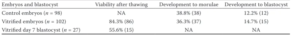
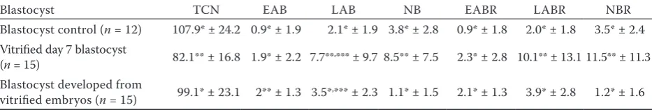
Most (84.3%) of the vitrified-warmed embryos were viable after warming and over one third of them (36.3%) developed further to morula. How-ever, only 14.7% reached the blastocyst stage in EC after warming. The overall viability of vitrified day 7 blastocyst was 55.6% (Table 1). The total number of blastomeres in blastocysts developed from vitrified-warmed embryos was similar to the control group (Table 2), but significantly reduced in day 7 blastocyst subjected to vitrification.
[image:4.595.61.535.686.744.2]Early and late apoptotic changes and NB were noted in blastocysts from all treatments. Vitrifi-cation significantly increased late apoptotic and
Table 1. Post warming survival and further development of day 3 embryos and viability of day 7 blastocyst subjected to vitrification-warming procedure (number and percentage)
necrotic changes in blastocysts subjected to rapid freezing and early apoptotic changes in blasto-cyst developed from vitrified-thawed embryos. In vitrified day 7 blastocysts, the number of early apoptotic blastomeres was similar to the control group, whereas the numbers of late apoptotic blastomeres and NB were higher than in the con-trol group. Moreover, the ratio of early apoptotic blastomeres in day 7 vitrified blastocyst was simi-lar, whereas the ratios of late blastomeres and NB were higher, when compared to control. On the other hand, blastocyst developed from vitrified-warmed embryos contained significantly more early apoptotic blastomeres than in the control group. However, the ratios of early, late, and ne-crotic blastomeres in blastocyst developed from vitrified embryos were similar to blastocysts from the control group (Table 2).
DISCUSSION
This study evaluated the implications of a vit-rification-warming procedure on the incidence of early and late apoptotic changes in blastocysts subjected to vitrification and in blastocysts de-veloped from previously vitrified embryos. The results demonstrate that vitrification-warming had a negative impact on blastocysts, in which it increased late apoptotic changes, as well as the number of NB. On the contrary, in blastocyst developed from vitrified-warmed embryos, cryo-preservation did not affect the incidence of late apoptotic or necrotic changes, but increased the number of early apoptotic blastomeres.
There are not many available reports on feline embryo cryopreservation. Although, vitrification
seems to be more efficient, cheaper, and easier than slow rate freezing, this procedure has not been well established yet (Elnahas et al. 2010). The reported success rate in embryo vitrification in cats is still very limited (Tsujioka et al. 2008; Pope et al. 2012). Crichton (2002) showed slightly higher cleavage resumption after warming in vit-rified 2- to 4-cell tiger (Panthera tigris altaica) embryos than in our study (46 vs 36%). Similarly, the reported blastocyst development from vitri-fied 2- to 4-cell domestic cat embryos was 29% (Pederson et al. 2009), whereas in our study it was 15%. The difference might have been related to the different embryo developmental stage undergoing vitrification, or the individual culture conditions.
[image:5.595.64.531.126.205.2]The only available report on successful vitrifi-cation of cat blastocysts was published by Tsui-joka et al. (2008), who showed 73% viability in vitrified day 6 and 66% viability in vitrified day 7 blastocysts, which was higher than in the current study. However, in our previous study (Ochota et al. 2016b) we showed that blastocyst viability after warming was markedly affected by the de-gree of expansion, in favour of compacted (75% post-warming viability) vs expanded blastocyst (47% post-warming viability). In the current study, blastocysts underwent vitrification on day 7 re-gardless of the degree of expansion, which could have affected the outcome. In our study, 55% of blastocyst survived vitrification, which corresponds with the results of Tsujoka et al. (2008) for day 7 blastocyst. Although, Tsujoka et al. (2008) did not find a statistical difference between day 6 vs day 7 blastocyst viability post-vitrification, their results also show a tendency of better resistance to sub-zero temperatures by less expanded (day 6) blastocysts.
Table 2. Mean number and ratio of early, late, and necrotic blastomeres in vitrified blastocyst and in blastocyst de-veloped from vitrified embryos in comparison to non-vitrified control group
Blastocyst TCN EAB LAB NB EABR LABR NBR
Blastocyst control (n = 12) 107.9* ± 24.2 0.9* ± 1.9 2.1* ± 1.9 3.8* ± 2.8 0.9* ± 1.8 2.0* ± 1.8 3.5* ± 2.4 Vitrified day 7 blastocyst
(n = 15) 82.1** ± 16.8 1.9* ± 2.2 7.7**,*** ± 9.7 8.5** ± 7.5 2.3* ± 2.8 10.1** ± 13.1 11.5** ± 11.3 Blastocyst developed from
vitrified embryos (n = 15) 99.1* ± 23.1 2** ± 1.3 3.5*,*** ± 2.3 1.1* ± 1.5 2.1* ± 1.3 3.9* ± 2.8 1.2* ± 1.6 TCN = total cell number, EAB = early apoptotic blastomeres, LAB = late apoptotic blastomeres, NB = necrotic blastomeres, EABR = percentage of early apoptotic blastomeres, LABR = percentage of late apoptotic blastomeres, NBR = percentage of necrotic blastomeres
In the study of Tsujioka et al. (2008), they also checked the total cell number in blastocysts sub-jected to vitrification and found no difference between fresh and vitrified blastocyst, which is in contrast to our study where vitrified blastocyst had significantly less blastomeres comparing to non-vitrified, control blastocysts. The observed, lower TCN in vitrified blastocysts was likely a result of the damage caused by cold storage, since we also observed an increase in late apoptotic and necrotic changes in comparison to the control group. A decrease of total cell count in blastocysts after cryostorage was also reported in other species and explained as impaired post-warming mitotic activ-ity and delayed proliferation (Marquez-Alvarado et al. 2004; Chrenek et al. 2014). It was also proven in vitrified porcine blastocyst that after a 24-hour culture, 2/3 of embryos showed normal, regener-ated morphology and an increase in TCN (Fabian et al. 2005a). Interestingly, the early apoptosis in the blastocysts subjected to vitrification was not different from control, suggesting the progres-sion of early apoptosis towards later stages in the response to vitrification-warming procedure without inducing new, early apoptotic changes. On the contrary, blastocyst developed from vitrified embryos had the same quality in terms of TCN as the control group.
Programmed cell death is a physiological process occurring spontaneously in many cell populations (Fabian et al. 2005b). During embryogenesis it plays an important role in eliminating abnormal, detrimental, or superfluous cells and control-ling embryo blastomere number (Handyside and Hunter 1986). Results of TUNEL labelling showed at least one apoptotic cell in all in vivo and in vitro derived bovine, human, and porcine blastocyst (Fabian et al. 2005b). Morphological features, such as chromatin condensation, marginalization, and nuclear fragmentation, were visible in 70–80% of in vitro produced mice and human embryos and in almost all cattle blastocyst (Handyside and Hunter 1986; Byrne et al. 1999; Hardy 1999). Ap-optosis seems to be one of the crucial mechanisms controlling all stages of embryonic development and influencing the survival and health of future offspring. However, at a higher occurrence, it has an adverse effect on embryo homeostasis, inducing developmental arrest or embryo death. As apoptosis can be induced by external stimuli, quantification of its occurrence may allow for the
evaluation of experimental procedures and condi-tions (Makarevich et al. 2008). All the blastocysts in our experiment exhibited some incidence of apoptosis, which affected individual cells through-out the embryo. Since apoptotic and necrotic blastomeres are common during normal embryo development, their increase in post-vitrified blas-tocyst may result from the vitrification process itself or exposure to vitrification media (Fabian et al. 2005b). There have only been a few studies performed regarding the relation between cryo-preservation and the incidence of apoptosis and none of them concerned cats. In bovine embryos, it was proven that cryopreservation increased the number of apoptotic cells (Marquez-Alvarado et al. 2004; Paschoal et al. 2017), the same as in mice (Ahn et al. 2002). Similarly, in our experiment we noted a significant increase of late and necrotic blastomeres in vitrified blastocyst and early apop-totic blastomeres in blastocysts developed from vitrified embryos. Based on these results, it could be assumed that the vitrification procedure led to the further progression of early apoptotic changes in blastocysts, turning those blastomeres into late apoptotic and necrotic ones. On the other hand, in blastocyst developed from vitrified embryos, the number of early apoptotic blastomeres was higher, but the ratio of early apoptotic cells to the total number of cells was not different from the control group. It suggests that post-vitrification embryo development was not affected by vitrifica-tion, and vitrified embryos were able to develop into good quality blastocyst, capable of limiting apoptosis at its early stages.
CONCLUSION
carrying the same fertilizing potential, embryos and blastocysts contain a limited number of cells each with a different biological function. There-fore, the damage of some cells in an embryo could affect its overall viability, reduce cryopreservation results, and, most importantly, prevent further development of such an embryo.
Acknowledgement. The authors are grateful to MSc Barbara Smalec for her technical assistance, Wroclaw Animal Shelter and local veterinary clin-ics for their help in collecting samples.
REFERENCES
Ahn H.J., Sohn I.P., Kwon H.C., Jo D.H., Park Y.D., Min C.K. (2002): Characteristics of the cell membrane fluidity, actin fibers, and mitochondrial dysfunctions of frozen-thawed two-cell mouse embryos. Molecular Reproduction and Development, 61, 466–476.
Anguita B., Vandaele L., Mateusen B., Maes D., Van Soom A. (2007): Developmental competence of bovine oocytes is not related to apoptosis incidence in oocytes, cumulus cells and blastocysts. Theriogenology, 67, 537–549. Betts D.H., King W.A. (2001): Genetic regulation of embryo
death and senescence. Theriogenology, 1, 171–191. Brison D.R., Schultz R.M. (1998): Increased incidence of
ap-optosis in transforming growth factor α-deficient mouse blastocysts. Biology of Reproduction, 59, 136–144. Byrne A.T., Southgate J., Brison D.R., Leese H.J. (1999):
Analysis of apoptosis in the preimplantation bovine em-bryo using TUNEL. Journal of Reproduction and Fertility, 117, 97–105.
Chrenek P., Marakevich A.V., Kubovicova E. (2014): Devel-opmental potential of vitrified rabbit embryos. Slovak Journal of Animal Science, 47, 198–201.
Crichton E.G. (2002): Efficacy of porcine gonadotropins for repeated stimulation of ovarian activity for oocyte retrieval and in vitro embryo production and cryopreser-vation in Siberian tigers (Panthera tigris altaica). Biology of Reproduction, 68, 105–113.
Dhali A., Anchamparuthy V., Butler S., Pearson R., Mul-larky I., Gwazdauskas F. (2009): Effect of droplet vitri-fication on development competence, actin cytoskeletal integrity and gene expression in in vitro cultured mouse embryos. Theriogenology, 71, 1408–1416.
Elnahas A., Alcolak E., Abu E., Elnahas T. (2010): Vitrifica-tion of human oocytes and different development stages of embryos: an overview. Middle East Fertility Society Journal, 15, 2–9.
Fabian D., Gjorret J.O., Berthelot F., Martinat-Botte F., Maddox-Hyttel P. (2005a): Ultrastructure and cell death of in vivo derived and vitrified porcine blastocysts. Mo-lecular Reproduction and Development, 70, 155–165. Fabian D., Koppel J., Maddox-Hyttel P. (2005b): Apoptotic
processes during mammalian preimplantation develop-ment. Theriogenology, 64, 221–231.
Fabian D., Makarevich A.V., Chrenek P., Bukovska A., Kop-pel J. (2007): Chronological appearance of spontaneous and induced apoptosis during preimplantation development of rabbit and mouse embryos. Theriogenology, 68, 1271–1281. Gjorret J.O., Knijn H.M., Dieleman S.J., Avery B., Larsson L.I., Maddox-Hyttel P. (2003): Chronology of apoptosis in bovine embryos produced in vivo and in vitro. Biology of Reproduction, 69, 1193–2000.
Handyside A.H., Hunter S. (1986): Cell division and death in the mouse blastocyst before implantation. Wilhelm Roux’s Archives of Developmental Biology, 195, 519–526. Hardy K. (1999): Apoptosis in the human embryo. Reviews
of Reproduction, 4, 125–134.
Kidson A., Rubio-Pomar F.J., Van Knegsel A., Van Tol H.T.A., Hazeleger W., Ducro-Steverink D.W., Colen-brander B., Dieleman S.J., Bevers M.M. (2004): Quality of porcine blastocysts produced in vitro in the presence or absence of GH. Reproduction, 127, 165–177.
Makarevich A.V., Chrenek P., Massanyi P., Lukac N., Pivko J. (2008): Apoptosis detection as a tool for the determina-tion of animal embryo quality. Slovak Journal of Animal Science, 41, 153–159.
Mandawala A.A., Harvey S.C., Roy T.K., Fowler K.E. (2016): Cryopreservation of animal oocytes and embryos: cur-rent progress and future prospects. Theriogenology, 86, 1637–1644.
Marquez-Alvarado Y.C., Galina C.S., Castilla B., Leon H., Moreno-Mendoza N. (2004): Evidence of damage in cryo-preserved and fresh bovine embryos using the Tunel tech-nique. Reproduction in Domestic Animals, 39, 141–145. Moussa M., Tremoleda J.L., Duchamp G., Bruyas J.F., Co-lenbrander B., Bevers M.M., Daels P.F. (2004): Evaluation of viability and apoptosis in horse embryos stored under different conditions at 5°C. Theriogenology, 61, 921–932. Nizanski W., Dejneka G.J., Klimowicz M., Dubiel A. (2005):
Evaluating some properties of domestic cat epididymal spermatozoa and their cryopreservation. Medycyna We-terynaryjna, 61, 173–178. (in Polish)
Ochota M., Pasieka A., Nizanski W. (2016a): Superoxide dismutase and taurine supplementation improves in vitro blastocyst yield from poor-quality feline oocytes. Theriogenology, 85, 922–927.
and blastocyst with and without artificially collapsed blastocoels cavity. Reproduction in Domestic Animals, 52, 281–287.
Pampfer S., Cordi S., Vanderheyden I., Van Der Smissen P., Courtoy P.J., Van Cauwenberge A., Alexandre H., Don-nay I., De Hertogh R. (2001): Expression and role of Bcl-2 in rat blastocysts exposed to high d-glucose. Diabetes, 50, 143–149.
Papis K. (2001): “Open“ vitrification methods and their ap-plication in mammalian oocyte and embryo cryopreserva-tion. Medycyna Weterynaryjna, 57, 547–551. (in Polish) Paschoal D.M., Sudano M.J., Schwarz K.R., Maziero R.R.,
Guastali M.D., Crocomo L.F., Magalhaes L.C., Martins Jr. A., Leal C.L., Landim-Alvarenga F.D. (2017): Cell ap-optosis and lipid content of in vitro-produced, vitrified bovine embryos treated with forskolin. Theriogenology, 87, 108–114.
Pederson M.J., Watson C.A., Blevins B.A., Loskutoff N.M. (2009): Domestic cat (Felis catus) embryo cryopreserva-tion: slow-slowing versus vitrification. Reproduction Fertility and Development, 21, 180.
Pope C.E., Gomez M.C., Galiguis J., Dresser B.L. (2012): Applying embryo cryopreservation technologies to the
production of domestic and black-footed cats. Reproduc-tion in Domestic Animals, 47, 125–129.
Rizos D., Fair T., Papadopoulos S., Boland M.P., Lonergan P. (2002): Developmental, qualitative, and ultrastructural differences between ovine and bovine embryos produced in vivo or in vitro. Molecular Reproduction and Develop-ment, 62, 320–327.
Saragusty J., Arav A. (2011): Current progress in oocyte and embryo cryopreservation by slow freezing and vitrifica-tion. Reproduction, 141, 1–19.
Tsujioka T., Otzdorff C., Braun J., Hochi S. (2008): Effect of post-IVF developmental kinetics on in vitro survival of vitrified-warmed domestic cat blastocysts. Theriogenol-ogy, 327, 323–327.
Yang M.Y., Rajamahendran R. (2002): Expression of Bcl-2 and Bax proteins in relation to quality of bovine oocytes and embryos produced in vitro. Animal Reproduction Science, 70, 159–169.