Nifedipine–pyrazine (2/1)
Nate Schultheiss,* Melanie Roe and Jared P. Smit
SSCI (a division of Aptuit), 3065 Kent Avenue, West Lafayette, IN 47909, USA Correspondence e-mail: [email protected]
Received 20 July 2010; accepted 6 August 2010
Key indicators: single-crystal X-ray study;T= 120 K; mean(C–C) = 0.001 A˚;
Rfactor = 0.043;wRfactor = 0.125; data-to-parameter ratio = 23.3.
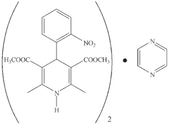
In the title compound, 2C17H18N2O6C4H4N2 [systematic name: 3,5-dimethyl 2,6-dimethyl-4-(2-nitrophenyl)-1,4-di-hydropyridine-3,5-dicarboxylate–pyrazine (2/1)], the complete pyrazine molecule is generated by crystallographic inversion symmetry. The center of the pyrazine ring lies on an inversion center. The nifedipine molecules are linked into chains along the caxis through N—H O hydrogen bonds, while the pyrazine molecules are organized in the structure through van der Waals interactions.
Related literature
Co-crystalline materials are of pharmaceutical interest due to their ability to alter the physicochemical properties of active pharmaceutical ingredients (APIs) (Schultheiss et al., 2009) and provide drug repositioning or life-cycle management (Trask, 2007). The corresponding crystal structure of nifedi-pine has been reported (Triggleet al., 2003) and it also forms chains through N—H O hydrogen bonds. Other crystalline forms also exist: polymorphs (Burger et al., 1996) solvates/ hydrates (Cairaet al., 2003) and a metal complex (Bontchevet al., 2003), as well as a non-crystalline, amorphous phase (Miyazakiet al., 2007).
Experimental
Crystal data
C19H20N3O6 Mr= 386.38 Monoclinic,P21=c a= 13.6278 (14) A˚
b= 9.1594 (9) A˚
c= 14.4432 (14) A˚ = 94.841 (4)
V= 1796.4 (3) A˚3 Z= 4
MoKradiation = 0.11 mm1 T= 120 K
0.240.180.10 mm
Data collection
Bruker APEXII CCD diffractometer
27572 measured reflections
6070 independent reflections 4916 reflections withI> 2(I)
Rint= 0.036
Refinement
R[F2> 2(F2)] = 0.043 wR(F2) = 0.125 S= 1.07 6070 reflections 261 parameters
H atoms treated by a mixture of independent and constrained refinement
max= 0.48 e A˚3
min=0.24 e A˚
3
Table 1
Selected torsion angles ().
[image:1.610.84.259.564.695.2]C12—C13—C14—C31 93.88 (10) C31—C14—C15—C16 93.78 (10)
Table 2
Hydrogen-bond geometry (A˚ ,).
D—H A D—H H A D A D—H A
N11—H11 O24i
0.906 (17) 1.942 (17) 2.8444 (12) 173.6 (15)
Symmetry code: (i)x;yþ3 2;zþ
1 2.
Data collection:APEX2(Bruker, 2007); cell refinement:SAINT
(Bruker, 2007); data reduction:SAINT; program(s) used to solve structure:SHELXS97(Sheldrick, 2008); program(s) used to refine structure: SHELXL97 (Sheldrick, 2008); molecular graphics:
PLATON (Spek, 2009); software used to prepare material for publication:SHELXTL (Sheldrick, 2008), PLATONand Mercury
(Macraeet al., 2006).
We would like to thank Dr John Desper (Kansas State Univeristy) for the data collection and structure solution. We also thank Mr Eyal Barash and Dr Richard McClurg for their careful review of this manuscript.
Supplementary data and figures for this paper are available from the IUCr electronic archives (Reference: KJ2152).
References
Bontchev, P. R., Mehandjiev, D. R., Ivanova, B. B. & Bontchev, R. P. (2003).
Transition Met. Chem.28, 745–748.
Bruker (2007).APEX2andSAINT. Bruker AXS Inc., Madison, Wisconsin, USA.
Burger, A. & Koller, K. T. (1996).Sci. Pharm.64, 293–301.
Caira, M. R., Robbertse, Y., Bergh, J. J., Song, M. & De Villiers, M. M. (2003).
J. Pharm. Sci.92, 2519–2533.
Macrae, C. F., Edgington, P. R., McCabe, P., Pidcock, E., Shields, G. P., Taylor, R., Towler, M. & van de Streek, J. (2006).J. Appl. Cryst.39, 453–457.
organic compounds
Acta Cryst.(2010). E66, o2297–o2298 doi:10.1107/S1600536810031703 Schultheisset al.
o2297
Acta Crystallographica Section E
Structure Reports
Online
Miyazaki, T., Yoshioka, S., Aso, Y. & Kawanishi, T. (2007).Int. J. Pharm.336, 191–195.
Schultheiss, N. & Newman, A. (2009).Cryst. Growth Des.9, 2950–2967. Sheldrick, G. M. (2008).Acta Cryst.A64, 112–122.
Spek, A. L. (2009).Acta Cryst.D65, 148–155. Trask, A. V. (2007).Mol. Pharm.4, 301–309.
supporting information
sup-1 Acta Cryst. (2010). E66, o2297–o2298
supporting information
Acta Cryst. (2010). E66, o2297–o2298 [https://doi.org/10.1107/S1600536810031703]
Nifedipine
–
pyrazine (2/1)
Nate Schultheiss, Melanie Roe and Jared P. Smit
S1. Comment
Designing, preparing, and characterizing cocrystalline materials is a rapidly growing area of research, especially in the
area of pharmaceutics, due to their ability to alter the physicochemical properties of active pharmaceutical ingredients
(APIs) (Schultheiss et al., 2009) and provide drug repositioning or life-cycle management (Trask, 2007). Cocrystals are
multi-component crystals where the individual, neutral molecules are typically held together through hydrogen-bonding.
Nifedipine (1,4-dihydro-2,6-dimethyl-4-(2-nitrophenyl) -3,5-pyridine dicarboxylic acid dimethyl ester),a calcium-channel
blocker, is known to exist in a variety of crystalline forms: polymorphs (Burger et al., 1996), solvates/hydrates (Caira et
al., 2003), and a metal complex (Bontchev et al., 2003), as well as a non-crystalline, amorphous phase (Miyazaki et al.,
2007). Suprisingly, examples of nifedipine cocrystals have yet to be published in the open literature, and thus we report
here the 2:1 cocrystal of nifedipine and pyrazine.
A view of the asymmetric unit of the title compound and its numbering scheme are displayed in Fig. 1. The material
crystallizes in a 2:1 (nifedipine:pyrazine) stoichiometric ratio, although the asymmetric unit contains the components in a
1:0.5 ratio, because the center of the pyrazine ring resides on an inversion center. It should also be noted that the
nitro-substituted phenyl ring is relatively orthogonal ("axial") to the dihydropyridine ring (Table 1) which is displayed in Fig. 1.
Nonetheless, the nifedipine molecules are linked into linear, one-dimensional chains with a graph set notation of C(6)
through N—H···O hydrogen bonds from the N—H moiety to a carbonyl moiety, Table 2. The hydrogen bonds are running
along the crystallographic c axis. Interestingly, the pyrazine molecules are not participating in hydrogen bonding with
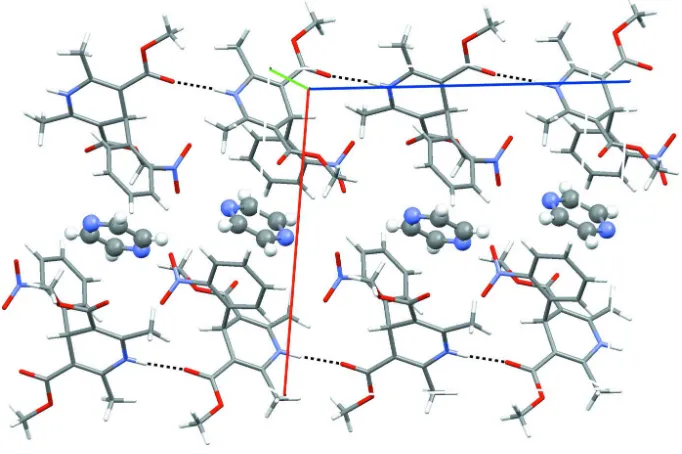
nifedipine, but are organized in between nifedipine rows through multiple van der Waals interactions (Fig. 2). Upon
extending the structure into three-dimensions, the organization of the pyrazine molecules within the crystal structure are
clearly shown. The pyrazine molecules are not only between one-dimensional rows of nifedipine, but also 'sandwiched′
between methyl-ester groups from neighboring nifedipine molecules.
S2. Experimental
The title compound was prepared by adding solid nifedipine to a nearly saturated solution of pyrazine in methanol and
allowed to stir for ~24 h at ambient temperature before filtering. Crystals of suitable size for single-crystal analysis were
obtained directly from the experiment.
S3. Refinement
The amino H-atom was located in a difference Fourier map. All other H-atoms were positioned geometrically and
Figure 1
The asymmetric unit of the title compound, with the atom labeling scheme and 50% probability displacement ellipsoids.
Figure 2
View down the b axis displaying the hydrogen bonding (black-dashed lines) between nifedipine molecules. The pyrazine
molecules (ball-and-stick mode) are positioned between the one-dimensional nifedipine rows (right). The direction of the
[image:4.610.135.476.346.573.2]supporting information
sup-3 Acta Cryst. (2010). E66, o2297–o2298
3,5-dimethyl 2,6-dimethyl-4-(2-nitrophenyl)-1,4-dihydropyridine-3,5-dicarboxylate–pyrazine (2/1)
Crystal data
C19H20N3O6
Mr = 386.38 Monoclinic, P21/c
Hall symbol: -P 2ybc a = 13.6278 (14) Å b = 9.1594 (9) Å c = 14.4432 (14) Å β = 94.841 (4)° V = 1796.4 (3) Å3
Z = 4
F(000) = 812 Dx = 1.429 Mg m−3
Mo Kα radiation, λ = 0.71073 Å Cell parameters from 9767 reflections θ = 2.6–31.7°
µ = 0.11 mm−1
T = 120 K Prism, colourless 0.24 × 0.18 × 0.10 mm
Data collection
Bruker APEXII CCD diffractometer
Radiation source: fine-focus sealed tube Graphite monochromator
φ and ω scans
27572 measured reflections 6070 independent reflections
4916 reflections with I > 2σ(I) Rint = 0.036
θmax = 31.8°, θmin = 2.6°
h = −20→19 k = −13→13 l = −17→21
Refinement
Refinement on F2
Least-squares matrix: full R[F2 > 2σ(F2)] = 0.043
wR(F2) = 0.125
S = 1.07 6070 reflections 261 parameters 0 restraints
Primary atom site location: structure-invariant direct methods
Secondary atom site location: difference Fourier map
Hydrogen site location: inferred from neighbouring sites
H atoms treated by a mixture of independent and constrained refinement
w = 1/[σ2(F
o2) + (0.070P)2 + 0.250P]
where P = (Fo2 + 2Fc2)/3
(Δ/σ)max < 0.001
Δρmax = 0.48 e Å−3
Δρmin = −0.24 e Å−3
Special details
Geometry. All e.s.d.'s (except the e.s.d. in the dihedral angle between two l.s. planes) are estimated using the full covariance matrix. The cell e.s.d.'s are taken into account individually in the estimation of e.s.d.'s in distances, angles and torsion angles; correlations between e.s.d.'s in cell parameters are only used when they are defined by crystal symmetry. An approximate (isotropic) treatment of cell e.s.d.'s is used for estimating e.s.d.'s involving l.s. planes.
Refinement. Refinement of F2 against ALL reflections. The weighted R-factor wR and goodness of fit S are based on F2,
conventional R-factors R are based on F, with F set to zero for negative F2. The threshold expression of F2 > σ(F2) is used
only for calculating R-factors(gt) etc. and is not relevant to the choice of reflections for refinement. R-factors based on F2
are statistically about twice as large as those based on F, and R- factors based on ALL data will be even larger.
Fractional atomic coordinates and isotropic or equivalent isotropic displacement parameters (Å2)
x y z Uiso*/Ueq
supporting information
sup-5 Acta Cryst. (2010). E66, o2297–o2298
Atomic displacement parameters (Å2)
U11 U22 U33 U12 U13 U23
N11 0.0221 (4) 0.0250 (4) 0.0100 (3) −0.0011 (3) 0.0008 (3) 0.0011 (3) C12 0.0176 (4) 0.0209 (4) 0.0119 (4) 0.0010 (3) 0.0011 (3) −0.0001 (3) C13 0.0159 (4) 0.0201 (4) 0.0116 (4) −0.0002 (3) 0.0012 (3) 0.0004 (3) C14 0.0169 (4) 0.0183 (4) 0.0109 (4) −0.0003 (3) 0.0015 (3) −0.0002 (3) C15 0.0170 (4) 0.0175 (4) 0.0147 (4) 0.0003 (3) 0.0025 (3) 0.0005 (3) C16 0.0199 (4) 0.0197 (4) 0.0145 (4) 0.0015 (3) 0.0032 (3) 0.0013 (3) C22 0.0200 (5) 0.0293 (5) 0.0143 (4) −0.0021 (4) −0.0014 (3) −0.0011 (4) C23 0.0174 (4) 0.0215 (4) 0.0132 (4) 0.0005 (3) 0.0014 (3) −0.0002 (3) O23 0.0211 (3) 0.0285 (4) 0.0141 (3) −0.0065 (3) 0.0031 (3) −0.0004 (3) O24 0.0279 (4) 0.0550 (6) 0.0111 (3) −0.0138 (4) −0.0004 (3) 0.0043 (3) C25 0.0182 (4) 0.0187 (4) 0.0178 (4) 0.0024 (3) 0.0029 (3) −0.0004 (3) O25 0.0246 (4) 0.0240 (4) 0.0170 (3) −0.0059 (3) −0.0004 (3) −0.0019 (3) O26 0.0308 (4) 0.0224 (4) 0.0247 (4) −0.0055 (3) 0.0037 (3) 0.0025 (3) C26 0.0300 (5) 0.0242 (5) 0.0173 (4) −0.0007 (4) 0.0043 (4) 0.0057 (4) C27 0.0270 (5) 0.0335 (6) 0.0196 (5) −0.0097 (4) 0.0066 (4) 0.0014 (4) C28 0.0273 (5) 0.0295 (5) 0.0256 (5) −0.0089 (4) 0.0003 (4) −0.0067 (4) C31 0.0166 (4) 0.0175 (4) 0.0131 (4) −0.0010 (3) 0.0018 (3) 0.0015 (3) C32 0.0188 (4) 0.0202 (4) 0.0135 (4) −0.0018 (3) 0.0001 (3) 0.0002 (3) N32 0.0221 (4) 0.0224 (4) 0.0142 (4) 0.0003 (3) −0.0022 (3) 0.0010 (3) O32 0.0270 (4) 0.0339 (4) 0.0155 (3) 0.0015 (3) 0.0033 (3) −0.0017 (3) O33 0.0249 (4) 0.0304 (4) 0.0245 (4) −0.0054 (3) −0.0067 (3) −0.0025 (3) C33 0.0204 (5) 0.0244 (5) 0.0204 (5) 0.0022 (4) −0.0012 (3) 0.0022 (4) C34 0.0252 (5) 0.0227 (5) 0.0237 (5) 0.0054 (4) 0.0014 (4) 0.0010 (4) C35 0.0276 (5) 0.0208 (4) 0.0197 (5) 0.0031 (4) 0.0017 (4) −0.0029 (4) C36 0.0223 (5) 0.0204 (4) 0.0149 (4) 0.0009 (3) 0.0002 (3) −0.0008 (3) N41 0.0291 (5) 0.0373 (5) 0.0247 (5) −0.0013 (4) 0.0058 (4) −0.0037 (4) C42 0.0283 (6) 0.0271 (5) 0.0327 (6) −0.0038 (4) 0.0101 (4) −0.0035 (5) C43 0.0313 (6) 0.0325 (6) 0.0264 (5) 0.0000 (5) 0.0093 (4) 0.0046 (5)
Geometric parameters (Å, º)
N11—C12 1.3682 (13) C27—H27A 0.9800 N11—C16 1.3759 (13) C27—H27B 0.9800 N11—H11 0.906 (17) C27—H27C 0.9800 C12—C13 1.3636 (13) C28—H28A 0.9800 C12—C22 1.4996 (14) C28—H28B 0.9800 C13—C23 1.4507 (13) C28—H28C 0.9800 C13—C14 1.5125 (13) C31—C32 1.3937 (13) C14—C15 1.5165 (13) C31—C36 1.3973 (13) C14—C31 1.5311 (13) C32—C33 1.3865 (14)
C14—H14 1.0000 C32—N32 1.4706 (13)
C15—C16 1.3566 (13) N32—O32 1.2180 (12) C15—C25 1.4651 (14) N32—O33 1.2257 (12) C16—C26 1.4989 (14) C33—C34 1.3778 (15)
C22—H22B 0.9800 C34—C35 1.3871 (15)
C22—H22C 0.9800 C34—H34 0.9500
C23—O24 1.2170 (12) C35—C36 1.3802 (14) C23—O23 1.3357 (12) C35—H35 0.9500 O23—C27 1.4342 (12) C36—H36 0.9500 C25—O26 1.2050 (12) N41—C42 1.3282 (17) C25—O25 1.3544 (12) N41—C43i 1.3311 (17)
O25—C28 1.4377 (13) C42—C43 1.3819 (18)
C26—H26A 0.9800 C42—H42 0.9500
C26—H26B 0.9800 C43—N41i 1.3311 (17)
C26—H26C 0.9800 C43—H43 0.9500
C12—N11—C16 123.46 (8) O23—C27—H27B 109.5 C12—N11—H11 118.4 (10) H27A—C27—H27B 109.5 C16—N11—H11 117.8 (10) O23—C27—H27C 109.5 C13—C12—N11 118.48 (9) H27A—C27—H27C 109.5 C13—C12—C22 127.75 (9) H27B—C27—H27C 109.5 N11—C12—C22 113.77 (8) O25—C28—H28A 109.5 C12—C13—C23 124.66 (9) O25—C28—H28B 109.5 C12—C13—C14 120.65 (8) H28A—C28—H28B 109.5 C23—C13—C14 114.68 (8) O25—C28—H28C 109.5 C13—C14—C15 109.88 (8) H28A—C28—H28C 109.5 C13—C14—C31 111.23 (8) H28B—C28—H28C 109.5 C15—C14—C31 109.79 (8) C32—C31—C36 115.33 (9) C13—C14—H14 108.6 C32—C31—C14 125.80 (8) C15—C14—H14 108.6 C36—C31—C14 118.66 (8) C31—C14—H14 108.6 C33—C32—C31 123.31 (9) C16—C15—C25 120.41 (9) C33—C32—N32 114.74 (9) C16—C15—C14 119.99 (9) C31—C32—N32 121.95 (9) C25—C15—C14 119.60 (8) O32—N32—O33 123.91 (9) C15—C16—N11 119.08 (9) O32—N32—C32 118.74 (9) C15—C16—C26 126.89 (9) O33—N32—C32 117.32 (9) N11—C16—C26 114.01 (9) C34—C33—C32 119.38 (10) C12—C22—H22A 109.5 C34—C33—H33 120.3 C12—C22—H22B 109.5 C32—C33—H33 120.3 H22A—C22—H22B 109.5 C33—C34—C35 119.32 (10) C12—C22—H22C 109.5 C33—C34—H34 120.3 H22A—C22—H22C 109.5 C35—C34—H34 120.3 H22B—C22—H22C 109.5 C36—C35—C34 120.14 (10) O24—C23—O23 121.36 (9) C36—C35—H35 119.9 O24—C23—C13 122.49 (9) C34—C35—H35 119.9 O23—C23—C13 116.15 (8) C35—C36—C31 122.51 (9) C23—O23—C27 115.24 (8) C35—C36—H36 118.7 O26—C25—O25 121.79 (9) C31—C36—H36 118.7 O26—C25—C15 127.27 (9) C42—N41—C43i 115.38 (11)
O25—C25—C15 110.91 (8) C42—N41—C42ii 106.16 (7)
C25—O25—C28 115.21 (8) C43i—N41—C42ii 137.11 (8)
supporting information
sup-7 Acta Cryst. (2010). E66, o2297–o2298
C16—C26—H26B 109.5 N41—C42—H42 118.9 H26A—C26—H26B 109.5 C43—C42—H42 118.9 C16—C26—H26C 109.5 N41i—C43—C42 122.46 (11)
H26A—C26—H26C 109.5 N41i—C43—H43 118.8
H26B—C26—H26C 109.5 C42—C43—H43 118.8 O23—C27—H27A 109.5
C16—N11—C12—C13 15.45 (15) C14—C15—C25—O26 −176.60 (10) C16—N11—C12—C22 −164.07 (9) C16—C15—C25—O25 −175.78 (9) N11—C12—C13—C23 −173.05 (9) C14—C15—C25—O25 5.23 (12) C22—C12—C13—C23 6.40 (17) O26—C25—O25—C28 −2.79 (14) N11—C12—C13—C14 7.81 (14) C15—C25—O25—C28 175.50 (8) C22—C12—C13—C14 −172.75 (9) C13—C14—C31—C32 138.67 (9) C12—C13—C14—C15 −27.91 (12) C15—C14—C31—C32 −99.50 (11) C23—C13—C14—C15 152.87 (8) C13—C14—C31—C36 −46.76 (11) C12—C13—C14—C31 93.88 (10) C15—C14—C31—C36 75.07 (11) C23—C13—C14—C31 −85.35 (10) C36—C31—C32—C33 −0.04 (14) C13—C14—C15—C16 28.86 (12) C14—C31—C32—C33 174.69 (9) C31—C14—C15—C16 −93.78 (10) C36—C31—C32—N32 −179.46 (9) C13—C14—C15—C25 −152.15 (8) C14—C31—C32—N32 −4.73 (15) C31—C14—C15—C25 85.21 (10) C33—C32—N32—O32 130.11 (10) C25—C15—C16—N11 171.13 (9) C31—C32—N32—O32 −50.42 (13) C14—C15—C16—N11 −9.89 (14) C33—C32—N32—O33 −48.13 (12) C25—C15—C16—C26 −7.30 (15) C31—C32—N32—O33 131.33 (10) C14—C15—C16—C26 171.68 (9) C31—C32—C33—C34 0.63 (16) C12—N11—C16—C15 −14.39 (15) N32—C32—C33—C34 −179.91 (9) C12—N11—C16—C26 164.24 (9) C32—C33—C34—C35 −0.67 (16) C12—C13—C23—O24 173.91 (11) C33—C34—C35—C36 0.14 (17) C14—C13—C23—O24 −6.90 (14) C34—C35—C36—C31 0.48 (16) C12—C13—C23—O23 −6.01 (15) C32—C31—C36—C35 −0.51 (14) C14—C13—C23—O23 173.19 (8) C14—C31—C36—C35 −175.64 (9) O24—C23—O23—C27 −0.71 (15) C43i—N41—C42—C43 −0.10 (19)
C13—C23—O23—C27 179.21 (9) C42ii—N41—C42—C43 −169.14 (10)
C16—C15—C25—O26 2.38 (16) N41—C42—C43—N41i 0.1 (2)
Symmetry codes: (i) −x+1, −y+1, −z+1; (ii) −x, −y, −z.
Hydrogen-bond geometry (Å, º)
D—H···A D—H H···A D···A D—H···A
N11—H11···O24iii 0.906 (17) 1.942 (17) 2.8444 (12) 173.6 (15)