SYSTEMS TO NOVEL VACCINE ADJUVANTS
Katarzyna Anna Morley
A Thesis Submitted for the Degree of PhD
at the
University of St Andrews
2016
Full metadata for this item is available in
St Andrews Research Repository
at:
http://research-repository.st-andrews.ac.uk/
Please use this identifier to cite or link to this item:
http://hdl.handle.net/10023/15670
M
ETAL
O
RGANIC
F
RAMEWORKS
:
F
ROM
A
NTICANCER MULTIFUNCTIONAL DRUG
DELIVERY SYSTEMS TO NOVEL VACCINE
ADJUVANTS
Katarzyna Anna Morley
A Thesis Submitted in application for
the Degree of PhD
in the School of Chemistry at the University of St
Andrews
I, Katarzyna Morley, hereby certify that this thesis, which is approximately 35 000
words in length, has been written by me, and that it is the record of work carried out
by me, or principally by myself in collaboration with others as acknowledged, and
that it has not been submitted in any previous application for a higher degree.
I was admitted as a research student in September, 2012 and as a candidate for the
degree of PhD in September, 2013; the higher study for which this is a record was
carried out in the University of St Andrews between 2012 and 2015.
Date signature of candidate
I hereby certify that the candidate has fulfilled the conditions of the Resolution and
Regulations appropriate for the degree of PhD in the University of St Andrews and
that the candidate is qualified to submit this thesis in application for that degree.
Date signature of supervisor
In submitting this thesis to the University of St Andrews I understand that I am
giving permission for it to be made available for use in accordance with the
regulations of the University Library for the time being in force, subject to any
copyright vested in the work not being affected thereby. I also understand that the
title and the abstract will be published, and that a copy of the work may be made and
supplied to any bona fide library or research worker, that my thesis will be
electronically accessible for personal or research use unless exempt by award of an
embargo as requested below, and that the library has the right to migrate my thesis
into new electronic forms as required to ensure continued access to the thesis. I have
obtained any third-party copyright permissions that may be required in order to allow
publication of this thesis:
PRINTED COPY & ELECTRONIC COPY
Embargo on all print copy and electronic copy for a period of 2 years on the
following ground:
Publication would be commercially damaging to the supervisor, or the University
Supporting statement for printed embargo request:
Chapter 6 – MOFs as an example for potential adjuvant applications, fully performed
at School of Pharmacy, UCL, with Dr Gareth Williams needs to be protected as there
is a plan for commercialisation of this idea and also some publishing will follow.
Date signature of candidate signature of supervisor
Courses Attended
Homogenous Catalysis Prof. Paul Kramer
Chemistry of phosphorus and related elements Dr A. Aitken
Functional materials Dr F. D. Morrison
Advanced polymer science Prof P. Wormald
I would like to thank my supervisor, Prof. Russell Morris for his help and
guidance throughout this complex project.
Also, I am grateful to Dr Gareth Williams for his advice, patience in explaining
the complexity of vaccine mechanism and contributing a lot in finding the ways of
solving the impossible and the possibility to extend my research and enriching my
thesis in the biological testing done for drug delivery systems as well as opening a
new avenue of applications for MOFs as vaccine adjuvants. Many thanks to Dr
Ilona Kubajewska for her patience and help with setting up the biological assays
and broad discussions on immune-response signals and cell work.
Thanks to my colleagues for their good advice and creating a good atmosphere in
the office: Sam Morris, Laura McCormick, Paul Wheatley, Yuyang Tien, Daniel
Firth. Thanks also to the other Morris group members who have helped with my
research. A special thanks goes to Mrs Sylvia Williamson for her patient and
helping me out with BET measurements.
Of course, I must thank my parents, Izabela and Jerzy, for their support and
encouragement throughout my PhD.
Lastly, I would like to thank my now husband Alexander Morley whom I met in
St Andrews and who supported me immensely through my PhD project, cheering
me up which gave me the motivation to finish the research that is illustrated in
this thesis.
Finally, thank you to the EPSRC for providing the funding that has allowed me to
Incorporation of cisplatin into the metal-organic frameworks UiO66-NH2 and UiO66 –
encapsulation vs. Conjugation.
for potential applications as drug delivery systems and novel vaccine adjuvants.
The opening chapter (Chapter 1) provides a reader with a background to the field, metal
organic frameworks synthesis routes and their potential applications in medicine and insight
into the basics of immunology and biological assays as well as the anti-cancer drugs
mechanisms and how it can be used to target the tumour cells.
Chapter 3 describes the experimental techniques used in this research combining biological
with chemical testing.
Investigation of Zr-MOFs (UiO66 and UiO66-NH2) and CPO – 27 (CPO Mg and CPO Ni) for
their potential application as drug delivery systems for cisplatin is presented and compared in
Chapter 4, as well as two different drug incorporation techniques (conjugation and physical
encapsulation) in the case of UiO66-NH2. The route proved to be more efficient when
compared with encapsulation for the same MOF, and overall the most promising candidate for
a drug delivery system for cisplatin, due to its large pores, was UiO66.
Chapter 5 focuses on the potential application of Zr-MOFs and CPO-27 (Mg and Ni) as a
drug delivery system for 5-Fluorouracil, a drug which because of its sparse solubility in water
is challenging when it comes to its administration in the human body. Chapter 7 investigates
the possibility to design multiple drug delivery systems utilising MOFs that contain cisplatin
and fluorouracil in their framework, loading them with nitric oxide to increase the anti-cancer
action and also to prevent thrombosis that may pose a danger to patients who undergo
anti-cancer therapies. Chapter 6 describes the new concept of investigating MOFs (Zr-MOFs and
Al-MOFs) as potential candidates as vaccine adjuvants. The research concluded that the
Chapter 1. Introduction ... 1
1.1. Metal-organic frameworks for biological applications ... 1
1.2. Nitric oxide – a friend or foe? ... 5
1.3. Anticancer drugs and drug delivery systems. ... 7
1.3.1. Cisplatin ... 11
1.3.2. 5-Fluorouracil ... 13
1.4. Adjuvants – integral part of every vaccine ... 14
1.5. References ... 19
Chapter 2. Aims ... 28
Chapter 3. Experimental Methods ... 30
3.1 Solvothermal and Microwave-assisted synthesis ... 30
3.2 X-ray diffraction ... 31
3.3 Microwave-Plasma – Atomic Emission Spectroscopy (MP-AES) ... 33
3.4. High Performance Liquid Chromatography (HPLC) ... 34
3.5 Biological Assays ... 36
3.5.1 Flow Cytometry ... 36
3.5.2 Viability by Alamar Blue ... 39
3.5.3 Cytokines’ secretion by ELISA ... 42
3.6. Nitric Oxide chemiluminescence ... 45
3.7 Fourier transform infrared spectroscopy (FTIR) ... 46
3.8 References...48
Chapter 4. MOFs as drug delivery carrier for cisplatin ... 50
4.1. Aims ... 50
4.2. Synthesis ... 51
4.2.1. One pot synthesis ... 54
4.3.1. Quantification of cisplatin content ... 57
4.3.2. Cisplatin Release ... 65
4.3.3. Viability assays ... 67
4.3.3.1. Viability of cells: Conjugation vs. Encapsulation...71
4.3.3.2. Viability of cells: Zr-MOFs and CPO-27...76
4.3.4. NPs of metallic Platinum as a result of one – pot synthesis. ... 82
4.4. Conclusions and Summary ... 85
4.5. References ... 88
Chapter 5. MOFs as a drug delivery carrier for Fluorouracil (5-FU)... 92
5.1. Aims ... 92
5.2. Encapsulation MOFs with 5-FU (UiO66, UiO66-NH2, CPO Mg, CPO Ni) ... 93
5.3. Results ... 94
5.3.1. MOF framework stability – X-ray of 5-FU encapsulated powders. ... 94
5.3.2. Presence of 5-FU in the pores of MOFs ... 98
5.3.2.1. Changing BET ... 98
5.3.2.2. FTIR spectra (ATR) ... 102
5.3.2.3. Drug release and challenges to measure it (HPLC and UV-VIS) ... 102
5.3.2.4. Traces of 5-FU in NMR spectra ... 106
5.3.3. Biological testing: Viability and Efficacy ... 107
5.4. Conclusions ... 112
5.5. References...113
Chapter 6. MOFs as potential vaccine adjuvants ... 116
6.1. Aims ... 116
6.2. Synthesis ... 119
6.3. Results ... 122
6.3.2.2 Immune response: Zr-MOFs ... 125
6.3.3. Immune responses measured by Flow Cytometry ... 127
6.3.4. Viability tests - Dendritic Cells and THP-1 differentiated macrophages ... 130
6.4. Conclusions ... 132
6.3. References...134
Chapter 7. MOFs as multifunctional drug delivery systems – nitric oxide release ... 137
7.1. Aims ... 137
7.2. Nitric Oxide loading ... 137
7.2.1. MOFs encapsulated with 5FU or Cisplatin ... 137
7.2.2. Zr-MOF with a conjugated prodrug ... 138
7.3. Results ... 138
7.3.1. NO release from Zr-MOF with a conjugated prodrug. ... 138
7.3.2. NO release from Mg-MOFs encapsulated with cisplatin and fluorouracil drugs. . 140
7.3.3. NO release from Ni-MOFs encapsulated with cisplatin and fluorouracil drugs. .. 142
7.3.4. NO release from Zr-MOFs encapsulated with cisplatin and fluorouracil drugs. .. 143
7.4. Conclusions ... 145
7.5. References ... 147
Chapter 8. Further Works...149
8.1. References ... 151
List of Abbrevation...153
Appendix 1. ...154
Appendix 2...170
1
Chapter 1.
Introduction
1.1.
Metal-organic frameworks for biological applications
The development of new materials is crucial for industry as well as for everyday
life. Metal–organic frameworks (MOFs) are a relatively new class of materials
that offers great potential in many applications1-3 e.g. in gas storage4-11, gas purification and separation12, heterogeneous catalysis13-15, imaging16-18, luminescence19 and biomedicine20-24. For more than 20 years, MOFs have been a hot topic in terms of their superb properties: large surface areas with pore sizes
that can be adjusted depending on the usage and adsorption capacities as well as
the fact that their density is low25, 26. These properties play a great role in the applications in catalysis, gas storage and gas separation. Their exceptional
properties (e.g. swelling of the crystallographic network in the presence of
different guest molecules26), are very interesting for some applications in which zeolites cannot be used and offer easy and non-complicated synthesis. In
comparison with the zeolite materials, an inorganic or organic template must be
used and its removal may cause the structure to collapse1. In contrast to zeolites, MOFs are easier to synthesise and do not require the generation of a template, as
they have an organic linker and a metal in their structure. It is important to
mention that a lot of names have been given to these porous materials (e.g. porous
coordination networks, porous coordination polymers, metal-organic frameworks
etc.). These materials are highly attractive to research as well as the industrial
world and the number of publications is remarkable1, 2. Especially important applications are in gas storage, drug storage and delivery27. There is a question left to answer, how many MOFs structures are there. This depends on how the
MOF is defined. Well, if we take the provisional recommendations on MOF
terminology from IUPAC that says a MOF is a coordination polymer with an
open framework containing potential voids28 then our search will be based on a transition metal that is bonded to a nitrogen or an oxygen via a polymeric bond. It
does not require a 3D framework only that it will be repeated. According to
2 Number of publications regarding MOFs sky-rocketed over the years as depicted
[image:12.595.152.464.119.348.2]in Figure 1-1 below.
Figure 1-1 Number of publications on MOFs30. Source: Web Science.
Discovered by Robson in 198931, metal-organic frameworks are a relatively young group of advanced porous materials that consists of a metal ion, the
inorganic part and the organic linker. The number of possible combinations is
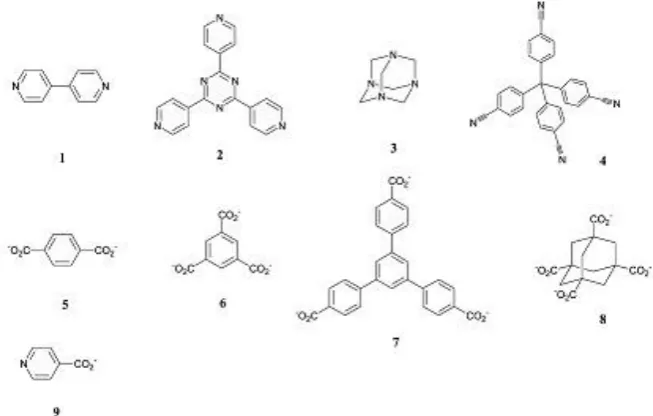
infinite. The most commonly used linkers are depicted in the Figure 1-2 below.
Figure 1-2 Some example of linkers that are used to synthesise MOFs. 4,4-bipyridine (1),1,3,5-Tris(4-pyridyl)-2,4,6-triazine (2), hexamethylenetetramine (3), tetra(4-cyanophenyl)methane (4). Terephthalic acid BDC 1,4- benzene dicarboxylate (5), benzene-1,3,5-tricarboxylic acid (6), 1,3,5-Tris(4-carboxyphenyl)benzene (7),
[image:12.595.144.471.455.663.2]3 One of the key properties of MOFs is the tuneable size of the pores that can
sometimes adjust their porosity upon the loading with gases or molecules; or also
as easily by exchanging the metal atom or the linker forming new structures20. A quite interesting property for MOFs as recently reported, is the so-called
reversible breathing or swelling effect, which gives rise to flexible pore size
depending on the guest molecules hosted by the porous structure26, 32. To give an example, the cell volume of the dry structures increases by 50-230% in case of the
MIL-53 (Cr) and (Fe) isoreticular family33. The term isoreticular was introduced by Yaghi34 to describe the structure formed with “the same preferred topology” with the only difference in the metal ion and the size of the links. In the same
framework it is possible to exchange the metal ions and keep the topology of the
material the same. This is not the case with the rigid bonds found in zeolites. The
cages and tunnels of MOFs are larger and thus, so are the specific surface areas of
500 – 6000 m2/g BET35 or even 6240 reported for MIL-20036, with low densities, generally 0.2-1 g/cm3 (density of Li metal 0.56 g/cm3)37 and with the exceptionally low density of 0.126 g/cm3 and big pores 8–43.2 Å21, 22, 30, 33, 34 recently reported for MOF-39934. Just recently, the reported surface areas of MOFs are about 10000 m2/g which makes them the materials for special missions and in this matter superior to zeolites and carbons38. These features give MOFs an edge in a variety of applications such as gas storage, catalysis, separation and
recently drug and bioactive gases loading with a controllable release39, 40. Also, thanks to the open metal sites of the inorganic part of the MOF, bioactive gases
such as NO, CO and H2S can be successfully loaded inside the molecule as a
result of their chemical nature41.
For bio-applications, the following criteria must be fulfilled – the MOFs should be
non-toxic, which requires both the linker and the metal to be inert; the release of
the drug or the bioactive gas must be controllable; and also the excretion of the
MOF after the drug administration should be possible24. That is why the endogeneous linker is the most preferable candidate, as it could have bioactive
functionality in the human body. What is meant by “endogeneous” is the fact of
being the part of or produced by the human body. An example of such a linker is
4 dose LD50=30g/kg) builds BioMIL-1 (Figure 1-5). Nicotinic acid that controls the
blood pressure and can control cholesterol and has antilipemic properties42.
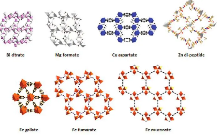
Figure 1-3 gives an overview how we cluster MOFs, and Figure 1-4 shows the
variety of MOF structures comprising of endogeneous linker, that may make them
more biocompatible. Coordination polymer is the most general term and MOFs
are best described as 3-dimentional coordination networks, IUPAC set the
[image:14.595.123.500.438.672.2]recommendations for nomenclature of coordination polymers and MOFs43.
Figure 1-3 Variety of the coordination polymer, the 3D ones are referred as MOFs44
5 Figure 1-5 A view of some MOFs with endogenous linkers. Here from the left: CPO-27 (Zn, Mg), MIL-100 (Fe), Bio-MOF-120.
1.2.
Nitric oxide – a friend or a foe?
Nitric oxide is a very interesting molecule thanks to its antithrombotic and
antibacterial properties, the discovery of which brought a Nobel Prize in 1998 for
Robert F Furchgott, Louis J Ignarro and Ferid Murad. It became the “molecule of
the year” six years earlier after the discovery of a wide spectrum of its
bio-properties like relaxation of the smooth muscle, prevention of platelet clotting and
neurotransmission45. The nitric oxide molecule has got 11 valance electrons, in which 5 electrons of Nitrogen [2s22p3] and 6 of oxygen [2s2p4] are responsible for bonding that emerges from sp-orbitals mixing leading to π-bonding. Figure 1-6
illustrates a schematic diagram of the molecular orbital in nitric oxide. The bond
order defined as the difference between bonding electrons and anti-bonding
electrons divided by two, for nitric oxide is 2.5.
[image:15.595.182.433.506.725.2]6 This free radical molecule (it has an unpaired electron in the 2π orbital) can
interact with transition metals and an unpaired electron could accept an electron
and thus acting as an anti-oxidant47. NO is an important gas-transmitter in a human
body and has a positive impact on blood platelet adhesion and cell division and is
responsible for vasodilation (relaxation of the blood vessels) and control of blood
pressure48, 49. It has been stipulated that biologically active amount of nitric oxide for certain applications may vary and for example to offer anti-thrombosis action,
only a small dose is necessary (4 ppm), however, for anti-bacterial protection - a 10
times higher dose of nitric oxide is required50. Antibacterial, wound healing and
anti-blood-clotting properties of NO have already been tested in vitroand in vivo to assess
its efficacy51. Several studies shown that NO assists with wound healing and
prevents forming of the blood clots35, 50, 51. NO – donor therapy is based on the generation of the accurate amount of NO in the right spot in the human body52. Not only have MOFs recently been used for NO delivery but also zeolites53 and functionalised nanoparticle silica54 and polymers55, 56. MOFs can deliver pure gas without any other oxides and the release of the gas can be controlled and triggered
by exposure of the MOF-powder to water. For example CPO-27: Ni showed the
exceptional uptake of NO up to 7 mmol/g, prolonged release under H2O over time
up to 3 weeks and after 2 years the loaded material showed no loss in NO
storage24, 41, which compared to HKUST-1 of 2 μmol/g at 298 K, gives the value 2500 times higher. This level of loading (HKUST-1) is still sufficient for
biological applications, but for antibacterial would not be enough57. The mechanism of action is still unknown and the NO dose that would suppress the
tumour growth must be yet identified, it is stipulated that above certain threshold
NO is carcinogenic58.
Not only anti-thrombotic and anti-bacterial properties of nitric oxide are of
interest but also its potential application as anti-cancer agent that may show higher
efficacy when compared to conventional therapies involving anti-cancer drugs59. In the human body, nitric oxide is produced by nitric oxide synthases (NOSs), a
family of enzymes with catalytic properties, in the process of L-Arginine
7 complex (nicotinamide adenine dinucleotide phosphate-oxidase) and oxygen and
as products, generates L-citrulline and NO60.
There are three forms of nitric oxide synthase enzyme: NOS-II, whose the main
function is cytotoxicity and defence of the host, and its primary site of action is
either in macrophages or tumour cells, NOS-I responsible for
neurotransmission/relaxation of smooth muscle and finally NOS-III, in charge of
smooth muscle proliferation control and minimising clotting of blood platelets.
NOS-II action can be regulated when stimulated by variety of cytokines and
ligands: TNF-α, interferon, IL-1 and endotoxins61 and is induced in different cell types for example macrophages and dendritic cells62.
Some research conducted, showed that NO can cause apoptotic death of cancer
cell at elevated concentrations63 or can lead to suppression of tumour early development64 and also of its growth and metastasis65 66, which could potentially open another avenue for therapies other than radio – and chemotherapies45.
1.3.
Anticancer drugs and drug delivery systems.
The interest in pharmaceuticals and therapies is seeing a substantial growth every
year with a lot of new drugs being approved by FDA (Food and Drug
Administration). Despite the fact that research is aided with computational
chemistry and biology helping in drug discovery immensely, drug development
that leads to successful commercialization is still a very time-consuming and
expensive process, taking between 10-15 years of development, clinical trials and
ca. $1.8 bn of cost per drug before it lands on the shelves of a drug store67, 68. Figure 1-7 depicts the total market sales in billion of $ and its growth over years.
The trend shows that starting from 2012 there is a growth potential over the last 2
years. Table 1-1 depicts the top 5 drugs that had quite an influence on the sales
revenues of the companies who made discoveries, however, the expiration dates
of patents is alarming, which is pushing the companies to maximize their revenues
within the time frame of an existing patent. As it is really tricky and
time-consuming to successfully come up with new drugs every time, there is a
paradigm that by preparing the formulations of these drugs and working on drug
8 Figure 1-7 Total market sales in pharmaceuticals/drugs sector according to IMS Health (American company providing information services and technology for the healthcare industry).
Table 1-1 Top 5 Medicines worldwide and the percentage of company sales as of 2010 that it generated68. *)the patent expired in 2010 in the US and in Europe in 2012.
The application of biocompatible and non-toxic polymers has contributed a lot to
modern medicine as it impacts on a vast field of biosciences including the areas
such as: tissue engineering, diagnostics and therapeutic strategies. Different
naturally-occurring and synthetic polymers are used to develop state-of-the-art
drug delivery systems, just to mention a few: chitosan, alginate, gelatin, dextran,
albumin. These polymers may help chemotherapy be more selective as it offers a
secure platform for controlled release that does not lead to overdose in some
human body regions67. Metal organic frameworks have found wide applications in many fields, but there is still one which needs to be more developed and further
research should be carried out in order to give satisfying results – it is the
bio-application of MOFs27. There is a challenge to navigate the cargo in the region invaded by cancer by using different targets in the cancer tissue69. These could be
0.00% 1.00% 2.00% 3.00% 4.00% 5.00% 6.00% 7.00% 8.00% 9.00% 10.00% - $ 200.0 $ 400.0 $ 600.0 $ 800.0 $ 1 000.0 $ 1 200.0 $
2005 2006 2007 2008 2009 2010 2011 2012 2013 2014
Total Market Growth %
Product Manufacturer Sales (USD
million)
% of company sales
Patent expire date
Lipitor Pfizer 12657 22.8 2011
Plavix Sanofi-Aventis/BMS 8817 17.3 2012
Seretide GSK 8469 25.2 2013
Nexium Astra Zeneca 8362 23.5 2014
[image:18.595.130.495.84.302.2]9 the changed mitochondria70, leaky cellular walls and lower pH values in the tumour tissue. Normal physiological conditions have a pH value of 7.2-7.4,
however, it becomes more acidic (5.0-6.5) in the extracellular space in the
endosomes and drops further to pH 4.0 in lysosomes (primary and secondary), in
the tumour cells the pH values are lower by 0.5-1.0 units than in the healthy tissue
as it is reported by non-invasive techniques55, 71. That feature could be used for selective targeting.
Figure 1-8 Endocytic pathway with different pH regions (left). To the right, the scheme that illustrates the normal cell and the one touched by the cancer with the possible ways for the drug delivery system migration shown 55.
As of now, only between 10-100 ppm of administered antibodies may reach their
targets in vivo72. Therapeutics that are available now, are quite limited with their non-selective distribution in the human body that leads to high administration
doses, fast excretion, poor pharmacokinetics (very fast release also called “burst
effect” that may lead to the overdose at certain spots) and side effects which in
case of anti-cancerous drugs might be very severe causing damage to the healthy
cells73, 74. Nanotechnology offers some ways to overcome these challenges and has received a lot of attention in the past 20 years72. The nano-carriers such as polymeric structures, micelles, liposomes, iron oxide and gold encapsulated with a
drug have been used for advanced therapies e.g. Abraxane and Doxil21. Research focused on encapsulation Fe3O4 nano-carriers with cisplatin and coated with
Poly(D,L-Lactide) showed the ability of iron oxide to absorb the drug and release
it within 70 hours from activation in PBS (phosphate buffer saline)75. Only recently MOFs, thanks to their interesting properties – large pores and porosity
10 7140 m2/g for NU-11076 (NU – Northwestern University), theoretically predicted to go as high as 10 000 m2/g or greater77, 78 and coordinative unsaturated sites upon dehydration, has turned the research world into converting them into drug
delivery systems.
MOFs offer the possibility to incorporate the Mn or Gd cations in the framework
that can work as imaging agent with an encapsulated cargo that the agent will
enable tracing to allow for a better understanding of the targeting and delivery
mechanisms. The Lin Group developed imaging agents – a nano-MOF containing
Manganese with a linker terephthalic acid (BDC) or trimesic acid (BTC)17. A gas or small molecules can serve as a drug. Some of the gases are highly toxic
in high amounts but paradoxically when dosed in a controllable way, are crucial
for life processes: NO, CO and H2S. The most recognised for its vasodilating
properties is nitric oxide but the two other gases recently have caught interest with
more research done on their controlled delivery35. The pores of MOFs are big enough to host small molecules of drugs, e.g. CPO-27 Ni (11Å). The presence of
nickel makes the entire structure quite toxic for bioaplications23 , however, it can still be used for anti-tumour drugs as they are very toxic20. Christian Serre’s group tested their MIL materials family (MIL-53, 88A, 88B, 89, 100, 101-NH2) for drug
loading: doxorubicin, cidovir, busulfan, caffeine, azidothimidine triphosphate
(antiviral for HIV)79. For better drug storage, derivatives of 2,5- dihydroxyterephthalic acid linkers were used that introduced different functional
groups in the framework which had great impact on the size of pores leading to
the formation of flexible iron (III) dicarboxylates22, 26, 33. This group also accomplished to synthesize a BioMOF called BioMIL based on a bioactive linker
which is nicotinic acid and a non-toxic iron (LD50 = 30g/kg). It is important to
mention that nicotinic acid is responsible in the human body for lowering
LDL-cholesterol and triglyceride levels and keeping HDL-LDL-cholesterol levels high and
also shows antibacterial and vasodilating properties42, and is a precursor for NADH coenzyme in living cells. Some other MOFs based on the nicotinic acid as
a linker were already reported but they all had a toxic cation in the framework
11 The anti-cancer drugs can be simply divided into 4 groups in regards with the
mechanism they present to fight the cancer cells, these are alkylating agents,
antimetabolites, anthracyclines and hormones. In this thesis, MOFs were
researched as potential drug delivery systems for two drugs representing
alkylating agent group – cisplatin and antimetabolites – 5- fluorouracil.
Alkylating agents have chemical groups capable of forming a covalent bond with
nucleophilic substances in the cell, and such a substance is DNA. Nitrogen in
guanine in the position N7 is strongly nucleophilic and as such a perfect target for
alkylation, nitrogen molecules in adenine (N1, N3 positions) and in cytosine (N3)
may also be a target of attack for the alkylating agents. These can bond in the
intra- or inter- chain positions, leading to cross-linking, which disturbs
transcription and replication of DNA, and ideally, the DNA-chain of the tumour
cells. In this group, we can list the following agents cisplatin, carboplatin,
busulfan hydroxycarbamide and carmustine81.
1.3.1. Cisplatin
Already in 196882 cytotoxicity against cancerous cells presented by cisplatin was recognized and in numerous intensive research projects over subsequent years
showed efficacy against several cancers in clinical trials83. Finally in 1978, it was approved by the FDA, although it has been known since 184574 in the world of chemistry. The mechanism of action of cisplatin is based on forming intrastrand
and interstrand cross-links with nucleic acids, which causes damage because by its
binding to the bases of DNA, the multiplication of DNA is impaired, leading to
cell death (apoptosis) caused by inability of DNA to proliferate84, 85.
Looking at the variety of cancers where cisplatin is used as a drug - the treatment
of head, neck, ovarian, cervical86, testicle, breast and bladder tumours87-89, one can state that cisplatin is the most commonly used anti-cancer drug. There are some
shortcomings of cisplatin therapy, as the drug affects all cells because it cannot
distinguish between cancerous and healthy cells. Even though its efficacy is quite
high, it comes with many side-effects: these include nephrotoxicity (kidneys
damage) and neurotoxicity (toxicity in the nervous system); drug resistance of
tumours also develops over time as well as may cause an acute thrombosis in
12 ongoing in order to circumvent the drug’s side-effects by introducing new ways in
which cisplatin is delivered88, 92, 93. This is achieved by the use of a variety of drug delivery systems, designed to release the drug inside the tumour cell and to leave
healthy cells untouched. Carbon nanotubes94, liposomes88, 95-99, polymers88, 100 and nano-sized metal phosphates87, oxides75 and hyaluronic acid nanoparticles101 are all under investigation. Recently MOFs have also begun to be explored in this
regard23, 102, 103.
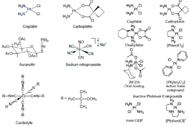
Below, in the Figure 1-9, reader may find the representation of different
derivatives of cisplatin that will follow the same mechanism of action in the
tumour cells, namely intrastrand cross-linking of DNA.
Figure 1-9 Different derivative of cisplatin reproduced from104 (left) and74 (right).
Only cis-platin is active in cancer treatment, as its geometric isomer trans-platin
did not show this feature in the clinical trials74. What makes the drug efficient, it is its affinity to DNA which acts here as a target to which cisplatin is bound which
causes damage to the helical structure followed by the inability for further
replication and transcription. That leads to the cell apoptosis104. Recently it was discovered that cisplatin does not enter the tumour cells by passive diffusion but a
copper transporter CTR1, which is a cell membrane protein and influences
cisplatin uptake. At the same time cisplatin causes degradation of CTR1 and in
[image:22.595.153.455.293.496.2]13 resistance of the tumour cell against cisplatin that occurs over the course of time is
linked to lack of the membrane protein in the cancer cell105.
1.3.2. 5-Fluorouracil
Fluorouracil (5-FU) is an example of another mechanism of action which aims to
replace uracil in purine biosynthesis. In contrary to uracil, 5-FU cannot be
converted to thymidylate and because of this blocks DNA synthesis and cell
proliferation. 5-FU is widely used mainly in topical applications to treat solid
tumours, however, some drawbacks e.g. variable efficacy and toxicity limit its
usage and FDA recommends not to administer 5-FU to patients with
dihydropyrimidine dehydroglenase deficiency (DPD metabolic disorder).
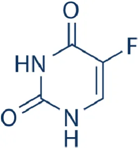
[image:23.595.242.382.325.475.2]The structure of 5-FU is depicted in Figure 1-10.
Figure 1-10 Structure of Fluorouracil, which is a derivative of uracil.
The drug was patented by Charles Heidelberg, who carried out research that
showed that 5-FU was capable to stop the propagation of cancer in mice106. 5-Fluorouracil is a hydrophilic drug what makes it challenging for preparations of
formulations as its solubility in water is very sparse, and can only be enhanced in
acidic or basic environments. It is though widely used as a part of therapies to
treat colorectal, bladder, head and neck and breast cancers107, 108. Likewise cisplatin, therapies comprising 5-FU are toxic to the healthy cells and the short
half-life (16 min) makes the therapy not very long-lasting with repetitions of
administered doses being necessary and as such these have to be well adjusted
depending on the individual109.
A new Zn-MOF (Zn-BDC) was reportedly synthesised of a surface area of
14 the level of 0.42 g/g. 5-FU release was monitored110. This piece of research did not provide any information on the drug delivery system efficacy in the in vitro
environment. ZSM-5 zeolite has been recently investigated for its potential
application as a drug delivery system for 5-FU111.
Another approach has been presented by researcher at School of Pharmacy, UCL,
to encapsulate 5-FU, by applying electro-spinning method. This technique
involved a preparation of a formulation for production of electrospun fibres112 that contained 5-FU. It has been shown that pH values had tremendous effect on the
speed of drug release and at pH =1, between 40% and 80% of cargo was released
and only increasing the pH value to 6.8 resulted in 100% release of the drug. The
ideal drug delivery system would keep the cargo intact at pH 7 and only allow for
a release at lower pH values to target the areas touched by cancer.
1.4.
Adjuvants – integral part of every vaccine
First vaccination procedure that was ever reported was the one performed by
Edward Jenner and this was 220 years ago. He realized that people whose job was
to milk cows, were not vulnerable to smallpox. Based on this observation he
inoculated a boy with a cowpox and as a result this gave him immune protection
against smallpox113, 114. Years later, it has been researched that the protection against disease can be provided by vaccination in a form of administration
live-attenuated virus, inactivated virus or a vaccines. The latter was the safest way of
offering a full protection, however the immune response induced was quite
insignificant. Live attenuated vaccines have a live form of the pathogen that was
modified so that it is in its non-virulent form, which can still multiply in the living
organism without causing the full form of the disease. The immune response is
very strong and the lifetime immunity to the virus can be achieved by
administration of only two doses (rubella, measles)115, 116. However, there is a risk associated with this type of the vaccines as the pathogen can reverse to its virulent
form which would pose a danger of the infection with the disease following
vaccination. The other possibility is to administer the inactivated vaccine, in
which the pathogen is dead and is unable to reproduce in the body as once dead, it
15 chemicals to inactivate the pathogen and it is of great importance to monitor such
a process to make sure that no live strains of pathogen was left in the vaccine. As
a result, such vaccines are much safer than live attenuated ones and just to name
an example is Salk polio vaccine. This type, however, can only induce relatively
weak immune response when compared to the former type, and in order to
achieve a long-term immune protection an administration of multiple doses is
necessary117.
Subunit vaccines contain only a part from the antigen, such as sugar of a virus
which would be something the immune system would respond to and record the
immune event. The antigen is either modified in a chemical or mechanical way,
that damages the cellular structure or by genetic engineering where the genomic
material has been removed from the pathogen118. Administration of such a non-virulent vaccines does not cause any infection in the organism and can be given to
patients with HIV or these undergoing chemotherapy. It is worth noting that
subunit vaccines as well as inactivated ones, to be efficient need to paired with an
adjuvant, which is a second component added to the formulation containing
antigen. “Adiuvare”, the verb in Latin means to help or to aid and is where the
word adjuvant originates from. It was observed and then reported by Alexander
Glenny nearly 100 years ago, that the antigen (toxoid) that was precipitated with
aluminium potassium sulfate was able to stimulate much stronger immune
response than the antigen itself and resulted in much higher number of antibodies
produced119. This experiment was performed “in vivo” on guinea pigs and proved that aluminum is capable of working as a powerful adjuvant and remained for
many years the only vaccine adjuvant one approved by FDA in the USA and
referred as alum and by far most widely used. Its role is to enhance the immune
response, by stimulating the production of the antibodies that was initially
triggered by the administration of antigen and would not reach the desired
threshold to be effective120. It has been researched that Al-containing adjuvants induce strong immune responses121. So far, there is no explanation to the mechanism of such action and trials are made in order to develop other adjuvants
that would contain other metals, or salts like calcium phosphate, which is used in
16 Special cells like dendritic, macrophages, B cells and T cells are involved in
creating an immune response. Dendritic Cell (DC) presents antigen to T cells
which activates a specific immune response that would be conveyed by a T cell
(pre-programmed by DC) to this particular pathogen that the antigen came from.
What happens is the following: if an antigen is detected in the body, dendritic cell
will travel to the lymph node in order to present the antigen to the T cell.
Activated T cells secrete cytokines and chemokines to recruit other immune cells
to fight the pathogen. By monitoring the response from the stimulated DCs, we
can predict how effectively DCs will activate T cells to respond to pathogen. T
cells assist other cells by speeding up the process of B cell-maturation, and are
called a T-helper (CD4+) and ascribed as Th. In an event of infection, the T and B
cells are “naïve”, meaning that they are not efficient to fight the infection and
need to adapt numerous cell proliferations must occur in the body to produce a
certain number of effectively working cells, which are able to produce cytokines
(protein mediators) or differentiate to cells able to secrete antibody to fight the
infection (B cells). The other type of T cells, CD8+ (cytotoxic T lymphocytes)
will act as a warhead to destroy infected cells, as well as tumour cells. The last
type of cells, important for this thesis are the B cells, which are able to produce
antibodies that are specific to the pathogen.
Generally, vaccine will stimulate one or both immune responses, recognized as
Th1 and Th2. Th2 is observed during cell stimulation by alum – coordination of
humoral response which is responsible for production of antibodies, and very
common in case of bacterial infections 123. Th1 response is cell-mediated and follows through activation of cytokines, which is promoting viral termination.
This kind of response involves mainly T cells that displays major
histocompatibility complex including cells infected by pathogens, tumour cells or
transplanted cells. The function of the vaccine is to create a supply of specific B
and T cells with the memory to the pathogen causing a disease, but in this case
without infecting the body. Th1 cells synthesise cytokines: IL-2, IL-18, IFN-γ also
promote cellular defence and inflammatory response. Th2 cells secrete the
cytokines: IL-4, IL-5, IL-6 and IL-13 as well as TNF-α (tumour necrosis factor).
17 responses in order to cover a wider spectrum of immune response and hence be
universal for bacterial and viral infections.
Cytokines are small proteins playing a pivotal role in signalling to the cells the
state of inflammation arising from the state of infection. Cytokines term, includes
interleukins, interferons, chemokines and tumour necrosis factors. These proteins
are secreted by B and T cells (lymphocytes), mast cells and macrophages. It is
worth noting that each cytokine may be produced by any of the cells. They are
important for immune system as they regulate cell population and maturation by
keeping in balance immune responses coming from humoral (Th2) and
cell-mediated (Th1) immune responses. Some can influence the way other cytokines
act and as such important to host immune responses to infection, inflammation
and tumour cells. Just to introduce some of the cytokines, IL-2 and IL-6 are
believed to treat cancer124, IFN-α found application to treat multiple sclerosis and hepatitis C. IL-4 activates T cells into Th2 cells. TNF-α is produced mainly by
macrophages and plays crucial role in regulating the immune cells, responsible for
triggering inflammatory response (fever) or capable of inducing apoptotic death.
The latter action is believed to give some hope in the cancer therapies125-127. Many materials come to interest when it comes to search after the potent adjuvant.
There was some research done, dedicated to the potential usage of Zinc oxide as
an adjuvant formulated in a nanoparticles form, where after 21 days after
vaccination performed on mice elevated amounts of IL-17, IL-4 and IL-5 were
reported128. Another study performed later, where nano-sized ZnO with particles smaller than 50 nm were used, confirmed these findings especially on enhanced
secretion of the following cytokines: IL-2, IL-4, IL-6, IL-17, however the
production of IL-10 and TNF-α decreased129. Also, an interesting study was performed in the 90’ to investigate the role of the surface area of the materials
used. This investigated SiO2, ZrO2, Fe-oxides (III and II+III), SnO2, Al2O3. Silica
was reported to promote antibody production, Aluminium and Iron oxides
triggered small response and others proved to be not effective130.
Au-nanoparticles came to interest of researchers as potential drug carriers for
18
132
. These studies showed that there are some cases for which the immune
response triggered is not sufficient and as research suggests the best results, to
stimulate the immune response, are obtained by introducing the second adjuvant
which is alum133.
The human immune system is highly complicated and this introduction does not
aim to describe it fully and the goal is to give the reader an overview to follow the
concepts of this thesis. For more detailed outlook into immunology, the reader is
19
1.5.
References
1. R. J. Kuppler, D. J. Timmons, Q.-R. Fang, J.-R. Li, T. A. Makal, M. D.
Young, D. Yuan, D. Zhao, W. Zhuang and H.-C. Zhou, Coordination
Chemistry Reviews, 2009, 253, 3042-3066.
2. S. L. James, Chemical Society Reviews, 2003, 32, 276-288. 3. G. Ferey, Chemical Society Reviews, 2008, 37, 191-214. 4. K. M. Thomas, Dalton transactions, 2009, 0, 1487-1505.
5. P. D. C. Dietzel, V. Besikiotis and R. Blom, Journal of Materials
Chemistry, 2009, 19, 7362-7370.
6. K. Sumida, D. L. Rogow, J. A. Mason, T. M. McDonald, E. D. Bloch, Z.
R. Herm, T.-H. Bae and J. R. Long, Chem Rev, 2011, 112, 724-781. 7. K. L. Mulfort and J. T. Hupp, J Am Chem Soc, 2007, 129, 9604-+.
8. D. Zhao, D. Yuan and H.-C. Zhou, Energy & Environmental Science,
2008, 1, 222-235.
9. R. E. Morris and P. S. Wheatley, Angew Chem Int Edit, 2008, 47, 4966-4981.
10. S. Ma and H.-C. Zhou, Chem. Commun., 2010, 46, 44-53.
11. L. J. Murray, M. Dinca and J. R. Long, Chemical Society Reviews, 2009,
38, 1294-1314.
12. J.-R. Li, J. Sculley and H.-C. Zhou, Chem Rev, 2011, 112, 869-932.
13. J. Lee, O. K. Farha, J. Roberts, K. A. Scheidt, S. T. Nguyen and J. T.
Hupp, Chemical Society Reviews, 2009, 38, 1450-1459.
14. A. Henschel, K. Gedrich, R. Kraehnert and S. Kaskel, Chem. Commun.,
2008, 0, 4192-4194.
15. A. Corma, H. García and F. X. Llabrés i Xamena, Chem Rev, 2010, 110, 4606-4655.
16. M. Kurmoo, Chemical Society Reviews, 2009, 38, 1353-1379.
17. K. M. L. Taylor, W. J. Rieter and W. Lin, J Am Chem Soc, 2008, 130, 14358-14359.
18. K. M. L. Taylor-Pashow, J. D. Rocca, Z. Xie, S. Tran and W. Lin, J Am
20 19. M. D. Allendorf, C. A. Bauer, R. K. Bhakta and R. J. T. Houk, Chemical
Society Reviews, 2009, 38, 1330-1352.
20. P. Horcajada, R. Gref, T. Baati, P. K. Allan, G. Maurin, P. Couvreur, G.
Ferey, R. E. Morris and C. Serre, Chem Rev, 2012, 112, 1232-1268.
21. R. C. Huxford, J. Della Rocca and W. Lin, Current Opinion in Chemical
Biology, 2010, 14, 262-268.
22. P. Horcajada, C. Serre, M. Vallet-Regí, M. Sebban, F. Taulelle and G.
Férey, Angewandte Chemie, 2006, 118, 5974-5978.
23. A. C. McKinlay, R. E. Morris, P. Horcajada, G. Ferey, R. Gref, P.
Couvreur and C. Serre, Angew Chem Int Ed Engl, 2010, 49, 6260-6266. 24. A. C. McKinlay, B. Xiao, D. S. Wragg, P. S. Wheatley, I. L. Megson and
R. E. Morris, J Am Chem Soc, 2008, 130, 10440-10444.
25. C.-D. Wu and W. Lin, Angewandte Chemie International Edition, 2005,
44, 1958-1961.
26. P. Horcajada, F. Salles, S. Wuttke, T. Devic, D. Heurtaux, G. Maurin, A.
Vimont, M. Daturi, O. David, E. Magnier, N. Stock, Y. Filinchuk, D.
Popov, C. Riekel, G. Férey and C. Serre, J Am Chem Soc, 2011, 133,
17839-17847.
27. D. Farrusseng, Metal-Organic Frameworks: Applications from Catalysis
and Gas Storage, Wiley-VCH Verlag GmbH & Co. KGaA, 2011.
28. L. Öhrström, RSC Publishing CrystEngComm Blog, 2013.
29. P. Wood, Highly Holey: MOFs with Record-Breaking Surface Areas,
2013.
30. S. Keskin and S. Kizilel, Ind Eng Chem Res, 2011, 50, 1799-1812. 31. B. F. Hoskins and R. Robson, J Am Chem Soc, 1989, 111, 5962-5964. 32. C. Serre, F. Millange, C. Thouvenot, M. Noguès, G. Marsolier, D. Louër
and G. Férey, J Am Chem Soc, 2002, 124, 13519-13526.
33. P. Horcajada, C. Serre, G. Maurin, N. A. Ramsahye, F. Balas, M.
Vallet-Regi, M. Sebban, F. Taulelle and G. Ferey, J Am Chem Soc, 2008, 130, 6774-6780.
34. H. Furukawa, Y. B. Go, N. Ko, Y. K. Park, F. J. Uribe-Romo, J. Kim, M.
21 35. P. K. Allan, P. S. Wheatley, D. Aldous, M. I. Mohideen, C. Tang, J. A.
Hriljac, I. L. Megson, K. W. Chapman, G. De Weireld, S. Vaesen and R.
E. Morris, Dalton transactions, 2012, 41, 4060-4066.
36. H. Furukawa, N. Ko, Y. B. Go, N. Aratani, S. B. Choi, E. Choi, A. O.
Yazaydin, R. Q. Snurr, M. O'Keeffe, J. Kim and O. M. Yaghi, Science,
2010, 329, 424-428.
37. M. Eddaoudi, J. Kim, N. Rosi, D. Vodak, J. Wachter, M. O'Keeffe and O.
M. Yaghi, Science, 2002, 295, 469-472.
38. H. Furukawa, K. E. Cordova, M. O’Keeffe and O. M. Yaghi, Science,
2013, 341.
39. T. Chalati, P. Horcajada, R. Gref, P. Couvreur and C. Serre, Journal of
Materials Chemistry, 2011, 21, 2220-2227.
40. G. Férey, C. Serre, C. Mellot-Draznieks, F. Millange, S. Surblé, J. Dutour
and I. Margiolaki, Angewandte Chemie International Edition, 2004, 43, 6296-6301.
41. N. J. Hinks, A. C. McKinlay, B. Xiao, P. S. Wheatley and R. E. Morris,
Micropor Mesopor Mat, 2010, 129, 330-334.
42. S. R. Miller, D. Heurtaux, T. Baati, P. Horcajada, J.-M. Greneche and C.
Serre, Chem. Commun., 2010, 46, 4526-4528.
43. R. Batten Stuart, R. Champness Neil, X.-M. Chen, J. Garcia-Martinez, S.
Kitagawa, L. Öhrström, M. O’Keeffe, M. Paik Suh and J. Reedijk, in Pure
Appl Chem, 2013, vol. 85, p. 1715.
44. J. J. Perry Iv, J. A. Perman and M. J. Zaworotko, Chemical Society
Reviews, 2009, 38, 1400-1417.
45. S. Huerta, S. Chilka and B. Bonavida, International Journal of Oncology,
2008, 33, 909-927.
46. P. K. Allan, Chemistry, University of St Andrews, Thesis, 2012.
47. G. A. Blaise, D. Gauvin, M. Gangal and S. Authier, Toxicology, 2005,
208, 177-192.
22 49. R. N. Anthony R Butler, Life, Death and Nitric Oxide, The Royal Society
of Chemistry, 2003.
50. M. R. Miller and I. L. Megson, British Journal of Pharmacology, 2007,
151, 305-321.
51. H. Zhu, B. Ka and F. Murad, World J. Surg., 2007, 31, 624-631.
52. K. D. Kroncke and C. V. Suschek, Journal of Investigative Dermatology,
2008, 128, 258-260.
53. P. S. Wheatley, A. R. Butler, M. S. Crane, S. Fox, B. Xiao, A. G. Rossi, I.
L. Megson and R. E. Morris, J Am Chem Soc, 2006, 128, 502-509.
54. J. H. Shin and M. H. Schoenfisch, Chemistry of Materials, 2007, 20, 239-249.
55. R. Haag and F. Kratz, Angewandte Chemie International Edition, 2006,
45, 1198-1215.
56. F. DeRosa, M. R. Kibbe, S. F. Najjar, M. L. Citro, L. K. Keefer and J. A.
Hrabie, J Am Chem Soc, 2007, 129, 3786-3787.
57. B. Xiao, P. S. Wheatley, X. B. Zhao, A. J. Fletcher, S. Fox, A. G. Rossi, I.
L. Megson, S. Bordiga, L. Regli, K. M. Thomas and R. E. Morris, J Am
Chem Soc, 2007, 129, 1203-1209.
58. J. R. Hickok and D. D. Thomas, Current pharmaceutical design, 2010, 16, 381-391.
59. B. Brune, Cell Death Differ, 2003, 10, 864-869.
60. S. Moncada and A. Higgs New England Journal of Medicine, 1993, 329, 2002-2012.
61. K. D. Kröncke, K. Fehsel and V. Kolb-Bachofen, Clinical and
Experimental Immunology, 1998, 113, 147-156.
62. C. Nathan, The Journal of Clinical Investigation, 100, 2417-2423.
63. S. Duan, S. Cai, Q. Yang and M. L. Forrest, Biomaterials, 2012, 33, 3243-3253.
64. S. P. Hussain, G. E. Trivers, L. J. Hofseth, P. He, I. Shaikh, L. E.
Mechanic, S. Doja, W. Jiang, J. Subleski, L. Shorts, D. Haines, V. E.
Laubach, R. H. Wiltrout, D. Djurickovic and C. C. Harris, Cancer
23 65. D. Wei, E. L. Richardson, K. Zhu, L. Wang, X. Le, Y. He, S. Huang and
K. Xie, Cancer Research, 2003, 63, 3855-3859.
66. X. Le, D. Wei, S. Huang, J. R. Lancaster and K. Xie, Proceedings of the
National Academy of Sciences of the United States of America, 2005, 102, 8758-8763.
67. C. Vilos and L. A. Velasquez, Journal of Biomedicine and Biotechnology,
2012.
68. S. Gaisford and M. Saunders, in Essentials of Pharmaceutical
Preformulation, John Wiley & Sons, Ltd, 2012, pp. 1-35.
69. S. H. van Rijt and P. J. Sadler, Drug discovery today, 2009, 14, 1089-1097.
70. E. Zhang, C. Zhang, Y. Su, T. Cheng and C. Shi, Drug discovery today,
2011, 16, 140-146.
71. R. Moreno-Sánchez, E. Saavedra, S. Rodríguez-Enríquez, J. C.
Gallardo-Pérez, H. Quezada and H. V. Westerhoff, Mitochondrion, 2010, 10,
626-639.
72. M. Ferrari, Nat Rev Cancer, 2005, 5, 161-171.
73. W. R. Sanhai, J. H. Sakamoto, R. Canady and M. Ferrari, Nat Nano, 2008,
3, 242-244.
74. E. R. Jamieson and S. J. Lippard, Chem Rev, 1999, 99, 2467-2498.
75. S. V. Devi and T. Prakash, Ieee Transactions on Nanobioscience, 2013,
12, 60-63.
76. H. Deng, S. Grunder, K. E. Cordova, C. Valente, H. Furukawa, M.
Hmadeh, F. Gándara, A. C. Whalley, Z. Liu, S. Asahina, H. Kazumori, M.
O’Keeffe, O. Terasaki, J. F. Stoddart and O. M. Yaghi, Science, 2012,
336, 1018-1023.
77. O. K. Farha, I. Eryazici, N. C. Jeong, B. G. Hauser, C. E. Wilmer, A. A.
Sarjeant, R. Q. Snurr, S. T. Nguyen, A. Ö. Yazaydın and J. T. Hupp, J Am
Chem Soc, 2012, 134, 15016-15021.
78. H. Furukawa, N. Ko, Y. B. Go, N. Aratani, S. B. Choi, E. Choi, A. Ö.
Yazaydin, R. Q. Snurr, M. O’Keeffe, J. Kim and O. M. Yaghi, Science,
24 79. P. Horcajada, T. Chalati, C. Serre, B. Gillet, C. Sebrie, T. Baati, J. F.
Eubank, D. Heurtaux, P. Clayette, C. Kreuz, J. S. Chang, Y. K. Hwang, V.
Marsaud, P. N. Bories, L. Cynober, S. Gil, G. Ferey, P. Couvreur and R.
Gref, Nature Materials, 2010, 9, 172-178.
80. J. Y. Lu and A. M. Babb, Inorganic Chemistry Communications, 2001, 4, 716-718.
81. H. P. Rang, J. M. Ritter, R. J. Flower and G. Henderson, Rang & Dale's
Pharmacology, 8th edn., Churchill Livingstone.
82. M. H. Greene, J Natl Cancer Inst, 1992, 84, 306-312.
83. O. Rixe, W. Ortuzar, M. Alvarez, R. Parker, E. Reed, K. Paull and T. Fojo,
Biochemical Pharmacology, 1996, 52, 1855-1865.
84. W. H. Ang, S. Pilet, R. Scopelliti, F. Bussy, L. Juillerat-Jeanneret and P. J.
Dyson, Journal of Medicinal Chemistry, 2005, 48, 8060-8069. 85. D. Gibson, Dalton transactions, 2009, 10681-10689.
86. P. G. Rose, B. N. Bundy, E. B. Watkins, J. T. Thigpen, G. Deppe, M. A.
Maiman, D. L. Clarke-Pearson and S. Insalaco, New England Journal of
Medicine, 1999, 340, 1144-1153.
87. A. Diaz, M. L. Gonzalez, R. J. Perez, A. David, A. Mukherjee, A. Baez,
A. Clearfield and J. L. Colon, Nanoscale, 2013, 5, 11456-11463.
88. H. Xiao, L. Yan, Y. Zhang, R. Qi, W. Li, R. Wang, S. Liu, Y. Huang, Y.
Li and X. Jing, Chem. Commun., 2012, 48, 10730-10732.
89. R. Wang, H. Xiao, H. Song, Y. Zhang, X. Hu, Z. Xie, Y. Huang, X. Jing
and Y. Li, Journal of Materials Chemistry, 2012, 22, 25453-25462.
90. D. D. Fernandes, M. L. Louzada, C. A. Souza and F. Matzinger, Current
Oncology, 2011, 18, 97-100.
91. T. Proverbs-Singh, S. K. Chiu, Z. Liu, S. Seng, G. Sonpavde, T. K.
Choueiri, C.-K. Tsao, M. Yu, N. M. Hahn, W. K. Oh and M. D. Galsky,
Journal of the National Cancer Institute, 2012.
92. V. Pinzani, F. Bressolle, I. Johanne Haug, M. Galtier, J. Blayac and P.
Balmès, Cancer Chemother Pharmacol, 1994, 35, 1-9.
93. Z. H. Siddik, D. R. Newell, F. E. Boxall and K. R. Harrap, Biochemical
25 94. A. H. Tamsyn and M. H. James, Nanotechnology, 2007, 18, 275704. 95. J. Yang, W. Mao, M. Sui, J. Tang and Y. Shen, Journal of Controlled
Release, 2011, 152, E108-E109.
96. H. Song, H. Xiao, Y. Zhang, H. Cai, R. Wang, Y. Zheng, Y. Huang, Y. Li,
Z. Xie, T. Liu and X. Jing, Journal of Materials Chemistry B, 2013, 1, 762-772.
97. A. Samad, Y. Sultana and M. Aqil, Current Drug Delivery, 2007, 4, 297-305.
98. E. A. Leite, C. M. Souza, A. D. Carvalho-Junior, L. G. V. Coelho, A. M.
Q. Lana, G. D. Cassali and M. C. Oliveira, International Journal of
Nanomedicine, 2012, 7, 5259-5269.
99. H. Chen, S. Pazicni, N. L. Krett, R. W. Ahn, J. E. Penner-Hahn, S. T.
Rosen and T. V. O'Halloran, Angewandte Chemie International Edition,
2009, 48, 9295-9299.
100. H. Xiao, R. Qi, S. Liu, X. Hu, T. Duan, Y. Zheng, Y. Huang and X. Jing,
Biomaterials, 2011, 32, 7732-7739.
101. Y.-I. Jeong, S.-T. Kim, S.-G. Jin, H.-H. Ryu, Y.-H. Jin, T.-Y. Jung, I.-Y.
Kim and S. Jung, Journal of Pharmaceutical Sciences, 2008, 97, 1268-1276.
102. C.-Y. Sun, C. Qin, X.-L. Wang and Z.-M. Su, Expert Opinion on Drug
Delivery, 2013, 10, 89-101.
103. W. J. Rieter, K. M. Pott, K. M. L. Taylor and W. Lin, J Am Chem Soc,
2008, 130, 11584-11585.
104. C. X. Zhang and S. J. Lippard, Current Opinion in Chemical Biology,
2003, 7, 481-489.
105. P. a. Ma, H. Xiao, X. Li, C. Li, Y. Dai, Z. Cheng, X. Jing and J. Lin,
Advanced Materials, 2013, n/a-n/a.
106. C. Heidelberger, N. K. Chaudhuri, P. Danneberg, D. Mooren, L.
Griesbach, R. Duschinsky, R. J. Schnitzer, E. Pleven and J. Scheiner,
Nature, 1957, 179, 663-666.
107. P. Li, Y. Wang, Z. Peng, F. She and L. Kong, Carbohydrate Polymers,
26 108. D. B. Longley, D. P. Harkin and P. G. Johnston, Nat Rev Cancer, 2003, 3,
330-338.
109. P. J. Ross, A. Webb, D. Cunningham, J. Prendiville, A. R. Norman and J.
Oates, Annals of Oncology, 1997, 8, 111-115.
110. Y. Zhu, Y. Wang, P. Liu, Y. Wu, W. Wei, C. Xia and J. Xie, New Journal
of Chemistry, 2015.
111. R. A. Al-Thawabeia and H. A. Hodali, Journal of Chemistry, 2015, 2015, 9.
112. U. E. Illangakoon, D.-G. Yu, B. S. Ahmad, N. P. Chatterton and G. R.
Williams, International Journal of Pharmaceutics, 2015, 495, 895-902. 113. M. Kool, K. Fierens and B. N. Lambrecht, Journal of Medical
Microbiology, 2012, 61, 927-934.
114. E. Jenner, Harvard Classics, 1909-14, 38.
115. A. Clem, Fundamentals of vaccine immunology, 2011.
116. S. Dhillon and M. P. Curran, Pediatric Drugs, 2008, 10, 337-347.
117. N. I. o. A. a. I. Diseases, NIAID Types of vaccines,
https://www.niaid.nih.gov/topics/vaccines/Pages/typesVaccines.aspx.
118. P. M. Moyle and I. Toth, ChemMedChem, 2013, 8, 360-376.
119. A. T. Glenny, C. G. Pope, H. Waddington and U. Wallace, The Journal of
Pathology and Bacteriology, 1926, 29, 31-40.
120. S. Calabro, M. Tortoli, B. C. Baudner, A. Pacitto, M. Cortese, D. T.
O’Hagan, E. De Gregorio, A. Seubert and A. Wack, Vaccine, 2011, 29,
1812-1823.
121. V. E. J. C. Schijns and E. C. Lavelle, Expert Review of Vaccines, 2011, 10, 539-550.
122. N. Goto, H. Kato, J.-i. Maeyama, M. Shibano, T. Saito, J. Yamaguchi and
S. Yoshihara, Vaccine, 1997, 15, 1364-1371.
123. K. Murphy, Janeway's Immunology, Garland Science, 2011.
124. M. Rincon, Trends in Immunology, 2012, 33, 571-577.
125. K. L. Honda, S. Lamon-Fava, N. R. Matthan, D. Wu and A. H.
Lichtenstein, Prostaglandins, Leukotrienes and Essential Fatty Acids
27 126. J. Zhao and M. W. Lawless, Cytokine, 2013, 64, 626-637.
127. A. Macciò and C. Madeddu, Cytokine, 2012, 58, 133-147.
128. M. Matsumura, M. Nagata, K. Nakamura, M. Kawai, T. Baba, K. Yamaki
and S. Yoshino, Immunopharmacology and Immunotoxicology, 2010, 32, 56-62.
129. R. Roy, S. Kumar, A. K. Verma, A. Sharma, B. P. Chaudhari, A. Tripathi,
M. Das and P. D. Dwivedi, International Immunology, 2014, 26, 159-172. 130. J. O. Naim, C. J. van Oss, W. Wu, R. F. Giese and P. A. Nickerson,
Vaccine, 1997, 15, 1183-1193.
131. S. Dhar, W. L. Daniel, D. A. Giljohann, C. A. Mirkin and S. J. Lippard, J
Am Chem Soc, 2009, 131, 14652-14653.
132. L. Xu, Y. Liu, Z. Chen, W. Li, Y. Liu, L. Wang, Y. Liu, X. Wu, Y. Ji, Y.
Zhao, L. Ma, Y. Shao and C. Chen, Nano Letters, 2012, 12, 2003-2012. 133. S. Parween, P. K. Gupta and V. S. Chauhan, Vaccine, 2011, 29,
2451-2460.
134. M. G. A. J;, Exploring Immunology: Concepts and Evidence Wiley, 2012.
135. W. E. Paul, Fundamental Immunology, 7th edn., Lippincott Williams and
Wilkins, 2012.
28
Chapter 2.
Aims
The main objective of this thesis was to research and investigate the potential
applications of MOFs as drug delivery carriers for anticancer drugs: cisplatin and
5-Fluorouracil (5-FU). The potential double-functionality will be looked into,
obtained thanks to NO (nitric oxide) loading, as the former has shown to play an
important role in the human organism particularly its beneficial cancer,
anti-thrombotic and anti-microbial properties. It is important for anti-cancer therapies
in which the treatment may suppress immune system and an extra anti-bacterial
protection is crucial.
Also, this thesis will investigate several MOFs for their potential to be candidates
for its use as adjuvants in novel vaccines, replacing “alum” that is commonly used
in today’s vaccination formulations.
The aim of this project was to synthesise MOFs which would be non-toxic to cells
and able to allow for drug encapsulation or incorporation and its release in the
in-vitro conditions for possible use in medicinal applications.
In the context of anti-cancer drug delivery systems research project, MOFs were
chosen on the basis of their bio-compatibility, pore sizes and the availability of the
functional groups to allow for incorporation of selected drugs: cisplatin and 5FU.
In terms of the selection of MOFs for candidates for adjuvants, these were
selected due to the bio-compatibility criteria as well as their porosity and in the
first instance the MOFs containing Al-metal were given preference as their
composition be closely related to alum. In the second instance, the search would
expand for MOFs with other metal centres. It is worth noting that till now the
mechanism of action for alum is unknown which makes research for novel
vaccines challenging.
Each chapter of the thesis gives the aims separately, to help the reader to follow
the thesis.
Chapter 3 contains Experimental Section, where the experimental methods used in
this thesis are described. Chapter 4 focuses on MOFs as drug carriers for cisplatin
encapsulation or incorporation in the framework and viability studies of such drug
29 successfully encapsulate Fluorouracil, its release up to the viability and
cytotoxicity studies. Chapter 6 investigates the potential application for MOFs as
adjuvants in the novel vaccines with necessary biological testing to assess the
immune response signals triggered in the macrophages and dendritic cells by their
stimulation with a selection of MOFs (Al- and Zr-MOFs). Chapter 7 describes the
research that was conducted in order to synthesise the multifunctional drug carrier
systems, capable to perform drug delivery of the anti-cancer drug coupled with
the release of nitric oxide. Chapter 8 gives some indications into the future work
that could follow up the investigations for medical applications of MOFs
conducted in this thesis.
Appendix 1 contains the cell parameters refinements performed in CELCheck
program and Appendix 2, the x-ray patterns of the Al-MOFs synthesised to test
their immune response properties. It also contains the x-ray patterns from the CIF