215
© 2017 by the Serbian Biological Society How to cite this article: Gačić J, Dimitrijević-Srećković V, Gačić D, Petričević S, Srećković B, Lazić B, Đorđević A, Đukanović B, Ranđelović T. Pre- and postoperative characteristics of metabolic syndrome in patients with colorectal cancer. Arch Biol Sci. 2017;69(2):215-22.
INTRODUCTION
Metabolic syndrome (MS) represents a cluster of sev-eral metabolic diseases with insulin resistance as a mutual underlying pathophysiological process. The incidence of MS in the general population is about 17-25%, whilst in the United States it goes up to 35% [1]. In Serbia, the frequency of MS, with regard to overweight and advanced age, goes up to 60% in some parts of the country [2]. It is recognized as a predis-posing factor for the development of many other diseases, predominantly for diabetes mellitus type 2
and cardiovascular diseases [3,4]. Previous studies de-scribed a potential association between MS presence and the risk for different cancer types evolving [5,6]. Furthermore, MS has been described as a potential risk factor for colorectal cancer (CRC) development [7]. The pathological mechanisms that link these two conditions are most probably related to abdominal obesity and insulin resistance [9,10].
CRC is one of the most frequent types of tumor worldwide [11]. In Serbia, CRC is the second most common cause of death, after lung cancer, in men, and the third most common cause of death, after breast and
Pre- and postoperative characteristics of metabolic syndrome in patients with colorectal
cancer
Jasna Gačić1,* Vesna Dimitrijević-Srećković2, Dragan Gačič1, Simona Petričević1, Branko Srećković1, Bratislav
Lazić3, Aleksandar Đorđević4, Blagoje Đukanović1 and Tomislav Ranđelović5
1Clinical Hospital Center “Bežanijska kosa”, Belgrade, Serbia
2Clinic for Endocrinology, Diabetes and Metabolic Diseases, Clinical Center of Serbia, Belgrade, Serbia
3Surgical Clinic, Faculty of Medicine, University of Priština, Kosovska Mitrovica, Priština-Gračanica, Serbia
4Clinic for Cardiac Surgery, Clinical Center of Serbia, Belgrade, Serbia
5Emergency Center, Clinical Center of Serbia, Belgrade, Serbia
*Corresponding author: [email protected]
Received: June 8, 2016; Revised: June 15, 2016; Accepted: June 16, 2016; Published online: September 14, 2016
Abstract: The pathological mechanisms that link the metabolic syndrome (MS) and colorectal cancer (CRC) are most
probably related to abdominal obesity and insulin resistance. This study aimed to assess the relationship between MS and its clinical characteristics, with CRC. We investigated the changes in the appearance of MS features three months after surgical treatment, and its relationship with the concentration of tumor and inflammation markers. The retrospective cohort study was performed on 193 patients who were diagnosed with CRC and consequently surgically treated (at the Department of General Surgery, Clinical Hospital Center “Bežanijska kosa”, Belgrade). The included patients were divided into two groups based on the presence of MS. Body mass index (BMI), waist circumference, blood pressure, blood glucose, triglycerides (TG), high density lipoproteins – cholesterol (HDL-C), carcinoembryonic antigen (CEA), α-fetoprotein (AFP), carbohydrate antigen 19-9 (CA 19-90) and C-reactive protein (CRP) were analyzed at the time when the CRC diagnosis was made and three months after surgery. We observed a significant decrease in the number of patients with MS three months postoperatively compared to the number of patients in the preoperative period (106 versus 81; p<0.001). CRP lev-els were significantly decreased postoperatively compared to the preoperative period in patients with MS (p<0.001). AFP concentrations were significantly decreased (p<0.001), while CEA and CA 19-9 were significantly increased postoperatively compared to preoperatively (p<0.001, p<0.001). Further studies should be conducted in order to examine the influence of MS and its characteristics solely on CRC prognosis and its overall effect on CRC treatment.
cervical cancer, in women. The standardized mortality rate for CRC is estimated as 16.6/100000 in the Serbian population, and is almost 2-fold higher in men [12]. Surgical treatment is the method of choice for cur-ing CRC patients. In many countries worldwide CRC screening was initiated in 1976, with an improving rate for detection over time [13]. To date, several tumor biomarkers have been used in the diagnosis of CRC [14]. Carcinoembryonic antigen (CEA), carbohydrate or cancer antigen 19-9 (CA 19-9) and α-fetoprotein (AFP) are one the most frequently exploited [15]. However, they have shown different levels of sensitiv-ity and specificsensitiv-ity depending on the stage of the CRC [16], use of chemotherapy [17], patients’ habits [18] and the presence of nonmalignant diseases [19].
Numerous previous studies have investigated MS as a risk factor for CRC development, but only a few of them have evaluated possible changes in MS pres-ence in CRC patients after tumor excision [9,10]. This study aimed to assess the relationship between MS and its clinical characteristic with CRC. Furthermore, we investigated changes in the appearance of MS fea-tures three months after the surgical treatment and its relationship with the concentration of tumor and inflammation markers.
MATERIALS AND METHODS Patient selection and data sampling
The retrospective cohort study was performed on 193 patients who were diagnosed with CRC and conse-quently treated surgically. The analyzed period was from September 2013 to March 2016. All necessary medical data were collected from the patients’ medi-cal records at the Department of General Surgery, Clinical Hospital Center “Bežanijska kosa”, Belgrade. The patients’ sociodemographic data, including age at diagnosis, sex, alcohol and tobacco use and medical history for tumors, obesity and cardiovascular dis-eases, were observed. The tumor differentiation grade, Dukes’ and Astler-Coller classification were employed for the staging of CRC. The study was approved by the Ethical Committee of the Faculty of Medicine, University of Belgrade.
The included patients were divided into two groups based on the presence of MS. MS was defined
by the National Cholesterol Education Program Adult Treatment Panel III (NCEP ATP III) (20). It was di-agnosed when three or more of the following criteria were met: (i) BMI > 25 kg/m2 with measurement of abdominal obesity: waist circumference >90 cm in men and >80 cm in women; (ii) triglycerides (TG) ≥150 mg/dL (≥1.7 mmol/L), or on drug treatment to decrease TG; (iii) high-density lipoprotein cholesterol (HDL-C) <40 mg/dL (<1 mmol/L) in men or <50 mg/ dL (<1.3 mmol/L) in women or during drug treatment to reduce HDL-C; (iv) blood pressure ≥130/85 mmHg, or during drug treatment for hypertension; and (v) fasting blood sugar ≥110 mg/dL (≥6.1 mmol/L), or during drug treatment to decrease blood glucose. The values of tumor markers CEA, CA 19-9 and AFP, and inflammation marker, CRP, were observed in the two study groups. The cut-off value for CEA was 4.7 ng/ mL, for AFP 5.8 IU/mL, for CA 19-9 39 U/mL and for CRP 10 mg/L. All investigated parameters were ana-lyzed at the time when the CRC diagnosis was made and three months after surgical procedures.
Statistical analysis
Statistical analysis was performed using IBM SPSS Statistics for Windows Software (Version 20.0< IBM Corp, Armonk, NY, USA). The χ2 analysis was
con-ducted to assess statistical significance between cat-egorical data. The Wilcoxon signed-rank or Student t-test were used to determine statistical significance between numerical data. All p values less than 0.05 were considered significant.
RESULTS
Sociodemographic characteristics of patients
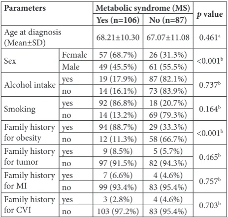
the group without MS (88.7% versus 11.3%; p<0.001). The mean age at diagnosis, alcohol intake and smok-ing, medical history for tumors and cardiovascular dis-eases did not show statistically significant differences between the groups with and without MS (Table 1).
The relationship between the degree of tumor differentiation, Dukes’ and Astler-Coller staging and the presence of MS are given in Table 2. The majority of CRC patients with (58.5%) and without (60.9%) MS had a G2 degree of tumor differentiation. The observed difference was not statistically significant (p=0.762). We observed that the half of the CRC patients with MS had a C stage of Dukes’ classifica-tion. On the other hand, an equal number of patients without MS were in Dukes’ stages B (46.0%) and C (46.0%). Additionally, the observed differences were not statistically significant (p=0.565). Astler-Coller classification revealed that more than half of CRC patients with MS were at stage C (50.9%). On the other hand, the majority of CRC patients without MS (48.0%) were at stage B of the Astler-Coller classifi-cation. The observed difference was not statistically significant (p=0.842) (Table 2).
The characteristics of MS in CRC patients before and after surgery
The characteristics of MS among the patients with CRC in the analyzed periods are given in Table 3. We observed a significant decrease in the number of pa-tients with MS after three months postoperatively as compared to the number of patients in the preopera-tive period (106 versus 81; p<0.001). The values of the anthropometric measures (BMI and waist circum-ference) were significantly decreased after treatment compared to the preoperative period in patients with MS (p<0.001, p<0.001, respectively). The levels of blood glucose and triglycerides were significantly de-creased, while the levels of HDL-C were significantly increased at the postoperative evaluation compared to the pretreatment status (p<0.001, p<0.001, p<0.001, respectively). Additionally, the values of systolic blood pressure were significantly decreased three months after surgery compared to the values before treatment (p=0.004). Statistical analysis did not show a signifi-cant difference in the change of diastolic blood pres-sure values among the analyzed periods in patients with MS (Table 3).
Levels of inflammatory and tumor markers in CRC patients before and after surgery
The changes in tumor and inflammation marker levels in CRC patients with and without MS in the analyzed periods are presented in Table 4. The values
Table 1. Sociodemographic characteristics of patients diagnosed with CRC.
Parameters Metabolic syndrome (MS) p value
Yes (n=106) No (n=87) Age at diagnosis
(Mean±SD) 68.21±10.30 67.07±11.08 0.461a
Sex Female 57 (68.7%) 26 (31.3%) <0.001b
Male 49 (45.5%) 61 (55.5%)
Alcohol intake yes 19 (17.9%) 87 (82.1%) 0.737b
no 14 (16.1%) 73 (83.9%)
Smoking yes 92 (86.8%) 18 (20.7%) 0.164b
no 14 (13.2%) 69 (79.3%)
Family history
for obesity yesno 94 (88.7%)12 (11.3%) 29 (33.3%)58 (66.7%) <0.001b Family history
for tumor yesno 97 (91.5%)9 (8.5%) 82 (94.3%)5 (5.7%) 0.465b Family history
for MI yesno 99 (93.4%)7 (6.6%) 83 (95.4%)4 (4.6%) 0.757b Family history
for CVI yesno 103 (97.2%) 83 (95.4%)3 (2.8%) 4 (4.6%) 0.703b
aStudent t-test; bChi-square test; Mn – mean, SD – standard deviation, n –
number of cases, MI – myocardial infarction, CVI – cerebrovascular insult
Table 2. The relationship between the degree of tumor differen-tiation, Dukes’ and Astler-Coller scales and the presence of MS.
Metabolic syndrome (MS) p value
Yes n (%) No n (%)
Degree of tumor differentiation
G1 35 (33.0%) 29 (33.3%)
0.762a G2 62 (58.5%) 53 (60.9%)
G3 9 (8.5%) 5 (5.7%)
Dukes’ classification of tumor
A 2 (1.9%) 5 (5.7%)
0.565a B 48 (45.3%) 40 (46.0%)
C 53 (50.0%) 40 (46.0%)
D 3 (2.8%) 2 (2.3%)
Astler-Coller classification of tumor
A 2 (1.9%) 3 (3.4%)
0.842a B 47 (44.3%) 42 (48.3%)
C 54 (50.9%) 40 (46.0%)
D 3 (2.8%) 2 (2.3%)
aChi-square test; G1 − well differentiated, G2 − moderately
of CRP were not significantly different between the CRC patients with MS compared to patients with-out MS in the preoperative period (15.73±32.70 ver-sus 8.21±16.38; p=0.204). On the other hand, three months after surgery, the CRP level was significantly decreased in patients without MS compared to pa-tients with MS (p<0.013).
The values of AFP and CEA were not significantly different among the CRC patients with regard to MS presence at the time of diagnosis and three months postoperatively (Table 4). Although the CA 19-9 concentrations were below the cut-off value in both groups (<39 U/mL), they were significantly higher in CRC patients with MS compared to patients
with-out MS in both analyzed periods (21.37±13.54 vs. 17.03±10.15, 29.82±19.36 vs. 25.22±23.35; p=0.046, p=0.005, respectively).
The trend of tumor and inflammation marker lev-els in CRC patients with MS pre- and postoperatively are shown in Table 5. The CRP level was significantly lower three months after surgery compared to the pre-operative level (p<0.001). Similarly, the AFP value was significantly lower postoperatively (p<0.001). On the other hand, values of CEA and CA 19-9 were signifi-cantly higher three months after treatment (p<0.001, p<0.001, respectively).
DISCUSSION
MS represents one of the major health problems worldwide with a growing incidence over time. This is mainly due to negative changes in life habits, such as an unbalanced diet and a lack of physical activity [21-23]. Additionally, these factors are recognized to be responsible for CRC development [24]. A previous study showed that regular physical exercise leads to a decrease in the presence of CRC [25].
The results of this study did not show a correla-tion between positive family history for myocardial in-farction (MI), cerebrovascular insult (CVI) or tumors with the presence of CRC. Only obesity was signifi-cantly more prevalent among CRC patients’ families, regardless of their MS status. In the twenty-first cen-tury, obesity is a disease of pandemic proportions that predominantly affects the developed world, but with increasing occurrence in third world countries [26]. Abdominal obesity is recognized as an independent
Table 3. Characteristics of the MS in patients with colorectal can-cer in pre- and postoperative periods.
Parameters
Metabolic syndrome (MS)
p value Preoperative
period (n=106) (Mn±SD)
Postoperative period (n=81)
(Mn±SD)
BMI (kg/m2) 25.68±3.64 24.74±2.95 <0.001a Waist
circumference (cm) 89.70±13.47 87.88±12.48 <0.001a Triglycerides
(mmol/L) 1.43±0.35 1.39±0.67 <0.001b
HDL (mmol/L) 1.44±0.33 1.8±0.32 <0,001a
Systolic BP
(mmHg) 134.96±19.40 133.87±19.13 0.004a
Diastolic BP
(mmHg) 81.22±10.53 81.40±9.99 0.401a
Blood glucose
(mmol/L) 6.86±2.86 5.48±1.46 <0.001a
aStudent t-test; bWilcoxon signed rank test; Mn − mean, SD − standard
deviation, n − number of cases, BMI − body mass index, HDL − high density lipoproteins, BP − blood pressure
Table 4. Pre- and postoperative levels of tumor and inflammation markers in colorectal cancer patients with and without MS.
Parameters Metabolic syndrome (MS) Before surgery Analyzed periods
(Mn±SD) P value 3 months postoperative(Mn±SD) P value
CEA (ng/mL) Yes 4.68±3.70 p=0.581a 5.00±5.80 p=0.435a
No 4.31±1.81 4.66±5.68
AFP (IU/mL) Yes 10.29±9.74 p=0.294a 5.95±7.46 p=0.377a
No 9.08±8.31 5.71±9.26
CA 19-9 (U/mL) Yes 21.37±13.54 p=0.046a 29.82±19.36 p=0.005a
No 17.03±10.51 25.22±23.35
CRP (mg/L) Yes 15.73±32.70 p=0.204a 14.91±31.31 p=0.013a
No 8.21±16.38 5.82±11.93
aMann-Whitney U test; Mn − mean, SD − standard deviation, n − number of cases, AFP − α-fetoprotein, CA 19-9 − cancer antigen 19-9, CEA −
risk factor for atherogenic and metabolic abnormali-ties in youth [27]. Furthermore, recent studies found an association between excessive fatness and the risk for MS and CRC [28].
At the time of CRC diagnosis, slightly more than half of our patients were also diagnosed with MS. Al-though there were more men with CRC, a significantly larger number of women met the criteria for MS. Three months after surgical treatment we found the percent-age of patients with three or more MS characteristics significantly decreased. Two earlier studies showed an increased risk of CRC mortality with an increase in MS features compared to their sole influence, suggesting their possible additive or synergistic effect [7,8]. Ad-ditionally, this study analyzed the potential effect of MS on the degree of tumor differentiation and level of tumor stage at the time of CRC diagnosis. Our re-sults showed no significant correlation between these investigated tumor parameters and the presence of MS. Such findings suggest that MS does not contribute to CRC invasiveness and the degree of differentiation.
Our results showed that the concentration of TG, HDL-C and blood pressure values were significantly different pre- and postoperatively. All of them showed significant amelioration after surgical treatment. Two earlier studies showed the correlation between un-regulated blood pressure and CRC [7,29]. Grossman et al. [30] reported that hypertension increased the risk of overall cancer mortality by 23%. Several studies showed a significant link between TG level and adeno-ma development, predominantly in Asian population [31-33]. In addition, Bayerdorffer et al. [34] found a 2- to 3-fold increased risk for CRC development in patients with low HDL-C concentration. Pooled
results of a meta-analysis [9] have shown that high concentrations of TG and low HDL-C, independently present a lower risk for CRC in comparison with MS. Other characteristics of the MS (increased blood glucose concentration, BMI and waist circumference) in our study were significantly increased before treat-ment. Esposito et al. [9] showed that individuals with hyperglycemia and/or a pathological waist circumfer-ence should be more closely observed during CRC screening. Also, our results are in accordance with pre-vious studies showing the possible role of increased glu-cose levels and insulin resistance in CRC pathogenesis [9,35]. Trevisan et al. [8] reported that, besides other components of MS, only increased glucose concentra-tions were associated with an increased risk of death in CRC patients. Other studies showed that elevated fasting insulin levels in patients without diabetes mel-litus are significantly and independently associated with the development of a different type of cancers, including CRC [36-39]. There are several mechanisms whereby insulin resistance may be responsible for CRC development: the ability of insulin to translocate the Ras protein on the cell membrane, the susceptibility of tumor cells to the growth effects of insulin in coopera-tion with insulin-like growth factor 1 (IGF-1) inhibi-tion of the synthesis of sex hormone-binding globulin (SHBG), which leads to increased bioavailability of androgens and estrogens, hormones that play a role in the pathogenesis of CRC [39,40]. Cancer cells have high demands for glucose and are recognized as cells with an accelerated metabolic state. Hyperglycemia leads to an environment of excess energy in the body, which promotes the proliferation of tumor cells [5]. Further-more, elevated glucose levels promote the formation of reactive oxygen species (ROS), which can damage DNA and thus lead to cancer development [41]. Additionally, it is suggested that hypertriglyceridemia may have an influence on excessive ROS production and thereby have its part in promoting carcinogenesis [42].
Calle et al. [40] described increased BMI (>25 kg/ m2) as being responsible for higher mortality rates in
many types of cancers, including CRC. BMI as a clini-cal feature has two imperfections: an inability to dis-tinguish between fat and lean body mass, and ignoring body fat distribution [43]. On the other hand, waist circumference is a direct reflection of the amount of
Table 5. Changes in tumor and inflammation marker levels in colorectal cancer patients with MS before and after treatment.
Parameters
Metabolic syndrome (MS)
P value Before surgery
(n=106) (Mn±SD)
3 months postoperative
(n=81) (Mn±SD)
CEA (ng/mL) 4.52±3.00 4.84±5.73 <0,001a
AFP (IU/mL) 9.74±9.12 5.77±8.49 <0,001a
CA 19-9 (U/mL) 19.41±12.29 27.43±22.14 <0,001a
CRP (mg/L) 15.36±32.00 6.79±13.93 <0.001a
aWilcoxon signed rank test; Mn − mean, SD − standard deviation, n −
abdominal obesity (a reflection of visceral fat distri-bution). The European Prospective Investigation into Cancer and Nutrition (EPIC) has proposed that ab-dominal obesity (measured as waist circumference or the waist-to-hip ratio) is an equally strong risk factor for colon cancer for both sexes [44].
Previous studies have shown abdominal or central obesity to be responsible for higher risk in colorectal adenoma development [45,46]. Visceral adipose tissue, which is physiologically more active than peripheral subcutaneous fat, is recognized as an endocrine tissue, leading to hormone and cytokine production with in-flammatory and metabolic potential [47]. Proinflam-matory cytokines, tumor necrosis factor α (TNF-α), interleukin 6 (IL-6) and adiponectin, which are pro-duced in visceral adipose tissue, promote the devel-opment of chronic subclinical inflammation [48]. In obese individuals, it provides an environment which favors the development of MS by inflammation [49]. TNF-α and IL-6 induce the liver to produce an acute-phase protein, the C-reactive protein (CRP) [50]. A link between colonic areas with chronic subclinical in-flammation and sporadic colorectal neoplasia has been suggested. Itzkowitz et al. [51] reported the evidence for chronic inflammation as the main predisposing factor for CRC in inflammatory bowel disease (IBD). Our findings showed a 2.26-fold decrease in CRP three months postoperatively. These results, together with the reduction in the number of patients with MS and significantly decreased BMI (by 1 kg/m2, respectively),
are in agreement with previous studies indicating that weight loss leads to the suppression of inflammatory processes. In addition, our results suggest that the pres-ence of CRC provokes the inflammatory response and consequently leads to an increment of CRP values. This is supported by the lower CRP levels and changes in colorectal mucosa [52,53].
Currently, there are a large number of tumor markers used for the early detection, diagnosis and prognosis of CRC, as well as prediction of the efficacy of different therapeutic protocols, survival rate and disease relapse. To date, none of the tumor biomark-ers shows enough specificity and sensitivity for CRC screening [14]. CEA is one of the most used tumor markers in CRC screening. Its specificity for diagnosis is shown to be around 90% and sensitivity between 40% and 75% [15]. However, the use of CEA has some
limitations. It could be produced by other malignant and benign epithelial tumors, as well as some inflam-matory processes, such as IBDs, pancreatitis, lung infections and liver diseases [54]. Herrera et al. [55] showed that a high preoperative level of CEA is an indicator of advanced stage and low differentiation grade of a tumor.
CA 19-9 was first recognized in 1979 as an ad-ditional marker for CRC follow-up [56]. Previous in-vestigations revealed the significantly lower specificity and sensitivity of CA 19-9 compared to CEA [57]. Unlike CEA, CA 19-9 is significantly lower in smok-ers [18]. Also, its values could be increased in patients with poorly controlled diabetes mellitus, regardless of tumor presence [19]. Unlike CEA and CA 19-9, the AFP biomarker is not commonly used in CRC screen-ing. Normally, it is produced during the fetal period by the liver and yolk sac [58]. It is mostly used for the diagnosis of hepatocellular cancer, hepatoblastoma and yolk sac tumors [59].
Our results showed a significant decrease in AFP values three months after treatment in patients with MS. On the other hand, CEA and CA 19-9 concen-trations were significantly higher after the treatment. This could be due to the rest of the tumor, undetect-able metastasis or some other inflammatory process which took place at the time of blood sampling post-operatively. We did not include some of the radiologi-cal imaging techniques that could give a more com-prehensive explanation of the patients’ postoperatively status. According to our results, pre- and postopera-tively, MS presence did not additionally influence AFP and CEA levels. On the other hand, even if its con-centrations were below the cut-off value, CA 19-9 was significantly higher in the MS group in both analyzed periods. Such findings might suggest the possible in-fluence of metabolic changes on CA 19-9 production. To date, there is no research regarding follow-up in tumor marker level changes pre- and postoperatively in CRC patients in the presence of MS. Future studies are needed to evaluate the correlation of tumor mark-ers and MS within CRC screening programs.
Authors’ contribution: All authors participated in all of the stages of manuscript preparation.
REFERENCES
1. Grundy SM, Cleeman JI, Daniels SR, Donato KA, Eckel RH, Franklin BA, Gordon DJ, Krauss RM, Savage PJ, Smith SC Jr, Spertus JA, Costa F. Diagnosis and management of the meta-bolic syndrome: An American Heart Association/National Heart, Lung, and Blood Institute scientific statement. Curr Opin Cardiol. 2006;21(1):1-6.
2. Parapid B, Ostojic MC, Lalic NM, Micic D, Damjanovic S, Bubanja D, Simic D, Lalic K, Polovina S, Marinkovic J, Milic NM. Risk factors clustering within the metabolic syndrome: a pattern or by chance? Hellenic J Cardiol. 2014;55(2):92-100.
3. DeFronzo RA, Ferrannini E. Insulin resistance. A multifac-eted syndrome responsible for NIDDM, obesity, hyperten-sion, dyslipidemia, and atherosclerotic cardiovascular dis-ease. Diabetes Care. 1991;14(3):173-94.
4. Lindsay RS Howard BV. Cardiovascular risk associated with the metabolic syndrome. Curr Diab Rep. 2004;4(1):63-8. 5. Cowey S, Hardy RW. The metabolic syndrome: A high-risk
state for cancer? Am J Pathol. 2006;169(5):1505-22. 6. Mendonça FM, de Sousa FR, Barbosa AL, Martins SC,
Araújo RL, Soares R, Abreu C. Metabolic syndrome and risk of cancer: which link? Metabolism. 2015;64(2):182-9. 7. Colangelo LA, Gapstur SM, Gann PH, Dyer AR, Liu K.
Colorectal cancer mortality and factors related to the insu-lin resistance syndrome. Cancer Epidemiol Biomarkers Prev. 2002;11(4):385-91.
8. Trevisan M, Liu J, Muti P, Misciagna G, Menotti A, Fucci F; Risk Factors and Life Expectancy Research Group. Markers of insulin resistance and colorectal cancer mortality. Cancer Epidemiol Biomarkers Prev. 2001;10(9):937-41.
9. Esposito K, Chiodini P, Capuano A, Bellastella G, Maiorino MI, Rafaniello C, Panagiotakos DB, Giugliano D. Colorectal cancer association with metabolic syndrome and its compo-nents: a systematic review with meta-analysis. Endocrine. 2013;44(3):634-47.
10. Jinjuvadia R, Lohia P, Jinjuvadia C, Montoya S, Liangpun-sakul S. The association between metabolic syndrome and colorectal neoplasm: systemic review and meta-analysis. J Clin Gastroenterol 2013;47(1):33-44.
11. Ferlay J, Soerjomataram I, Dikshit R, Eser S, Mathers C, Rebelo M, Parkin DM, Forman D, Bray F Cancer incidence and mortality worldwide: sources, methods and major patterns in GLOBOCAN 2012. Int J Cancer. 2015;136(5):E359-E386.
12. Krivokapić Z, Ćeranić M, Šaranović Đ, Stanojević G, Tripković I, Bulajić M, Galun D, Radosavljević D, Brebe-rina M. Nacionalni vodič dobre kliničke prakse za dijagnos-tikovanje i lečenje raka kolona i rektuma. Beograd: Agencija za akreditaciju zdravstvenih ustanova Srbije; 2012. Available from: http://www.zdravlje.gov.rs/showpage.php?id=145. 13. Yang DX, Gross CP, Soulos PR, Yu JB. Estimating the
mag-nitude of colorectal cancers prevented during the era of screening: 1976 to 2009. Cancer. 2014;120(18):2893-901. 14. Newton KF, Newman W, Hill J. Review of biomarkers in
colorectal cancer. Colorectal Dis. 2012;14(1):3-17.
15. Duffy MJ, van Dalen A, Haglund C, Hansson L, Klapdor R, Lamerz R, Nilsson O, Sturgeon C, Topolcan O. Clinical
utility of biochemical markers in colorectal cancer: Euro-pean Group on Tumour Markers (EGTM) guidelines. Eur J Cancer. 2003;39(6):718-27.
16. Tarantino I, Warschkow R, Schmied BM, Güller U, Mieth M, Cerny T, Büchler MW, Ulrich A. Predictive Value of CEA for Survival in Stage I Rectal Cancer: a Population-Based Propensity Score-Matched Analysis. J Gastrointest Surg. 2016;20(6):1213-22.
17. Li YH, An X, Xiang XJ, Wang ZQ, Wang FH, Feng F, Jiang WQ, He YJ, Xu RH. Clinical significance of a transient increase in carcinoembryonic antigen and carbohydrate antigen 19-9 in patients with metastatic colorectal cancer receiving chemotherapy. Ai Zheng. 2009;28(9):939-44. 18. Sajid KM, Parveen R, Durr-e-Sabih RM, Chaouachi K,
Naeem A, Mahmood R, Shamim R. Carcinoembryonic anti-gen (CEA) levels in hookah smokers, cigarette smokers and non-smokers. J Pak Med Assoc. 2007;57(12):595-9. 19. Nakamura N, Aoji O, Yoshikawa T, Mori K, Kajiyama S,
Kitagawa Y, Kanatsuna T, Kondo M. Elevated serum CA19-9 levels in poorly controlled diabetic patients. Jpn J Med. 1986;25(3):278-80.
20. Lorenzo C, Williams K, Hunt JK, Haffner MS. The National Cholesterol Education Program-Adult Treatment Panel III, International Diabetes Federation, and World Health Orga-nization Definitions of the Metabolic Syndrome as Predic-tors of Incident Cardiovascular Disease and Diabetes. Dia-betes Care. 2007;30(1):8-13.
21. Souza MR, Diniz M, Medeiros-Filho JE, Araujo MS. Meta-bolic syndrome and risk factors for nonalcoholic fatty liver disease. Arq Gastroenterol. 2012;49(1):89-96.
22. Savva SC, Lamnisos D, Kafatos AG. Predicting cardiometa-bolic risk: waist-to-height ratio or BMI. A meta-analysis. Diabetes Metab Syndr Obes. 2013;6:403-19.
23. Yamada T, Hara K, Kadowaki T. Chewing betel quid and the risk of metabolic disease, cardiovascular disease, and all-cause mortality: a meta-analysis. PLoS One. 2013; 8(8):e70679.
24. American Cancer Society. Cancer Facts & Figures 2012. Atlanta: American Cancer Society; 2012.
25. Friedenreich CM. Physical activity and cancer prevention: from observational to intervention research. Cancer Epide-miol Biomarkers Prev. 2001;10(4):287-301.
26. Ogden C, Carroll M, Kit B, Flegal K. Prevalence of obesity and trends in body mass index among US children and ado-lescents, 1999-2010. JAMA. 2012;307(5):483-90.
27. Sowers JR. Obesity as a cardiovascular risk factor. Am J Med. 2003;115(Suppl 8A):37S-41S.
28. Kim JY, Jung YS, Park JH, Kim HJ, Cho YK, Sohn CI, Jeon WK, Kim BI, Choi KY, Park DI. Different risk factors for advanced colorectal neoplasm in young adults. World J Gas-troenterol. 2016;22(13):3611-20.
29. Ahmed RL, Schmitz KH, Anderson KE, Rosamond WD, Folsom AR. The metabolic syndrome and risk of incident colorectal cancer. Cancer. 2006;107(1):28-36.
31. Park SK, Joo JS, Kim DH, Kim YE, Kang D, Yoo KY. Associa-tion of serum lipids and glucose with the risk of colorectal adenomatous polyp in men: a case-control study in Korea. J Korean Med Sci. 2000;15(6):690-5.
32. Tabuchi M, Kitayama J, Nagawa H. Hypertriglyceridemia is positively correlated with the development of colorectal tubular adenoma in Japanese men. World J Gastroenterol. 2006;12(8):1261-4.
33. Bird CL, Ingles SA, Frankl HD, Lee ER, Longnecker MP, Haile RW. Serum lipids and adenomas of the left colon and rectum. Cancer Epidemiol Biomarkers Prev. 1996;5(8):607-12. 34. Bayerdorffer E, Mannes GA, Richter WO, Ochsenkühn
T, Seeholzer G, Köpcke W, Wiebecke B, Paumgartner G. Decreased high-density lipoprotein cholesterol and increased low-density cholesterol levels in patients with colorectal adenomas. Ann Intern Med. 1993;118(7):481-7. 35. Larsson SC, Orsini N, Wolk A. Diabetes mellitus and risk
of colorectal cancer: a meta-analysis. J Natl Cancer Inst. 2005;97(22):1679-87.
36. Berstein LM, Kvatchevskaya JO, Poroshina TE, Kovalenko IG, Tsyrlina EV, Zimarina TS, Ourmantcheeva AF, Ashra-fian L, Thijssen JH. Insulin resistance, its consequences for the clinical course of the disease, and possibilities of cor-rection in endometrial cancer. J Cancer Res Clin Oncol. 2004;130(11):687-93.
37. Giovannucci E. Insulin, insulin-like growth factors and colon cancer: a review of the evidence. J Nutr. 2001;131(11Suppl):3109S-3120S.
38. Goodwin PJ, Ennis M, Pritchard KI, Trudeau ME, Koo J, Madarnas Y, Hartwick W, Hoffman B, Hood N. Fasting insulin and outcome in early-stage breast cancer: results of a prospective cohort study. J Clin Oncol. 2002;20(1):42-51. 39. Hammarsten J, Hogstedt B. Clinical, haemodynamic,
anthropometric, metabolic and insulin profile of men with high-stage and high-grade clinical prostate cancer. Blood Press. 2004;13(1):47-55.
40. Calle EE, Rodriguez C, Walker-Thurmond K, Thun MJ. Overweight, obesity, and mortality from cancer in a pro-spectively studied cohort of U.S. adults. N Engl J Med. 2003;348(17):1625-38.
41. Valko M, Izakovic M, Mazur M, Rhodes CJ, Telser J. Role of oxygen radicals in DNA damage and cancer incidence. Mol Cell Biochem. 2004;266(1-2):37-56.
42. Cejas P, Casado E, Belda-Iniesta C, De Castro J, Espinosa E, Redondo A, Sereno M, Garcia-Cabezas MA, Vara JA, Dominguez-Caceres A, Perona R, Gonzalez-Baron M: Implications of oxidative stress and cell membrane lipid peroxidation in human cancer. Cancer Causes Control. 2004;15(7):707-19.
43. Okorodudu DO, Jumean MF, Montori VM, Romero-Corral A, Somers VK, Erwin PJ, Lopez-Jimenez F. Diagnostic per-formance of body mass index to identify obesity as defined by body adiposity: a systematic review and meta-analysis. Int J Obes (Lond). 2010; 34(5):791-9.
44. Pischon T, Lahmann PH, Boeing H, Friedenreich C, Norat T, Tjonneland A, Halkjaer J, Overvad K, Clavel-Chapelon F, Boutron-Ruault MC, Guernec G, Bergmann MM, Lin-seisen J, Becker N, Trichopoulou A, Trichopoulos D, Sieri S, Palli D, Tumino R, Vineis P, Panico S, Peeters PH,
Bueno-de-Mesquita HB, Boshuizen HC, Van Guelpen B, Palmqvist R, Berglund G, Gonzalez CA, Dorronsoro M, Barricarte A, Navarro C, Martinez C, Quirós JR, Roddam A, Allen N, Bingham S, Khaw KT, Ferrari P, Kaaks R, Slimani N, Riboli E. Body size and risk of colon and rectal cancer in the Euro-pean Prospective Investigation Into Cancer and Nutrition (EPIC). J Natl Cancer Inst. 2006; 98(13):920-31.
45. Kim BC, Shin A, Hong CW, Sohn DK, Han KS, Ryu KH, Park BJ, Nam JH, Park JW, Chang HJ, Choi HS, Kim J, Oh JH. Association of colorectal adenoma with compo-nents of metabolic syndrome. Cancer Causes Control. 2012;23(5):727-35.
46. Liu CS, Hsu HS, Li CI, Jan CI, Li TC, Lin WY, Lin T, Chen YC, Lee CC, Lin CC. Central obesity and atherogenic dyslip-idemia in metabolic syndrome are associated with increased risk for colorectal adenoma in a Chinese population. BMC Gastroenterol. 2010;10:51.
47. Galic S, Oakhill JS, Steinberg GR. Adipose tissue as an endo-crine organ. Mol Cell Endocrinol. 2010;316(2):129-39. 48. Trayhurn P, Beattie JH. Physiological role of adipose tissue:
white adipose tissue as an endocrine and secretory organ. Proc Nutr Soc. 2001;60(3):329-39.
49. Wisse BE. The inflammatory syndrome: the role of adipose tissue cytokines in metabolic disorders linked to obesity. J Am Soc Nephrol. 2004;15(11):2792-800.
50. Roberts DL, Dive C, Renehan AG. Biological mechanisms linking obesity and cancer risk: new perspectives. Annu Rev Med. 2010;61:301-16.
51. Itzkowitz SH. Cancer prevention in patients with inflam-matory bowel disease. Gastroenterol Clin North Am. 2002;31(4):1133-44.
52. Fayh AP, Lopes AL, da Silva AM, Reischak-Oliveira A, Friedman R. Effects of 5% weight loss through diet or diet plus exercise on cardiovascular parameters of obese: a ran-domized clinical trial. Eur J Nutr. 2013;52(5):1443-50. 53. Pendyala S, Neff LM, Suarez-Farinas M, Holt PR.
Diet-induced weight loss reduces colorectal inflammation: implications for colorectal carcinogenesis. Am J Clin Nutr. 2011;93(2):234-42.
54. Rockall TA, McDonald PJ. Carcinoembryonic antigen: its value in the follow-up of patients with colorectal cancer. Int J Colorectal Dis. 1999;14(1):73-7.
55. Herrera MA, Chu TM, Holyoke ED. Carcinoembryonic antigen (CEA) as a prognostic and monitoring test in clini-cally complete resection of colorectal carcinoma. Ann Surg. 1976;183(1):5-9.
56. Koprowski H, Steplewski Z, Mitchell K, Herlyn M, Herlyn D, Fuhrer P. Colorectal carcinoma antigens detected by hybrid-oma antibodies. Shybrid-omatic Cell Genet. 1979;5(6):957-71. 57. Filella X, Molina R, Grau JJ, Pique M, Garcia-Valdecasas JC,
Astudillo E, Biete A, Bordas JM, Novell A, Campo E, Ballesta AM. Prognostic value of CA 19.9 levels in colorectal cancer. Ann Surg. 1992;216(1):55-9.
58. Abelev GI, Perova SD, Khramkova NI, Postnikova ZA, Irlin IS. Production of embryonal alpha-globulin by transplant-able mouse hepatomas. Transplantation. 1963;1:174-80. 59. McIntire KR, Waldmann TA, Moertel CG, Go VL. Serum