LOWBUSH BLUEBERRY EXTRACT PROTECTS NORMAL
HUMAN (HOMO SAPIENS L.) EPIDERMAL KERATINOCYTES
FROM DAMAGE BY UV-B IRRADIATION
Seiji Yamasaki,
[a]*Kenji Mizoguchi,
[a]Naohiro Kodama
[a]and Joji Iseki
[a]Keywords: flavonoids, normal human epidermal keratinocytes (NHEKs), polyphenols, proanthocyanidins, ultraviolet-B (UV-B).
To search for natural materials that protect human skin from damage caused by ultraviolet-B (UV-B) irradiation, we prepared water extracts of the following four dry berries: lowbush blueberry (LBB) (Vaccinium angustifolium L.), highbush blueberry (HBB) (Vaccinium corymbosum L.), cranberry (CB) (Vaccinium macrocarpon Ait.) and grape (Vitis vinifera L.). Normal human epidermal keratinocytes (NHEKs) were pretreated with each of the extracts and then subjected to transient UV-B irradiation (15.0 W m-2) for 80 s. The results showed that pretreatment of NHEKs with LBB extract significantly attenuated UV-B-induced damage. The concentrations of proanthocyanidins, total flavonoids and total polyphenols in LBB extract were higher than those in the other three berry extracts. Therefore, these compounds might mediate the protection of NHEKs from UV-B-induced damage.
* Corresponding Author Tel: +81-940-35-1386 Fax: +81-940-35-1386
E-Mail: [email protected]
[a] Laboratory of Plant Physiology, Department of Science Education, Faculty of Education, University of Teacher Education Fukuoka, 1-1 Akamabunkyomachi, Munakata, Fukuoka 811-4192, Japan
INTRODUCTION
Chlorofluorocarbons cause continual depletion of the stratospheric ozone layer, which increases the amount of solar ultraviolet-B (UV-B; 280−320 nm) irradiation [UV-B (+)] passing through to the earth’s surface.1 UV-B (+) is harmful to living organisms. UV-B is the chief cause of skin reddening and sunburn, which damages the skin’s more superficial epidermal layers. Direct absorption of UV-B by DNA induces mutations and leads to the development of carcinomas in cutaneous spinous cells.2,3 Protection from UV-B (+) is crucial and is primarily achieved by the application of topical sunscreen treatments. Sunscreen contains UV-light absorbers and UV-light scattering agents. UV-light absorbers are usually phenolic compounds found in various oils whereas UV-light scattering agents are minerals such as titanium oxide or zinc oxide. However, these components sometimes cause allergies. Thus, it is essential to identify natural and safe compounds that do not cause allergies and that still protect skin from UV-B-mediated damage.
Red and purple berries, such as lowbush blueberry (LBB) (Vaccinium angustifolium L.), highbush blueberry (HBB) (Vaccinium corymbosum L.), cranberry (CB) (Vaccinium macrocarpon Ait.) and grape (Vitis vinifera L.) provide a readily available source of dietary phenolic compounds. Berries also contain many flavonoids.4-6 A flavonoid fraction from CB extract inhibits the proliferation of human tumor cell lines.7 Flavonoids from LBB and CB inhibit matrix metalloproteinases (MMPs), which degrade the extracellular cell matrix (ECM) during cancer metastasis8,9 and in human prostate cancer cells.10,11 Monomeric flavonoids that form oligomers and polymers are called
proanthocyanidins or condensed tannins. Feeding proanthocyanidins to hairless mice reduced UV-B-induced skin carcinogenesis.12 These combined results suggest that flavonoids and proanthocyanidins have beneficial dietary activities that promote health. However, the full effects of these berry compounds have not been elucidated. Plant flavonoids absorb UV irradiation;13-15 therefore, berry extracts may absorb UV irradiation when added to a topical treatment. Our aim is to determine whether the addition of berry extracts to topical treatments could protect human skin from UV-B (+).
Although water extracts of living tissues can result in low recovery rates, they are simple to prepare, easy to use and provide a safe and biologically compatible matrix for bioactive compounds. We previously prepared water extracts from the following four dry berries: LBB, HBB, CB and grape (designated as the ‘berry extract’ in the present study). We showed that these four berry extracts efficiently absorb UV radiation, primarily UV-B.16 Pretreatment of cucumber (Cucumis sativus L.) cotyledons with LBB, HBB and CB extracts attenuates UV-B-induced damage without causing any side effects.16 We used high-performance liquid chromatography (HPLC) to analyze 51 polyphenols in the four berry extracts. The LBB extract contained primarily chlorogenic acid, caffeic acid and protocatechuic acid; the HBB extract contained primarily chlorogenic acid, caffeic acid and syringic acid; and the CB extract contained primarily protocatechic acid, myricetin and p-coumaric acid.16 We proposed that these compounds contributed to the protection of cucumber plants against UV-B-induced damage.16
Similar to the UV-B protective effects of LBB, HBB and CB extracts in cucumber cotyledons, pretreatment of human skin with these extracts might also offer protection against UV-B-induced damage without any side effects. With the aim of developing a safe sunscreen for use in humans in the present study, we investigated whether the four berry extracts could protect human cells from UV-B-induced damage. First, we pretreated normal human (Homo sapiens
NHEK survival. Second, we performed a spectrophotometric analysis to determine the concentrations of proanthocyanidins, total flavonoids and total polyphenols in the four berry extracts. Finally, we discuss the potential defense mechanisms elicited by the berry extracts in NHEKs in response to UV-B (+).
EXPERIMENTAL
Preparation of berry extracts
Commercially produced dry fruits of lowbush blueberry (LBB) (Vaccinium angustifolium L.), highbush blueberry (HBB) (Vaccinium corymbosum L.), cranberry (CB) (Vaccinium macrocarpon Ait.) and grape (Vitis vinifera L.) were purchased from the local supermarket Aeon (Aeon Co., Ltd., Chiba, Japan) in Fukuoka prefecture on September 27, 2012 and July 11, 2013, and were used before their expiry dates. For each of the four species, 5 g dried berries were ground using a mortar and pestle at room temperature, and then 15 ml of distilled water (pH 7.5) was added. Samples were placed at 4 °C, and the ground tissue was extracted into distilled water for 14 h. Then, samples were centrifuged at 6,000 rpm for 15 min at 4 °C. The crude supernatant was transferred to a new tube and centrifuged at 12,000 rpm for 10 min at 4 °C, and the clear supernatant was collected. This clear supernatant is designated as the berry extract. The Brix value of the four berry extracts was measured with a pocket sugar content meter (APAL-J; Atago Co., Ltd., Tokyo, Japan). The Brix values of LBB, HBB, CB and grape extracts were 19.4 %, 18.7 %, 22.4 % and 21.6 %, respectively. The extraction efficiencies [(collected extract weight / dried fruit weight) Brix] of LBB, HBB, CB, and grape extracts were 45.7 %, 42.4 %, 43.25 and 56.1 %, respectively. The four berry extracts were stored at 4 °C during the experiment.
Analysis of berry extract dose-response effects on normal human epidermal keratinocytes (NHEKs) under normal light radiation [UV-B (−)]
The maximum amount of each berry extract that could be added to NHEKs (KK-4109; Kurabo Industries, Ltd., Osaka, Japan) without affecting cell viability under UV-B (−) was investigated by ACEL, Inc. (Kanagawa, Japan). NHEKs (2
104) were transferred to 100 µl keratinocyte basal medium (KBM, HuMedia-KG2; Kurabo Industries, Ltd., Osaka, Japan) in 32 wells of a 96-well culture plate (MS-8096F; Sumitomo Bakelite Co., Ltd., Tokyo, Japan), and cells were cultured in a dark humidified (5 % CO2 atmosphere) incubator for 24 h at 37 °C. Cell growth and proliferation were confirmed. Then, 100 µl KBM per culture well was replaced with 100 µl KBM supplemented with0 % (defined as a control in this study), 0.00064 %, 0.0032 %, 0.016 %, 0.08 %, 0.4 %, 2 % or 10 % (v/v) of each berry extract (n=4). One culture plate was used for each of the four berry extracts (a total of four culture plates were used for the experiment). The cells were cultured for 24 h at 37 °C before assessing cell viability using the Cell Count Reagent SF colorimetric assay (Nacalai Tesque, Inc., Kyoto, Japan). Briefly, 10 µl Cell Count Reagent SF was added to each well and incubated for 2 h at 37 °C. Then, cell viability was assessed colorimetrically by measuring the optical density at
450 nm (OD450) using a microplate reader (Precision Microplate Reader; Molecular Devices Co., Tokyo, Japan). In this assay, the relative number of viable cells was expressed as the change in OD450 over a period of 1 h (OD450 h-1).
Investigation of UV-B-induced lethality in NHEKs
The effect of transient UV-B irradiation [UV-B (+)] on NHEK survival was tested by ACEL, Inc. (Kanagawa, Japan). First, the time required for UV-B (+) to cause cell death in approximately 50 % of the NHEKs (LD50) was determined as follows. The NHEKs (2 104) were transferred to 100 µl KBM per well in 6 wells in the middle of a 96-well culture plate, and cells were cultured in a dark humidified (5 % CO2 atmosphere) incubator for 24 h at 37 °C. Cell growth and proliferation were confirmed. Then, the culture plates were placed 8 cm below a UV lamp (UVLMS-38; Funakoshi, Co., Ltd., Tokyo, Japan) with the following intensities of UV-A, UV-B and UV-C: 2.4, 15.0 and 1.7 W m-2, respectively. UV-A, UV-B and UV-C intensities were measured with a digital UV intensity meter (UVX radiometer; Funakoshi, Co., Ltd.) equipped with a sensor for UV-A (Radiometer Sensor UVX-36, 365 nm; Funakoshi, Co., Ltd.), UV-B (Radiometer Sensor UVX-31, 310 nm; Funakoshi, Co., Ltd.) or UV-C (Radiometer Sensor UVX-25, 254 nm; Funakoshi, Co., Ltd.). Thus, the UV lamp primarily radiates UV-B. This UV lamp was adopted as a transient emitter of UV-B (+) for the present study. The culture plates were irradiated with the UV lamp for 0, 20, 100, 200 or 1,000 s, and then cultured again in a dark humidified (5 % CO2 atmosphere) incubator for 24 h at 37 °C. Therefore, a total of five culture plates were needed for the study. Cell viability was evaluated after 24 h using the Cell Count Reagent SF colorimetric assay described in the previous section.
Effect of transient UV-B (+) on NHEKs that were pretreated with berry extracts
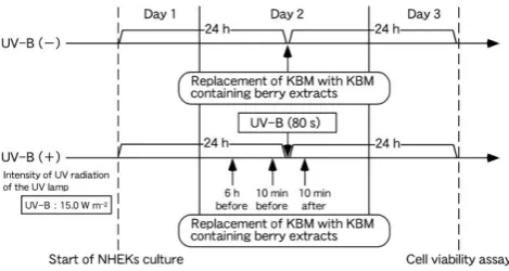
cultured in 100 µl KBM per well in 16 wells in the middle of a 96-well culture plate in a dark humidified (5 % CO2 atmosphere) incubator at 37 °C (Figure 1). Then, the culture medium in 4 wells was replaced with 100 µl KBM supplemented with berry extract at the maximum concentration that did not affect viability (as determined above) at each of three different times [6 h before, 10 min before and 10 min after transient UV-B (+)], and cells were again cultured under the same conditions (Figure 1). Therefore, a total of 12 wells were needed. The remaining four wells were used as controls. Because there were four berry extracts, a total of four culture plates were required. Cell viability was evaluated using the Cell Count Reagent SF colorimetric assay as described above.
Figure 1. Illustration of the experimental protocol for assessing the effects of UV-B (−) and transient UV-B (+) on the viability of NHEKs that were treated with berry extracts. Standard KBM medium was removed and replaced with KBM containing berry extracts 6 h before, 10 min before and 10 min after transient UV-B (+) for 80 s.
Quantitative spectrophotometric analysis of proanthocyanidin concentrations in berry extracts
Proanthocyanidin concentrations in the berry extracts were analyzed according to a modified method which was reported previously.17 Briefly, 1 ml berry extract was added to 4 ml of 0.07 % (w/v) iron (II) sulfate solution, and the reactions were incubated at 95 °C for 1 h. Then, 4 ml of 0.6 N hydrochloric acid:n-butanol (1:1, v/v) was added, and the sample absorbance was measured at 550 nm with ultraviolet and visible spectrophotometry (UVIDEC-4; Jasco Co., Tokyo, Japan). Delphinidin solution was used to prepare a standard calibration curve, and proanthocyanidin concentration was calculated with respect to the delphinidin calibration curve (µg µl-1). The proanthocyanidin concentration in each berry extract was expressed as the average of three replicates.
Quantitative spectrophotometric analysis of the total flavonoid concentration in berry extracts
The total flavonoid concentration was analyzed according to the method which was reported previously.18 Briefly, 1 ml berry extract was added to 4 ml distilled water and 0.3 ml of 5 % (w/v) sodium nitrite solution, and the reaction was allowed to stand for 5 min at room temperature. Then, 0.3 ml of 10 % (w/v) aluminum chloride solution was added and the tube was vortexed. After 1 min, 2 ml of 1.0 M sodium hydroxide solution was added, and the tube was vortexed
again. Then, 2.4 ml of distilled water was added, and the sample absorbance was measured at 510 nm with ultraviolet and visible spectrophotometry (UVIDEC-4; Jasco Co., Tokyo, Japan). A calibration curve was prepared using (+)-catechin, and the total flavonoid concentration was calculated with respect to the (+)-catechin calibration curve (µg µl-1). Total flavonoid concentration in each berry extract was expressed as the average of three replicates.
Quantitative spectrophotometric analysis of the total polyphenol concentration in berry extracts
The total polyphenol concentration was analyzed according to the method of Folin-Denis.19 Briefly, 50 µl berry extract was added to 4 ml distilled water and 1 ml phenol reagent (a 5-fold dilution of Folin-Ciocalteu reagent with distilled water). After vortexing, 1 ml of 10 % (w/v) sodium carbonate solution was added and the tube was vortexed again. Then, the samples were allowed to stand in the dark for 1 h at room temperature. Sample absorbance was measured at 760 nm with ultraviolet and visible spectrophotometry (UVIDEC-4; Jasco Co., Tokyo, Japan). A calibration curve was prepared using (+)-catechin solution, and the total polyphenol concentration was calculated with respect to the (+)-catechin calibration curve (µg µl-1). Total polyphenol concentration in each berry extract was expressed as the average of three replicates.
Statistical analysis
NHEK cell viability and the concentrations of proanthocyanidins, total flavonoids and total polyphenols in each berry extract were expressed as the mean ± standard error. Statistically significant differences were assessed by Dunnett’s test (http://www.gen-info.osaka-u.ac.jp/MEPHAS/dunnett.html, March 6, 2017).
RESULTS
Pretreatment of NHEKs with berry extract protects against transient UV-B-induced cell death.
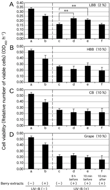
First, we determined the maximum concentrations of berry extracts (expressed as % of pure extract) that did not reduce NHEK cell viability more than 70 % of the value in control NHEKs under UV-B (−). The maximum concentrations of LBB, HBB, CB and grape extracts that did not affect NHEK cell viability were 2 %, 10 %, 10 % and 10 % (v/v), respectively. Because excessive dilution of the KBM media inhibits cell growth, the maximum percentage of extract that could be added to the cells was limited to 10 %. Second, the time for UV-B (+) that caused cell death in approximately 50 % of the NHEKs (LD50) was determined; this time was 80 s (Figure 2).
Figure 2. Effect of transient UV-B (+) at 15.0 W m-2 on NHEK viability. Values represent the average of six replicates (six wells in the culture plate).
The decline in cell viability of NHEKs treated with transient UV-B (+) (Figures 3Ac−Dc) was less than 50 % of that of NHEKs treated with UV-B (−) (Figures 3Aa−Da). Cell viability of NHEKs pretreated with 2 % LBB extract 6 h before transient UV-B (+) (Figure 3Ad) was higher than that of NHEKs treated with transient UV-B (+) (P < 0.01) (Figure 3Ac).
Cell viability of NHEKs pretreated with 2 % LBB extract 10 min before transient UV-B (+) (Figure 3Ae) was higher than that of NHEKs treated with transient UV-B (+) (P < 0.01) (Figure 3Ac). By contrast, cell viability of NHEKs treated with 2 % LBB extract 10 min after transient UV-B (+) (Figure 3Af) was not significantly different from that of NHEKs treated with transient UV-B (+) (Figure 3Ac).
Cell viability of NHEKs treated with 10 % HBB extract at 6 h and 10 min before (Figure 3Bd, Be) and 10 min after (Figure 3Bf) transient UV-B (+) was not higher than that of NHEKs treated only with transient UV-B (+) (Figure 3Bc). Similarly, cell viability of NHEKs treated with 10 % CB extract 6 h and 10 min before (Figure 3Cd, Ce) and 10 min after (Figure 3Cf) transient UV-B (+) was not more significant than that of NHEKs treated only with transient UV-B (+) (Figure 3Cc). Cell viability of NHEKs treated with 10 % grape extract 6 h and 10 min before (Figure 3Dd, De) and 10 min after (Figure 3Df) transient UV-B (+) was not higher than that of NHEKs treated only with transient UV-B (+) (Figure 3Dc). These results indicate that pretreatment of NHEKs with LBB extract protected the cells from UV-B-induced damage.
Spectrophotometric analysis of compounds in the berry extracts
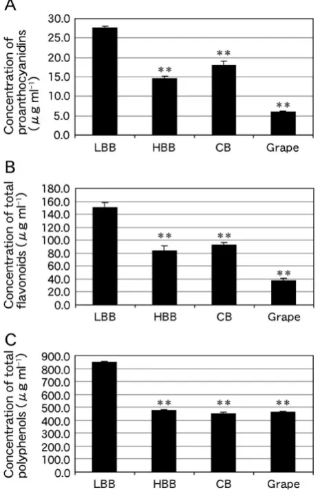
To evaluate the presence of compounds that absorb UV radiation in the berry extracts, the concentrations of proanthocyanidins, total flavonoids and total polyphenols were analyzed. The proanthocyanidin concentrations in LBB, HBB, CB and grape extracts were 27.7 ± 0.44, 14.5 ± 0.63, 18.0 ± 1.1 and 6.1 ± 0.2 µg ml-1, respectively (Figure 4A). Therefore, the proanthocyanidin concentration in LBB extract was 1.91-, 1.54- and 4.57-fold greater than those in HBB (P < 0.01), CB (P < 0.01) and grape (P < 0.01) extracts, respectively. The total flavonoid concentrations in LBB, HBB, CB and grape extracts were 150.8 ± 7.8, 83.2 ±
7.9, 92.4 ± 4.3 and 37.4 ± 3.5 µg ml-1, respectively (Figure 4B). Therefore, the total flavonoid concentration in LBB extract was 1.81-, 1.63- and 4.03-fold greater than those in HBB (P < 0.01), CB (P < 0.01) and grape (P < 0.01) extracts, respectively.
Figure 3. Effect of transient UV-B (+) (80 s) on the viability of NHEKs treated with extracts of 2 % LBB (A), 10 % HBB (B), 10 % CB (C) and 10 % grape (D) 6 h before, 10 min before and 10 min after UV-B (+). The vertical axis of the graph indicates relative numbers of viable cells expressed as OD450 h-1. Values represent the average of four replicates (four wells in the culture plate). (a) NHEK viability under UV-B (−); (b) NHEK viability after treatment with 2 % LBB (A), 10 % HBB (B), 10 % CB (C) or 10 % Grape (D) extract under UV-B (−); (c) NHEK viability after transient UV-B (+); (d) NHEK viability after pretreatment with 2 % LBB (A), 10 % HBB (B), 10 % CB (C) or 10 % Grape (D) extract 6 h before transient UV-B (+); (e) NHEK viability after pretreatment with 2 % LBB (A), 10 % HBB (B), 10 % CB (C) or 10 % Grape (D) extract 10 min before transient UV-B (+); and (f) NHEK viability after treatment with 2 % LBB (A), 10 % HBB (B), 10 % CB (C) or 10 % Grape (D) extract 10 min after transient UV-B (+). Statistically significant differences were determined by Dunnett’s test (**P < 0.01 vs. column c).
< 0.01), CB (P < 0.01) and grape (P < 0.01) extracts, respectively. This concentration of total polyphenols is consistent with that of our previous study.16 These combined results indicate that the concentrations of proanthocyanidins, total flavonoids and total polyphenols in the LBB extract were all substantially higher than those in the other three berry extracts.
Figure 4. Concentrations of (A) proanthocyanidins, (B) total flavonoids and (C) total polyphenols in LBB, HBB, CB and grape extracts. Statistically significant differences were determined by Dunnett’s test (**P < 0.01 vs. column LBB).
DISCUSSION
In this study, we prepared water extracts from four dry berries, LBB, HBB, CB and grape. Although the four berry extracts efficiently absorb UV-B primarily,16 they showed slight toxicity against NHEKs under UV-B (−) (Figure 3Aa, Ab−Da, Db). Of the four berry extracts, pretreatment of NHEKs with LBB extract attenuated UV-B-induced damage (Figure 3A). These results indicate that LBB extract confers protection against UV-B-induced damage to human epidermal cells despite its cell toxicity.
We showed that concentrations of proanthocyanidins, total flavonoids and total polyphenols in the LBB extract were higher than those in the other three berry extracts (Figure 4). Therefore, the concentrations of proanthocyanidins, total flavonoids and total polyphenols are positively correlated
with protection against UV-B-induced damage to NHEKs. In higher plants, flavonoids and proanthocyanidins are major UV-absorbing compounds.13-15 Therefore, it is possible that flavonoids and proanthocyanidins in the LBB extract absorb UV-B irradiation, thereby protecting NHEKs from UV-B-induced damage. Proanthocyanidins also have high antioxidant and radical scavenging activity,20-22 which is usually higher than that of vitamins C and E, the antioxidant and radical scavenging gold standards. It is conceivable that UV-B-induced ROS oxidize proanthocyanidins and this protects NHEKs from excess ROS, thereby attenuating UV-B-induced damage. To confirm the exact roles of proanthocyanidins and total flavonoids in protecting NHEKs from UV-B-induced damage, it will be necessary to analyze the effect of UV-B irradiation on NHEKs treated with these substances at concentrations similar to those found in LBB extract.
Alternatively, if the exact concentrations of proanthocyanidins and total flavonoids found in LBB extract are the cause to protect NHEKs from UV-B-induced damage, pretreatment of NHEKs with diluted LBB extract containing these substances at concentrations similar to those found in HBB, CB or Grape extract would not attenuate UV-B-induced damage. Such analysis might also provide useful information.
The LBB extract may contain other unidentified polyphenol compounds that attenuate UV-B-induced damage in NHEKs. We previously conducted an HPLC analysis of 51 polyphenols in the four berry extracts and showed that the most abundant compounds in LBB extract were caffeic acid, protocatechuic acid, syringic acid, vanillic acid and quercetin.16 It is known that these compounds efficiently absorb UV-B light.23-25 Caffeic acid is a nonflavonoid catecholic compound and is present in many plants.26 Catecholic acids are reported to have anti-inflammatory, antimutagenic, antioxidant and anticarcinogenic activities.26 Syringic acid and vanillic acid have been identified as antioxidants in medicinal mushroom (Inonotus obliquus).27 Quercetin is one of the most abundant natural flavonoids and is a powerful antioxidant and metal ion chelator.24 Thus, caffeic acid, syringic acid, vanillic acid and quercetin have verified antioxidant activities. It is possible that these five compounds contribute to the attenuation of UV-B-induced damage in NHEKs by a mechanism similar to that of proanthocyanidins as described above.
ingestion of LBB and LBB extracts is not known to have toxic side effects, a patch test of LBB extract on the human skin should be conducted. This work has potential applications for developing a topical treatment to protect against UV-B radiation in humans.
CONCLUSION
To search for natural materials that protect human skin from damage caused by UV-B irradiation, we prepared water extracts of the following four dry berries: LBB, HBB, CB and grape. Pretreatment of NHEKs with LBB extract significantly attenuated UV-B-induced damage. The concentrations of proanthocyanidins, total flavonoids and total polyphenols in LBB extract were higher than those in the other three berry extracts. These compounds might mediate the protection of NHEKs from UV-B-induced damage.
ACKNOWLEDGEMENTS
This work was partially supported by the Japan Society for the Promotion of Science, a Grant-in-Aid for Scientific Research (C) (no. 15K07292 to S.Y.), the 5th Nissan Science Foundation, and the Saito Gratitude Foundation.
REFERENCES
1Rozema, J., van de Staaij, J., Björn, L. O., Caldwell, M., Trends
Ecol. Evol., 1997, 12(1), 22-28. https://doi.org/10.1016/S0169-5347(96)10062-8
2Wikonkal, N. M., Brash, D. E., Dermatol. Symp. Proc., 1999, 4(1), 6-10. https://doi.org/10.1038/sj.jidsp.5640173
3Lisby, S., Gniadecki, R., Wulf, H. C., Exp. Dermatol., 2005, 14(5), 349-355. https://doi.org/10.1111/j.0906-6705.2005.00282.x
4Singh, A. P., Wilson, T., Kalk, A. J., Cheong, J., Vorsa, N., Food
Chem., 2009, 116(4), 963-968.
https://doi.org/10.1016/j.foodchem.2009.03.062
5Ivanova, V., Stefova, M., Chinnici, F., J. Serb. Chem. Soc., 2010,
75(1), 45-59. http://eprints.ugd.edu.mk/id/eprint/314
6Rodriguez-Mateos, A., Cifuentes-Gomez, T., Tabatabaee, S., Lecras, C., Spencer, J. P., J. Agric. Food Chem., 2012,
60(23), 5772-5778.
https://pubs.acs.org/doi/10.1021/jf203812w
7Ferguson, P. J., Kurowska, E., Freeman, D. J., Chambers, A. F., Koropatnick, D. J., J. Nutr., 2004, 134(6), 1529-1535. https://doi.org/10.1093/jn/134.6.1529
8Stetler-Stevenson, W. G., Yu, A. E., Semin. Cancer Biol., 2001,
11(2), 143-152. https://doi.org/10.1006/scbi.2000.0365 9Pupa, S. M., Menard, S., Forti, S., Tagliabue, E., J. Cell. Physiol.,
2002, 192(3), 259-267. https://doi.org/10.1002/jcp.10142 10Matchett, M. D., MacKinnon, S. L., Sweeney M. I.,
Gottschall-Pass, K. T., Hurta, R. A. R., Biochem. Cell Biol., 2005, 83(5), 637-643. https://doi.org/10.1139/o05-063
11MacLean, M. A., Matchett, M. D., Amoroso, J., Neto, C., Hurta, R., FASEB J., 2007, 21(6), 791.5. https://www.islandscholar.ca/islandora/object/ir:892
12Mittal, A., Elmets, C. A., Katiyar, S. K., Carcinogenesis, 2003,
24(8), 1379-1388. https://doi.org/10.1093/carcin/bgg095 13Bieza, K., Lois, R., Plant Physiol., 2001, 126(3), 1105-1115.
https://doi.org/10.1104/pp.126.3.1105
14Hada, H., Hidema, J., Maekawa, M., Kumagai, T., Plant Cell
Environ., 2003, 26(10), 1691-1701. https://doi.org/10.1046/j.1365-3040.2003.01087.x
15Fujibe, T., Saji, H., Arakawa, K., Yabe, N., Takeuchi, Y., Yamamoto, K. T., Plant Physiol., 2004, 134(1), 275-285. https://doi.org/10.1104/pp.103.033480
16Yamasaki, S., Mizoguchi, K., Kodama, N., Iseki, J., J. Agr. Res.
Quart., 2017, 51(3), 241-250.
https://doi.org/10.6090/jarq.51.241
17Kayano, S., Fukutsuka, N., Suzuki, T., J. Agr. Food. Chem., 2003,
51(5), 1480-1485.
https://pubs.acs.org/doi/pdf/10.1021/jf025929c
18Kim, D. O., Chun, O. K., Kim, Y. J., J. Agr. Food. Chem., 2003,
51(22), 6509-6515.
https://pubs.acs.org/doi/abs/10.1021/jf0343074
19Ono, K., Huang, S. A., Res. Bull., Gifu City Women’s College,
2001, 51, 135-138. https://ci.nii.ac.jp/els/contents110000473993.pdf?id=ART00 00857907
20Baguchi, D., Garg, A., Krohn, R. L., Baguchi, M., Tran, M. X., Stohs, S. J., Res. Commun. Mol. Pathol. Pharmacol., 1997,
95(2), 179-189.
https://www.ncbi.nlm.nih.gov/pubmed/9090754
21Ho, K. Y., Huang, J. S., Tsai, C. C., Lin, T. C., Hsu, Y. F., Lin, C. C., J. Pharm. Pharmacol., 1999, 51(9), 1075-1078. https://doi.org/10.1211/0022357991773410
22Beninger, C. W., Hosfield, G. L., J. Agric. Food Chem., 2003,
51(27), 7879-7883.
https://pubs.acs.org/doi/full/10.1021/jf0304324
23Pearl, I. A., J. Am. Chem. Soc., 1949, 71(7), 2331-2333. https://pubs.acs.org/doi/pdfplus/10.1021/ja01175a700
24Svobodová, A., Psotová, J., Walterová, D., Biomed. Papers, 2003,
147(2), 137-145. http://mefanet.upol.cz/BP/2003/2/137.pdf 25Savić, S. R., Stanojević, J. S., Marković, D. Z., Petronijević, Ž.
B., Hem. Ind., 2013, 67(3), 411-418. http://www.ache.org.rs/HI/2013/No3/HEMIND_Vol67_No3 _p411-418_Maj-Jun_2013.pdf
26Moridani, M. Y., Scobie, H., Jamshidzadeh, A., Salehi, P., O’brien, P. J., Drug Metab. Dispos., 2001, 29(11), 1432-1439.
http://dmd.aspetjournals.org/content/dmd/29/11/1432.full.pdf
27Kumagai, A., Koizumi, Y., Kawagoe, M., Koyota, S., Sugiyama, T., Akita J. Med., 2013, 40, 113-119.
http://hdl.handle.net/10295/2341