Putu Irma Wulandari et al JMSCR Volume 06 Issue 12 December 2018 Page 508
Diagnostic Reference Levels: A Review
Authors
Putu Irma Wulandari
1, Kirey B Talumantak
1, Maghfirotul Iffah
1, Dea Ryangga
1,
Cokorda Istri Ariwidiastuti
1, Triningsih
21
Akademi Teknik Radiodiagnostik dan Radioterapi Bali, 2Sanglah Hospital Corresponding Author
Putu Irma Wulandari
Akademi Teknik Radiodiagnostik dan Radioterapi (ATRO) Bali Email: [email protected]
Abstract
The radiation dose delivered to patients undergoing specific medical x-ray examinations might vary among practices due to the difference in energy, filtration, and technique used to acquire images. In order to minimize this variation, the Diagnostic Reference Level (DRL) was introduced by ICRP in 1996 in publication no.73 as a standard reference dose in medical imaging practice. DRL can be used to promote an optimum range of doses to acquire diagnostic images for specified procedures. The implementation of DRL will be effective only if dose monitoring is regularly conducted, and corrective actions are performed if the doses consistently exceed DRL values. While many studies on DRLs have been published, the concept of DRLs and its implementation might not be familiar to all medical radiation professionals, especially for those who do not have national standard. Therefore, this article is expected to give comprehensive and useful guidelines for medical radiation practitioners regarding DRL in order to promote the establishment and effective implementation of DRL. This article will explore the current literature related to the definition of DRL, steps in establishing DRL, various studies on DRLs, and DRL in paediatric.
Keywords: Diagnostic Reference Level, dose optimisation.
Introduction
The radiation protection principle in medical imaging, known as ALARA (as low as reasonably achievable), emphasizes that a diagnostic image must be achieved with minimum dose[1]. However, the radiation dose delivered to patients undergoing certain medical x-ray examinations might vary amongst practices[2][3], due to different sources of radiation and techniques used to acquire images[4]. Previous investigation found a large variation of entrance skin dose (ESD) delivered to patients undergoing the same type of examination, by factor of 10, or even higher for common examinations such as chest x-ray (PA projection) and abdomen
(AP Projection), by factor of 50 and 100 respectively[4]. Obviously, this variation should not exist, as there is no justification for giving extremely higher doses to patients undergoing the same examination with the same purposes.
In order to minimize the dose variation on practice, the Diagnostic Reference Level (DRL) was introduced by ICRP in 1996 in publication no 73. ICRP defines the DRL as “a form of investigation
level, apply to an easily measured quantity, usually the absorbed dose in air, or in a tissue equivalent material at the surface of a simple standard phantom or representative patient”[5]
.
www.jmscr.igmpublication.org Impact Factor (SJIF): 6.379
Index Copernicus Value: 79.54 ISSN (e)-2347-176x ISSN (p) 2455-0450
Putu Irma Wulandari et al JMSCR Volume 06 Issue 12 December 2018 Page 509 DRL can be used to prevent excessive unnecessary
radiation exposure to patients[5]. This can be accomplished by promoting an optimum range of doses to acquire diagnostic images for specified procedures[6]. The implementation of DRL will be effective only if dose monitoring is regularly performed, and corrective actions are taken if the doses consistently exceed DRL values[6].
Considering the importance of DRL implementation in underpinning radiation safety practice, it is expected that all medical radiation professionals are familiar with the basic concept of DRL and its effective implementation. This article, therefore, will provide the medical radiation professionals and all related bodies with a comprehensive guideline related to the concept of DRL and the key points in establishing diagnostic reference level.
Materials and Methods
The literatures were explored in various databases with multiple keywords combination, such as ALARA principle, diagnostic reference levels, DRL in various countries, diagnostic reference levels in Australia, diagnostic reference levels in Europe, diagnostic deference levels in the UK, establishing diagnostic reference levels, guidance on diagnostic reference levels, etc.
Only full text journal articles, official government websites, reports and textbooks published in English were included in this study.
DRL Definition
DRL can be referred as the dose level for standard-sized patients, undergoing typical examinations in diagnostic radiography practice for broadly defined type of equipment[4]. It is hoped that this level should not be exceeded when performing standard procedures in normal practice. Regarding this definition, it is important to emphasize that DRL is not dose limit, but rather a guidance value[5]. As the purpose of diagnostic radiography is to provide diagnostic images, DRL should never restrict this provision[4]. Therefore, DRL should be implemented with flexibility, allowing higher doses (exceeding DRL values) when the clinical judgement indicates
to do so[7]. However, if this value is consistently exceeded within a diagnostic centre, an investigation on the causal agents for this high dose should be performed[8], followed by corrective actions[9].
Also, DRL is unique for specific population (standard-sized patients). This means that if the patient is larger than the normal size, the exposure may be higher than the DRL[4]. However, the term “normal size” would be different across countries, and as a result, DRL cannot be simply adopted from other countries. In other words, each country has to establish their own national standard, which is based on national survey, known as National Diagnostic Reference Levels (NDRLs).
Establishing Diagnostic Reference Levels
As mentioned in the previous section that the DRL is unique for specific population, all medical radiation practitioners and related stakeholders should collaborate in establishing their own national DRLs. While this activity could be comprehensive and require much effort, establishing a local or even facilities diagnostic reference levels (fDRLs) can be considered as a starting point. This will be beneficial to promote the effective implementation of DRL. When practitioners have already familiar with the basic concept of fDRL and its implementation, then it would be easier afterwards to establish the national standard.
While the establishment of DRL can be done in various approach, the following considerations might be a useful guidance in establishing diagnostic reference level.
a. Define the type of the examination
While all radiography studies using ionizing radiation are expected to have dose references, this seems impossible to be achieved instantly on practice, as enormous data and effort are required for this[4]. However, the DRL establishment could be done periodically, started with the most common procedures such as chest x-ray.
Putu Irma Wulandari et al JMSCR Volume 06 Issue 12 December 2018 Page 510 be CT scan examinations, fluoroscopy,
mammography, lumbar spine lateral, pelvis, abdomen x-ray, etc.
b. Define the type and number of hospitals
The number diagnostic centres to be included in the survey should sufficiently represent the number of populationin the surveyed area. The type of centres must also vary from small clinics to large hospitals, so that we could get a variety of data that can reflect the current practice in broad perspectives[4].
c. Define the patient’s size and number
As stated in the DRL definition by ICRP, the survey could be conducted either in patients or phantoms. However, it is slightly difficult to identify whether images acquired with phantom are diagnostically acceptable in the real clinical situations[9][4]. Therefore, the DRL study with patients might be a better option. Generally, at least 10-20 patients per examination per room would be sufficient for DRL establishment[4][9].
Additionally, the size of patients used for setting up a DRL must represent the average size of population in the surveyed area/country. For example, if the average/standard-sized patients in UK is around 70 kg, the patients to be included in the survey will be within 70kg + 10kg. Asian, however, might be smaller than this. This highlights that the “standard-sized” patients would be different across countries.
d. Define the dose quantities and measurements
Once the types of examination and hospital to be investigated in the survey have been carefully defined, the next stage is to define the dose quantities and measurement methods, as these will vary between procedures. The dose quantities used to set up DRL should be easily measured[5]. In general radiography examinations, for example, the dose could be measured in the form of Entrance Surface dose (ESD) or Dose Area Product (DAP)[4][9].
ESD can be measured using thermo luminescent dosimeter (TLD), placed on the patient surface within the irradiated area, resulting in a value in
mGy. This method can record both primary radiation and backscattered radiation entering the patient. However, TLD is prone to error and must be calibrated regularly[4]. TLD also requires careful placements for every single projection, therefore, it cannot be used when the patient and/or the tube is moving during the procedure, such as in contrast studies or fluoroscopy examinations[4].
Dose Area Product (DAP), on the other hand, is measured by DAP meter. It is relatively more convenient to use, as it is attached to x-ray tube, recording the primary radiation exiting the tube, then multiplying the dose by irradiated area (Gy.cm2). This measurement is effective for fluoroscopy examination. However, DAP only measures the dose from primary beam, excluding the backscattered radiation.
Obviously, more complex procedures such as mammography, computed tomography and nuclear medicine will have different techniques to acquire images. This will result in different dose quantities, and obviously dose measurements, compared with general x-ray procedures. The following are dose quantities and units for DRL in different procedures
Table 1 Dose quantities for different procedures[9]
Type of Procedure Dose Quantities
General Radiography Entrance Surface dose (ESD) in mGy, or
Dose Area Product (DAP) in mGy.cm2
Mammography Mean Glandular Dose, in mGy
CT Scan CT Dose Index (CTDI) in mGy, or
Dose Length Product (DLP) in mGy.cm
Nuclear Medicine Administered activity, in mBq
e. Collection of additional data
Putu Irma Wulandari et al JMSCR Volume 06 Issue 12 December 2018 Page 511
f. DRL Calculation
The main data used to calculate DRL is the measured dose. In general radiography, for example, if the ESD and/or DAP for specific examinations were recorded per room/per hospital, the mean value per room should be firstly calculated, then presented graphically. The DRL could be any of these values, for example the 50th or 95th percentile of the values. These values can be retrieved from statistical package software. However, the 75th percentile is generally preferred.
The 75th percentile has been adopted in many DRL establishments as it is more reasonable and acceptable[4]. As an illustration, if 75th percentile were chosen, there would be only 25% centres delivering doses beyond the dose reference. On the other hand, if 50th percentile were chosen, there would be a half of the surveyed centres requiring corrective actions. This might be excessive and difficult to be adapted by many hospitals[4]. Therefore, even though 75th percentile is not a universal choice, it might be more acceptable in practical situation in order to promote the effective implementation of DRL on practice.
Studies on DRLs
Various works regarding DRL in local and national levels have been published across the globe in order to promote dose optimisation on practice. The following are the comparison of the number of DRLs successfully established in various countries.
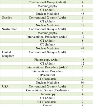
Table 2 Number of established DRLs in various countries [4]
Country Procedures Number of
DRLs
Australia CT (Adult 15+years) 6
CT (Paediatric) 6
France Conventional X-rays (Adult) 9
Conventional X-rays (Paediatric) 9
CT (Adults) 4
Nuclear Medicine 4
Germany Conventional X-rays (Adult) 12
Fluoroscopy (Adult) 5
CT (Adult) 7
Interventional Procedure (Adult) 2
Conventional X-rays (Paediatric) 6
Fluoroscopy (Paediatric) 1
Nuclear Medicine 17
Greece Mammography-Nuclear Medicine 12
CT 7
Italy Conventional X-rays (Adult) 7
Conventional X-rays (Infant) 4
Mammography 1
CT (Adult) 4
Nuclear Medicine 48
Sweden Conventional X-rays (Adult) 6
CT (Adult) 4
Nuclear Medicine 19
Switzerland Conventional X-rays (Adult) 9
Mammography 1
Interventional Procedure (Adult) 12
CT (Adult) 8
CT (Infant) 4
Nuclear Medicine 47
United Kingdom
Conventional X-rays (Adult) 13
Fluoroscopy (Adult) 15
CT (Adult) 12
Interventional Procedure (Adult) 5
Interventional Procedure (Paediatric)
3
CT (Paediatric) 2
Nuclear Medicine 96
USA Conventional X-rays (Adult) 3
Conventional X-rays (Paediatric) 2
Fluoroscopy 4
CT (Adult) 3
CT (Paediatric) 2
Dental 5
The table 2 indicates that in terms of procedures, different countries shows different approach to their DRL set up. While the prioritized examinations might be different across countries, it is not surprising that all of the listed countries have DRLs for CT scan. This might be because of the premise that CT scan has been associated with relatively high radiation exposure and responsible for high total population dose[10][11], increasing the risk of radiation-induced cancers. Therefore, radiation safety precautions on CT scan examination is highly demanded.
Also, in terms of CT technology, there has been significant advancement in CT scan, especially since the invention of multidetector technology, allowing for a better image resolution, faster scan time and longer scan coverage[12]. Additionally, various image reconstruction methods available in current CT technology have significantly improve image quality and reduce radiation dose.
Putu Irma Wulandari et al JMSCR Volume 06 Issue 12 December 2018 Page 512 of the effective implementation of DRL. Therefore,
there have been continuing works regarding the establishment and implementation of DRLs. Some countries have regularly upgraded their NDRLs. UK, for example, has upgraded their DRL values for 3-periode of reviews, showing a strong commitment in dose optimisation by regularly updating their standard. Recently, Australian Government through ARPANSA has just released their updated MDCT DRLs on 1th July 2018, superseding the previous 2012 version[13]. The following tables are the old (2012) and updated version (2018) of Australian MDCT DRLs for adults.
Table 3 Australian adult MDCT DRLs-2012 (superseded) [14]
Adult Protocol CTDI.vol
(mGy)
DLP (mGy.cm)
Head 60 1000
Neck 30 600
Chest 15 450
Abdomen pelvis 15 700
Chest abdomen pelvis 30 1200
Lumbar spine 40 900
Table 4 Australian adult MDCT DRLs-2018 (new version)[13]
Scan region Description (e.g. indication)
CTDIvol (mGy)
DLP (mGy.cm) Head Non-contrast brain
(trauma/headache)
52 880
Cervical spine Non-contrast (trauma) 23 470 Soft-tissue neck Post contrast (oncology) 17 450 Chest Post contrast (oncology) 10 390 Abdomen-pelvis Post contrast (oncology) 13 600
Kidney-ureter-bladder
Non-contrast (suspected renal colic)
13 600
Chest-abdomen-pelvis
Post contrast (oncology) 11 940 Lumbar spine Non-contrast (degenerative
pain)
26 670
As can be seen from the tables, much lower dose levels were apparent in all 2018 Australian adult MDCT protocols compared to those in 2012 DRLs, with the most significant reduction was shown in chest-abdomen-pelvis examination, from 30 mGy in 2012 to 11 mGy in 2018. This significant dose reduction might be due to the development of current technology, allowing lower doses to be more achievable on practice. Additionally, there is also a possibility that the effective implementation of previous DRLs have enabled dose reduction on practice. However, further evidence is required to proof this premise.
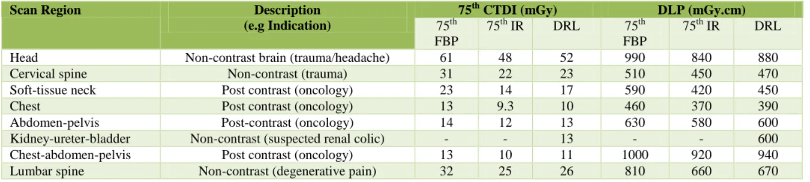
Another important point with this new DRLs is an additional feature of clinical indication. Clinical indication is a good consideration when comparing the fDRLs to NDRLs, as different levels of image quality would be demanded for different pathologies, requiring different dose levels to acquire images. Interestingly, ARPANSA also released the comparison of 75 percentile distributions of fRLs for different reconstruction methods, which are Iterative Reconstruction (IR) and Filetered Back Projection (FBP), as shown on the table below.
Table 5 Comparison between the 75th percentile FRL resulting from filtered back projection (FBP) and iterative reconstruction (IR), and Diagnostic Reference Levels (DRLs)[13]
Scan Region Description
(e.g Indication)
75th CTDI (mGy) DLP (mGy.cm)
75th FBP
75th IR DRL 75th
FBP
75th IR DRL
Head Non-contrast brain (trauma/headache) 61 48 52 990 840 880
Cervical spine Non-contrast (trauma) 31 22 23 510 450 470
Soft-tissue neck Post contrast (oncology) 23 14 17 590 420 450
Chest Post contrast (oncology) 13 9.3 10 460 370 390
Abdomen-pelvis Post-contrast (oncology) 14 12 13 630 580 600
Kidney-ureter-bladder Non-contrast (suspected renal colic) - - 13 - - 600
Chest-abdomen-pelvis Post contrast (oncology) 13 10 11 1000 920 940
Putu Irma Wulandari et al JMSCR Volume 06 Issue 12 December 2018 Page 513 The table 5 shows how dose varies in different
reconstruction methods. It is clearly seen from the table that the 75th percentile of FRLs with Iterative reconstruction was lower than those acquired with Filtered Back Projection. This highlights the importance of DRL evaluation in order to improve the standard in accordance with recent update in technology. This activity also reflects strong commitment in maintaining ALARA principle, through sustainable activity and effective implementation of Diagnostic reference levels.
Paediatric DRL
The ALARA principle must be applied to all patients regardless their gender or age, including the paediatric. Paediatric can be defined as children aged 16 years old or bellow. The paediatric are prone to ionizing radiation due to developing tissues, which are highly sensitive to radiation, requiring careful consideration and rigorous approach in terms of radiation protection. Therefore, DRLs for paediatrics are highly required on practice. However, there has been limited studies regarding DRL for paediatrics. While only few countries have set up NDRL for paediatrics, most of their values were adopted from other countries instead of conducting a national survey.
Establishing DRLs for paediatric could be challenging as there is a large variation of patient’s size not only in different age group, but also in a given age group. Thus, it is highly recommended that the paediatric patients are classified into several groups. European Commission recommends paediatric classifications into the following groups: age 0 - < 3 months, 3 months - < 1 y, 1 - < 6 y, >6 years old[15]. In addition to age, the grouping is also divided into the following size intervals, size intervals: <5kg,5-<15kg,15-<30kg,30-< 50 kg[15]. While European Commission (2018) recommend the grouping by size and age, Australian paediatric DRLs were calculated from 2 groups: baby (0-4 years old) and children (5-14 years old) [6]. Noting that the DRL set up could be done in various approach, the grouping system should be deliberately chosen considering the current situation
and the ease of data collection in the surveyed area. The most important point of this activity is to promote both the establishment and implementation of paediatric DRLs itself, while regular evaluation and improvement could be done afterwards.
Conclusion
Diagnostic Reference Level has been considered as an optimisation tool diagnostic radiography practice, in order to make sure that low doses are used to achieve diagnostic images. Therefore, all medical radiation practitioners should familiar with the basic concept of DRL and its implementation.
It is important to note that DRL can only be implemented in diagnostic radiography purposes, not radiation therapy. Also, DRL is unique for specific population and specified procedure, therefore it cannot be simply adopted from other countries. Medical radiation professionals and related stakeholders should actively contribute to the establishment of national DRLs, based on national survey, for both adult and paediatric DRLs. While this activity might be comprehensive and time-consuming, there are some critical points to be considered in establishing national DRLs.
The study on DRLs is a continuous work. Once it has been established, the DRL should be effectively implemented and regularly updated to adjust with the recent development of technology and techniques in medical imaging.
References
1. ICRP, “Radiological Protection in Medicine. Annals ICRP Publication 105,” 2007.
2. UNSCEAR, “Sources and Effects of Ionising Radiation,” United Nations, New York, 2000.
3. D. A. Johnston and P. C. Brennan, “Reference dose levels for patients undergoing common diagnostic x-ray examinations in Irish hospitals,” The british Journal Of Radiology, pp. 396-402, 2000. 4. E. Seeram and P. Brennan, Radiation
Putu Irma Wulandari et al JMSCR Volume 06 Issue 12 December 2018 Page 514 5. ICRP, “Radiological Protection and safety in
medicine: ICRP Publication 73,” 1996. 6. ARPANSA, “National Diagnostic Reference
Level Service in more detail,” 17 November
2018. [Online]. Available:
https://www.arpansa.gov.au/research-and- expertise/surveys/national-diagnostic-reference-level-service/in-more-detail. 7. Medical Council, “Diagnostic Reference
Level: Position Paper,” 18 November 2004.
[Online]. Available:
https://www.medicalcouncil.ie/About-
Us/Legislation/Medical-Ionising-Radiation/Diagnostic-Reference-Levels.pdf. 8. E. Commission, “European guidelines on
diagnostic criteria,” 1996.
9. J. Vassileva and Madan Rehani, “Diagnostic Reference level,” American Journal of Radiology, pp. 204 W1-W3, 2015.
10.d. G. A. Berrington, M. Mahesh, K. P. Kim, M. Bhargavan, R. Lewis and F. Mettler, “Projected cancer risks from computed tomographic scans performed in the United States in 2007,” Arch Intern Med, pp. 2071-2077, 2009.
11.E. Hall and D. J. Brenner, “Cancer risks from diagnostic radiology,” British Journal of Radiology, pp. 362-378, 2008.
12.S. J. Foley, M. F. McEntee and L. A. Rainford, “Establishment of CT diagnostic reference levels in Ireland,” The British Journal of Radiology, pp. 1390-1397, 2012. 13.ARPANSA, “Current Australian national
diagnostic reference levels for multi detector computed tomography,” 17 November 2018.
[Online]. Available:
https://www.arpansa.gov.au/research-and-
expertise/surveys/national-diagnostic- reference-level-service/current-australian-drls-update/mdct.
14.ARPANSA, “Multi detector computed tomography statistics,” 17 November 2012.
[Online]. Available:
https://www.arpansa.gov.au/research-and- expertise/surveys/national-diagnostic-
reference-level-service/mdct/statistics#superseded.
15.European Commission, Radiation Protection No.185, Louxembourg: European Union, 2018.