CHARACTERIZING
SALMONELLA
TYPHIMURIUM'S EVASION
OF INNATE IMMUNITY
Farhan Lakhani—Miao Lab
APRIL 2, 2020
UNC CHAPEL HILL1 Abstract
Inflammasomes play an important role in defense against infection. The NOD-like Receptors CARD Domain Containing 4 (NLRC4) inflammasome detects the Type Three Secretion System (T3SS) system rod and needle proteins, leading to caspase-1 activation. Active caspase-1 induces pro-interleukin-1β
2 Introduction
The innate immune system is the first line of defense against infection. Although it is often
described as nonspecific, the innate immune system can successfully discriminate between
commensal bacteria and pathogens to spare the former which are essential for food digestion and
other metabolic function, while eliminating pathogens that cause disease.
The innate immune system achieves this function by detecting pathogen-associated virulence
through sets of transmembrane receptors such as Toll-Like Receptors (TLRs) or cytosolic
sensors like Nod-Like Receptors (NLRs). NLRs like NLRP3 or NLRC4 are composed of a
sensor domain, a nucleotide-binding oligomerization domain (NOD), and a signaling domain
(CARD or Pyrin) signaling domains1. NLRC4 detects type three secretion system (T3SS)
proteins, rod and needle. After recognizing its agonist, NLRC4 monomers oligomerize to form a
large platform which can recruit and activate caspase-12. Active caspase-1 cleaves
pro-interleukin-1B (pro-IL-1β) and pro-IL-18 to their mature form. In addition, active caspase-1
triggers a form of lytic programed cell death called pyroptosis. Pyroptosis is a highly
inflammatory process, which allows inflammatory cytokines like IL-1β and IL-18 to be released
into extracellular space. These cytokines recruit other immune cells to clear infected sites such as
neutrophils3. Therefore, pyroptosis can benefit the host by eliminating potential replication
niches for intracellular pathogens and recruiting other immune cells to eliminate the pathogen.
Many Gram-negative pathogens use T3SSs as virulence factors; T3SS functions like a
hypodermic syringe to inject effector proteins directly into the host’s cytoplasm, allowing
bacteria to reprogram host cells. Salmonella enterica serovar Typhimurium (S. Typhimurium)
uses two T3SSs to establish an intracellular niche: Salmonella pathogenicity island 1 (SPI-1) and
3 (macrophages are naturally phagocytic and thus take up the bacteria into their vacuoles). Once
inside the vacuole of either epithelial cells or macrophages, S. Typhimurium switches off SPI-1
and expresses the SPI-2, whose injected effector proteins redirect resources to the vacuole to
enable bacterial replication.5
Several proteins compose the T3SSs: the two proteins that form the rod and needle component of
the syringe are sometimes unintentionally injected into infected cells6. Rod and needle can be
detected in the cytosol of immune cells by the NLRC4 inflammasome. Miao et. al7,8 previously
showed that rod and needle proteins expressed by the SPI-1 T3SS systems, PrgJ and PrgI, are
detected by NLRC4 while SsaI, a needle protein expressed by the SPI-2 T3SS remains
undetected by NLRC4. Although SPI-2 expressing S. Typhimurium are not detected by NLRC4,
it had never been experimentally shown whether the SPI-2 needle (SsaG) protein evaded
detection. We hypothesized that like SsaI, SsaG would also evade detection by NLRC4. This is
consistent with literature as it would cause S. Typhimurium to remain undetected while
activating SPI-2 in vivo as demonstrated previously5. The goal of this study is to determine
whether and how SsaG is detected in vitro by WT macrophages.
Methods
Mice
Wild-type (WT) C57BL/6 and Nlrc4–/– mice used to prepare bone marrow derived macrophages
(BMMs) were 8–12 weeks old, male or female, and housed under specific pathogen free facility.
All protocols were approved by the Institutional Animal Care and Use Committee at the
University of North Carolina at Chapel Hill and met the humane care of animals guidelines of
4 Cloning
Polymerase Chain Reaction (PCR) was performed on S. Typhimurium to amplify the following
genes using primers noted in Table 1: prgJ, prgI, ssaI, ssaG. Each gene was inserted into the
cloning vector pMXsIG, containing an IRES-GFP (IG) element, by first being digested (8 total)
with restriction enzymes encoded in the respective gene’s primer by applying a ratio of 1 µg
DNA, 1 µL restriction enzyme 1, 1 µL restriction enzyme 2, 5 µL 10X Buffer, and nuclease-free
water to 50 µL. The mixture was incubated at 37° Celsius (C) for 2 hours to ensure digestion.
Thereafter, ligation was performed by adding 1:3 insert to vector (vector at 100 ng) to 1 µL of T4
DNA ligase, 1µL T4 DNA ligase buffer, and nuclease-free water to 10 µL. Concentrations of
nucleic acid were determined by NanoDrop® ND-1000 Spectrophotometer. The resulting
ligations were transformed via electroporation into electrocompetent XL1-Blue Escherichia coli
(E. coli) at a ratio of 1 µL ligation: 40 µL competent cells. Transformations were plated on
Luria-Bertani (LB) plates containing 100 µg/mL ampicillin (an LB+amp plate), to screen for
transformants and grown for 16 hours at 37° C. Transformed, single colonies of each strain
(pMXs-IG-prgJ, for example) were collected, grown overnight in liquid LB+amp solution (2
mL), and flash frozen in a 20% glycerol solution to be maintained at -80° C.
Plasmid Preparation
Each transformant containing desired genes were incubated overnight in LB+amp solutions (50
mL) for 16 hours at 37° C. The plasmids were extracted from solution using the Qiagen Midi Kit
(as outlined by the manufacturer). Restriction digests were performed and examined via gel
electrophoresis. Positive plasmids were verified by next generation sequences performed by Eton
5 Retroviral Lethality Assay
Immortalized bone marrow macrophages (myc macrophages AKA iBMMs) were generated as
previously described9 using C57BL/6 mice, WT and Nlrc4–/–. Ecotropic Phoenix cells (American
Type Culture Collection) were grown in Dulbecco's Modified Eagle Medium (DMEM) with
10% Fetal Bovine Serum (FBS) while macrophages were grown in DMEM with 10% FBS and
10% L-cell supernatant. All cell lines were grown in 37° C and 5% CO2 in tissue culture (TC)
treated flasks. Phoenix cells were passaged 3-4 times using trypsin at approximately 90%
confluency while iBMMs (WT and Nlrc4–/–) were passaged using Phosphate-Buffered Saline
with 10 mM Ethylenediaminetetraacetic acid (PBS+EDTA) twice at approximately 90%
confluency before use. In the afternoon (Day 1), Phoenix cells were harvested with 5 mL of
trypsin by incubating in growth conditions for 2-3 minutes. The cells were pelleted at 1000 rpm
at room temperature (RT) for 5 minutes and resuspended in growth media to seed 5 mL of media
into TC treated 10 cm dishes (4) with 1*107 cells. The DNA transfection mix was prepared by
mixing 12 µg of DNA with 1.5 mL of Opti-MEM media (OPTI) and mixing 36 µL of
Lipofectamine 2000 with 1.5 mL of OPTI separately. After 5 minutes at RT, the separate
mixtures were combined. After 20 minutes, 3 mL of transfection mix was added to the 5 mL of
Phoenix cells dropwise (this infects 2 wells of macrophages; 1 WT and 1Nlrc4–/–). Media from
transfected Phoenix cells was aspirated and replaced with 10 mL of growth media the following
morning (Day 2). WT and Nlrc4–/– iBMMs were seeded at 2.5 * 105 cells in TC treated, 6 well
plates in the afternoon of Day 3. The supernatant of the Phoenix cells was collected and filtered
through a 0.4 µm syringe filter on Day 4. Polybrene was added to 5 mg/mL, producing the
retroviral supernatant. Media was aspirated from macrophages and the retroviral supernatant was
6 macrophages were placed in growth conditions for 1 hours and then media was replaced with
normal growth media. Macrophages were pelleted 2 days later (Day 6) at 300 RCF at RT and
was resuspended in 200 µL of 2% Paraformaldehyde (PFA) in the dark. After 30 minutes,
macrophages were spun in the same way for ten minutes and suspended in 200 µL FACS buffer
(1% bovine serum albumin in PBS). Flow cytometry was performed to identify GFP-positive
(GFP+) WT and Nlrc4–/– singlet macrophages.
Competitive Index Assay
Competitive index assays were performed as described elsewhere10 with WT and Casp1–/– or
Casp1/11–/– mice using WT S. Typhimurium (AmpR) and ΔsseF S. Typhimurium (KanR) for a 48
hour infection.
Protein Transfection
Proteins of interest with 6xHIS tags were purified using Talon Beads (Clontech). The proteins
were originally solubilized in urea and either used in that solvent or dialyzed to PBS. Proteins
(quantities noted in figure descriptions) were transfected for 1 hour using Profect P1 (Targeting
Systems). Transfection was enhanced by “spin-fection”, where after application of the
transfection mix, macrophages are spun at 1250 RPM for 15 minutes at room temperature (RT).
Cells were incubated at 37 degrees, 5% CO2 for 1 hour. Lactate DeHydrogenase (LDH) Assays
and Enzyme-Linked Immunosorbent Assays (ELISA) were performed as previously described.
Immunoblot was also performed as previously described except that the cellular compartment
and supernatant/extracellular components were treated separately for analysis. Additionally,
7 Results & Discussion
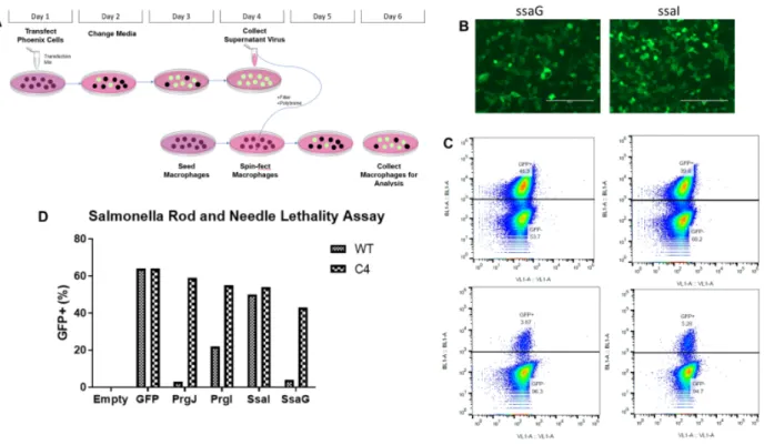
Figure 1. SsaG activates NLRC4 -/-, leading to cell death. (A) Schematic of retroviral lethality assay where phoenix cells were transfected with DNA encoding rod and needle proteins. The virus generated was later used to transduce iBMMs and were marked by GFP. (B) Microscopic images of Phoenix cell transfection efficiency for ssaG (left) and ssaI (right). More intense green translates to higher virus expression. (C) Flow cytometry data for both WT and Nlrc4-/-
macrophages infected with a retrovirus encoding for SsaG. Singlet Nlrc4-/- (upper two graphs) and WT macrophages (lower two graphs) were selected by appropriate gating. The same virus was applied to both WT and Nlrc4-/- macrophages. (D) Quantitative summary of all flow cytometry data is displayed here. Identical gating was applied in generating both C and D. NLRC4 detects SsaG, but not SsaI, and initiates cell death upon detection
We used a retroviral lethality assay to determine if SsaI and SsaG are detected by NLRC4. PrgJ
and PrgI detection were previously demonstrated using a similar assay 7. In this assay,
retro-virally transduced iBMMs expressed rod or needle proteins and we tracked this expression by
detecting GFP (Figure 1A). In Nlrc4–/– iBMMs, rod and needle proteins are not detected
therefore they should not undergo pyroptosis allowing us to measure GFP expression, while WT
8 WT iBMMs undergo pyroptosis, resulting in loss of GFP expression. Our results show
successful transfection occurred—phoenix cell images transfected with ssaG and ssaI are
provided as examples (Figure 1B). We show population level data on both replicates of Nlrc4–/–
and WT macrophages transduced with ssaG (Figure 1C). The WT macrophages show
significantly less GFP expression (average of 4.48% GFP+) than their Nlrc4–/– counterparts
(average of 43.05% GFP+). This indicates that SsaG, similar to PrgJ and PrgI, was detected by
NLRC4 to induce pyroptosis, resulting in loss of GFP expression. In contrast, WT and Nlrc4–/–
iBMMs transduced with ssaI show similar expression, indicating that SsaI was not detected by
NLRC4 and therefore pyroptosis was not induced. Comparisons take into account the positive
control of solely inducing GFP expression in both WT and Nlrc4–/– iBMMS (Figure 1D).
Rod proteins have been shown to be detected by NAIP2-NLRC4 inflammasome while needle
proteins have been shown to be detected by NAIP1-NLRC4 inflammasome12. Since NAIP1
expression is lower compared to NAIP2 in WT iBMMs, we found a higher percentage of GFP+
WT iBMMs expressing PrgI, indicating that less pyroptosis took place. Therefore, our inability
to detect SsaI via GFP expression may be due to the reduced expression of NAIP1 in WT
iBMMs. To clarify potentially confounding data, we will upregulate expression of NAIP1 in
9 Figure 2. SsaG triggers pyroptosis via an NLRC4-dependent, Caspase-1 pathway. (A) Schematic representation of workflow of protein generation provided. All proteins generated were found in inclusion bodies. (B) Western blot of both inclusion bodies and soluble portions of cellular lysate are provided. Anti-His tag antibody was applied to determine where the proteins were produced. (C) LDH Assay was performed on the supernatant of cells, WT and Nlrc4-/-, receiving protein transfections. 50ng/uL of the relevant protein dialyzed into PBS from 8M Urea were used. 5*10^4 cells were affected in a 96-well plate and were primed with PAM3CSK4 at 500 ng/mL. Lysis wells were generated using 10X Triton diluted in PBS to 1X in an identical volume to the protein-affected wells. (D) A series of Western blots are provided, displaying a dose-dependent relationship in both caspase-1 processing and location and IL-1β maturation and location. Antibodies detect both immature and mature caspase-1 and IL-1β. For the Nlrc4-/- protein-affected samples, each sample received the maximum dosage of 100ng of protein. Priming was performed identically to C (E) ELISA data is displayed from an independent experiment,
demonstrating a quantitative, dose-dependent relationship between amount of protein and IL-1β produced. Priming was performed in the same way.
SsaG is detected via caspase-1 mediated pyroptosis in a dose-dependent manner
To better replicate rod and needle proteins’ route of entry and obtain mechanistic detail on how
pyroptosis occurs, we ran assays on protein transfections performed on macrophages. Rod and
needles proteins were expressed and purified under the associated scheme (Figure 2A) and found
10 solvent of urea because this best mimics how the unfolded protein enters the host cells; although
cytotoxicity was found in the way that was expected and consistent (Figure 2C), we are unsure if
the protein is detected while it is unfolded or folded. Literature7 has shown that rod and needle
proteins of SPI-1 trigger caspase-1 cleavage and ensuing IL-1β release from the lysed cell. To
intracellular and extracellular effects, we examined both the cellular component and supernatant
of macrophages post-transfection for caspase-1 and IL-1β via western blot. We also performed
the assay in at varying dosages to examine how the signaling cascade varies with agonist
concentration. We expected to find increasing amounts of cleaved caspase-1 in the supernatant
alongside decreasing amounts of pro-caspase-1 in the cellular components in cells that detected
the rod or needle protein. With detection, we also expected to find increasing amounts of IL-1β
in the supernatant alongside decreasing amounts of pro-IL-1β in the cellular component. This
assay (Figure 2D) when combined with the cytotoxicity assay demonstrates that SsaG induces
pyroptosis via caspase-1 cleavage and IL-1β release in a dosage-dependent fashion. With
cytotoxicity assays, normalization of proteins loaded into the western blots can be elusive. Total
protein concentration was normalized for these samples. After noting difficulties in illustrating
IL-1β presence by western blot, we decided to more accurately quantify it by ELISA (Figure
11 Figure 3. SseF and SlrP do not exhibit chaperone-like behavior. (A) Schematic representation is provided. (B) 48 hours post infection by a 1:1 ratio of ΔsseF and WT S. Typhimurium, mice spleens were harvested and plated to determine total CFUs from each strain. In WT mice, the average value is -1.42 while in Casp1–/– mice the average value is -1.13. P-values were
determined by a two-tailed t-test (P < 0.05). (C) SlrP was tested similarly however the difference between competitive index values between WT mice and Casp1/11–/– were not statistically
significant.
Neither SseF nor SlrP Inhibit SsaG Detection by NLRC4.
Our in vitro results suggest that S. Typhimurium expressing SPI-2 T3SS should be detected by
hosts since SsaG is detected by NLRC4, resulting in caspase-1 mediated pyroptosis. The
induction of pyroptosis should clear the pathogen during in vivo infection. However, Miao et
al.13 previously showed that S. Typhimurium remains undetected while expressing SPI-2. This
shows the opposite result – SsaG is not detected in vivo. How can we reconcile these
contradictory results? We hypothesized that SsaG would actually be detected in vivo but a SPI-2
effector protein inhibits SsaG detection. The first candidate we tested as an inhibitor was SseF.
12 assay, comparing its growth to WT S. Typhimurium in WT and Casp1–/– mice (Figure 1A). If our
hypothesis is true, then WT S. Typhimurium would outcompete the sseF mutant Salmonella in
WT mice. This is because the mutant sseF bacteria would permit SPI-2 needle protein detection
by NLRC4 and therefore be at a competitive disadvantage compared to WT S. Typhimurium.
However, in Casp1–/– mice, the competition between the two strains of bacteria should be equal,
due to the macrophages’ inability to induce pyroptosis.
Our results in Figure 3B show a minimal difference in growth between WT and ΔsseF S.
Typhimurium across both strains of mice, resulting in similar competitive index values of -1.42
in WT mice and -1.13 in Casp1–/– mice. Therefore, SseF is likely not the protein responsible for
SsaG evading detection of NLRC4. Negative values (Figure 3B) indicate that mutant S.
Typhimurium are at a disadvantage compared to WT S. Typhimurium. In WT mice, ΔsseF S.
Typhimurium is at the greatest disadvantage while in Casp1–/– mice, ΔsseF S. Typhimurium is
only slightly competitively closer to WT bacteria. Had a chaperoning protein been identified that
assisted in SsaG evading NLRC4, the competition between WT and mutated S. Typhimurium in
Casp1–/– mice would have been nearly equal.
We tested SLRP—another virulent effector— in the same way (Figure 3C). We expected for
there to be a strong negative value for the competitive index in WT mice and near-zero value in
Casp1/11–/–. However, we found both competitive index values were near-zero. Therefore,
whether S. Typhimurium encodes an inhibitor of SsaG detection remains unknown. We have
13 Future Directions
The NAIP that detects SsaG can be studied using Naip1–/– and Naip2–/– macrophages. If SsaG is
conclusively determined to be detected by NLRC4, then this is reason to believe that S.
Typhimurium has a mechanism to block the detection of its needle protein. Likely, an effector
protein has a chaperoning function, assisting in the needle’s evasion of inflammasomes. Further
in vivo competitive index assays of S. Typhimurium strains with certain effectors removed
compared to WT S. Typhimurium could identify such an inhibitor.
In the future we would like to study an environmental pathogen with a similar T3SS virulence
mechanism to promote support for a novel hypothesis termed the “Red Pawn hypothesis”. This
draws reference from Alice's Adventures in Wonderland, where the Red Queen taunts Alice
saying that it would take all the running she could do to stay in the same place. Evolutionarily
speaking, this parallels host-adapted pathogens; S. Typhi is a Red Queen because it has evolved
with us to continually subvert evolved innate immune defenses. However, there are also
pathogens that could not keep running, so to speak, which we term as Red Pawns. An example of
such a Red Pawn is Chromobacterium Violaceum (C. Violaceum) commonly found in soil.
Inflammasomes in WT mice have required a 100,000-fold higher dose of C. Violaceum to
succumb compared to immune-deficient mice. The scientific community considers pathogens
like this environmental pathogens because they are generally ubiquitous and do not take hold in
immune-competent systems. This is because inflammasomes have evolved to effectively shut
those pathogens out with high efficacy, indicating that perhaps that is the true purpose of
inflammasomes: to defend against environmental pathogens. It is only by coincidence and much
more poorly that inflammasomes defend against host-adapted pathogens. Comparing the two
14 Acknowledgments
15 Appendix
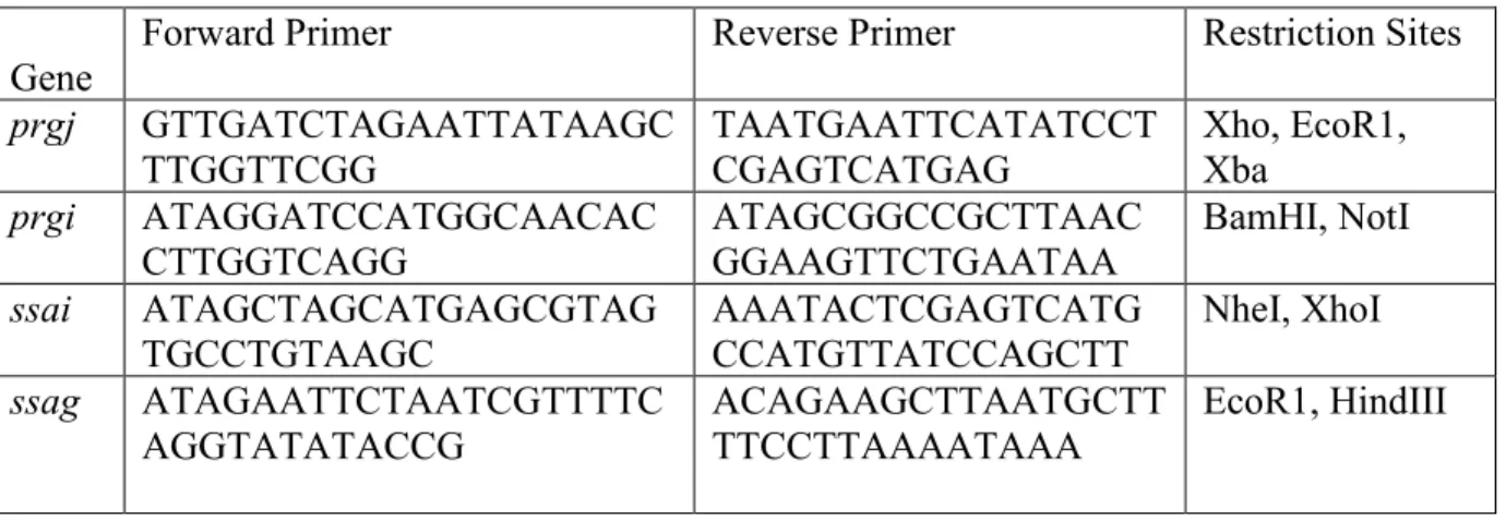
Table 1. PCR supplemental information.
Forward and reverse primers are listed for the following genes cloned into pMXs-IG. Restriction sites are also noted.
Gene Forward Primer Reverse Primer Restriction Sites
prgj GTTGATCTAGAATTATAAGC
TTGGTTCGG TAATGAATTCATATCCTCGAGTCATGAG Xho, EcoR1, Xba
prgi ATAGGATCCATGGCAACAC
CTTGGTCAGG ATAGCGGCCGCTTAACGGAAGTTCTGAATAA BamHI, NotI
ssai ATAGCTAGCATGAGCGTAG
TGCCTGTAAGC AAATACTCGAGTCATGCCATGTTATCCAGCTT NheI, XhoI ssag ATAGAATTCTAATCGTTTTC
16 References
1. Miao, E. A., Rajan, J. V & Aderem, A. Caspase-1-induced pyroptotic cell death. Immunol. Rev. 243, 206–14 (2011).
2. Franchi, L., Eigenbrod, T., Muñoz-Planillo, R. & Nuñez, G. The inflammasome: a caspase-1-activation platform that regulates immune responses and disease pathogenesis. Nat. Immunol. 10, 241–7 (2009).
3. Jorgensen, I., Rayamajhi, M. & Miao, E. A. Programmed cell death as a defence against infection. Nature Reviews Immunology 17, 151–164 (2017).
4. Jones, M. A., Hulme, S. D., Barrow, P. A. & Wigley, P. The Salmonella pathogenicity island 1 and Salmonella pathogenicity island 2 type III secretion systems play a major role in pathogenesis of systemic disease and gastrointestinal tract colonization of Salmonella enterica serovar Typhimurium in the chicken. Avian Pathol. 36, 199–203 (2007).
5. Coombes, B. K., Wickham, M. E., Lowden, M. J., Brown, N. F. & Finlay, B. B. Negative regulation of Salmonella pathogenicity island 2 is required for contextual control of virulence during typhoid. Proc. Natl. Acad. Sci. 102, 17460–17465 (2005).
6. Notti, R. Q. & Stebbins, C. E. The Structure and Function of Type III Secretion Systems. Microbiol. Spectr. 4, (2016).
7. Miao, E. A. et al. Innate immune detection of the type III secretion apparatus through the NLRC4 inflammasome. Proc. Natl. Acad. Sci. 107, 3076–3080 (2010).
8. Rayamajhi, M., Zak, D. E., Chavarria-Smith, J., Vance, R. E. & Miao, E. A. Cutting Edge: Mouse NAIP1 Detects the Type III Secretion System Needle Protein. J. Immunol. 191, 3986–3989 (2013).
9. GANDINO, L. & VARESIO, L. Immortalization of Macrophages from Mouse Bone Marrow. Exp. Cell Res. 188, 192–198 (1990).
10. Jorgensen, I., Zhang, Y., Krantz, B. A. & Miao, E. A. Pyroptosis triggers pore-induced intracellular traps (PITs) that capture bacteria and lead to their clearance by efferocytosis. J. Exp. Med. 213, 2113–28 (2016).
11. Miao, E. A. et al. Cytoplasmic flagellin activates caspase-1 and secretion of interleukin 1b via Ipaf. (2006). doi:10.1038/ni1344
12. Fusco, W. G. & Duncan, J. A. Novel aspects of the assembly and activation of
inflammasomes with focus on the NLRC4 inflammasome. doi:10.1093/intimm/dxy009 13. Miao, E. A. & Rajan, J. V. Salmonella and Caspase-1: A complex Interplay of Detection
and Evasion. Front. Microbiol. 2, 85 (2011).