Physical activity, immobilization
and
Cover: Motionless water in a pool made for physical activity
Cover design: K.J. van Stralen & J. van der Ahé
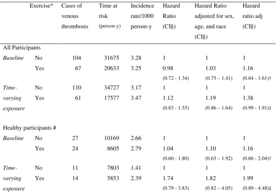
© 2008 K.J. van Stralen
Lay-Out: Y. Souverein
ISBN: 978-90-9022857-0
and
the risk of venous thrombosis
Proefschrift
ter verkrijging van
de graad van Doctor aan de Universiteit Leiden,
op gezag van Rector Magnificus prof.mr. P.F. van der Heijden,
volgens besluit van het College voor Promoties
te verdedigen op donderdag 3 april 2008
klokke 15.00 uur door
Karlijn Janneke van Stralen
geboren te Delft
Promotiecommissie
Promotor:
Prof. Dr. F.R. Rosendaal
Copromotor:
Dr. C.J.M. Doggen
Referent:
Prof. Dr. B.M. Psaty (University of Washington, Seattle, USA)
Overige leden: Prof. Dr. J.P. Vandenbroucke
Prof. Dr. H.R. Büller (University of Amsterdam)
Dr. S. Le Cessie
The work described in this thesis was performed at the department of clinical
epidemiology, Leiden University Medical Center, Leiden, the Netherlands, and the
Cardiovascular Health and Research Unit, University of Washington, Seattle, USA.
This research was supported by the Netherlands Organization for Scientific
Research (912-033| 2003) and the Leducq foundation, Paris, France for the
development of transatlantic networks of excellence in cardiovascular research.
Financial support by the Netherlands Heart Foundation and the J.E. Jurriaanse
Stichting for the publication of this thesis is gratefully acknowledged.
Chapter 1 7 Introduction
Chapter 2 15
The tortuous history of the implementation of early ambulation after delivery
Chapter 3 31
Regular sports activities decrease the risk of venous thrombosis
Chapter 4 49
The relationship between exercise and risk of venous thrombosis in elderly people
Chapter 5 65
Strenuous sport activities involving the upper extremities increase the risk of venous thrombosis of the arm
Chapter 6 73
Minor injuries as a risk factor for venous thrombosis
Chapter 7 89
Mechanisms of the factor V Leiden paradox
Chapter 8
Discussion & Summary 105
Samenvatting 117
Dankwoord 125
Chapter 1
9
Venous thrombosis is a common disease affecting millions of individuals every year1.
Approximately 80 percent of the thrombi originate in the leg. Thrombi can detach resulting
in a pulmonary embolism. This embolism is fatal in approximately 10 percent of the cases2.
Most patients with a fatal pulmonary embolism die within two hours after the onset of the
symptoms3. For this reason, it is important to focus on the identification of risk factors as
this may lead to the prevention of venous thrombosis. In 1856, Virchow described
thrombosis as a disease caused by clotting of the blood4. He wrote “Wir können auch
künftig die mehr mechanische Formen der Thrombose, wie sie bei der Blutstockung
vorkommen [stasis], von den mehr chemischien [blood composition] or physikalischen
Formen, wie sie durch direkte Sauerstoff-Einwirkung oder veränderte Flächenanziehung zu
Stande kommen [vessel wall], unterscheiden”. This has been interpreted as the now famous
“Triad” with three major causes of thrombosis e.g. slowing down of the bloodstream the
so-called stasis, changes in the blood composition and damage to the vessel wall. It is
generally believed that only the first two causes are involved in the occurrence of venous
thrombosis.
As humans walk upright, blood from the feet and lower legs has to overcome gravity for
over a meter before it reaches the heart. It is therefore easy to imagine how stasis occurs in
the veins of the lower extremities. This may lead to the formation of blood clots in the
bloodstream. Two important systems assist the blood in streaming upwards. First, veins
contain valves which prevent the reflux of blood after it passes the valves. When the valves
are damaged, due to for example varicose veins, the risk of venous thrombosis increases5.
Secondly, the blood flow is stimulated by the pump function of the leg muscles. Altered
muscle function due to immobilization is known to cause venous thrombosis. In the Second
World War, increased rates of pulmonary embolism were reported in individuals who had
sought shelter in the underground and had sat cramped positions for hours during the
bombings on London6 (see figure 1). Nowadays more “modern” forms of immobilization
have been reported to cause venous thrombosis such as the economy class syndrome due to
Chapter 1
10
Figure 1. People sheltering from air raids in the Aldwych underground station, London, UK.
Stimulation of the blood flow by increasing calf muscle movement through physical
activity is therefore likely to decrease the risk of venous thrombosis. Mild forms of physical
activity such as ambulation after surgery or giving birth may reduce the risk of venous
thrombosis. In the early 1900s women were advised to stay in bed for 28 days after
delivery9 and venous thrombosis rates post-partum were very high; up to 8 per 1000
deliveries10. Nowadays, women usually leave the bed on the same day as the delivery and
venous thrombosis rates post-partum have decreased to approximately 1 per 1000
births11;12. Although many other factors have changed since then, these figures suggest that
ambulation soon after delivery decreases the risk of venous thrombosis. Up till now, it was
unknown whether the drop in venous thrombosis rates was actually due to the change in
ambulation policy. Therefore the reasons and implications of this transition will be
described in chapter 2.
Besides ambulation more rigorous forms of physical activity such as sports activities may
affect the risk of venous thrombosis. Only a few studies have been conducted on this
association and conflicting results have been found. One cohort study has shown increased
venous thrombosis rates after sports activities13, while a case-control study has shown a
11
composition of the blood in relation to physical activity. On one hand, during and shortly
after exercise the blood is in a procoagulant state15;16. However, after a training period of
several months daily levels of procoagulant factors are reduced and there is less of a
prohrombotic state than in individuals who are untrained17;18. This suggests that performing
sports activities on a regular basis results in a beneficial coagulant balance that may reduce
the risk of venous thrombosis. However, performing sports activities also increases the risk
of injuries19 which may result in immobilization and lead to venous thrombosis. Up till now
not much was known on the association of sports activities and venous thrombosis risk. For
this reason the risk of venous thrombosis associated with sports activities was determined in
two separate studies. Results from a case-control study, the Multiple Environmental and
Genetic Assessment of risk factors for venous thrombosis (MEGA study), are described in
chapter 3. The results obtained in a cohort study (Cardiovascular Health Study or CHS) are
described in chapter 4.
The physicians Sir James Paget (London, 1875)20 and Leopold von Schrötter (Vienna,
1884) described a second mechanism for an increased risk of venous thrombosis after
sports activities, which is called the Paget-Schrötter syndrome21 or effort induced
thrombosis. It was later shown that overdevelopment of the scalene muscle can compress
the subclavian vein resulting in a rare form of venous thrombosis, i.e. venous thrombosis of
the arm (figure 2). Many case reports of venous thrombosis of the arm have been published
regarding athletes who intensively use their arms, such as weight lifters22 and wrestlers23.
So far risk estimates for performing sports involving the arms have not been made.
Therefore in chapter 5, we assessed whether arm sports increase the risk of venous
thrombosis of the arm compared with sports mainly involving the leg and performing no
Chapter 1
12
Figure 2. The Paget-Schrötter syndrome; the subclavian artery, vein and scalene muscle all are fixed between the clavicle and first rib. Overdevelopment of the scalene muscle therefore leads to compression of the vein, as the
results in decreased blood flow through the vein and could therefore lead to venous thrombosis of the arm.
As described previously, sports activities
venous thrombosis. Major trauma, for example
known to increase venous thrombosis risk to a large exten
showed that pulmonary embolism was the c
Without prophylaxis, venous thrombosis
half of the trauma patients25. For this reason,
hospitalized, have surgery or plaster cast are provided prophylactic
However, not much is known about the effect of minor injuries that
hospitalization. In general, when someone has an ankle sprain or knee twist, no prophylaxis
is provided. As it is unclear what the risk is and
association of minor injuries with the risk of venous thr
Schrötter syndrome; the subclavian artery, vein and scalene muscle all are fixed between the clavicle and first
rib. Overdevelopment of the scalene muscle therefore leads to compression of the vein, as the artery can not be compressed. This
results in decreased blood flow through the vein and could therefore lead to venous thrombosis of the arm.
sports activities may lead to injuries which can increase the risk of
Major trauma, for example as caused by car accidents, has long been
known to increase venous thrombosis risk to a large extent. Autopsy studies in the 1930s
showed that pulmonary embolism was the cause of death in 38 percent of fatal injuries24.
Without prophylaxis, venous thrombosis, mainly asymptomatic, occurred in approximately
. For this reason, many patients with major injuries who are
surgery or plaster cast are provided prophylactic anticoagulant treatment.
is known about the effect of minor injuries that do not require
hospitalization. In general, when someone has an ankle sprain or knee twist, no prophylaxis
what the risk is and whether this regimen is appropriate, the
13
Finally, some risk factors have been shown to differentially affect the risk of deep vein
thrombosis of the leg and pulmonary embolism. Factor V Leiden, a genetic risk factor for
venous thrombosis, has a clear effect on the risk of deep venous thrombosis but little or no
effect on pulmonary embolism risk 26;27. Several causes for this difference have been
proposed and will be studied in chapter 7. Mechanisms under study are, among others, an
effect of the factor V Leiden mutation on the location of the thrombus in the leg, the
number of affected veins, and the speed of thrombus formation i.e. the time between the
formation of the thrombus and the actual diagnosis.
In the final chapter, chapter 8, the results in this thesis will be summarized and their
validity and implications will be discussed. Furthermore, some suggestions for additional
research will be presented.
References
1. Heit JA, Silverstein MD, Mohr DN, Petterson TM, O'Fallon WM, Melton III LJ. Risk factors for deep vein thrombosis and pulmonary embolism: a population-based case-control study. Arch Intern Med, 2000, 160: 809-815.
2. Kearon C. Natural history of venous thromboembolism. Circulation, 2003, 107: I22-I30.
3. Stein PD, Henry JW. Prevalence of acute pulmonary embolism among patients in a general hospital and at autopsy. Chest, 1995, 108: 978-981.
4. Virchow R. Phlogose und Thrombose im Gefässystem. Gesammelte Abhandlungen zur Wissenschaftlichen Medizin. Frankfurt, Staatsdruckerei. 1856. 525.
5. White JV, Ryjewski C. Chronic venous insufficiency. Perspect Vasc Surg Endovasc Ther, 2005, 17: 319-327.
6. Simpson K. Shelter deaths from pulmonary embolism. Lancet, 1945, 744.
7. Cannegieter SC, Doggen CJM, van Houwelingen HC, Rosendaal FR. Travel-related venous thrombosis: results from a large population-based case control study (MEGA study). PLoS Med, 2006, 3: e307. 8. Beasley R, Raymond N, Hill S, Nowitz M, Hughes R. eThrombosis: the 21st century variant of venous
thromboembolism associated with immobility. Eur Respir J, 2003, 21: 374-376.
9. Rush J, Chalmers I, Enkin M. Care of the new mother and baby. In: Chalmers I, Enkin M, Keirse MJNC, editors. Effective care in pregnancy and childbirth. 1989. 1333-1346.
10. Ashton WE, McGlinn JA. Sanders' Question Compends: essentials of obstetrics., 7 ed. Philadelphia and London, W.B. Sanders Company. 1911. 231.
11. Bonnar J. Venous thrombo-embolism and pregnancy. Clinical Obstetrics and gynaecology, 1981, 8: 455-473.
12. Treffers PE, Huidekoper BL, Weenink GH, Kloosterman GJ. Epidemiological observations of thrombo-embolic disease during pregnancy and in the puerperium, in 56,022 women. Int J Gynaecol Obstet, 1983, 21: 327-331.
13. Glynn RJ, Rosner B. Comparison of risk factors for the competing risks of coronary heart disease, stroke, and venous thromboembolism. Am J Epidemiol, 2005, 162: 975-982.
14. Sidney S, Petitti DB, Soff GA, Cundiff DL, Tolan KK, Quesenberry CP, Jr. Venous thromboembolic disease in users of low-estrogen combined estrogen-progestin oral contraceptives. Contraception, 2004, 70: 3-10.
Chapter 1
14
16. El Sayed MS, Lin X, Rattu AJ. Blood coagulation and fibrinolysis at rest and in response to maximal exercise before and after a physical conditioning programme. Blood Coagul Fibrinolysis, 1995, 6: 747-752.
17. Burg van den PJ, Hospers JE, Mosterd WL, Bouma BN, Huisveld IA. Aging, physical conditioning, and exercise-induced changes in hemostatic factors and reaction products. J Appl Physiol, 2000, 88: 1558-1564.
18. Burg van den PJ, Hospers JE, van Vliet M, Mosterd WL, Bouma BN, Huisveld IA. Effect of endurance training and seasonal fluctuation on coagulation and fibrinolysis in young sedentary men. J Appl Physiol, 1997, 82: 613-620.
19. Jones BH, Cowan DN, Knapik JJ. Exercise, training and injuries. Sports Med, 1994, 18: 202-214. 20. Paget J. Clinical lectures and essays. 1877.
21. Hughes, ESR. Venous obstruction in the upper extremity (Paget-Schroetter's syndrome). A review of 320 cases. International abstracts of surgery 1949. 88: 89-127.
22. McGlinchey PG, Shamsuddin SA, Kidney JC. Effort-induced thrombosis of the subclavian vein--a case of Paget-Schroetter syndrome. Ulster Med J, 2004, 73: 45-46.
23. Medler RG, McQueen DA. Effort thrombosis in a young wrestler. A case report. J Bone Joint Surg Am, 1993, 75: 1071-1073.
24. Fitts Jr. WT, Leher HB, Bitner RL, Spelman JW. An analysis of 950 fatal injuries. Surgery, 1964, 56: 663-668.
25. Geerts WH, Code KI, Jay RM, Chen E, Szalai JP. A prospective study of venous thromboembolism after major trauma. N Engl J Med, 1994, 331: 1601-1606.
26. Emmerich J, Rosendaal FR, Cattaneo M et al. Combined effect of factor V Leiden and prothrombin 20210A on the risk of venous thromboembolism--pooled analysis of 8 case-control studies including 2310 cases and 3204 controls. Study Group for Pooled-Analysis in Venous Thromboembolism. Thromb Haemost, 2001, 86: 809-816.
1
Chapter 2
The tortuous history of the implementation of
early ambulation after delivery.
KJ van Stralen*, EM Terveer*, CJM Doggen,
FM Helmerhorst, JP Vandenbroucke.
Journal of the Royal Society of Medicine, 2007 feb; 100 (2):90-96
17
Abstract
At the beginning of the twentieth century, venous thrombosis was a major complication
during puerperium. It occurred in almost eight out of 1000 postnatal women and was fatal
in about a third of the cases. Around 1900 women were told to stay in bed until the 28th
day. Nowadays women are advised to get out of bed as early as possible in order to prevent
thrombosis. We therefore studied what led to early mobilisation after delivery.
Published studies identified via searches of literature databases MEDLINE, EMBASE,
Web of Science, Scopus, Index Medicus, Dutch Central Catalogue, consecutive editions of
generally used British, American and Dutch obstetrics and gynaecology textbooks, old
volumes of The Lancet and the Dutch ‘Nederlands Tijdschrift voor Geneeskunde’
In 1878, the German gynaecologist Küstner promoted early ambulation, which was
embraced by other German gynaecologists. After a short period of cautious
implementation, the practice of early mobilisation disappeared. This was due to new
theoretical arguments and anecdotal cases of fatal pulmonary embolisms upon mobilisation.
The Second World War and the baby-boom meant that there was pressure on hospital
maternity beds, resulting in practical reasons for early discharge of the mother. After WWII
the reserved attitude against early ambulation began to disappear. Nevertheless, it took until
the 1980s before the practice of early mobilisation was universally applied.
Even though a reduction in venous thrombosis and overall morbidity were the primary
reasons for implementation of early ambulation, no accurate risk estimations of its effect
have been made. The final implementation was mainly due to practical reasons.
Introduction
One of earliest known risk factors for venous thrombosis is pregnancy. As long ago as 1718
Mauriceau described the "milk leg". He suspected that in pregnant women a venous
thrombosis in the leg was caused by "redundancy and metastasis" of breast milk causing the
swelling and pain.1,2 This idea lasted for more than a century and is generally accepted as
the first description of a venous thrombosis.2 It was not until the 1850s that people realised
venous thrombosis was not only a disease of women during or after pregnancy.2 By the
beginning of the 20th century venous thrombosis occurred in approximately eight out of
Chapter 2
18
For a long period, even until after the Second World War, most clinicians believed that
venous thrombosis was an infectious disease and could be contagious.4 In 1856 Virchow
described venous thrombosis as a disease caused by clotting of the blood.5 He developed
the now famous 'Triad of Virchow' in which he described three major causes for venous
thrombosis - damage to the vessel wall, changes in the blood composition, and slowing
down of the bloodstream. Currently it is believed that the latter two risk factors are the most
important for venous thrombosis. During pregnancy and shortly after delivery coagulation
factors are increased which ensures that bleeding during delivery is not prolonged.6 At the
end of the pregnancy, the velocity of the bloodstream also falls by 50 % 7,8 due to
compression of the inferior vena cava.7,9 To ensure that the circulation returns to normal
after childbirth, women are nowadays stimulated to get out of bed as early as possible.
However, even though stasis had been postulated as a potential cause of venous thrombosis
as early as 1856, early ambulation only became widely accepted after the Second World
War, whilst immediate ambulation after delivery has only become a general rule since the
1980s. In the Netherlands gymnastic exercises starts on the first day after delivery.10
Women are advised to get out of bed early and bruises, piles, and stitches are no reason not
to participate. After a caesarean delivery exercises start on day two.10
Given that Virchow postulated the risk of venous stasis in 1856, and pregnancy was known
to be an important risk factor for venous thrombosis, why was it not until the 1980s before
early ambulation after delivery was generally implemented in hospitals? On what evidence
was early mobilisation based? These questions are the focus of our extensive literature
search, the results of which are reported here.
Methods
A literature search was being performed using Medline containing one of the following
words: pregnancy, puerperium, postpartum, post-partum, obstetric*, maternity, in
combination with either ambulation, mobilisation, mobilization, bed rest, bedrest, exercise,
move, moved, rise, rising or discharge. Medline was also checked for entries for “milk leg”
and “phlegmasia alba dolens”. Similar terms were used in other bibliographic databases
19
Tijdschrift voor Geneeskunde” and The Lancet. Furthermore, Index Medicus (1879-1950),
and the Dutch Central Catalogue were searched using the terms gynaecology, gynaecologie,
obstetrics and verloskunde. References cited in other articles were checked. Whenever
available, German, English, French, Dutch and Czech articles were read in their original
language. Consecutive editions of widely used American, British and Dutch obstetrics and
gynaecology textbooks, published between 1930 and 1975, were read and references cited
in these books were traced if the topic concerned immobilisation after childbirth. We
interviewed well-known Dutch obstetricians, namely Prof. Dr J.F. Schutte (in practice from
1930 to 1975) and Prof. Dr H.J. Huisjes (in practice from 1960 to 1990), Prof. Dr P.E.
Treffers (who remains in practice, having started in 1965), and Prof. Dr J.
Bennebroek-Gravenhorst (who remains in practise, having started in 1968). Most research in obstetrics
and gynaecology at the end of the 19th and beginning of the 20th century was performed in
Germany and other European countries. The discussion emerged in the US literature only
shortly before the Second World War.
1777 to First World War
In 1777 an English obstetrician, by the name of Charles White, in his treatise on the Arrest
of Puerperal Fever, recommended early mobilisation after delivery.2,11 However this
recommendation was not followed by other obstetricians and disappeared.2,12 Gooch, also
from Great Britain, held the opposite opinion in 1820; this professor of obstetrics cautioned
his student obstetricians not to allow their patients out of bed before the 21st day after
delivery.11 At the end of the 19th century women were advised to stay in bed for 28 days.13
Around 1900 German gynaecologists started early mobilisation. This was based on the
finding of Küstner in 1878.14,15 He wanted to reduce the risk of infection in women after
childbirth. He wondered whether this risk could be reduced if women had the same “bed
regimen” after giving birth as healthy individuals, so he decided to encourage women to get
out of bed at an early stage. He found less fever in these women and moreover did not find
any deep venous thrombosis in 600 women who were mobilised on the first day after
delivery, when eight cases would have been expected.16 After this promising result other
German obstetricians started to mobilise women at an early stage. The firsts to follow were
Chapter 2
20
a group of 416 women in his maternity clinic who were mobilized on the first day, no
venous thrombosis or pulmonary embolism occurred (0 %). Amongst 146 women who
stayed in until at least the 11th day, five had a venous thrombotic event (3.4%).2 This led to
the suggestion by Krönig that venous thrombosis was mainly caused by disturbances of the
circulation.16
Bumm confirmed these results in 1907; he did not find any venous thrombosis among 900
women mobilized early.2,16 Around 1911, Klein found no cases of venous thrombosis in
2524 women who were mobilized between the first and third day, whereas in 2500 women
who stayed in bed until at least the ninth day, four venous thromboses and one fatal
pulmonary embolism occurred.2 Gauss found similar results among 600 women. He did not
find a single case of venous thrombosis among women who had been mobilized early,
compared with eight cases of venous thrombosis among the women who had remained in
bed for at least six days, however it is not known how many women remained in bed for
that period.16 In all these studies, however relatively healthy women were allowed out of
bed at an early stage, while the women who had fever and other complications were
generally kept in bed for a longer period.
After these results many clinics, mainly German, adopted early mobilization, although
every clinic had its own definition of early ambulation. While one clinic advised its patients
to get out of bed on the first day after delivery, other clinics still spoke of “early
ambulation” when women stayed in bed until the eighth day.2 Prevention of venous
thrombosis was not always the reason for early ambulation. In 1908, Hüffell, for example,
mobilised his relatively healthy patients after four days to make it easier for them to return
to daily life.16 Before this change in practice, women were required to stay in bed until the
eight day and went home the ninth day. At home the daily activities had to be resumed
leaving women little time to re-acclimatise to normal life.16
Besides the positive effects of prevention of venous thrombosis and acclimatisation, some
physicians like Hüffell16, Velits18, Simon21 and Alvensleben20 also saw other beneficial
effects of early ambulation on general morbidity.16,21 Among these postulated effects were
21
retroflexions, 16,21 less fever,18 fewer pneumonias,2 less blood in the lochia18 and a better
state of mind2.
Despite these beneficial results in the early 1900s, European obstetricians became more
careful in prescribing early ambulation after 1910. Four important reasons accounted for
their reluctance. The main reason was that the abdominal organs were thought to be
loosened by childbirth, and would put too much pressure on the uterus, increasing the risk
of prolapses (Huisjes, Schutte, personal communication 11). Our review of the literature did
not show any evidence that staying in bed prevented prolapses. However, until long after
the Second World War, this was the main reason for not to implementing early
ambulation.22
A second reason was a publication by Fromme, head of the university maternity clinic in
Halle, Germany. He described a single lethal case of pulmonary embolism due to, in his
opinion, premature ambulation.17 In 1908, after the experiences of Krönig and others, he
had allowed women without fever or other complications to sit up in bed on the first day.
One early ambulated woman died of a severe pulmonary embolism shortly after early
mobilization. Since Fromme had never seen a lethal pulmonary embolism among his 6600
patients who had the old bed rest policy, he strongly advised caution in promoting early
ambulation until more was known on the cause of pulmonary emboli.17 This report was
influential: most obstetricians acted less enthusiastically in prescribing early ambulation
after delivery.19 A third reason was that, although some German gynaecologists were
convinced of the beneficial effects of early ambulation, some were afraid that a policy
change would force women from the working class to return to their usual physical
activities too early.12,20,21 Finally, not all gynaecologists were convinced of the beneficial
effects of early ambulation, as in most studies only the healthy women were allowed to get
out of bed early.21
Gynaecologists in the United States were also reticent about early ambulation. In 1910
Mosher surveyed views about early ambulation among many important American
obstetricians.12 Most obstetricians did not allow women to get out of bed before the tenth
Chapter 2
22
allowed to move freely in bed and to eat in the sitting position.12,23,24 Most clinicians in the
United States had heard about the German practice. Nevertheless, they did not believe it
would be useful and they thought it could be dangerous. They reasoned that “as the practice
(by White) did not find many imitators, it was not found advantageous”.12 The appearance
of several cases of lethal pulmonary emboli and the ideas that "rest is best" and "the
American women of the better class were no comparison to the German peasantry"
(Mosher, page 624) resulted in a more conservative approach in the United States.12
First to Second World War
Probably the first semi-randomised controlled trial for women during puerperium was
proposed by Baird around 1930 when he worked as an assistant obstetrician in Glasgow,
Great Britain. The legs of the women in that hospital were tied together for 14 days to
prevent infection. Baird questioned the rationale of this regime and proposed that he might
try, on alternate women after giving birth, not to tie the legs with binders and see what
would happen.25,26 According to one textbook, subsequent comparison of these women
with those who had their knees tied did not show a benefit of tying the legs and the practice
was discontinued.26 However a second textbook suggests that this experiment was only
proposed by Baird and that it is uncertain whether it was performed.25
Wichmann wrote in 1938 a manuscript promoting early ambulation after surgery and
delivery.19,27 He obtained his ideas from the studies done by Küstner and Krönig, as well as
new studies done by Scherf.19 Scherf had found in an autopsy series that deep veins were
more often thrombotic in women who had a long bed rest compared to those with a short
bed rest.19 Wichmann implemented early ambulation in his clinic and saw many beneficial
effects. Women themselves preferred it, and less overall morbidity was found. Eight
months after the policy change not a single woman had experienced a venous thrombosis or
pulmonary embolism.19 In Helsinki, Finland, 4447 out of 4657 women were allowed out of
bed within 48 hours after delivery between January 1938 and June 1939. Fewer cases of
venous thrombosis (0.11%) were found compared to women who had to remain in bed for
23
For a long time after the Second World War gynaecologists and obstetricians faced a
dilemma. As it was becoming more and more accepted that early ambulation prevented the
risk of venous thrombosis, they were also afraid that premature ambulation might lead to
increased risks of prolapses of the uterus, bladder and even rectum (Huisjes,
Bennebroek-Gravenhorst, personal communication28). This dilemma resulted in different approaches in
different countries and hospitals. After a plea by Chalier, an advocate of early ambulation, a
group of French clinics implemented early ambulation in the late 1930s.19 In Britain and the
United States it was usual to let the women stay in bed for approximately 7 to 14 days,
however women were allowed to move freely in bed.29-33 A remarkable fact is that
gynaecologists in these countries did not advise elastoplast strapping or binders anymore,
whilst in the Netherlands this was still common practice.
Second World War to 1950
New reasons to practice early ambulation arose during the Second World War. During the
Blitz in 1940, women in a maternity hospital in London were encouraged to get out of bed
on the first day, so that in case of a bombing they would be able to walk to the air-raid
shelter themselves. Less morbidity, better involution and considerable less venous
thrombosis occurred, although the latter was ascribed to the increased use of elastoplast
strapping. It was believed that “a possible increased risk of prolapse was justified under
these unusual circumstances”.34
In the United States a shortage of hospital beds occurred in the beginning of the Second
World War. A wartime baby boom occurred, because women wanted to have children by
their husbands before they went overseas. This resulted in an increase in births from 18.4
per 1000 population in the 1930s to 22.7 per 1000 at the height of the baby boom in
1943.35,36 The shortage of hospital beds became even more problematic, as not only rich
women delivered in hospital, but other social classes could also afford a hospital stay. In
1935, 24.4% of the births took place in hospital, while this increased to 78.8% in 1945.
Supported by findings of the London hospitals during the Blitz, the only way to solve the
shortage problem was believed to be early discharge. However, since early discharge was
Chapter 2
24
show that early ambulation was safe. In a hospital in Baltimore 150 women with no
complications after normal childbirth were allowed out of bed on the third or fourth day
postpartum. Women who got out of bed earlier, had better involution and a similar
morbidity rate compared women with similar characteristics in a second hospital, in which
the old bed rest regimen was practiced.11,36
The baby boom in Europe started after the Second World War, but it resulted in the same
problems as had been experienced a few years earlier in the United States. In Britain there
were too few maternity beds in hospital. As women were sent home on the fifth day it was
important that they were able to do easy tasks themselves.37 Half a day after delivery,
women were stimulated to sit on the bedside and move their legs. Both physicians and
patients eagerly accepted this policy. A survey showed that most general practitioners
(69%) were in favour of early ambulation.37 No differences in the occurrence of venous
thrombosis were found in the new practice compared to the old regimen.37
Even though the acute shortage of hospital beds was an important problem for hospitals, not
all agreed with the idea of early discharge. Hospitals were advised in the Journal of the
American Medical Association not to discharge patients before the seventh or eight day and
with printed instructions about their future care.38
1950 to 1980
Around 1950 the attitude of physicians and clinics to women giving birth changed. In
Britain and the United States a transition occurred from late to early ambulation. Where
some were reluctant to prescribe early ambulation39-43 others were more progressive.44-50
Women were no longer regarded as patients, and were restricted less.13 Babies were
allowed to be in the same rooms as their mothers, visitors were welcomed, and women
were discharged at an early stage.13 Many physicians allowed women to move in bed, and
gave them a say when to get up. Most women left their bed on the first day to sit in a chair.
After this transitional period, most British and American obstetricians were convinced of
the negative effects of stasis of the blood on the risk of venous thrombosis and adjusted
their policies. In the fifties it was common practice to leave bed on the first or second day
25
In contrast, in 1953, Mayes in Australia described early ambulation as a controversial, very
old idea which had been abandoned years previously.51 He required women to remain in
bed for four or fivedays, probably because he was afraid that early mobilization would
increase the risk of prolapses. Not withstanding this, he thought full ambulation at the
earliest reasonable time after confinement was responsible for greatly reducing the
morbidity of venous thrombosis.51
Some Dutch textbooks, such as that by Amesz published in 1963, still referred to mainly
negative effects of early ambulation, such as prolapses and mentioned only a few negative
effects after bed rest lasting eight to nine days.52 However, during this time period more
hospitals started implementing early ambulation in daily practice.53-56 From 1958 onwards,
women were advised to get out of bed at an early stage. From the first hours after delivery,
women were allowed to move freely. From the second day onwards she was allowed to get
out of bed for short periods, while later on women were allowed out of bed for longer
periods of time. 53,54,57-60 Binders or elastoplast strapping were less often prescribed and
usually bound not as tightly as in the old days.56,61 From 1973 onwards binders were no
longer advised.55,60 Moreover, deliveries among otherwise healthy women with
uncomplicated pregnancies in the Netherlands increasingly took place at home, and it can
safely be assumed that all kinds of restrictions will have been somewhat less strict.
Therefore, when a woman gave birth at home she would probably not have been in bed for
the prescribed period. As only very healthy women gave birth at home, the occurrence of
venous thrombosis in this group cannot be compared to that of those who gave birth in the
hospital.
Bonnar showed that the number of lethal pulmonary emboli after delivery decreased in
England and Wales between 1972 and 1981 compared with the situation in 1952. This was
ascribed, among other reasons, to the policy changes regarding early ambulation, since the
number of lethal pulmonary emboli during pre-partum period remained stable (figure 1).7
A similar study by Treffers, also found a remarkable decrease in thromboembolic disease in
the post partum period over the years. The main decrease of cases of venous thrombosis
was seen in the years 1973 to 1979. However, the early ambulation policy had already been
Chapter 2
26
and 1963-1967 periods did not show any decrease in the occurrence of venous thrombosis
(figure 1).62 This led to the conclusion by van Bouwdijk-Bastiaanse that early ambulation
did not help in reducing the risk of venous thrombosis.63 The decrease in the seventies was
ascribed to a decreasing age of pregnant women and to the provision of anticoagulant
therapy to women who had a caesarean section.62
In Czechoslovakia, Dvorak performed a study in 1977 on the effects of early ambulation
after delivery, much like the German obstetricians in the beginning of that century by
comparing two time periods. From 1955 to 1964, 9774 women were kept in bed for six
weeks. Two percent of them experienced a venous thrombosis: 0.09% a pulmonary
embolism, 0.66% a deep venous thrombosis and 1.34% a superficial thrombophlebilis.
From 1970 to 1975, 10235 women were mobilised within 24 hours after delivery. No deep
vein thrombosis or pulmonary embolisms occurred, while only 0.34% of the women got a
thrombophleblitis.64 However, as with the other studies, no corrections for other changes in
27
1980 - to date
Nowadays it is generally accepted that early mobilisation has mainly advantages.
Nevertheless, a number of new studies have been performed over the last few years, since
the discussion started whether bedridden pregnant women should be prescribed
prophylactic anticoagulants or stasis-reducing treatment. Since it is not justified anymore to
keep healthy women in bed after giving birth to a child, most studies on early ambulation
are performed with pregnant women who have to remain in bed for diseases or
complications.65 Small increased risks of venous thrombosis of extended bed rest have been
found.65-67 However, similar to studies performed in the beginning of the twentieth century,
women who are obligatorily bedridden most often have a lesser health status, which results
in a higher risk of venous thrombosis, compared to women who are allowed to leave the
bed at an early stage. For this reason, still no accurate risk estimations have been made for
comparable groups of women.
Discussion
After Virchow described venous stasis as a risk factor for venous thrombosis, German
obstetricians started encouraging women to get out of bed early after childbirth. The first
individual promoting this practice was probably Küstner in 1878. He was followed by only
a few German obstetricians. However, other countries and obstetricians were reluctant for
several reasons, of which fear of prolapses was the most important. Therefore the practice
of early ambulation virtually disappeared.
The Second World War and the accompanying baby boom led to a shortage of hospital
beds, resulting in a strong practical reason for early ambulation. Early ambulation was
implemented in many hospitals. Since no negative effects were found, there was no reason
to return to the old practice. After the Second World War a decrease was found in cases of
post partum venous thrombosis. However, besides early ambulation, other factors, like
anticoagulation and the age of child-bearing women, changed as well. Therefore it is not
known whether early ambulation was responsible for this decrease.
We performed an extended literature review and we did not find studies which provided
Chapter 2
28
this article was old, mostly performed before the Second World War, it is possible that we
may have missed some studies concerning this topic. However, we did check all the
relevant references in articles and handbooks. Therefore we believe that if these studies
have been performed, their impact was likely to be limited.
Mainly practical reasons, and not profound scientific arguments, were the most important
factor in changing the treatment of child bearing women. We do not suggest that more
research is needed to study whether extended bed rest is more beneficial than early
ambulation as nothing indicates that the former might be better. However, we believe that it
is important to note that other factors than evidence based practice have played the major
role in the past in shaping the best, currently used, practice.
Acknowledgement
We are in great dept to the (former) obstetricians and gynaecologists who helped us with
this project. Prof. Dr H.J. Huisjes and Prof. Dr J.F. Schutte had time for personal interviews
and were of great help. Prof. Dr P.E. Treffers and Prof. Dr J. Bennebroek-Gravenhorst also
made time for discussions on this topic. We would like to thank J.W. Schoones for helping
us with the literature search in the library. We thank Prof. Dr F.R. Rosendaal and Sir I.
Chalmers for reading the manuscript carefully.
References
1. White, C. An inquiry into the nature and cause of that swelling, in one or both of the lower extremities, which sometimes happens to lying-in women together with an examination into the property of drawing the breasts of those who do and also of those who dot give suck. 1784. Warringhton: Dilly. 2. van Vugt D. Bijdrage tot de aetiologie, kliniek en therapie van de phlegmasia alba dolens.1929. 3. Ashton WE, McGlinn JA. Sanders'Question Compends: essentials of obstetrics. Philadelphia and
London: W.B. Sanders Company, 1911.
4. de Snoo K. Leerboek der verloskunde. Groningen: Wolters, 1933.
5. Virchow. Phlogose und Thrombose im Gefässystem. Gesammelte Abhandlungen zur Wissenschaftlichen Medizin. Frankfurt: Staatsdruckerei, 1856.
6. Heineman MJ, Bleker OP, Evers LH, Heintz APH. Obstetrie & Gynaecologie. Obstetrie en Gynacologie. De voortplanting van de mens. Maarssen: Elsevier Gezondheidszorg, 2001.
7. Bonnar J. Venous thrombo-embolism and pregnancy. Clinical Obstetrics and gynaecology 1981;8: 455-473.
8. Kerr DB, Scott DB, Samuel E. Studies of the inferior vena cava in late pregnancy. Br Med J 1964: 532-533.
9. Toglia MR, Weg JG. Venous thromboembolism during pregnancy. N Engl JMed 1996;335: 108-114. 10. Huisjes HJ. Inleiding tot de obstetrie. Alphen aan den Rijn: Samsan Stafleu, 1987.
29
12. Mosher GC. Posture of the lying-in patient. Am J Obstet Gynecol 1911;44: 617-625.
13. Rush J, Chalmers I, Enkin M. Care of the new mother and baby. In: Chalmers I, Enkin M, Keirse MJNC (eds) Effective care in pregnancy and childbirth. 1989.
14. Küstner O. An welchem Tage soll die Wöchnerin das Bett verlassen? Berliner Klinische Wochenschrift 1878; 23.
15. Koller TH, Haefeli H, Merz. Sofortaufstehen im Wochenbett zur Prophylaxe thrombo-embolischer Erkrankungen. Gynaecologia 1968;166: 10-19.
16. Hüffel. Zum frühaufstehen der Wochnerinnen. Zentralblad für Gynäkologie 1909; 33: 764-769. 17. Fromme. Thrombose und frühaufstehen im wochenbett. Zentralblad für Gynäkologie 1908;33:15-21. 18. Velits. Uber das frühaufstehen der wochnerinnen. Zentralblad für Gynäkologie 1910;34: 845-848. 19. Wichmann. Über die bedeutung des frühaufstehens in der prophylaxe der Thrombose und Embolie.
Acta soc medic fenn douecim 1938; Ser B fase 1-2 art 2.27;1: 1-11.
20. Alvensleben V. Das aufstehen der Wöchnerinnen in den ersten Tagen des Wochenbettes. Zentralblad für Gynäkologie 1907; 36: 1184-91.
21. van de Poll CN. Nog eens: "rust in het kraambed". Medisch Weekblad 1910. 22. Pereira-D'Olivera E. Het mobiliseren van de kraamvrouw. NTVG 1948;92: 4207. 23. de Lee JB. The principles and practice of obstetrics. Philadelphia: Saunders, 1915. 24. Grandin EH, Jarman JG. A text-book on practical obstetrics. Philadelphia: Saunders, 1900.
25. Dennis J. The physiology and management of the puerperium. In: Sir Alec Turnball, Chamberlain G (eds) Obstetrics. Edingburgh, London, Melborne, New York: Churcill Livingstone, 1989.
26. Grant A, Sleep J. Relief of the perineal pain and discomfort after childbirth. In: Chalmers I, Enkin M, Keirse MJNC (eds) Effective care in Pregnancy and childbirth. Oxford: Oxford University Press, 1989. 27. Vara P. Beobachtungen über das "Frühaufstehen" nach gynäkologischen Operationen bezw.
Entbindungen. Acta Obstet Gynecol Scand 1941; 21: 168-79. 28. Redactie. Vraag en antwoord. NTVG 1948;92: 3422.
29. Curtis AH. Obstetrics and gynecology. Philadelphia: Saunders, 1933. 30. Tweedie EH, Falkiner NM, Salomons B. Practical obstetrics. London: 1937.
31. Carnac RL, Queen Charlotte's maternity hospital. The Queen Charlotte's textbook of obstetrics. London: 1943.
32. Irving FC. Outline of normal obstetrics. Boston: Mass, 1944. 33. Browne O. A manual of practical obstetrics. London: Bristol, 1948. 34. Basden M. A Maternity hospital at the home front. BMJ 1940; 788: 453. 35. Shorter hospital period after child birth. JAMA 1942; 120: 631.
36. Temkin E. Driving through: postpartum care during World War II. Am J Public Health 1999;89: 587-595.
37. Soldenhoff de R, Edin MB. Early ambulation in obstetric and gynaecological cases. The Lancet 1948: 961-964.
38. Bed rest and exercise restrictions after childbirth. JAMA 1942;120: 801. 39. Baird D. Combined textbook of obstetrics and gynaecology. Edinburgh: 1950.
40. Bergqvist D, Lowe G. Venous thromboembolism in patients undergoing laparoscopic and arthroscopic surgery and in leg casts. Arch Intern Med 2002;162: 2173-2176.
41. Strachan GI. Textbook of obstetrics. London: Lewis, 1947.
42. Stander HJ. Textbook of obstetrics; designed for the use of students and practitioners. New York: Appleton-Century, 1945.
43. de Lee JB, Greenhill JP. The principles and practice of obstetrics. Philadelphia: Saunders, 1945. 44. Greenhill JP. Obstetrics in general practice. Chicago: Year Book Publishers, 1945.
45. Williams JW, Eastman NJ. Obstetrics. New York: 1950. 46. Mengert WF. Postgraduate obstetrics. New York: London, 1947.
47. Carter B, Davis M. Gynecology and obstetrics. Hagerstown: W.F. Prior, 1947. 48. Dobbie BMW. Obstetrics and gynaecology: a synoptic guide to treatment. London:1948. 49. Claye AM. Management in obstetrics. London: 1948.
50. Browne FJ. Postgraduate obstetrics and gynaecology. London: Butterworth, 1950. 51. Mayes BT. A textbook of obstestrics. Sydney: 1953.
52. Amesz HJ. Verloskunde. Lochem: 1963.
Chapter 2
30
55. Assche van A. De voortplanting van de mens: Leerboek voor obstetrie en gynaecologie. Bussum: Centen, 1973.
56. Eskes TKAB. Gynaecologie & Obstetrie. Leiden: Spruyt, van Mantgem en de Does, 1968. 57. Berge BS. Leerboek der Verloskunde. Bussum: van Dishoeck, van Holkema & Warendorf, 1967. 58. Berge BS. Leerboek der Verloskunde. Amsterdam: van Holkema & Warendorf, 1963.
59. Cunningham FG, Grant NF, Leveno KJ. Williams Obstetrics. New York: McGraw-Hill, Medical Publishing Division, 2001.
60. Kloosterman GJ. De voortplanting van de mens: leerboek voor obstetrie en gynaecologie. Bussum: Centen, 1974.
61. Eskes TKAB. Gynaecologie & Obstetrie. Leiden: Spruyt, van Mantgem en de Does, 1973.
62. Treffers PE, Huidekoper BL, Weenink GH, Kloosterman GJ. Epidemiological observations of thrombo-embolic disease during pregnancy and in the puerperium, in 56,022 women. Int J Gynaecol Obstet 1983;21: 327-331.
63. van Bouwdijk Bastiaanse MA, ten Berge BS, Holmer AJM et al. Leerboek der Vrouwenziekten. Amsterdam: Scheltema & Holkema, 1965.
64. Dvorak V, Novotny A. Prevention of thromboembolism in the puerperium. Cesk Gynekol 1977;42: 697-698.
65. Danilenko-Dixon DR, Heit JA, Silverstein MD et al. Risk factors for deep vein thrombosis and pulmonary embolism during pregnancy or post partum: a population-based, case-control study. Am J Obstet Gynecol 2001;184: 104-110.
66. Kovacevich GJ, Gaich SA, Lavin JP et al. The prevalence of thromboembolic events among women with extended bed rest prescribed as part of the treatment for premature labor or preterm premature rupture of membranes. Am J Obstet Gynecol 2000;182: 1089-1092.
67. Carr MH, Towers CV, Eastenson AR, Pircon RA, Iriye BK, Adashek JA. Prolonged bedrest during pregnancy: does the risk of deep vein thrombosis warrant the use of routine heparin prophylaxis? J Matern Fetal Med 1997;6: 264-267.
68. Heit JA, Kobbervig CE, James AH, Petterson TM, Bailey KR, Melton LJ, III. Trends in the incidence of venous thromboembolism during pregnancy or postpartum: a 30-year population-based study. Ann Intern Med 2005; 143:697-706.
Chapter 3
Regular sports activities decrease the risk of venous
thrombosis
KJ van Stralen, S Le Cessie, FR Rosendaal, CJM Doggen
33
Abstract
Background: Stasis of the blood has been postulated as a major cause of venous thrombosis.
However, little is known about the effect of stimulating the blood flow in order to prevent
venous thrombosis through for example sports activities.
Objectives: In a large population-based case-control study (MEGA-study) we studied
whether participating in sports activities on a regular basis was associated with venous
thrombosis risk.
Patients/Methods: Consecutive patients with a first venous thrombosis of the leg or a
pulmonary embolism, and control subjects, consisting of partners of the patients and
randomly selected control subjects from the general population, were asked to participate.
Sports activities and other risk factors for venous thrombosis were reported in a
standardized mailed questionnaire. Participants with malignancy were excluded.
Results: 1136 out of 3608 patients (31.5%) and 1686 out of 4252 control subjects (39.7%)
participated in sports activities. Participating in sports activities reduced the risk of venous
thrombosis compared with not participating in sports activities (odds ratio (OR) 0.64; 95%
confidence interval (CI) 0.58-0.71). Risk reductions were similar after adjustment for sex,
age and body mass index (ORadj 0.71; 95% CI 0.64-0.78) and when the analysis was
restricted to healthy individuals (ORadj 0.67; 95% CI 0.58-0.78). No differences in risk were
found for various frequencies, intensities and types of sport.
Conclusion: Regular sports activities reduce the risk of venous thrombosis.
Introduction
The incidence of a first venous thrombosis is about 1 to 3 per 1000 individuals per year
[1,2]. The disorder commonly manifests as a deep vein thrombosis in the legs. Often
embolisation occurs resulting in pulmonary embolism which can lead to death in about 1 to
2 percent. Venous thrombosis is a multicausal disease and several risk factors such as
malignancy, oral contraceptive use and genetic mutations have been identified [3]. Apart
from changes of the composition of the blood and damage of the vessel wall, stasis of the
blood has been postulated by Virchow in 1856 as one of the three main causes of
thrombosis [4]. Immobilization and physical restrictions are well-known causes of venous
Chapter 3
34
i.e. physical activity [8]. In arterial disease, beneficial effects of physical activity have been
observed for stroke, cardiac failure and myocardial infarction [9,10]. Furthermore, several
studies have shown more beneficial coagulant state in individuals who exercise on a regular
basis suggesting a possible beneficial effect on venous thrombosis risk [8,11-15].
We previously showed a beneficial effect of sports activities involving the legs on the risk
of venous thrombosis of the arm [16], and one case-control study noted a reduced risk of
venous thrombosis of the leg and pulmonary embolisms in young women [17]. On the other
hand, two follow-up studies observed that participating in sports activities was associated
with a small increased venous thrombosis risk [18,19]. However, in all studies data
regarding type, frequency and intensity of sport were scarce, and numbers of venous
thrombosis events were small. Therefore, in this study, we set out to investigate in detail
whether participating in sports activities on a regular basis influences the risk of venous
thrombosis.
Patients and Methods
All analyses were performed as part of the Multiple Environmental and Genetic
Assessment of risk factors for venous thrombosis (MEGA-study), which is a large
population-based case-control study. Since March 1999 until September 2004, all
consecutive patients with a first venous thrombosis were recruited from six anticoagulation
clinics in the Netherlands.
These clinics monitor the anticoagulant treatment of all patients within a well-defined
geographical area. All patients were between 18 and 70 years of age and had their first
episode of venous thrombosis in the deep veins of the leg or had a pulmonary embolism. A
participant was considered ineligible when he could not read Dutch or had severe
psychiatric problems. 280 Patients died before they were able to fill in a questionnaire (see
below) while 82 patients were at the end stage of disease. Out of the remaining 5969
35
Figure 1 Flow chart of participating patients and control subjects for inclusion in the analyses.
Malignancy 546 MD 171 Freq SA<1week 241
Short questionnaire 212 Pilot or Short
questionnaire 235 Pilot or Short
questionnaire 487
Malignancy 101 MD 130 Freq SA<1week 193
Malignancy 116 MD 89 Freq SA<1week 247
Patients 5053 Partner control group 2985 Returned questionnaire 4566 Returned questionnaire 2750 Patients eligible for analyses 3608
Partners eligible for analyses 2326
Matched analyses in 1916 patient-partner pairs
RDD control group 3000
Returned
questionnaire 2788
RDD eligible for analyses 2336
Unmatched analyses in 3608 patients
2336 RDD
MD = missing data regarding sports activities, frequency of sports activities or body mass index Freq SA<1 week = frequency of sports activities of less than once a week
Chapter 3
36
Partners of the participating patients were asked to serve as a control group. Those with a
history of venous thrombosis were excluded. Of the participating patients, 3657 had an
eligible partner of whom 2985 participated (81.6%). A second control group was collected
in the same geographical area as the patients via random digit dialing, a method developed
by Waksberg [20]. This method is frequently used to gather a random control group. A
specific individual from the telephoned household, based on sex and age-range of the
patient group, was asked to participate to avoid a selective response from healthy control
subjects. In this way it was possible to obtain a random control group frequency matched
for age and sex to the patients. Of the 4350 contacted eligible individuals 3000 (69%) were
willing to participate. Collection of the random control group started in January 2002 and
continued until December 2004. All eligible control subjects were between 18 and 70 years
at time of their first contact.
Participants gave a written informed consent. This study was approved by the Ethics
Committee of the Leiden University Medical Center, Leiden, the Netherlands.
Data collection
Sports activities, frequency of performing sports, weight, standing height, surgery, plaster
cast, minor injury, pregnancy, malignancy, bed rest for more than 4 consecutive days,
chronic illness, hormone use and other risk factors for venous thrombosis covering a period
of one year prior to the indexdate were reported in a standardized mailed questionnaire. The
indexdate was the date of diagnosis of the thrombotic event for the patients and the date of
filling in the questionnaire for the control subjects. Body mass index (BMI) was calculated
by self-reported weight (kg) divided by height squared (m)2. Minor injuries, such as sprains
and small muscle ruptures, had to occur within the three months prior to indexdate. During
the first months of the study, a pilot questionnaire was used. When the participant was
unable to fill in the questionnaire questions were asked by phone using a standardized short
version. As neither of those questionnaires contained information on regular sports
37
Individuals who participated in sports activities at least once a week were considered to
exercise. Those who participated in sports activities but with an unreported frequency or
with a frequency of less than once a week were excluded, figure 1. A person was
considered not to exercise when the activity he performed was not considered a sport
activity. Six investigators independently scored whether an activity mentioned in the
questionnaire could be considered a sport activity. An activity was considered a sports
activity when it was scored as such by three or more investigators. For instance gardening,
walking (not for exercise) and bowling were not considered sports activities. Activities
were categorized in three different types of sport; endurance, interval, and power sports.
The intensity of a specific sport was categorized according to metabolic intensity scores
(METS) [21,22] into very strenuous intensity (METS≥ 8), strenuous intensity (METS≥ 6 -
8), and moderate intensity (METS≥ 3 - 6). To study whether the effect of performing sports
activities had a local or systemic effect, sports activities were divided into arm and other
sports. Bodybuilding, canoeing, climbing, judo, push-ups, rowing, swimming and wrestling
were considered as sports mainly involving the arm, so called arm-sports. Other sports
activities were categorized as other sports. We further made a distinction between
gravitational sports versus non-gravitational sports. Football, volleyball, hockey, athletics,
karate, Tai Kwando were considered gravitational sports, while swimming, waterpolo,
hydrotherapy, aqua jogging, aqua aerobics and scuba diving were considered
non-gravitational sports. Finally, based on the reported incidence density of number injuries per
1000 hours of sports activities[23], sports activities that have a high risk of injuries (more
than two injuries per 1000 hours of football, volleyball, hockey, basketball, athletics, karate
or Tai Kwando) were compared with sports activities with a low risk of injuries (less than
one injury per 1000 hours of fitness, horse riding, swimming or aerobics).
Analysis
Odds ratios (OR) were calculated as estimates of the relative risk of thrombosis with 95%
confidence intervals (CI). Odds ratios (ORadj) were adjusted for sex, age and body mass
index. Partners were matched to their patients to adjust for lifestyle factors resulting in 1916
eligible couples in a matched analysis (Mantel-Haenszel estimator), while all 3608 patients
Chapter 3
38
analysis, figure 1. For calculation of the overall risk we calculated a pooled odds ratio
combining the odds ratio of the matched analysis with the odds ratio of the unmatched
analysis. This included an adjustment for the patients who were included in both the
matched and the unmatched analysis. When analyzing the risk in men and women
separately only random control subjects were used, as in most couples partners were of
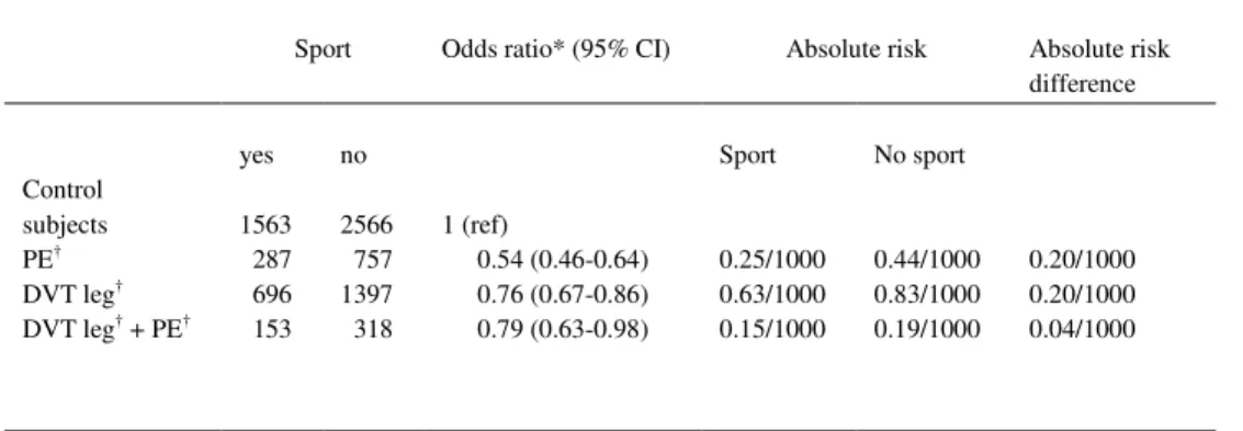
opposite sex. Absolute risk differences were calculated using absolute risks as previously
reported in a prospective study on the incidence of venous thrombosis in France [2].
To remove confounding by health status as much as possible, all participants with a
malignancy were excluded, figure 1. Furthermore, we performed an analysis for the risk of
idiopathic thrombosis in which only individuals were included without clinical risk factors,
e.g. surgery, plaster cast, minor injury, pregnancy, malignancy, bed rest for more than 4
consecutive days or chronic illness in the year prior to indexdate. A multivariate analysis
was performed among those who participated in sports activities regularly to determine
whether type, frequency and intensity of sports activities were independently related to
venous thrombotic risk. All analyses were performed in SAS 9.1 (SAS institute Inc, Cary,
NC, USA).
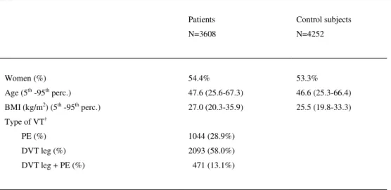
Table 1. Characteristics of study population
Patients N=3608
Control subjects N=4252
Women (%) 54.4% 53.3%
Age (5th -95th perc.) 47.6 (25.6-67.3) 46.6 (25.3-66.4) BMI (kg/m2) (5th -95th perc.) 27.0 (20.3-35.9) 25.5 (19.8-33.3) Type of VT†
PE (%) DVT leg (%) DVT leg + PE (%)
1044 (28.9%) 2093 (58.0%) 471 (13.1%)
†
PE = pulmonary embolism
39
Results
Overall 3608 patients and 4252 control subjects were included in the present analysis. Their
characteristics are shown in table 1. Of the patients 1136 (31.4%) participated regularly in
sports compared with 1686 (39.6%) control subjects. Performing sports reduced the risk of
venous thrombosis (OR 0.64; 95% CI 0.58-0.71). Adjustment for sex, age and body mass
index did marginally change the odds ratio (ORadj 0.71; 95% CI 0.64-0.78) (table 2). The
odds ratios were slightly closer to one when the partner control group was used (ORadj 0.86;
95% CI 0.71-1.02) compared with using the random digit dialing control group (ORadj 0.67;
95% CI 0.60-0.75).
Among participants without an injury in the preceding three months the risk of venous
thrombosis was 0.7 fold decreased (ORadj 0.66; 95% CI 0.59-0.74) for those who
participated in sports activities compared with those who did not participate in sports
activities. When the analysis was restricted to idiopathic thrombosis i.e. individuals without
clinical risk factors for venous thrombosis the risk reduction was similar to the overall risk
(ORadj 0.67; 95% CI 0.58-0.78). Further exclusion of women receiving oral contraceptives
or hormone replacement therapy did not lead to a different point estimate (ORadj 0.66; 95%
CI 0.54-0.79).
Table 2. Association between participating in sports activities and the risk of venous thrombosis
Patients Control subjects OR (95%CI)
ORadj* (95%CI)
No sport 2472 (68.6%) 2566 (60.3%) 1 1
Sports 1136 (31.4%) 1686 (39.7%) 0.64 (0.58-0.71) 0.71 (0.64-0.78) Total 3608 4252
*Adjusted for sex, age and body mass index OR = odds ratio
Chapter 3
40
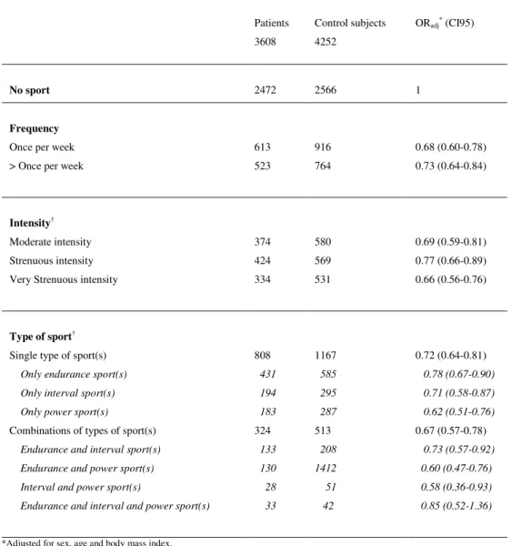
Among those participating regularly in sports activities, the frequency of performing a sport
did not affect the risk of venous thrombosis. Different types and intensities of sports were
equally beneficial (table 3).
Table 3. Relation between frequency, intensity and type of sports activities and risk of venous thrombosis Patients
3608
Control subjects 4252
ORadj* (CI95)
No sport 2472 2566 1
Frequency
Once per week 613 916 0.68 (0.60-0.78) > Once per week 523 764 0.73 (0.64-0.84)
Intensity†
Moderate intensity 374 580 0.69 (0.59-0.81) Strenuous intensity 424 569 0.77 (0.66-0.89) Very Strenuous intensity 334 531 0.66 (0.56-0.76)
Type of sport†
Single type of sport(s) 808 1167 0.72 (0.64-0.81)
Only endurance sport(s) 431 585 0.78 (0.67-0.90) Only interval sport(s) 194 295 0.71 (0.58-0.87) Only power sport(s) 183 287 0.62 (0.51-0.76)
Combinations of types of sport(s) 324 513 0.67 (0.57-0.78)
Endurance and interval sport(s) 133 208 0.73 (0.57-0.92) Endurance and power sport(s) 130 1412 0.60 (0.47-0.76) Interval and power sport(s) 28 51 0.58 (0.36-0.93) Endurance and interval and power sport(s) 33 42 0.85 (0.52-1.36)