4-year radiographic & esthetic evaluation of peri-implant tissue in
immediate implants replacing single teeth in the esthetic zone
Theresa C. Wang, D.D.S.
A thesis submitted to the faculty of the University of North Carolina at Chapel Hill in partial fulfillment of the requirements of the Master of Science in the Department of
Prosthodontics, School of Dentistry.
Chapel Hill 2012
Approved by
Lyndon F. Cooper, D.D.S., Ph.D. Gustavo Mendonca, D.D.S., Ph.D.
Abstract
THERESA C. WANG: 4-year radiographic & esthetic evaluation of peri-implant tissue in immediate implants replacing single teeth in the esthetic zone
(Under the direction of Lyndon F. Cooper D.D.S., Ph.D., Gustavo Mendonca D.D.S., Ph.D., Salvador Nares D.D.S., Ph.D.)
I would like to thank and acknowledge the efforts of:
Lyndon Cooper Gustavo Mendonca
Salvador Nares Christopher Vo Sheng Zhong
Table of Contents
List of Tables ... viii
List of Figures ... viii
Part 1
4-year radiographic & esthetic evaluation of peri-implant tissue in immediately loaded implants replacing single teeth in the esthetic zone.Abstract ... 1
Background ... 2
Materials and Methods ... 7
Patient Selection ... 7
Analysis of Radiographs... 7
Analysis of Photographs ... 9
Statistical Analysis ... 10
Results ... 11
Discussion ... 24
References ... 29
Part 2
The roles of tissue architecture, surgical procedures, implant components and prosthetic decisions on the esthetic outcomes of single tooth implants in the esthetic zone: a comprehensive review. Abstract ... 34Background ... 36
Materials & Methods ... 39
Types of studies ... 39
Types of participants ... 39
Interventions ... 40
Outcome measures ... 41
Search strategy ... 41
Quality assessment ... 42
Data extraction and analysis ... 42
Results ... 45
Implant Survival and Success ... 45
Complications ... 46
Radiographic evaluation... 47
Patient satisfaction ... 49
Peri-implant Structures ... 50
Esthetics ... 52
Bony architecture ... 53
Discussion ... 58
List of Tables
Part I
Table 1 PES subcategory average scores ± SD at 1 year and 4 year………..15 Part II
List of Figures
Part I
Fig 1. Radiographic measurements……….9
Fig 2. 1 year and 4 year mean Pink Esthetic Score (PES)subcategories.………....10
Fig 3. Mesial proximity of fixture to tooth and change in bone level…………..………….….13
Fig 4. Distal proximity of fixture to tooth and change in bone level………..14
Fig 5. Box plot of mean PES score at 1 year and 4 year follow up……….….16
Fig 6. Mesial proximity of fixture to tooth vs. PES……….….17
Fig 7. Distal proximity of fixture to tooth vs. PES.………....…...18
Fig 8. Mesial change in bone level vs. PES……….…...19
Fig 9. Distal change in bone level vs. PES ……….…20
Fig 10. Mesial change in bone level vs. mesial papilla score ……….21
Fig 11. Distal change in bone level vs. distal papilla score ………....22
Fig 12. Mesial proximity to tooth vs. mesial papilla score ……….…23
Fig 13. Distal proximity to tooth vs distal papilla score……….……..24
Part 1: 4-year radiographic & esthetic evaluation of peri-implant
tissue in immediately loaded implants replacing single teeth in the
esthetic zone.
Abstract
Interproximal bone levels are major determinants of implant esthetics. It is suggested that encroachment of the implant-abutment interface (IAI) to the existing tooth (< 1.5mm), negatively affects bone levels. The objective of this study is to compare the crestal bone and esthetic outcome of dental implants replacing single teeth in the esthetic zone at 1 year versus 4 years. Two calibrated examiners evaluated 44 implants. Data was obtained from patients (n=38) enrolled in a prospective clinical trial with an immediate provisionalization protocol. Bone levels were measured from IAI on periapical radiographs using digital methods. Pink Esthetic Scores (PES) were assigned using digital dental photography. Mean mesial and distal change in bone was 0.20 ±1.00 mm and 0.20 ±0.74 mm respectively. Current data fails to indicate a relationship between IAI–tooth distances and crestal bone changes at 4 years. There
is a statistically significant (p<0.001) relationship between smaller IAI–tooth
Background
A missing front tooth has a profound effect on the social and psychological health of an individual (Elias & Sheiham 1998; Abu Hantash et al. 2006). When replacing a front tooth, not only does the esthetic result matter, but it also becomes the critical factor in determining whether treatment can be considered successful. This most often applies to the maxillary anterior dentition, which is visible during daily function and social activities. Fixed tooth replacement in the esthetic zone is not only important for self-esteem but also associated with social perceptions of an individual’s well being (Burkhardt et al. 2000; Willis et al. 2008).
(Chen et al. 2004; DeKok et al. 2006; Lindeboom et al. 2006). Thus, the increasing demand for and use of dental implants in the replacement of missing anterior teeth underscores the importance of understanding and managing the various parameters that influence the esthetic outcome.
natural soft tissue contours will maximize esthetic results (Evans & Chen 2008; Cooper 2008).
Various approaches have been employed to maximize marginal tissue maintenance including hard and soft tissue augmentation, immediate placement protocols, and the use of implants with various configurations that promote the sustainability of tissue (Meijndert et al. 2007; Morris et al. 2004; den Hartog et al. 2008, Stein et al. 2009; Sanz 2009). Furthermore, dental implant components that promote natural visual results, such as ceramic custom abutments, have been employed (Ekfeldt et al. 2011). Other modifications to components, including surface technology to promote cell adhesion, have also been proposed (Nevins et al. 2008). Regardless of the techniques used to maximize marginal tissue, the underlying architecture must be present and biology must be respected to optimize our esthetic goals.
minimize crestal bone loss (Buser et al. 2004). Single implant cases benefit from the hard and soft tissue of adjacent dentition. It has been determined that the interproximal bone of a tooth-bound dental implant is dependent on the level of bone at the adjacent tooth (Avivi-Arber & Zarb 1996; Grunder et al. 2000). Thus, it is stipulated that the presence of papillae is primarily influenced by the interproximal bone level of the adjacent tooth (Jemt et al. 1997; Choquet et al. 2001; Kan et al. 2003; Cardaropoli et al. 2006). Finally, many studies indicate that the interproximal tissue volume increases following crown placement, but the buccal tissue tends to diminish during the first year (Jemt & Lekholm 2003; Cardaropoli et al. 2006; Raes et al 2011). We therefore cannot rule out the influence of the remodeling process that occurs following tooth extraction and surgical trauma (Gargiulo et al. 1961).
Materials and Methods
Patient SelectionThis study included subjects that were enrolled in a longitudinal prospective clinical trial at the University of North Carolina School of Dentistry. The study was approved by the University of North Carolina Institutional Review Board (IRB) and informed consent was obtained from all subjects. All patients were non-smokers and healthy (without systemic disease). The treatment modalities for implant placement (Astra Tech, Osseospeed, Mölndal, Sweden), provisonalization, and final crown delivery
are described in detail in a previous publication (Cooper et al. 2010). A total of 44
subjects with 56 implants were assessed for inclusion in this observational radiographic and photographic study. Due to poor image quality or incomplete data, 38 subjects with 44 implants were included for the final analysis.
Analysis of Radiographs
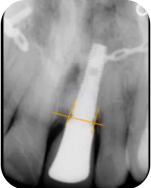
using Image J image processing software (National Institute of Health). All radiographs were calibrated using the known implant diameter as a reference. The implant abutment interface (IAI) was chosen as a reference point, as it was easily recognized. The distance between the reference point to the crestal bone level (My, Dy) and adjacent tooth (Mx, Dx) was measured in millimeters at the mesial and distal of each implant using magnification (x7) to the nearest .01 mm (Figure 1). Two independent examiners not related to the patients’ treatment analyzed all radiographs.
Analysis of Photographs
Intraoral digital photographs were used to assign Pink Esthetic Scores (PES) at 1 year and 4 year follow up (Furhauser et al. 2005). PES is composed of 7 parameters that can be scored 0-1-2, with 2 being the best and 0 being the worst score (Figure 2). Papillae are evaluated for completeness, and the other variables are assessed by comparison with a reference tooth. Cosyn and collegues (2010) defined a PES score of equal or less than 7 to be an esthetic failure, greater than 8 to be acceptable, and greater or equal to 12 to be almost perfect. PES were recorded by two calibrated independent examiners not involved in any treatment. All photographs were scored twice with an interval of 1 week.
Statistical Analysis
Results
Intra/interexaminer Reliability
Near perfect interexaminer agreement was confirmed using the Cronbach Coefficient Alpha Test for the radiographic measurements (α=0.98). PES also demonstrated near perfect inter- and intra- examiner agreement using the Cronbach Coefficient Alpha test (α=0.98).
Description of Sample
In total, 56 implants sites in 44 subjects were eligible for evaluation. 2 patients
were lost to follow up and 16 were not included due incomplete data or poor image
quality. 44 implant sites in 38 subjects provided adequate data for analysis. From this
cohort, there were not any implant failures or complications reported.
Radiographic Analysis
Photographic Analysis
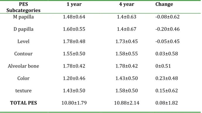
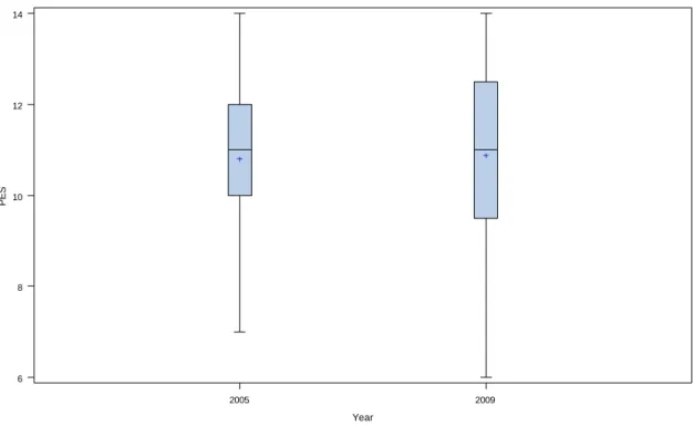
Table 1 demonstrates the mean PES subcategories at 1 year and 4 years. Mean PES increased from 10.80 (SD=1.79) at 1 year to 10.88 (SD=2.14) at 4 years follow up. At 1 year 30% of patients scored near perfect, 68% were considered acceptable, while
only 2% were considered esthetic failures. Similarly, at 4 years 45% of patients scored
near perfect, 48% were considered acceptable, while only 7% were considered esthetic
failures. The changes were not found to be statistically significant (Figure 5).
Table 1 PES subcategory average scores ± SD at 1 year and 4 year. PES
Subcategories 1 year 4 year Change
M papilla 1.48±0.64 1.4±0.63 -0.08±0.62
D papilla 1.60±0.55 1.4±0.67 -0.20±0.46
Level 1.78±0.48 1.73±0.45 -0.05±0.45
Contour 1.55±0.50 1.58±0.55 0.03±0.58
Alveolar bone 1.78±0.42 1.78±0.42 0±0.51
Color 1.20±0.46 1.43±0.50 0.23±0.48
texture 1.43±0.50 1.58±0.50 0.15±0.62
Fig 5. Box plot of mean PES score and distribution at 1 year and 4 year follow up.
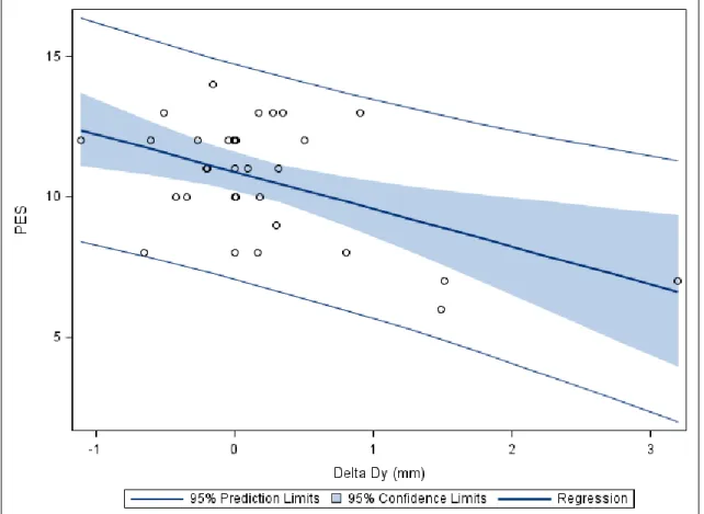
Bone levels and PES
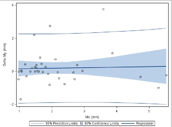
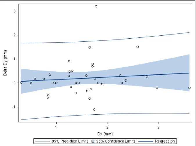
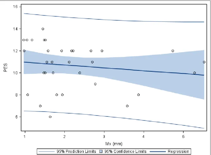
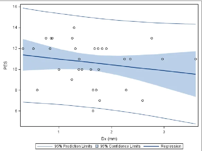
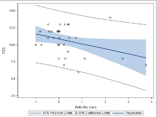
Interestingly, the proximity of the fixture to the tooth from both mesial and distal is significantly associated with PES (p<.0001). The data suggests that the closer the fixture is to the tooth, the lower the PES (Figure 6, 7). Change in bone level over 3 years on the distal side of the fixture is significantly associated with PES (p=0.004). However, this relationship was not identified from the mesial aspect of the fixture (Figure 8, 9). Regression analysis showed that more bone loss on the distal side of the fixture correlated with a lower PES.
2005 2009
6 8 10 12 14
PES
Fig 9. Distal change in bone level vs. PES (p=0. 0040).
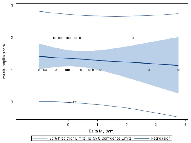
Radiographic measurements and papilla fill
Discussion
The survival rate for the implants included in this study was 100% (38/38) which is in accordance with the survival rates of other studies that had utilized an immediate provisionalization protocol (Kan et al. 2011; DeKok et al. 2006).
The data indicates that the mean change in bone level (0.20 mm) on both aspects of the fixture was consistent with those found in previous studies (Engquist et al. 2002; Wennstrom et al. 2005). Furthermore, bone levels reported are well within the parameters of success criteria described by Albreksson and colleagues (1986).
acceptable to near perfect result (8-14) with only 7% considered failures (≤7). Mean PES for this study at 1 and 4 years were 10.80±1.79 and 10.88 ±2.14 respectively. This is similar to results published by Raes and colleagues (2011) that examined immediately loaded dental implants in the esthetic zone. Other studies using PES have demonstrated PES with almost perfect scores (≥12) in 19-39% of the cases (Juodzbalys and Wang 2007; Chen et al. 2009; Cosyn et al. 2010, Raes et al. 2011). In this study, cases with almost perfect PES scores were demonstrated in 45% of the subjects, which is slightly higher than previous reports. Our results also demonstrated that the PES at the two different time points was found to not be statistically significant. This could be due to the fact that after 1- 1.5 years following surgical intervention, remodeling of the soft tissues has typically stabilized (Johnson et al. 1969; Gargiulo et al. 1961).
Regarding fixture to tooth distance and vertical bone loss, our findings are in agreement with a 3-year retrospective study that reported a lack of relationship between inter-unit distance and longitudinal marginal bone loss (Cardaropoli 2003). Esposito and colleagues (1993) described increased bone loss at the tooth with decreased distance to the implant between the time of implant placement and final crown placement. Since this study did not assess the radiographs at these time points, our study results cannot be compared directly to this previous report.
therapy. The reason for this is unclear and a larger sample size with a comprehensive evaluation of sample characteristics may contribute to our understanding of this finding.
It is worth mentioning that the implants used for this study have a connection that has an under-dimensioned abutment when compared to fixture diameter. It has been postulated that this type of connection moves the inflammatory infiltrate that is present at the implant abutment junction away from the bone (Lazzara et al. 2006). The microgap has been investigated in animal studies, and authors concluded that it is the stability of the interface rather than the size of the microgap that influences bone loss around dental implants (Herman et al. 2001; King et al. 2002). Furthermore, Cardaropoli and colleagues (2005) examined non-platform switched implants and had similar results in regard to bone loss. The influence of the implant abutment interface still needs further examination.
Our study does not demonstrate a relationship between IAI–tooth distances and crestal bone changes at 4 years. However, there is a statistically significant
References
Abu Hantash RO, AL-Omiri MK, AL-Wahadni AM. Psychological impact on implant patients’ oral health- related quality of life. Clinical Oral Implantology Res. 2006;17: 116–123.
Albrektsson T, Zarb G, Worthington P, Eriksson AR. The long-term efficacy of currently used dental implants: a review and proposed criteria of success. Int J Oral Maxillofacial Implants. 1986;1(1):11-25.
Belser UC, Buser D, Hess D, Schmid B, Bernard JP, Lang NP. Aesthetic implant restorations in partially edentulous patients – a critical appraisal. J Periodontology. 2000;17:132–150.
Belser UC, Grutter L, Vailati F, Bornstein MM, Weber HP, Buser D. Outcome evaluation of early placed maxillary anterior single-tooth implants using objective esthetic criteria: a cross-sectional, retrospective study in 45 patients with a 2- to 4-year follow-up using pink and white esthetic scores. J Periodontology. 2009;80:140–151.
Belser U, Martin W, Jung R, Hammerle C, Schmid B, Morton D, Buser D. ITI Treatment Guide, Volume 1: Implant Therapy in the Esthetic Zone Single Tooth Replacements. Berlin: Quintesseence Publishing Co Ltd, 2007.
Belser UC, Schmid B, Higginbottom F, Buser D. Outcome analysis of implant restorations located in the anterior maxilla: a review of the recent literature. Int J Oral Maxillofacial Implants. 2004;19 Suppl:30-42.
Berglundh T, Lindhe J, Ericsson I, Marinello CP, Liljenberg B, Thomsen P. The soft tissue barrier at implants and teeth. Clinical Oral Implants Res. 1991 Apr-Jun;2(2): 81-90. Burkhardt R, Andreoni C, Marinello C. Psychological and social effects of implant sup- ported reconstructions. ACTA Medicinae Dentium Helvetica 2000;5: 1–8.
Cardaropoli G, Lekholm U, Wennstrom JL. Tissue alterations at implant-supported single-tooth replacements: a 1-year prospective clinical study 2006 Apr;17(2):165-71. Chen ST, Wilson TG, Hammerle CH Immediate or early placement of implants following tooth extraction: review of biologic basis, clinical procedures, and outcomes. The International Journal of Oral & Maxillofacial Implants. 2004;19 (Suppl.): 12–25.
Cooper LF, Raes F, Reside GJ, Garriga JS, Tarrida LG, Wiltfang J, Kern M, deBruyn H. Comparison of radiographic and clinical outcomes following immediate provisionalization of single-tooth dental implants placed in healed alveolar ridges and extraction sockets. Int J Oral Maxillofacial Implants. 2010;25:1222–1232.
Cosyn J, Eghbali A, De Bruyn H, Dierens M, De Rouck T. Single implant treatment in healing versus healed sites of the anterior maxilla: an aesthetic evaluation. Clin Implant Dent Relat Res 2010. (ahead of print)
Den Hartog L, Slater JJ, Vissink A, Meijer HJ, Raghoebar GM. Treatment outcome of immediate, early and conventional single-tooth implants in the aesthetic zone: a systematic review to survival, bone level, soft-tissue, aesthetics and patient satisfaction. J Clin Periodontol. 2008 Dec;35(12):1073-86.
De Kok IJ, Chang SS, Moriarty JD, Cooper LF. A retrospective analysis of peri-implant tissue responses at immediate load/provisionalized microthreaded implants. Int J Oral Maxillofac Implants. 2006;21:405–412.
De Smet E, Jacobs R, Gijbels F, Naert I. The accuracy and reliability of radiographic methods for the assessment of marginal bone level around oral implants. Dentomaxillofacial Radiol. 2002;31:176–181.
Ekfeldt A, Furst B, Carlsson GE. Zirconia abutments for single-tooth implant restorations: a retrospective and clinical follow-up study. Clin Oral Implants Res. 2011 Nov;22(11):1308-14.
Elias AC, Sheiham A. The relationship between satisfaction with mouth and number and position of teeth. J Oral Rehabil. 1998 Sep;25(9):649-61.
Engquist B, Astrand P, Dahlgren S, Engquist E, Feldmann H, Grondahl K. Marginal bone reaction to oral implants: a prospective comparative study of Astra Tech and Branemark System implants. Clin Oral Implants Res. 2002 Feb;13(1):30-7.
Oral Implants Res. 2010 Jan;21(1):22-9.
Furhauser R, Florescu D, Benesch T, Haas R, Mailath G, Watzek G. Evaluation of soft tissue around single-tooth implant crowns: the pink esthetic score. Clin Oral Implants Res. 2005 Dec;16(6):639-44.
Gargiulo MS, Frank MW, Orban B. Dimensions and relations of the dentoginigval junction in humans. J Periodontology. 1961;32: 261–267.
Grunder, U. Stability of the mucosal topography around single-tooth implants and adjacent teeth: 1-year results. Int J Periodontics Restorative Dent. 2000;20, 11–17. Hermann JS, Buser D, Schenk RK, et al. Biologic width around titanium implants. A physiologically formed and stable dimension over time. Clinical Oral Implants Research 2000;11:1–11.
Hermann JS, Schoolfield JD, Schenk RK, Buser D, Cochran DL. Influence of the size of the microgap on crestal bone changes around titanium implants. A histometric evaluation of unloaded non-submerged implants in the canine mandible. J Periodontol. 2001 Oct;72(10):1372-83.
Huynh-Ba G, Pjetursson BE, Sanz M, Cecchinato D, Ferrus J, Lindhe J, Lang NP.
Analysis of the socket bone wall dimensions in the upper maxilla in relation to immediate implant placement. Clinical Oral Implants Res. 2010 Jan;21(1):37-42.
Johnson K. A study of the dimensional changes occurring in the maxilla following tooth extraction. Australian Dental Journal 1969; 14: 241–244.
Jung RE, Pjetursson BE, Glauser R, Zembic A, Zwahlen M, Lang NP A systematic review of the survival and complication rates of implant supported single crowns (SCs) after an observation period of at least 5 years. Clin Oral Implants Res. 2008;19(2):119-30.
Kan JY, Rungcharassaeng K, Umezu K, Kois JC. Dimensions of peri-implant mucosa: an evaluation of maxillary anterior single implants in humans. J Periodontol. 2003 Apr;74(4):557-62.
King GN, Hermann JS, Schoolfield JD, Buser D, Cochran DL. Influence of the size of the microgap on crestal bone levels in non-submerged dental implants: a radiographic study in the canine mandible. J Periodontol. 2002 Oct;73(10):1111-7.
Lazzara RJ, Porter SS. Platform switching: a new concept in implant dentistry for controlling post restorative crestal bone levels. Int J Periodontics Restorative Dent. 2006;26:9-17.
prospective randomized study with BioComp implants. J Oral Maxillofacial Surg 2006;64:936–942.
Martegani P, Silvestri M, Mascarello F, Scipioni T, Ghezzi C, Rota C, Cattaneo V. Morphometric study of the interproximal unit in the esthetic region to correlate anatomic variables affecting the aspect of soft tissue embrasure space. J Periodontol. 2007 Dec;78(12):2260-5.
Meijer HJ, Stellingsma K, Meijndert L, Raghoebar GM. A new index for rating aesthetics of implant-supported single crowns and adjacent soft tissues—the Implant Crown Aesthetic Index. Clin Oral Implants Res. 2005 Dec;16(6):645-9.
Meijndert L, Meijer HJ, Stellingsma K, Stegenga B, Raghoebar GM. Evaluation of aesthetics of implant-supported single-tooth replacements using different bone augmentation procedures: a prospective randomized clinical study. Clin Oral Implants Res. 2007 Dec;18(6):715-9.
Nevins M, Nevins ML, Camelo M, Boyesen JL, Kim DM. Human histologic evidence of a connective tissue attachment to a dental implant. Int J Periodontics Restorative Dent. 2008 Apr;28(2):111-21.
Pjetursson BE, Bragger U, Lang NP, Zwahlen M. Comparison of survival and complication rates of tooth supported fixed partial dentures and implant supported fixed partial dentures and single crowns. Clin Oral Implants Res. 2007;18(Suppl.3):97– 113.
Pjetursson BE and Lang NP. Prosthetic treatment planning on the basis of scientific evidence. J Oral Rehab. 2008; 3 (Suppl. 1): 72–79.
Raes F, Cosyn J, Crommelinck E, Coessens P, De Bruyn H. Immediate and conventional single implant treatment in the anterior maxilla: 1-year results of a case series on hard and soft tissue response and aesthetics. J Clin Periodontol. 2011; 38:385–394.
Tomasi C, Sanz M, Cecchinato D, Pjetursson B, Ferrus J, Lang NP, Lindhe J. Bone dimensional variations at implants placed in fresh extraction sockets: a multilevel multivariate analysis. Clin Oral Implants Res. 2010 Jan;21(1):30-6.
Wennstrom JL, Ekestubbe A, Grondahl K, Karlsson S, Lindhe J. Implant-supported single-tooth restorations: a 5-year prospective study. J Clin Periodontol. 2005 Jun;32(6):567-74.
Willis MS, Esqueda CW, Schacht RN. Social perceptions of individuals missing upper front teeth. Percept Mot Skills. 2008 Apr;106(2):423-35.
Part II: The roles of tissue architecture, surgical procedures, implant
components and prosthetic decisions on the esthetic outcomes of
single tooth implants in the esthetic zone: a comprehensive review.
Abstract
Objective: The purpose of this review was to examine how tissue architecture, surgical
procedures, implant components and prosthetic factors contribute collectively to esthetic success in the maxillary anterior and premolar region (esthetic zone).
Materials and methods: Pubmed, Embase and the Cochrane electronic databases were
a collective manner. However, the results of this review suggest that there is not a standardized measure of esthetic success in dental implant therapy.
Conclusions: The literature included in this review suggests that implant esthetic
Background
The progressive nature of dental implants treatment has been facilitated by outstanding survival rates from several long-term human clinical control trials (Jung et al. 2008, den Hartog et al. 2008). This has been accompanied by the establishment of treatment guidelines, improvements in surgical protocol, and the development of innovative implant components and surface technologies that have contributed to improving the quality of care in dental implant treatment (Belser et al. 2007, Shalabi et al. 2006).
Esthetic concerns apply when the implant restoration and the surrounding soft tissues are visible during daily functional activities and in social settings. The increasing use of dental implant therapy to replace missing teeth in the esthetic zone underscores the importance of evaluating esthetic success. However, scientific literature describing reproducible esthetic parameters is considerably insufficient (Belser et al. 2004). In the esthetic zone, success is highly dependent on the long-term esthetic results that can be achieved. Optimally, the goal is to match the peri-implant soft tissue with the soft tissue of the adjacent natural teeth; the implant crown should blend naturally in size, contour and shape with the adjacent teeth (Meijer et al 2005). Yet, the most unpredictable determining factor in establishing esthetics is peri-implant soft and hard tissue support. Not only do clinicians have to consider existing tissue architecture, another important factor is the surgical positioning of the implant and subsequent remodeling process of the bone (Cardaropoli et al 2006).
Materials & Methods
Types of studiesAll prospective human clinical trials of dental implants replacing single teeth in the esthetic zone were considered for this review. Study designs included randomized control trials, controlled trials, randomized trials and prospective cohort studies. Retrospective studies were not included. No time limitations were implemented. Language was restricted to papers published in English.
Types of participants
Interventions
The factors examined in the articles being reviewed can be grouped into the following categories:
A. Anatomical considerations
1) Bone wall dimensions (Ferrus et al. 2009, Huynh-Ba et al. 2009, Tomasi et al. 2009)
B. Surgical interventions
1) Early versus delayed implant placement (Gotfredsen et al. 2004) 2) Simultaneous hard tissue augmentation and implant placement
i. Type of grafting material (Meijndert et al. 2007) ii. Graft versus no graft (Chen et al. 2007)
C. Implant components 1) Type of implant
i. Cylinder versus taper (Sanz et al. 2009)
D. Prosthetic decisions 1) Loading protocols
i. Immediate versus delayed load in a flapless surgical approach (Oh et al. 2008)
Outcome measures
Implant survival, defined as the presence of the implant at the time of follow-up
Complications: biological and technical
Changes in marginal bone level assessed by radiographs
Assessment of peri-implant structures
o Papilla Index (Jemt et al. 1997)
o Probing depth, plaque index, bleeding on probing
Aesthetic indexes
o Pink Esthetic Score (Furhauser et al. 2005)
o Implant Crown Esthetic Index (Meijer et al. 2005)
Patient satisfaction/self-evaluation of esthetics
Measurements of bony architecture at time of extraction, implant placement or second stage surgery
Search strategy
This review consisted of a search of the literature utilizing PUBMED and EMBASE and was supplemented with a search of systematic reviews in the Cochrane Central Register of Controlled Trials (CENTRAL).
Two examiners scanned the titles and abstracts found in the search. Full-text articles were obtained and examined by two independent reviewers.
Quality assessment
All studies that were not relevant to the topic in review were not included for full text analysis. For example, studies examining dental implants replacing multiple missing teeth, with dentures or fixed partial dentures as ultimate prosthesis, were excluded. Those examining posterior teeth and studies with improper study design were not included. Following full-text analysis, methodological quality was assessed (Table 1).
Data extraction and analysis
Table 1 Studies excluded after quality assessment and reasons for exclusion.
Study Study design Reason for exclusion
Cochran et al. (2009)
Prospective Multicenter Human Clinical Trial
Not exclusive to esthetic zone, not exclusive to single tooth
Martegani et al. (2007)
Prospective Multicenter Human Clinical Trial
Evaluates natural teeth, not implants
Sunitha et al. (2008) Prospective Human Clinical Control Trial
Not exclusive to esthetic zone
Bianchi et al. (2004) Prospective Human Randomized
Clinical Control Trial
Not exclusive to esthetic zone
Lee & Hasegawa (2008)
Prospective Human Clinical Trial Small sample size, too many variables
Yilmaz et al. 1998 Prospective Human Clinical Control
Trial
Not exclusive to single tooth
Johnson & Persson (2001)
Prospective Human Clinical Trial Sites included not specified
Kemppainen et al. (1997)
Prospective Human Randomized Clinical Trial
Not exclusive to esthetic zone
Morris et al. (2004) Prospective Multicenter Human
Clinical Trial
Not exclusive to esthetic zone
Zembic et al. (2009) Prospective Human Randomized
Clinical Control Trial
Posterior teeth evaluated
Sailer et al. (2009) Prospective Human Randomized
Clinical Control Trial
Posterior teeth evaluated
Sethi et al. (2000) Prospective Human Clinical Trial Sites included not specified, not
exclusive to single tooth
Kastenbaum et al. (1998)
Prospective Human Clinical Trial Not exclusive to esthetic zone, not
exclusive to single tooth
Malo et al. (2003) Prospective Multicenter Human
Clinical Trial
Results
Implant Survival and SuccessOf the selected articles, authors that reported on survival considered the presence of the implant at follow-up as survival and followed the criteria for successful osseointegration proposed by Smith and Zarb (1989).
Gotfredsen and colleagues (2004) reported 100% implant survival rate for both their “early” (4 wee s following extraction) and conventional placement groups.
Meijndert et al. 2007 performed conventional placement with bone augmentation. All of their cases were grafted with an autogenous chin graft or xenograft (BioOss®) and resorbable collagen membrane. They had two implants that were mobile at the start of the prosthetic procedures, and thus did not include them for aesthetic evaluations. However, these authors were able to successfully re-operate with reimplantation and bone augmentation.
In a flapless approach, Oh and colleagues (2006) reported that at 6 months, they achieved a survival rate for delayed load and immediate load implants at 100% and 75%, respectively. Interestingly, all failures in their immediately loaded group were in the premolar region.
implants that were provisionalized, in both immediately and conventionally placed implants, yielded excellent survival of 96-100% at 1 year follow up (Hall et al. 2007; Lindeboom et al. 2006, De Rouck et al. 2008, De Rouck et al. 2009).
Failure of implants generally occurred in the early stages following dental implant placement. Some studies involved implants that showed mobility after 2-3 weeks (Lindeboom et al. 2006) while others proved to be more dispersed in regards to time of failure. In 2008, De Rouck and colleagues had one implant fail after one month with concurrent pain, mobility and discomfort. Likewise, in 2009, De Rouck, et al. had one implant lost due to mobility after the first month, one lost due to mobility after three months, and one was lost with concurrent pain after three months. In all immediately placed implants, failures occurred for all restorative treatment groups: delayed restorations, immediately provisionalized and immediately loaded implants. However, all treatment groups demonstrated good-excellent success rates of 92%, 88-97% and 92% respectively. (Lindeboom et al. 2006; De Rouck et al. 2008; De Rouck et al. 2009).
Complications
complications include fistulas (Gotfredsen et al. 2004; De Rouck et al. 2008), soft tissue dehiscence (Gotfredsen et al. 2004), peri-implant mucositis, (Chen et al. 2007) and abscess formation (Chen et al. 2007). Gotfredsen and colleagues (2004) reported that there was a gap at the implant abutment interface in the case that had a fistula. All biologic complications did not result in implant removal and were treated with local debridement and antibiotics.
Technical complications included loosened abutment screws (Gotfredsen et al. 2004; Lindeboom et al. 2006), porcelain fractures (Gotfredsen et al. 2004; Lindeboom et al. 2006) and loss of crown retention (De Rouck et al. 2008). Loose screws were tightened and occasionally accessed through cemented crowns. The technical complications were managed on a case by base basis, with regards to fractured porcelain. The porcelain fractures were resolved through crowns replacement or smoothing with polishing burs. The broad array of complications throughout the selected articles lacked association with implant placement strategies or restorative protocol.
Radiographic evaluation
In addition, mean bone loss did not differ significantly between immediately loaded and immediately provisionalized groups (Lindeboom et al. 2006). This group reported that the mesial marginal bone loss over a 12-month evaluation was minimal being 0.27(SD=0.2) mm for the immediate load group versus 0.28(SD=0.22) mm for the immediate placement group. Likewise, the distal marginal bone loss over 12 months was 0.19(SD=0.15) mm for the immediate load group versus 0.2(SD=0.11) mm for the immediate placement group.
Patient satisfaction
Of the 12 included studies, only four incorporated a self-report to reflect patient satisfaction. Each of these four studies utilized a 10 cm Visual Analog Scale (VAS) to reflect the degree of patient satisfaction, and high scores were readily apparent in all cases. One study determined that there lacked correlation between the clinicians’ objective means of scoring the implant restoration (Implant Crown Aesthetic Index) and the patients’ satisfaction (Meijndert et al, 2007). Interestingly, the Implant crown Aesthetic Index yielded acceptable result for 66% of the cases while the patient satisfaction questionnaire revealed an acceptable result for 100% of the cases. There was a correlation between the patients’ and professionals’ opinion of the peri-implant mucosa, however.
Peri-implant Structures
Six of the chosen studies measured papilla fill or utilized a papilla index to reflect the changes in interproximal tissue volume during the duration of the study. Gotfredsen and colleagues (2004) reported that papilla shrinkage was not significant during their 5-year evaluation for both early and delayed placement groups. Overall, they found an increased in papillary fill over time. Oh and colleagues (2006) performed a flapless approach on all subjects and demonstrated an increase in papilla index from baseline to 6 months, however it was only statistically significant for the immediate load group and not the delayed load group. Furthermore, there were not any significant differences between treatment groups at each time point.
index was not statistically different. Furthermore, the papilla index was poorly correlated to radiographic measurements of the implant position to the adjacent tooth. The largest amount of papilla loss was found at the 3 month assessment in both immediately restored and delayed restoration groups (DeRouck et al. 2008). Mean papilla shrinkage was demonstrated to be twice as high for the delayed restoration group when compared to the immediate restoration group. They also reported significant papilla regeneration in the delayed restoration group at 1 year. Finally, De Rouck and colleagues (2009) found that the largest reduction in papilla height was found at the 3-month follow up visit for implants that were immediately placed and provisionalized. They also demonstrated a trend of recovery in papilla height following 3 months of healing, although this was not statistically significant. Overall, the selected articles for this review demonstrate that there is not a difference in papilla fill when various surgical and prosthetic protocols are utilized and the most change occurs in the first 3 months following implant placement.
Peri-implant Health
Lindeboom et al. (2006) incorporated the gingival margin level in their clinical analysis and concluded that 100% of the implants in the immediately loaded and 91% of the implants in the immediately provisionalized (non-loaded) group had an ideal buccal margin.
Bleeding on probing (BOP) was recorded in a few of the studies as an indication of peri-implant health. Two studies determined there to be no difference between immediately loaded and delayed groups (Oh et al. 2006, De Rouck et al. 2009). In addition, it was determined that BOP scores consistently decline with time. In 2008, De Rouck et al. found that BOP scores were 54% at 1 month and 41% at 1 year with an immediately placement and provisional protocol. Additionally, Gotfredson et al. (2004) found that at the 3-year follow-up 54% had a bleeding score of 0 while at the 5-year follow-up 62% had a bleeding score of 0.
Esthetics
study that utilized an objective measurement index (Implant Crown Aesthetic Index). They reported an acceptable result in 66% of the cases, however, this did not correlate with the patients’ self-evaluation using VAS.
Bony architecture
As previously discussed, the vast majority of the chosen articles measured marginal bone level changes in their analysis. No trend can be drawn in terms of whether mesial or distal bone loss was more common, as it varied with each study. However, as a general trend, the largest amount of bone loss was observed in the first 3 months. After this initial period, the amount of bone loss notably declined (De Rouck et al. 2008; De Rouck et al. 2009, Chen et al. 2007). For example, De Rouck and colleagues (2008) found that marginal bone loss in the first 3 months was 0.58 mm mesially and 0.47 mm distally. After the span of one year, levels of marginal bone loss were recorded to be 0.98mm mesially and 0.78 distally.
In addition, bone level changes between experimental and control groups proved to be relatively consistent. In three studies (De Rouck et al. 2009; Lindeboom et al. 2006; Hall et al. 2007) it was determined that after 1 year, there was not a long-term difference between bone loss in immediately loaded versus comparison group (delayed, non-loaded provisionalized, or conventional). One study concluded that dimensional changes were not significant between cylindrical or tapered implant configurations (Sanz et al. 2010).
outcomes were consistently dependent on the baseline characteristics. Three studies conducted by this group agree that sites with thick boney walls corresponded to a greater degree of bone fill. Furthermore, sites with a thick buccal bone crest experienced smaller degrees of vertical resorption (Huynh-Ba et al. 2010; Ferrus et al. 2010; Tomasi et al. 2010). Implants that were more apically positioned experienced less thread exposure than implants that were positioned closer to the alveolar crest, suggesting that anterior sites are more susceptible to ridge alterations (Ferrus et al. 2010; Tomasi et al. 2010). Finally, there is a negative correlation between the size of the vertical residual gap and the vertical position of the bone crest opposite the implant. A positive correlation exists between size of the horizontal gap, the horizontal residual distance, and the residual depth. Essentially, the larger the horizontal gap (>1mm), the greater the amount of newly formed bone. (Ferrus et al. 2010; Tomasi et al. 2010; Huynh-Ba et al. 2010; Sanz et al. 2010).
55
56
Discussion
This comprehensive review assessed implant supported single tooth replacements and esthetic outcomes. There were a variety of parameters that the randomized control trials and clinical control trials identified as variables to evaluate their treatment modalities. We were able to identify outcome measures from the selected studies to include survival, complications, radiographic evaluation of crestal bone changes, patient satisfaction, peri-implant structures, peri-implant tissue health, esthetics, and bony architecture. This review included immediate placement and immediate provisionaliation protocols, in addition to conventional approaches. Although there is still insufficient long-term data regarding dental implants in the esthetic zone and esthetics, there are promising trends in the outcomes thus far. Currently, the available controlled clinical trials suggest that immediate and early placement protocols fair just as well as the conventional approach as treatment options. This is particularly important in that immediate restoration of a tooth in the esthetic zone may have profound effects for the patients’ self esteem and satisfaction with treatment.
positive results. However, authors have mentioned that careful evaluation of the clinical scenario is necessary. Proper diagnostics and treatment planning with an understanding of the peri-implant architecture is key in optimizing results. The studies found that implant loss mostly occurred within the first year of placement and early failures have been reported to be due to lack of primary stability and surgical factors.
The most consistent finding throughout the studies evaluated was that in both soft and hard tissue, the most changes were reported to occur at the 3-month follow up. This can be most readily explained by the fact that tissue remodeling occurs mostly within the first 6-8 weeks following treatment. The studies also indicated that following this 3-month mark, the changes in bone and soft tissue were not only much less in magnitude but also not statistically significant.
to be used in conjunction with PES so that the both soft tissue factors and the restorative crown can be scored independently, and ultimately combined to assess overall esthetic outcome. Since these indices are relatively new, there are not many studies that have utilized them to evaluate esthetics. However, it is important to be able to objectively measure esthetic outcomes as we can compare esthetic scores with patient based outcomes such as VAS or OHIP scores. Although the patients’ opinion may depend highly on personal expectations and on baseline esthetics, being able to identify the threshold for patient satisfaction is key in becoming clinically efficient.
Further studies concerning patient specific factors, including health status and tobacco/alcohol use, are imperative for our continued understanding of dental implant therapy. From the studies included in this discussion, Tomasi and collegues (2010) found through multilevel modeling that other factors, including age and smoking, were important in successful vertical gap fill in immediately placed implants. Age has been associated with decreased vascularization and bone formation in animal models (Lu et al. 2008). Furthermore, smoking has been described as a risk factor in dental implant placement. Through retrospective studies, smokers have been found to be at an increased risk of complications, have peri-implant mucositis, peri-implantitis and implants lost (Bain and Moy 1993; Rodriguez-Argueta et al. 2011; Cavalcanti et al. 2011).
References
Bain CA, Moy PK. The association between the failure of dental implants and cigarette smoking. Int J Oral Maxillofac Implants. 1993;8(6):609-15.
Belser U, Martin W, Jung R, Hammerle C, Schmid B, Morton D, Buser D. ITI Treatment Guide, Volume 1: Implant Therapy in the Esthetic Zone Single Tooth Replacements. Berlin: Quintesseence Publishing Co Ltd, 2007.
Belser UC, Schmid B, Higginbottom F, Buser D. Outcome analysis of implant restorations located in the anterior maxilla: a review of the recent literature. Int J Oral Maxillofac Implants. 2004;19 Suppl:30-42.
Cardaropoli G, Lekholm U, Wennstrom JL. Tissue alterations at implant-supported single-tooth replacements: a 1-year prospective clinical study. Clin Oral Implants Res. 2006 Apr;17(2):165-71.
Cavalcanti R, Oreglia F, Manfredonia MF, Gianserra R, Esposito M. The influence of smoking on the survival of dental implants: a 5-year pragmatic multicentre retrospective cohort study of 1727 patients. Eur J Oral Implantol. 2011 Spring;4(1):39-45.
den Hartog L, Slater JJ, Vissink A, Meijer HJ, Raghoebar GM. Treatment outcome of immediate, early and conventional single-tooth implants in the aesthetic zone: a systematic review to survival, bone level, soft-tissue, aesthetics and patient satisfaction. J Clin Periodontol. 2008 Dec;35(12):1073-86.
Ferrus J, Cecchinato D, Pjetursson EB, Lang NP, Sanz M, Lindhe J. Factors influencing ridge alterations following immediate implant placement into extraction sockets. Clin Oral Implants Res. 2010 Jan;21(1):22-9.
Huynh-Ba G, Pjetursson BE, Sanz M, Cecchinato D, Ferrus J, Lindhe J, Lang NP. Analysis of the socket bone wall dimensions in the upper maxilla in relation to immediate implant placement. Clin Oral Implants Res. 2010 Jan;21(1):37-42.
Jung RE, Pjetursson BE, Glauser R, Zembic A, Zwahlen M, Lang NP A systematic review of the survival and complication rates of implant supported single crowns (SCs) after an observation period of at least 5 years. Clin Oral Implants Res. 2008 (in press).
Lang NP, Berglundh T, Heitz-Mayfield LJ, Pjetursson BE, Salvi GE, Sanz M. Consensus statements and recommended clinical procedures regarding implant survival and complications. Int J Oral Maxillofac Implants. 2004;19 Suppl:150-4.
Martegani P, Silvestri M, Mascarello F, Scipioni T, Ghezzi C, Rota C, Cattaneo V. Morphometric study of the interproximal unit in the esthetic region to correlate anatomic variables affecting the aspect of soft tissue embrasure space. J Periodontol. 2007 Dec;78(12):2260-5.
Meijer HJ, Stellingsma K, Meijndert L, Raghoebar GM. A new index for rating aesthetics of implant-supported single crowns and adjacent soft tissues--theImplant Crown Aesthetic Index. Clin Oral Implants Res. 2005 Dec;16(6):645-9.
Meijndert L, Meijer HJ, Stellingsma K, Stegenga B, Raghoebar GM. Evaluation of aesthetics of implant-supported single-tooth replacements using different bone augmentation procedures: a prospective randomized clinical study. Clin Oral Implants Res. 2007 Dec;18(6):715-9.
Morris HF, Ochi S, Crum P, Orenstein IH, Winkler S. AICRG, Part I: A 6-year multicentered, multidisciplinary clinical study of a new and innovative implant design. J Oral Implantol. 2004;30(3):125-33.
Pjetursson BE, Bragger U, Lang NP, Zwahlen M. Comparison of survival and complication rates of tooth supported fixed partial dentures and implant supported fixed partial dentures and single crowns. Clin Oral Implants Res. 2007;18(Suppl.3):97– 113.
Pjetursson BE and Lang NP. Prosthetic treatment planning on the basis of scientific evidence. J Oral Rehab 2008; 3 (Suppl. 1): 72–79.
Rodriguez-Argueta OF, Figueiredo R, Valmaseda-Castellon E, Gay-Escoda C. Postoperative complications in smoking patients treated with implants: a retrospective study. J Oral Maxillofac Surg. 2011 Aug;69(8):2152-7.