STRUCTURE-FUNCTION RELATIONSHIPS OF LONG NON-CODING RNAS IN LIVING CELLS
Matthew James Smola
A dissertation submitted to the faculty at the University of North Carolina at Chapel Hill in partial fulfillment of the requirements for the degree of Doctor of Philosophy in the Department of Chemistry
Chapel Hill 2015
ABSTRACT
Matthew James Smola: Structure-Function Relationships of Long Non-Coding RNAs in Living Cells (Under the direction of Kevin M. Weeks)
“When we recognize our place in an immensity of light-years and in the passage of ages, when we grasp the intricacy, beauty, and subtlety of life, then that soaring feeling,
ACKNOWLEDGEMENTS
This work represents the culmination of over two decades of formal education and would not have been possible without the enduring love, support, and encouragement of my family. From the early days of science fair projects, illegible negative signs, and misplaced calculators to more recent experiences in experimental troubleshooting and code debugging, my parents have always been my strongest and most vocal supporters. They are the giants upon whose shoulders I now stand.
At its best, scientific research is immensely fulfilling, though at its worst it can be incredibly frustrating. Through all the highs and lows of graduate school, I have been lucky to be surrounded by my fellow Weeks Lab members. I am grateful for the constant encouragement, advice, and exquisitely well-timed comic relief that all of you have provided over the years.
It has been a privilege to experience graduate school alongside a group of close-knit friends. Kathleen, Fatima, Gregg, Tim, and Hannah, I am grateful to have had each of you in my life these past few years. I am certain that you will each have a positive impact on our world and I cannot wait to see what you accomplish.
I would like to thank Mauro Calabrese for inadvertently sparking my interest in lncRNA biology by proposing the Xist project. I am grateful to have you as a mentor and have enjoyed the casual nature of our interactions, especially the many enlightening, exciting, and frank discussions that we have had. Best of luck as you establish your own research group!
TABLE OF CONTENTS
LIST OF TABLES ... ix
LIST OF FIGURES ... x
LIST OF ABBREVIATIONS AND SYMBOLS ... xi
CHAPTER 1: THE RELATIONSHIP BETWEEN RNA STRUCTURE AND FUNCTION ... 1
Hierarchy of RNA structure ... 1
The rise of lncRNAs as effectors of genetic regulation ... 3
Methods for interrogating RNA secondary structure ... 6
Coupling RNA structure probing to massively parallel sequencing ... 9
Research overview ... 10
Perspective ... 13
References ... 14
CHAPTER 2: ACCESSING SHAPE-MAP PROFILES OF RARE RNA TRANSCRIPTS ... 19
Introduction ... 19
Experimental design ... 22
RNA folding and modification ...22
Mutational Profiling (MaP) ...25
Library construction and sequencing ...25
Results ... 28
Small RNA workflow – TPP riboswitch ...28
Amplicon workflow – Mouse ribosomal RNA ...28
Conclusion ... 31
Methods ... 33
RNA extraction and modification ...33
SHAPE-MaP ...34
SHAPE profile generation ...34
References ... 35
CHAPTER 3: DETECTION OF RNA-PROTEIN INTERACTIONS IN LIVING CELLS WITH SHAPE ... 37
Introduction ... 37
Results ... 41
Comparison of SHAPE-MaP and icSHAPE ...41
Validation of the ΔSHAPE approach ...43
Application of ΔSHAPE to RNase MRP ...48
Discussion ... 50
Methods ... 52
In cellulo modification ...52
Ex vivo RNA extraction and modification ...52
Denaturing control ...53
U1, SRP, and 5S SHAPE-MaP ...53
Whole-transcriptome SHAPE-MaP ...54
Sequencing and SHAPE profile generation ...54
SHAPE reactivity normalization ...55
icSHAPE profile generation ...55
Calculating ΔSHAPE, Z-factors, and standard scores to determine binding sites ...56
References ... 58
CHAPTER 4: SHAPE ANALYSIS REVEALS TRANSCRIPT-WIDE CELLULAR INTERACTIONS AND STABLE STRUCTURAL DOMAINS WITHIN THE XIST lncRNA ... 61
Introduction ... 61
Results ... 64
Ex vivo structure probing ...64
The effects of the cellular environment on Xist structure ...67
Localized cellular effects on Xist structure ...71
Conclusion ...76
Methods ... 78
In-cell RNA modification ...78
Ex vivo RNA extraction and modification ...78
Denaturing control ...79
Xist SHAPE-MaP ...79
Sequencing and SHAPE profile generation ...79
Structure modeling ...80
SNP analysis ...80
Conservation analysis ...81
Computing regions of large absolute reactivity changes ...81
Identifying protein binding sites and conformational changes with ΔSHAPE ...81
HuR RNA immunoprecipitation and sequencing ...82
Identification of sequence motifs among ΔSHAPE-identified interaction sites ...82
Clustering Fus-localized positive ΔSHAPE sites by pairing probability ...82
Evaluation of TARDBP antisense knock-down data ...83
LIST OF TABLES
LIST OF FIGURES
Figure 1.1 Elements and heirarchy of RNA structure ...2
Figure 1.2 Example mechanisms of lncRNA gene regulation ...5
Figure 1.3 Examples of RNA structure-probing reagents. ...8
Figure 2.1 SHAPE chemistry and useful SHAPE reagents ...20
Figure 2.2 Overview of SHAPE-MaP workflows ...23
Figure 2.3 Representative library size distributions as a function of workflow ...27
Figure. 2.4 Example of results obtained with the small RNA workflow ...29
Figure. 2.5 Example of results obtained with the amplicon workflow ...30
Figure 2.6 Example of results obtained with the randomer workflow ...32
Figure 3.1 Experimental and analytical framework for detecting SHAPE-MaP reactivity differences ...39
Figure 3.2 Comparison of SHAPE-MaP and icSHAPE reactivities ...42
Figure 3.3 Identification of protein binding sites by ΔSHAPE analysis ...44
Figure 3.4 Summary of results obtained for the SRP RNA ...45
Figure 3.5 Summary of results obtained for the 5S rRNA ...47
Figure 3.6 In-cell analysis of RNase MRP RNA interactions ...49
Figure 4.1 Predicted structural architecture of the Xist lncRNA ...65
Figure 4.2 Structural features of repeat regions A and E ...68
Figure 4.3 Effects of the cellular environment on Xist lncRNA structure ...70
Figure 4.4 Sequence motifs identified among ΔSHAPE sites ...72
LIST OF ABBREVIATIONS AND SYMBOLS 1M6 1-methyl-6-nitroisatoic anhydride
1M7 1-methyl-7-nitroisatoic anhydride
A adenosine
ARE A-U rich element BzCN benzoyl cyanide
C cytosine
cDNA complimentary DNA
CMCT 1-cyclohexyl-3-(2-morpholinoethyl)carbodiimide metho-p-toluenesulfonate cryo-EM cryo-electron microscopy
DMS dimethyl sulfate DMSO dimethyl sulfoxide DNA deoxyribonucleic acid dsRNA double-stranded RNA
EDTA ethylenediaminetetraacetic acid
G guanosine
g gram
HEPES N-2-hydroxyethylpiperazine-Nʹ′-ethanesulfonic acid
kb kilobase
KCl potassium chloride
kethoxal 1,1-dihydroxy-3-ethoxy-2-butanone lncRNA long non-coding RNA
M molar
min minute
ml milliliter
mM millimolar
mol mole
MPS massively parallel sequencing
ng nanogram
nM nanomolar
NMIA N-methylisatoic anhydride
nt nucleotide
PCR polymerase chain reaction
pmol picomole
RMRP RNase MRP
RNA ribonucleic acid
rRNA ribosomal RNA
RT reverse transcription SDS sodium dodecyl sulfate
SHAPE selective 2ʹ′-hydroxyl acylation analyzed by primer extension SRA steroid receptor RNA activator
SRP signal recognition particle ssRNA single-stranded RNA TF transcription factor TPP thiamine pyrophosphate TSC trophoblast stem cells
U uracil
Xi inactive X chromosome Xist X-inactive specific transcript
°C degree Celsius
µ mean
µg microgram
µl microliter
µM micromolar
CHAPTER 1: THE RELATIONSHIP BETWEEN RNA STRUCTURE AND FUNCTION Hierarchy of RNA structure
Ribonucleic acid (RNA) has been the focus of an extended scientific renaissance that has been gradually building momentum over the past 25 years (1). While originally thought to act as a simple, passive intermediate between genetic information stored in the nucleus and the protein synthesizing components of the cytoplasm, we now know that RNA is actively and critically involved in modulating gene expression through a variety of mechanisms (2). These modes of action can be cis- or trans-acting and can be carried out by the RNA alone or require additional protein partners. It has been discovered that RNA can regulate gene expression through alternative RNA splicing (3), RNA interference (4, 5), and by the action of riboswitches (6) and long non-coding RNA (lncRNA) (7).
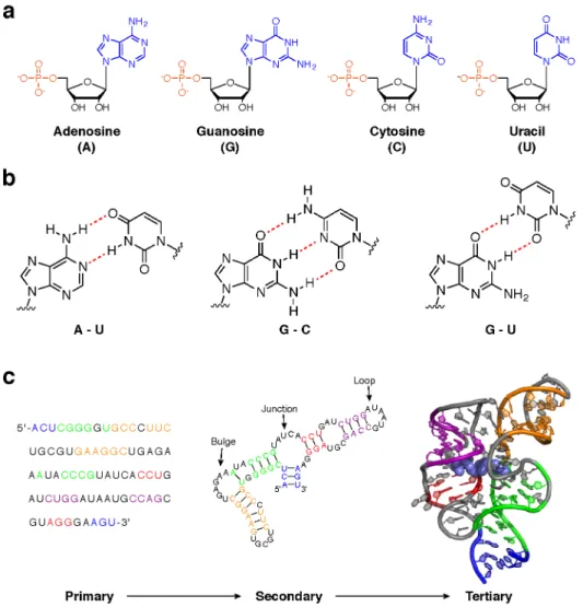
Underlying many of these functions is the ability of RNA molecules to adopt complex structures (8). RNA structure is hierarchical: structural elements at one level provide the foundation for higher-order structures (9). The simplest, primary structure of an RNA is defined by the specific linear sequence of individual nucleotides that comprise the full-length molecule. This sequence can be any combination of the four RNA nucleotides: adenosine (A), cytosine (C), guanosine (G), and uracil (U) (Fig. 1.1a). These are often described as the “letters” of the RNA “alphabet.” In some roles, RNA function is driven largely by primary structure (e.g. RNA silencing). However, higher levels of RNA structure can both tune these functions and enable more complex activities.
and, in many RNAs, multiple helical elements are connected by unpaired nucleotides to form loops, bulges, and junctions (Fig. 1c). In most cases, the secondary structure of an RNA strongly affects its tertiary structure This highest level of RNA structure is defined in three dimensions and is modulated by the length and placement of helices, loops, bulges, and other structural elements (9) (Fig. 1c). Tertiary structures can be stabilized by and, in many cases, facilitate interaction with proteins, small molecule ligands, and metal ions (10). RNA tertiary structures often play critical roles in biology. For example, riboswitches are a class of RNA elements that fold into well-defined tertiary structures and specifically recognize small metabolites (6). The binding and release of these metabolites induces dramatic three-dimensional structure changes that regulate gene expression.
Since RNA structure is closely linked to function, structural characterization of an RNA is often the first step to understanding how it operates. Experimentally determining the tertiary structure of an RNA can be an arduous undertaking, and the computational tools needed to predict such structures de novo are both resource-intensive and imperfect. However, even secondary structure can be highly informative of the function of an RNA (11), and several efforts to improve RNA secondary structure prediction have been successful (12). For this reason, and because secondary structure provides the foundation for tertiary structure, most investigations into RNA structure-function relationships focus on secondary structure. However, until recently, most RNA structure work has focused on abundant RNAs. Although biologically critical in many cases, rarer transcripts have been largely ignored in favor of those that are more easily accessible.
The rise of lncRNAs as effectors of genetic regulation
transfer RNA, and small nuclear RNA have been well-known for years, new families of non-coding RNAs are being continually discovered (15). Of particular interest are the long non-coding RNAs (lncRNAs), defined as transcripts longer than 200 nucleotides with little to no coding potential (7). This class of RNA is frequently associated with epigenetic regulation of chromatin (16), but the list of cellular roles ascribed to lncRNAs now includes transcriptional regulation, modulation of protein activity, organization of protein complexes, and intercellular signaling (17). lncRNAs exhibit cell type-specific expression patterns (18) and are associated with several diseases, including cancer (19). As a result, lncRNAs have been the focus of intense research in the last decade.
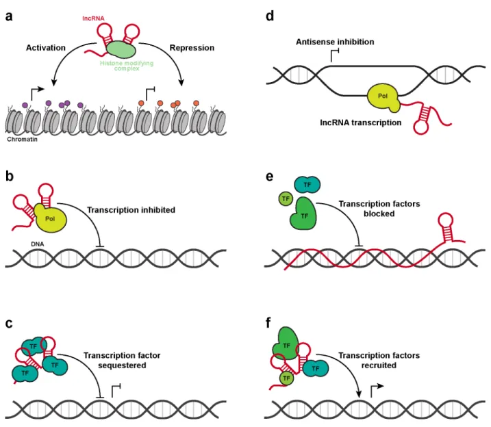
In performing various regulatory functions, lncRNAs often interact with protein complexes and genomic DNA (7, 17, 20, 21). The nature of these interactions can vary greatly. For example, lncRNAs such as Xist, HOTAIR, and Kcnq1ot1 act as molecular scaffolds to coordinate histone modifying complexes and silence specific regions of the genome (22-24) (Fig. 1.2a). In other cases, lncRNAs can act as molecular sponges and sequester RNA polymerase II or transcription factors, reducing gene transcription (25, 26) (Fig. 1.2b-c). Occasionally the lncRNA itself is not important; rather, the process of lncRNA transcription negatively affects antisense genes at the same locus (27) (Fig. 1.2d). Alternatively, some lncRNAs have been found to interact directly with genomic DNA, inhibiting the binding of transcription complexes (28) (Fig. 1.2e). Finally, not all lncRNA actions are repressive. For example, Evf-2 recruits a transcriptional activator to specific DNA regulatory elements, leading to increased transcription at those loci (29) (Fig. 1.2e).
are 0.87 and 2.1 kilobases (kb) long, respectively, and indeed adopt modular structures. However, they represent simple systems relative to other lncRNAs. For example, the Xist lncRNA is roughly an order of magnitude greater in length and associates with more than 50 proteins in order to silence an entire chromosome in a process called X-chromosome inactivation (XCI) (24, 32-35). Although XCI has been researched for more than 50 years (36) and the central role of Xist in this process was discovered 20 years ago (24, 35), the structural architecture of this lncRNA has yet to be determined. As an archetype of lncRNA-mediated genome silencing, structural characterization of Xist would provide an excellent foundation for understanding structure-function relationships in lncRNAs.
Methods for interrogating RNA secondary structure
For many years prior to the realization that RNA can form functional structures, most biochemical structure studies were performed on proteins using high-resolution (and labor-intensive) methods such as x-ray crystallography. For many proteins, the diverse array of hydrophobic and hydrophilic surfaces enables formation of suitable crystals. Unfortunately, the relatively uniform surface features and flexibility of RNA create significant challenges for crystallographers, so much so that many alternative structure-probing methods have been developed.
The goal of most RNA structure probing strategies is to discriminate, experimentally, base-paired nucleotides from single-stranded nucleotides. Generally, this is accomplished by using structure-selective RNases or small chemical probes. RNases are protein enzymes that recognize and cleave RNA; several RNases with structure-specific behaviors have been identified (37). Although once widely used for RNA studies, the activity of these bulky enzymes can be biased by factors unrelated to RNA structure, decreasing the accuracy of experimental results (37).
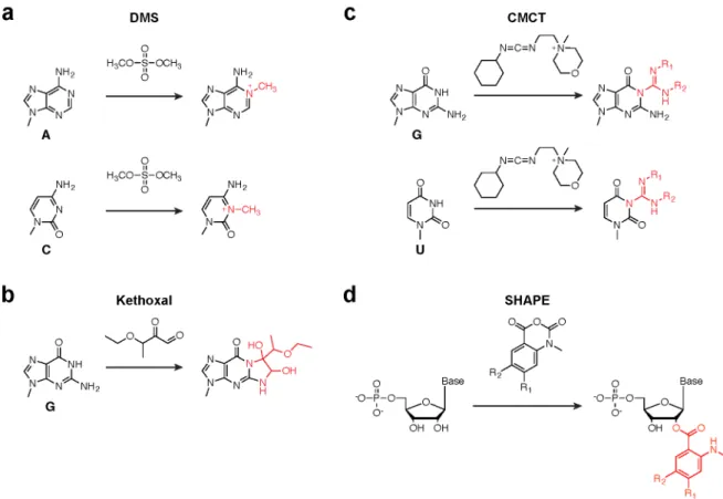
early experiments react with the nucleobase moieties of RNA and include dimethyl sulfate (DMS), which reports on A and C nucleotides (38, 39) (Fig. 1.3a); 1,1-dihydroxy-3-ethoxy-2-butanone (kethoxal), which reports on G nucleotides (40) (Fig. 1.3b); and 1-cyclohexyl-3-(2-morpholinoethyl)carbodiimide metho-p-toluenesulfonate (CMCT), which reports on G and U nucleotides (41) (Fig. 1.3c). While together these reagents yield structure information on all four RNA nucleotides, a major disadvantage is that obtaining a complete dataset for all four nucleobases requires optimization of reaction conditions for each reagent, including incubation time, buffer conditions, reagent concentration, and reaction quenching. In many cases, researchers settle for either DMS or CMCT, as these each yield data for two of the four RNA bases.
In the last decade, a suite of chemical reagents that react specifically with the 2ʹ′-hydroxyl moiety of flexible RNA nucleotides has been developed (42-44) (Fig. 1.3d). Unlike base-specific probes, these acylating reagents report on all four RNA bases since every nucleotide carries a 2ʹ′ hydroxyl. This approach to RNA chemical probing, known as SHAPE (selective 2ʹ′-hydroxyl acylation analyzed by primer extension), typically makes use of isatoic anhydride derivatives, including 1-methyl-7-nitroisatoic anhydride (1M7), 1-methyl-6-nitroisatoic anhydride (1M6), and N-methylisatoic anhydride (NMIA). 1M7 is generally considered the “workhorse” of SHAPE reagents and accurately reports on local nucleotide flexibility (43), while 1M6 and NMIA are often used in combination with 1M7 to reveal nuanced details of RNA structure (44, 45).
Perhaps one of the greatest advantages of SHAPE reagents over other chemical probes is their ability to accurately constrain computational predictions of RNA secondary structure. For very small RNAs, computer algorithms can use thermodynamic parameters to model generally accurate structures. However, as the size of the RNA increases, the number of possible base-pair combinations also increases and computational prediction accuracy decreases dramatically. Since SHAPE chemical probing data report on RNA structure, they can be used to guide structure prediction software and result in highly accurate structure models (45, 56, 57). In addition, structure modeling of large RNA transcripts has been robustly automated such that data-driven structure models and analysis can be easily generated within a few hours (50, 58).
Coupling RNA structure probing to massively parallel sequencing
Until very recently, the results of RNA chemical probing experiments were read out by annealing a labeled primer to the RNA of interest and extending the primer with reverse transcriptase to create a complimentary DNA (cDNA) strand. Under normal conditions, reverse transcriptase enzymes are blocked by chemical adducts or RNase cleavage sites. Thus, when a sample of RNA molecules is probed and analyzed by primer extension, the length and abundance of each cDNA species relates to the position and intensity of chemical modification or cleavage, respectively. Using labeled primers, these cDNAs are resolved and quantified using gel or capillary electrophoresis.
While a small amount of sample multiplexing is possible with this approach, experiments read out by electrophoresis are usually limited to studying a single RNA at a time. Due to signal decay, multiple primers must be used to study large RNAs, which introduces the additional challenge of accurately merging data from individual reactions into a contiguous data set. As the number of primer extension reactions increases, the material requirements also increase; a 2009 study of the ~9,000 nucleotide HIV RNA genome required nearly 300 µg of RNA purified from 19 L of virus culture (47).
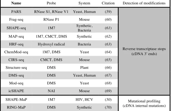
of interest, modern instruments sequence sample libraries containing millions of individual DNA fragments simultaneously. Most commercial platforms require specific adaptor sequences on the 5ʹ′ and 3ʹ′ ends for initial recognition by the instrument. Thus, it would appear as though all that is needed to couple RNA structure probing with MPS technology is to construct sequencing libraries that quantitatively preserve the 3ʹ′ termini of cDNAs generated during primer extension. Several research laboratories have developed methods that aim to achieve this type of massively parallel structure probing (Table 1.1). However, most of these methods rely on ligation steps that are biased in unpredictable ways, making them difficult to correct (71, 72). As a result, sequencing libraries generated in this fashion may not preserve the original chemical probing data with high fidelity.
To date, only one approach, called mutational profiling (MaP), offers an alternative strategy for quantifying the position and intensity of RNA structure probe reactivity. Instead of performing conventional primer extension in which reverse transcriptase is blocked by adducts, MaP exploits the ability of reverse transcriptase enzymes to proceed through chemical adducts in the presence of divalent manganese. Under MaP conditions, when reverse transcriptase encounters a site of modification it is more likely to incorporate a nucleotide non-complimentary to the original RNA sequence in the nascent cDNA strand. These sequence mutations are thus permanently and securely embedded within the cDNA, rendering MaP experiments impervious to common downstream library preparation biases. MaP has been coupled with SHAPE and DMS chemical probing (50, 70). Data generated by the SHAPE-MaP approach are of as high (or higher) quality than data obtained by prior gold-standard capillary electrophoresis methods (50) and, unlike other massively parallel structure probing approaches, can be used to accurately predict RNA secondary structures (50, 58).
Research overview
Name Probe System Citation Detection of modifications PARS RNase S1, RNase V1 Yeast, Human (59)
Reverse transcriptase stops (cDNA 3ʹ′ ends)
Frag-seq RNase P1 Mouse (60)
SHAPE-seq 1M7 Synthetic,
Bacteria (61)
MAP-seq 1M7, CMCT, DMS Synthetic (62)
HRF-seq Hydroxyl radical Bacteria (63)
ChemMod-seq 1M7, DMS Yeast (64)
CIRS-seq CMCT, DMS Mouse (65)
Structure-seq DMS Plant (66)
DMS-seq DMS Yeast, Human (67)
Mod-seq DMS Yeast (68)
icSHAPE NAI Mouse (69)
SHAPE-MaP 1M7 HIV, HCV (50) Mutational profiling
(cDNA internal mutations)
RING-MaP DMS Synthetic (70)
role in coordinating and modulating protein interactions in cells. In the course of this work, it was necessary to invent new solutions in order to obtain the necessary data. As such, my overall work toward understanding lncRNA structure has led to new experimental and computational methods for obtaining and analyzing SHAPE data.
In Chapter 2, I outline a novel approach for studying the structures of low-abundance RNAs with SHAPE-MaP. To properly characterize RNA structure by chemical probing, it is necessary to sufficiently sample the original pool of RNA molecules. While many RNAs are abundant enough that probing total cellular RNA yields high-quality structure data, the vast majority of transcripts are present at low levels and must be enriched or isolated prior to study. I show that PCR amplification prior to library construction can be used to extract chemical probing data for RNAs of interest. This allows for rapid and efficient analysis of rare, native RNA transcripts without the need for specialized pull-downs and with relatively low input material requirements.
In Chapter 3, I address the challenge of rigorously evaluating changes in SHAPE reactivity between two experimental conditions, focusing on detecting effects of the cellular environment on RNA structure. Using the built-in error estimates of SHAPE-MaP, I outline an analytical framework that identifies strong, significant changes in SHAPE reactivity. I validate this approach, called ΔSHAPE, with the U1 snRNA, 5S rRNA, and signal recognition particle, showing that ΔSHAPE analysis results in robust and correct identification of RNA-protein interaction sites. Finally, I apply ΔSHAPE to the RNA component of RNase MRP and show that this analysis detects previously-known RNA-protein interactions and identifies new contacts unaccounted for by current models.
by the cellular environment. Cross-referencing these sites with protein binding databases, I then identify several new protein interaction domains within Xist. Overall, this work provides many new insights on the relationship between RNA structure and function in lncRNAs. It shows that lncRNAs can adopt stable structures, that these structures modulate protein interactions in several specific ways, and that RNA chemical probing can be used to identify novel regions of interest even in Xist, an RNA that has been extensively studied for twenty years.
Perspective
The notion that form follows function is pervasive in all of biology. At every scale, from opposable thumbs, to the valves and chambers of the heart, down to the intricate atomic arrangement of an enzyme active site, the physical structures of biology are tightly linked to their functions. In this work, I apply principles from molecular biology, biochemistry, and biophysics in order to broaden our understanding of this structure-function relationship in the Xist lncRNA. In doing so, I have developed broadly useful approaches for accessing the chemical probing profiles of rare RNAs and for rigorously detecting chemical probing differences ex vivo and in living cells. In applying these advances to Xist, I have created a useful model that highlights how structure and function converge in the context of this RNA.
REFERENCES
1. T. R. Cech, RNA World research--still evolving. RNA. 21, 474–475 (2015). 2. P. A. Sharp, The Centrality of RNA. Cell. 136, 577–580 (2009).
3. Z. Wang, C. B. Burge, Splicing regulation: from a parts list of regulatory elements to an integrated splicing code. RNA. 14, 802–813 (2008).
4. A. Fire et al., Potent and specific genetic interference by double-stranded RNA in Caenorhabditis elegans. Nature. 391, 806–811 (1998).
5. V. Ambros, The functions of animal microRNAs. Nature. 431, 350–355 (2004).
6. B. J. Tucker, R. R. Breaker, Riboswitches as versatile gene control elements. Curr. Opin. Struct. Biol. 15, 342–348 (2005).
7. J. L. Rinn, H. Y. Chang, Genome Regulation by Long Noncoding RNAs. Annu. Rev. Biochem. 81, 145–166 (2012).
8. J. A. Cruz, E. Westhof, The Dynamic Landscapes of RNA Architecture. Cell. 136, 604–609 (2009).
9. N. B. Leontis, A. Lescoute, E. Westhof, The building blocks and motifs of RNA architecture. Curr. Opin. Struct. Biol. 16, 279–287 (2006).
10. S. E. Butcher, A. M. Pyle, The molecular interactions that stabilize RNA tertiary structure: RNA motifs, patterns, and networks. Acc. Chem. Res. 44, 1302–1311 (2011).
11. Y. Wan, M. Kertesz, R. C. Spitale, E. Segal, H. Y. Chang, Understanding the transcriptome through RNA structure. Nat. Rev. Genet. 12, 641–655 (2011).
12. J. T. Low, K. M. Weeks, SHAPE-directed RNA secondary structure prediction. Methods. 52, 150– 158 (2010).
13. S. Djebali et al., Landscape of transcription in human cells. Nature. 489, 101–108 (2012). 14. E. S. Lander et al., Initial sequencing and analysis of the human genome. Nature. 409, 860–921
(2001).
15. J. S. Mattick, Non-coding RNA. Hum. Mol. Genet. 15, R17–R29 (2006).
16. B. K. Dey, A. C. Mueller, A. Dutta, Long non-coding RNAs as emerging regulators of differentiation, development, and disease. Transcription. 5, e944014 (2014).
17. S. Geisler, J. Coller, RNA in unexpected places: long non-coding RNA functions in diverse cellular contexts. Nat. Rev. Mol. Cell Biol. 14, 699–712 (2013).
18. T. R. Mercer, M. E. Dinger, S. M. Sunkin, M. F. Mehler, J. S. Mattick, Specific expression of long noncoding RNAs in the mouse brain. Proc. Natl. Acad. Sci. U.S.A. 105, 716–721 (2008).
1, 391–407 (2011).
20. T. R. Mercer, J. S. Mattick, Structure and function of long noncoding RNAs in epigenetic regulation. Nat. Struct. Mol. Biol. 20, 300–307 (2013).
21. A. Fatica, I. Bozzoni, Long non-coding RNAs: new players in cell differentiation and development. Nat. Rev. Genet. 15, 7–21 (2014).
22. J. L. Rinn et al., Functional demarcation of active and silent chromatin domains in human HOX loci by noncoding RNAs. Cell. 129, 1311–1323 (2007).
23. C. Kanduri, Kcnq1ot1: A chromatin regulatory RNA. Semin. Cell Dev. Biol. 22, 343–350 (2011). 24. Y. Marahrens, B. Panning, J. Dausman, W. Strauss, R. Jaenisch, Xist-deficient mice are defective
in dosage compensation but not spermatogenesis. Genes Dev. (1997).
25. P. D. Mariner et al., Human Alu RNA is a modular transacting repressor of mRNA transcription during heat shock. Mol. Cell. 29, 499–509 (2008).
26. T. Kino, D. E. Hurt, T. Ichijo, N. Nader, G. P. Chrousos, Noncoding RNA Gas5 Is a Growth Arrest- and Starvation-Associated Repressor of the Glucocorticoid Receptor. Sci. Signal. 3, ra8 (2010).
27. P. A. Latos et al., Airn transcriptional overlap, but not its lncRNA products, induces imprinted Igf2r silencing. Science. 338, 1469–1472 (2012).
28. I. Martianov, A. Ramadass, A. Serra Barros, N. Chow, A. Akoulitchev, Repression of the human dihydrofolate reductase gene by a non-coding interfering transcript. Nature. 445, 666–670 (2007). 29. J. Feng, The Evf-2 noncoding RNA is transcribed from the Dlx-5/6 ultraconserved region and
functions as a Dlx-2 transcriptional coactivator. Genes Dev. 20, 1470–1484 (2006).
30. I. V. Novikova, S. P. Hennelly, K. Y. Sanbonmatsu, Structural architecture of the human long non-coding RNA, steroid receptor RNA activator. Nucleic Acids Res. 40, 5034–5051 (2012). 31. S. Somarowthu et al., HOTAIR Forms an Intricate and Modular Secondary Structure. Mol. Cell.
58, 353–361 (2015).
32. C. Chu et al., Systematic Discovery of Xist RNA Binding Proteins. Cell. 161, 404–416 (2015). 33. A. Minajigi et al., A comprehensive Xist interactome reveals cohesin repulsion and an
RNA-directed chromosome conformation. Science (2015), doi:10.1126/science.aab2276.
34. C. A. McHugh et al., The Xist lncRNA interacts directly with SHARP to silence transcription through HDAC3. Nature. 521, 232–236 (2015).
35. G. D. Penny, G. F. Kay, S. A. Sheardown, S. Rastan, N. Brockdorff, Requirement for Xist in X chromosome inactivation. Nature. 379, 131–137 (1996).
37. C. Ehresmann et al., Probing the structure of RNAs in solution. Nucleic Acids Res. 15, 9109–9128 (1987).
38. P. Brookes, P. D. Lawley, The reaction of mono-and di-functional alkylating agents with nucleic acids. Biochem. J. 80, 496–503 (1961).
39. P. D. Lawley, P. Brookes, Further studies on the alkylation of nucleic acids and their constituent nucleotides. Biochem. J. 89, 127–138 (1963).
40. M. Litt, V. Hancock, Kethoxal—A Potentially Useful Reagent for the Determination of
Nucleotide Sequences in Single-Stranded Regions of Transfer Ribonucleic Acid. Biochemistry. 6, 1848–1854 (1967).
41. N. W. Ho, P. T. Gilham, Reaction of pseudouridine and inosine with N-cyclohexyl-N'-beta-(4-methylmorpholinium)ethylcarbodiimide. Biochemistry. 10, 3651–3657 (1971).
42. E. J. Merino, K. A. Wilkinson, J. L. Coughlan, K. M. Weeks, RNA structure analysis at single nucleotide resolution by selective 2'-hydroxyl acylation and primer extension (SHAPE). J. Am. Chem. Soc. 127, 4223–4231 (2005).
43. S. A. Mortimer, K. M. Weeks, A fast-acting reagent for accurate analysis of RNA secondary and tertiary structure by SHAPE chemistry. J. Am. Chem. Soc. 129, 4144–4145 (2007).
44. K.-A. Steen, G. M. Rice, K. M. Weeks, Fingerprinting noncanonical and tertiary RNA structures by differential SHAPE reactivity. J. Am. Chem. Soc. 134, 13160–13163 (2012).
45. G. M. Rice, C. W. Leonard, K. M. Weeks, RNA secondary structure modeling at consistent high accuracy using differential SHAPE. RNA. 20, 846–854 (2014).
46. K. A. Wilkinson et al., High-throughput SHAPE analysis reveals structures in HIV-1 genomic RNA strongly conserved across distinct biological states. PLoS Biol. 6, e96 (2008).
47. J. M. Watts et al., Architecture and secondary structure of an entire HIV-1 RNA genome. Nature. 460, 711–716 (2009).
48. C. Gherghe et al., Definition of a high-affinity Gag recognition structure mediating packaging of a retroviral RNA genome. Proc. Natl. Acad. Sci. U.S.A. 107, 19248–19253 (2010).
49. E. J. Archer et al., Long-Range Architecture in a Viral RNA Genome. Biochemistry. 52, 3182– 3190 (2013).
50. N. A. Siegfried, S. Busan, G. M. Rice, J. A. E. Nelson, K. M. Weeks, RNA motif discovery by SHAPE and mutational profiling (SHAPE-MaP). Nat. Methods. 11, 959–965 (2014).
51. D. M. Mauger et al., Functionally conserved architecture of hepatitis C virus RNA genomes. Proc. Natl. Acad. Sci. U.S.A. 112, 3692–3697 (2015).
52. R. C. Spitale et al., RNA SHAPE analysis in living cells. Nat. Chem. Biol. 9, 18–20 (2012). 53. J. Tyrrell, J. L. McGinnis, K. M. Weeks, G. J. Pielak, The cellular environment stabilizes adenine
54. J. L. McGinnis, K. M. Weeks, Ribosome RNA assembly intermediates visualized in living cells. Biochemistry. 53, 3237–3247 (2014).
55. J. L. McGinnis et al., In-cell SHAPE reveals that free 30S ribosome subunits are in the inactive state. Proc. Natl. Acad. Sci. U.S.A. 112, 2425–2430 (2015).
56. K. E. Deigan, T. W. Li, D. H. Mathews, K. M. Weeks, Accurate SHAPE-directed RNA structure determination. Proc. Natl. Acad. Sci. U.S.A. 106, 97–102 (2009).
57. C. E. Hajdin et al., Accurate SHAPE-directed RNA secondary structure modeling, including pseudoknots. Proc. Natl. Acad. Sci. U.S.A. 110, 5498–5503 (2013).
58. M. J. Smola, G. M. Rice, S. Busan, N. A. Siegfried, K. M. Weeks, Selective 2′-hydroxyl acylation analyzed by primer extension and mutational profiling (SHAPE-MaP) for direct, versatile, and accurate RNA structure analysis. Nat. Protoc. 10, 1643–1669 (2015).
59. M. Kertesz et al., Genome-wide measurement of RNA secondary structure in yeast. Nature. 467, 103–107 (2010).
60. J. G. Underwood et al., FragSeq: transcriptome-wide RNA structure probing using high-throughput sequencing. Nat. Methods. 7, 995–1001 (2010).
61. J. B. Lucks et al., Multiplexed RNA structure characterization with selective 2'-hydroxyl acylation analyzed by primer extension sequencing (SHAPE-Seq). Proc. Natl. Acad. Sci. U.S.A. 108, 11063–11068 (2011).
62. M. G. Seetin, W. Kladwang, J. P. Bida, R. Das, Massively parallel RNA chemical mapping with a reduced bias MAP-seq protocol. Methods Mol. Biol. 1086, 95–117 (2014).
63. L. J. Kielpinski, J. Vinther, Massive parallel-sequencing-based hydroxyl radical probing of RNA accessibility. Nucleic Acids Res. 42, e70 (2014).
64. R. D. Hector et al., Snapshots of pre-rRNA structural flexibility reveal eukaryotic 40S assembly dynamics at nucleotide resolution. Nucleic Acids Res. 42, 12138–12154 (2014).
65. D. Incarnato, F. Neri, F. Anselmi, S. Oliviero, Genome-wide profiling of mouse RNA secondary structures reveals key features of the mammalian transcriptome. Genome Biol. 15, 491 (2014). 66. Y. Ding et al., In vivo genome-wide profiling of RNA secondary structure reveals novel
regulatory features. Nature. 505, 696–700 (2014).
67. S. Rouskin, M. Zubradt, S. Washietl, M. Kellis, J. S. Weissman, Genome-wide probing of RNA structure reveals active unfolding of mRNA structures in vivo. Nature. 505, 701–705 (2015). 68. J. Talkish, G. May, Y. Lin, J. L. Woolford, C. J. McManus, Mod-seq: high-throughput sequencing
for chemical probing of RNA structure. RNA. 20, 713–720 (2014).
69. R. C. Spitale et al., Structural imprints in vivo decode RNA regulatory mechanisms. Nature. 519, 486–490 (2015).
U.S.A. 111, 13858–13863 (2014).
71. K. M. Weeks, RNA structure probing dash seq. Proc. Natl. Acad. Sci. U.S.A. 108, 10933–10934 (2011).
CHAPTER 2: ACCESSING SHAPE-MAP PROFILES OF RARE RNA TRANSCRIPTS1 Introduction
RNA plays many fundamental biological roles and interacts with small-molecule ligands, proteins, and other RNAs (1). In these roles, RNA molecules must adopt specific secondary and tertiary structures, the details of which are often difficult or impossible to characterize from sequence alone. Chemical probing techniques have proven to be powerful tools for understanding the critical features of RNA structure at both small and large scales. SHAPE (selective 2ʹ′-hydroxyl acylation analyzed by primer extension) uses small hydroxyl-selective electrophilic reagents to probe the reactivity of the RNA ribose 2ʹ′-OH group. SHAPE reactivities are insensitive to base identity and measure local nucleotide flexibility and dynamics (2-4) because flexible residues sample a wide range of conformations, a subset of which enhance the reactivity of the 2ʹ′-hydroxyl (5) (Fig. 2.1a).
SHAPE chemistry makes it possible to examine RNA structure in an especially thorough way because, with the exception of some post-transcriptionally modified RNAs, all RNA nucleotides carry a 2ʹ′-hydroxyl group. SHAPE reactions are self-inactivating via a competing hydrolysis reaction with water (Fig. 2.1b) and thus require no specific quench step. Because few compounds have a net reactivity as high as 55 M water, intrinsic SHAPE reactivities are largely insensitive to the presence of (additional) competing small molecules, ligands, and proteins. SHAPE experiments work robustly when performed in complex environments including those inside virions (6-8) and in living cells (9, 10). By careful choice of SHAPE reagent (9-11) and experimental design, nucleotide flexibilities can be compared under
different experimental or environmental conditions, including cell-free versus in-cell and as a function of ligand and protein binding. SHAPE reactivity information used as constraints in RNA modeling algorithms results in accurate secondary structure models (12-14).
To enable identification of sites of SHAPE modification using high-throughput sequencing, we developed a strategy termed mutational profiling or MaP. In MaP, specialized primer extension conditions allow reverse transcriptase to read through SHAPE-modified nucleotides without termination of the nascent cDNA strand (15). The bulky 2ʹ′-O-adduct at modified RNA positions induces incorporation of a nucleotide non-complimentary to the original RNA sequence at the corresponding position in the newly synthesized cDNA. Thus, during reverse transcription, the positions and relative frequencies of SHAPE adducts are directly and permanently encoded as mutations in the cDNA sequence. Two control experiments are performed in parallel (15): (i) a no-reagent control to characterize the background mutations resulting from the MaP procedure and (ii) a denaturing control, in which the RNA is modified roughly evenly along its length, to measure position-specific differences in adduct detection. Ultimately, MaP is largely impervious to the substantial sequence- and structure-based biases that are introduced during construction of the libraries required for massively parallel sequencing. Because the reverse transcriptase enzyme reads through the chemical adducts, the MaP approach is also insensitive to single-strand breaks or background degradation, and does not exhibit signal decay or drop-off, effects that result in significant noise in other high-throughput sequencing-based strategies for detecting chemical modification of RNA.
MaP allows RNAs of virtually any size to be analyzed in a single experiment, facilitates high levels of multiplexing, and permits fully automated data analysis (15). Because the region under interrogation is completely sequenced in each read, sequence differences are revealed directly; therefore, the effects of sequence polymorphism and co-existing ribosnitches (15) can be evaluated in single experiments. The MaP experiment includes a DNA amplification step; therefore, individual RNAs present in scarce amounts, or in complex mixtures, can be examined. In sum, SHAPE-MaP yields robust nucleotide-resolution RNA structural information, enables accurate secondary structure modeling, can deconvolute sequence polymorphisms in a single experiment, readily allows analysis of low-abundance RNAs, and scales gracefully from short RNAs to transcriptome-wide analyses. We anticipate that SHAPE-MaP will contribute to deep understandings of the relationships between RNA structure and function.
Experimental design
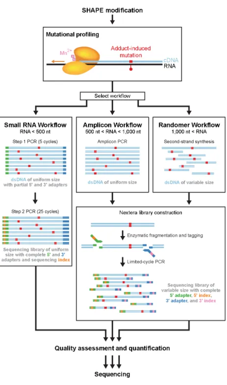
SHAPE-MaP yields quantitative SHAPE reactivity data for nearly every position in an RNA by combining the well-validated SHAPE acylation reaction with specialized reverse transcriptase conditions and deep sequencing. Once the MaP step is complete the RNA structure information is converted to DNA sequence information that is largely immune to biases and artifacts introduced during the steps required to convert the original cDNAs into the libraries required by any specific sequencing platform. Here we describe protocols for SHAPE probing, mutational profiling, and three library construction approaches based on Illumina sequencing (Fig. 2.2). The relative merits of the library construction approaches are governed by the length and amount of the RNA of interest.
RNA folding and modification
Methods for extraction, and purification of large, complex RNAs from virions (6-8, 18) and cells (10, 12) under native-like conditions have been described. RNA should be maintained under conditions likely to retain pre-existing RNA secondary and tertiary structure and use of denaturants, divalent ion chelators, or elevated temperature should be avoided. Direct interrogation of RNA structure directly inside cells by SHAPE is also well-established (9, 10, 19). This protocol emphasizes simple folding procedures for interrogating native-like and deproteinized RNAs, but any procedure that folds an RNA into an informative state can be used, provided the pH is in the 7.4-8.3 range.
Any SHAPE reagent can be used to modify RNA in a SHAPE-MaP experiment. In this protocol, we emphasize the use 1-methyl-7-nitroisatoic anhydride (1M7). Essentially identical approaches can be used with reagents 1-methyl-6-nitroisatoic anhydride and N-methyl-isatoic anhydride (1M6 and NMIA, respectively; Fig. 2.1c) (14). Reactions with 1M6 and NMIA are selective for nucleotides in which one face of the nucleobase is available for stacking and that undergo relatively slow conformational changes, respectively. These reagent-specific reactivities can be used both to identify residues that participate in non-canonical interactions and to improve RNA secondary structure modeling (14, 20). In addition, the MaP strategy can also be used to follow time-resolved RNA processes, in 1-sec snapshots, using the benzoyl cyanide (BzCN) reagent (21). The modest solubility and rapid hydrolysis of these reagents make over-modification of RNA samples virtually impossible.
Thus, a complete SHAPE-MaP experiment consists of three reactions: plus-reagent (+), minus-reagent (−), and denaturing control (DC).
Mutational Profiling (MaP)
After SHAPE modification of RNA, reverse transcriptase is used to create a mutational profile. This step encodes the positions and relative frequencies of SHAPE adducts as mutations in the cDNA sequence. Mutational profiling is efficient, with roughly 50% of SHAPE adducts detected as mutations in the cDNA (15). Whereas the reverse transcription reaction conditions are the same for any RNA, the researcher may choose one of two options regarding the type of DNA primer used. RNAs that are small enough to be sequenced end-to-end in a single massively parallel sequencing read (currently read lengths up to 600 nts are possible) can be subjected to reverse transcription with sequence-specific DNA primers. Specific primers can also be used when a specific sub-region of a large RNA is of interest (Fig. 2.2, small RNA and amplicon workflows). Use of gene- or region-specific primers also makes it possible to: (i) analyze a specific, relatively rare RNA in a complex mixture of RNAs or (ii) interrogate very rare, low abundance, RNAs. For analysis of large RNAs or the constituents of entire transcriptomes or multi-component ribonucleoprotein and long noncoding RNA assemblies, random primers facilitate even coverage of complex RNA states in a single experiment (Fig. 2.2, randomer workflow). Following mutational profiling with appropriate primers, one of three workflows is used for library construction, as detailed below.
Library construction and sequencing
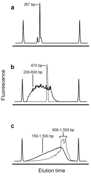
including sequence indices for sample multiplexing. After purification, sequencing libraries are of uniform size and each DNA molecule contains the entire sequence of interest (Fig. 2.3a).
The amplicon workflow is well suited for large, low-abundance RNAs or sub-regions of larger RNAs that cannot be sequenced end-to-end by a single deep sequencing read. After reverse transcription, purified cDNA is amplified via PCR with sequence-specific primers. The resulting dsDNA is then enzymatically fragmented and tagged with platform-specific adaptors and multiplexing indices. Sequencing libraries constructed in this way are of variable size, with each molecule containing a fragment of the original amplicon (Fig. 2.3b). Typically, when the amplicon workflow is used to construct a sequencing library, reverse transcription is primed with sequence-specific primers. However, if the researcher wishes to generate a sequencing library for a specific region of an RNA that was previously reverse transcribed with random primers, the amplicon workflow allows for targeted “re-construction” of libraries.
Results
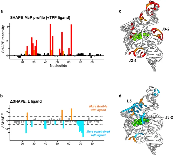
Small RNA workflow – TPP riboswitch
A SHAPE profile for the aptamer domain of the TPP riboswitch was readily obtained using the small RNA workflow. Using these data, secondary structure modeling for this riboswitch RNA improved from a base pair prediction accuracy of 73%, obtained using a nearest-neighbor thermodynamic algorithm alone, to 96%, using SHAPE-directed modeling (15). Observed reactivities correspond closely to those expected based on the local nucleotide flexibilities for the ligand-bound RNA (Fig. 2.4a-c). Reactive nucleotides fall in conformationally flexible single-stranded regions, especially the L3 loop and the J2-4 and J3-2 strands. Overall, relatively few nucleotides are reactive by SHAPE, consistent with the highly constrained conformation of this RNA. SHAPE-MaP also reveals fine differences corresponding to changes induced upon binding by the TPP ligand (Fig. 2.4b-d). Ligand interactions induce a large structural organization in the L5 loop and in the J3-2 elements in the ligand-binding pocket.
Amplicon workflow – Mouse ribosomal RNA
Randomer workflow – Bacterial ribosomal RNA
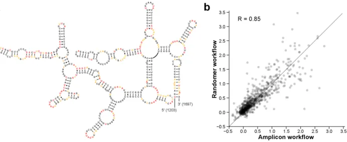
Large RNAs like the bacterial small and large ribosomal subunit RNAs (16S and 23S, respectively) are readily examined by applying the randomer workflow to total E. coli RNA (Fig. 2.6). Using random primers, both RNAs can be studied simultaneously with fully automated analysis involving approximately 3 days of hands-on experimental effort. The major post-processing requirement is that the per-nucleotide hit level be sufficiently high to permit full recovery of the underlying SHAPE data. In general, the hit level should be 5 or greater, corresponding to a read depth of 1-2,000 (15).
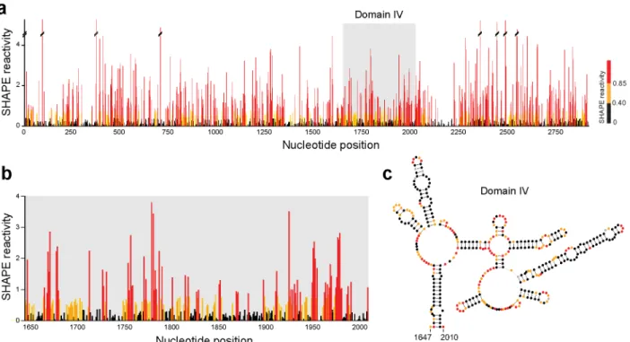
The 23S rRNA subunit alone represents ~2,900 nucleotides of SHAPE reactivity information after computational data processing (Fig. 2.6a). Comparing the SHAPE reactivities for domain IV of the 23S rRNA with the accepted sequence covariation-derived structural model (Fig. 2.6b-c) shows good agreement. Regions involved in canonical base pairs have low SHAPE reactivity, indicating that they are structurally constrained. Conversely, single-stranded loop and bulge regions have high SHAPE reactivity, indicating structural flexibility. Because of the inherent scalability of the MaP approach, these data – spanning several thousand nucleotides – are as accurate at single-nucleotide resolution as are data from a short RNA, like the TPP riboswitch.
Conclusion
Methods
SHAPE-MaP data for the TPP riboswitch and E. coli 16S rRNA were obtained previously (22) using the small RNA and randomer workflows, respectively. Data for the mouse 18S rRNA was obtained as described below.
RNA extraction and modification
To obtain SHAPE-MaP data for the mouse 18S rRNA, trophoblast stem cells (TSCs) were cultured as described (23). Approximately 106 TSCs were washed and pelleted in ice-cold PBS, resuspended in 2.5 ml Lysis Buffer [40 mM Tris, pH 7.9, 25 mM NaCl, 6 mM MgCl2, 1 mM CaCl2, 256 mM sucrose, 0.5% Triton X-100, 1,000 U/ml RNasin (Promega), 450 U/ml DNase I (Roche)], and rotated at 4 °C for 5 minutes. Cells were then pelleted at 4 °C for 2 minutes at 2250 g, resuspended in 40 mM Tris pH 7.9, 200 mM NaCl, 1.5% SDS, and 500 μg/ml of Proteinase K, and rotated at 20 °C for 45 minutes. RNA was then extracted twice with phenol:chloroform:isoamyl alcohol (24:24:1) pre-equilibrated with 1× Folding Buffer (100 mM HEPES, pH 8.0, 100 mM NaCl, 10 mM MgCl2), followed by one extraction with chloroform. RNA was exchanged into 1.1× Folding Buffer using a desalting column (PD-10, GE Life Sciences) and incubated at 37 °C for 20 minutes. Approximately 3 μg RNA was then added to a one-ninth volume of 1M7 in neat DMSO (10 mM final concentration), and then incubated at 37 °C for 5 minutes. Modified RNA was purified (RNeasy Midi spin column, Qiagen) and eluted in approximately 50 μl H2O. No-reagent negative control RNA was prepared in the same way except that neat DMSO was substituted for SHAPE reagent.
SHAPE-MaP
Mutational profiling reverse transcription reactions were primed with 2 pmol of an rRNA-specific primer (5ʹ′-ACCATCCAATCGGTAGTAGC-3ʹ′) (22). The resulting cDNA was purified (G-50 spin column, GE Healthcare) and eluted in 50 µl H2O. cDNAs were then used as templates for for either by second strand synthesis (40 µl input, NEBNext Second Strand Synthesis Module, NEB) or PCR (0.5 µl input) with rRNA-specific primers (forward: 5ʹ′-GAGGTGAAATTCTTGGACCG-3ʹ′, reverse: 5ʹ′- ACCATCCAATCGGTAGTAGC-3ʹ′). PCR reactions were performed in 50 µl volumes (1× Q5 Reaction Buffer, 200 µM dNTPs, 0.5 µM each primer, 0.02 U/µl Q5 high-fidelity DNA polymerase) using a touchdown format: 98 °C for 30 s, 25 cycles of [98 °C for 10 s, 72 °C for 30 s (decreasing by 1 °C per cycle until 60 °C), 72 °C for 30 s], 72 °C for 2 min. The resulting dsDNA was purified (Agencourt XP beads, Beckman Coulter) before construction of high-throughput sequencing libraries (Nextera XT, Illumina). Libraries were purified (Agencourt XP beads, Beckman Coulter) prior to sequencing on an Illumina Miseq instrument, generating 2 × 250 paired-end reads.
SHAPE profile generation
REFERENCES
1. P. A. Sharp, The Centrality of RNA. Cell. 136, 577–580 (2009).
2. E. J. Merino, K. A. Wilkinson, J. L. Coughlan, K. M. Weeks, RNA structure analysis at single nucleotide resolution by selective 2'-hydroxyl acylation and primer extension (SHAPE). J. Am. Chem. Soc. 127, 4223–4231 (2005).
3. C. M. Gherghe, Z. Shajani, K. A. Wilkinson, G. Varani, K. M. Weeks, Strong correlation between SHAPE chemistry and the generalized NMR order parameter (S2) in RNA. J. Am. Chem. Soc. 130, 12244–12245 (2008).
4. K. A. Wilkinson et al., Influence of nucleotide identity on ribose 2'-hydroxyl reactivity in RNA. RNA. 15, 1314–1321 (2009).
5. J. L. McGinnis, J. A. Dunkle, J. H. D. Cate, K. M. Weeks, The mechanisms of RNA SHAPE chemistry. J. Am. Chem. Soc. 134, 6617–6624 (2012).
6. K. A. Wilkinson et al., High-throughput SHAPE analysis reveals structures in HIV-1 genomic RNA strongly conserved across distinct biological states. PLoS Biol. 6, e96 (2008).
7. C. Gherghe et al., Definition of a high-affinity Gag recognition structure mediating packaging of a retroviral RNA genome. Proc. Natl. Acad. Sci. U.S.A. 107, 19248–19253 (2010).
8. E. J. Archer et al., Long-Range Architecture in a Viral RNA Genome. Biochemistry. 52, 3182– 3190 (2013).
9. J. Tyrrell, J. L. McGinnis, K. M. Weeks, G. J. Pielak, The cellular environment stabilizes adenine riboswitch RNA structure. Biochemistry. 52, 8777–8785 (2013).
10. J. L. McGinnis, K. M. Weeks, Ribosome RNA assembly intermediates visualized in living cells. Biochemistry. 53, 3237–3247 (2014).
11. S. A. Mortimer, K. M. Weeks, A fast-acting reagent for accurate analysis of RNA secondary and tertiary structure by SHAPE chemistry. J. Am. Chem. Soc. 129, 4144–4145 (2007).
12. K. E. Deigan, T. W. Li, D. H. Mathews, K. M. Weeks, Accurate SHAPE-directed RNA structure determination. Proc. Natl. Acad. Sci. U.S.A. 106, 97–102 (2009).
13. C. E. Hajdin et al., Accurate SHAPE-directed RNA secondary structure modeling, including pseudoknots. Proc. Natl. Acad. Sci. U.S.A. 110, 5498–5503 (2013).
14. G. M. Rice, C. W. Leonard, K. M. Weeks, RNA secondary structure modeling at consistent high accuracy using differential SHAPE. RNA. 20, 846–854 (2014).
15. N. A. Siegfried, S. Busan, G. M. Rice, J. A. E. Nelson, K. M. Weeks, RNA motif discovery by SHAPE and mutational profiling (SHAPE-MaP). Nat. Methods. 11, 959–965 (2014).
17. J. L. McGinnis, C. D. S. Duncan, K. M. Weeks, High-throughput SHAPE and hydroxyl radical analysis of RNA structure and ribonucleoprotein assembly. Meth. Enzymol. 468, 67–89 (2009). 18. J. M. Watts et al., Architecture and secondary structure of an entire HIV-1 RNA genome. Nature.
460, 711–716 (2009).
19. R. C. Spitale et al., RNA SHAPE analysis in living cells. Nat. Chem. Biol. 9, 18–20 (2012). 20. K.-A. Steen, G. M. Rice, K. M. Weeks, Fingerprinting noncanonical and tertiary RNA structures
by differential SHAPE reactivity. J. Am. Chem. Soc. 134, 13160–13163 (2012).
21. S. A. Mortimer, K. M. Weeks, Time-resolved RNA SHAPE chemistry. J. Am. Chem. Soc. 130, 16178–16180 (2008).
22. N. A. Siegfried, S. Busan, G. M. Rice, J. A. E. Nelson, K. M. Weeks, RNA motif discovery by SHAPE and mutational profiling (SHAPE-MaP). Nat. Methods. 11, 959–965 (2014).
CHAPTER 3: DETECTION OF RNA-PROTEIN INTERACTIONS IN LIVING CELLS WITH SHAPE
Introduction
Nearly all RNAs, regardless of function, interact with one or more protein partners in order to function properly (1, 2). Characterizing ribonucleoprotein (RNP) complexes is thus an important step in understanding RNA function. Several well-validated approaches have been developed to explore RNP complexes (3). These methods provide many valuable insights but often have a limited scope due to affinity purification steps that require prior knowledge about the RNA or protein of interest. As RNA structure studies expand to 'omics scales, direct and accurate approaches for uncovering sites of interaction between the transcriptome and the proteome will become increasingly important.
A wide variety of RNA structure probing methods have been proposed (15, 16), most of which depend on accurately identifying and quantifying cDNA ends created when reverse transcriptase enzymes encounter a chemical adduct or cleavage site. These methods all involve a critical adapter-ligation step. In principle, these methods make it straightforward to perform RNA structure probing on the entire contents of a given transcriptome; in practice, it is currently almost impossible to perform the adapter-ligation step quantitatively (17, 18). Moreover, transcriptome-wide experiments are strongly subject to the classic multiple and sparse measurement problems such that many measurements are unlikely to be statistically significant (6) and do not survive follow-up validation (19). Thus, an important challenge in large-scale and in-cell RNA structure analyses is to robustly detect significant structural changes.
We hypothesized that most RNA-protein interactions would affect the flexibility of nucleotides at the binding site and that by comparing SHAPE reactivities of deproteinized RNA (ex vivo) with reactivities obtained by probing RNA in living cells (in cellulo), it would be possible to characterize sites of RNP interactions (Fig. 3.1a). We developed an analysis framework that enables detection of RNP interactions based upon three principles: (i) RNA-protein interactions strongly affect SHAPE reactivity, either positively or negatively; (ii) due to measurement errors and the large number of reactivity measurements made, not all apparent reactivity changes are significant; and (iii) most RNA-protein interaction sites (20) will span sites of five or more nucleotides in primary sequence.
obtained with SHAPE-MaP. Thus, we chose 1M7 for its short half-life, ability to accurately report RNA secondary structure ex vivo (4-7, 27) and in living cells (9-11), and because in-cell reactivity of 1M7 is sufficiently robust that downstream enrichment is not required.
Differences in SHAPE reactivities (ΔSHAPE) were calculated by subtracting in cellulo SHAPE reactivities from ex vivo reactivities (Fig. 3.1b, upper left) and averaging over a three-nucleotide sliding window to reduce local signal fluctuation. By this definition, positive ΔSHAPE values indicate protection from modification in the cellular environment, and negative ΔSHAPE reports enhanced reactivity in cells. In a SHAPE-MaP experiment, discrete mutation events contribute to the overall reactivity at each nucleotide and are well modeled by a Poisson distribution (6). The standard error in the SHAPE reactivity measurement can therefore be estimated for every nucleotide (6). We used these error estimates to perform a modified Z-factor test (6, 28) for all positions in a given RNA (Fig. 3.1b, upper right). This test compares the magnitude of ΔSHAPE with the associated ex vivo and in cellulo measurement errors, identifying nucleotides for which the magnitudes of the errors are too large for the ΔSHAPE values to be significant. We formulated the Z-factor test such that the underlying ex vivo and in cellulo SHAPE reactivities must differ by more than 1.96 standard deviations (Z-factor > 0), ensuring that the 95% confidence intervals of each measurement do not overlap.
a Z-factor > 0 and an absolute standard score ≥ 1, those three (or more) nucleotides were considered to have significant cell-induced changes in SHAPE reactivity.
In this work, we show that biochemical RNA structure probing data generated with the well-validated SHAPE-MaP approach can be used to identify significant, meaningful changes in RNA structure between two states. Here, these states are the RNA in healthy mouse trophoblast stem cells and the same RNAs gently extracted from cells. We validate our approach with the abundant and well-characterized U1 small nuclear RNA (snRNA), 5S rRNA, and signal recognition particle (SRP) RNP complexes, illustrating that the statistical filters implemented in our analysis robustly identify sites of protein interactions. We then examine RNase MRP, an important RNP complex whose in-cell architecture is relatively poorly understood. Our analysis confirms several reported RNA-protein interactions within the complex, and also characterizes the underlying molecular phenotype of many disease-associated mutations.
Results
Comparison of SHAPE-MaP and icSHAPE
between icSHAPE and SHAPE-MaP was consistently poor, with correlation coefficients ranging from 0.1-0.3.
Validation of the ΔSHAPE approach
We used SHAPE-MaP to analyze three model RNAs ex vivo and in cellulo. The U1 snRNA is localized in the nucleus and forms the U1 snRNP complex upon binding several proteins: U1A, U1C, U1-70K, and the heteroheptameric Sm ring. Comparison of U1 snRNA ex vivo and in cellulo SHAPE reactivities revealed distinct qualitative reactivity differences throughout the RNA (Fig. 3.3a). Due to differences in the number of individual mutation events observed relative to the times a given nucleotide was sequenced, the estimated errors vary as a function of nucleotide position and are much greater for some reactivity measurements than others. This is a feature shared by all RNA structure probing experiments read out by massively parallel sequencing but is explicitly and uniquely measured using the MaP strategy. If a naïve approach had been taken that ignored these errors, multiple regions would have been (incorrectly) identified as having significant SHAPE reactivity differences (Fig. 3.3b, grey and green shading). Only a subset of these regions are involved in true RNA-protein interactions; the remainder are analysis artifacts caused by the measurement uncertainties that occur in any experiment, especially those read out by massively parallel sequencing. When we applied the complete analysis framework in which the Z-factor test is used to account for these errors, only three regions of significant ΔSHAPE were identified (Fig. 3.3b, green shading only). The locations of these positive ΔSHAPE values correspond precisely to known interactions sites of U1-70K, U1A, and the Sm ring proteins (Fig. 3.3c).
to these two domains, consistent with a lack of protein binding in the central helix.
In the Alu domain, we observed in cellulo protection at the SRP9/14 binding site (nts 24-26). We also detected enhanced in cellulo reactivity at nucleotides 35-37 and 46-48, consistent with protein-induced tertiary structure changes (Fig. 3.4b). In the S domain, we observed extensive in cellulo protection where SRP19 and SRP54 bind (Fig. 3.4c). Binding by SRP68/72 involves insertion of an α– helix into the major groove of the central helix, causing an adjacent asymmetric internal loop to open (24). Consistent with this observation, we detect enhanced in cellulo reactivity on the opened side of this loop at positions 230-232 (Fig. 3.4c). The interaction between SRP RNA and the complete SRP68/72 heterodimer has not been characterized at high resolution; however, cryo-electron microscopy data provide evidence that a portion of SRP68/72 interacts with the central helix at an internal “hinge” loop comprised of nucleotides 97-104 and 249-253 (25). In-cell SHAPE supports this observation, as enhanced in cellulo reactivity was noted on both sides of the loop at nucleotides 99-101 and 251-253, and suggests a local conformational change also occurs at nucleotides 230-232. Overall, every region of significant in cellulo protection in the SRP RNA identified by our analysis framework corresponds to sites of direct protein binding.
Application of ΔSHAPE to RNase MRP
We next applied the ΔSHAPE analysis framework to in-cell analysis of the RNA component of mouse RNase MRP (RMRP). This RNA forms a complex with 10 proteins in eukaryotes that functions in rRNA processing and mitochondrial replication (32). In humans, numerous mutations within RMRP RNA cause a spectrum of autosomal recessive skeletal diseases ranging from cartilage-hair hypoplasia (CHH) to anauxetic dysplasia (AD) (33). The structure of and protein interactions with the RNA component of RMRP have been investigated in vitro using affinity selection, chemical probing, and crosslinking experiments (32, 34-36). A recent cryo-EM study has revealed the overall three-dimensional architecture of the complex in yeast (37). However, the precise binding sites of proteins and interactions with substrate have not been examined natively in cells.
Multiple regions of the RMRP RNA have statistically significant enhanced reactivity or protection in cellulo (Fig. 3.6a) and many of these can be attributed to interaction with protein components. These include in the P3 domain, a functionally critical element (Fig. 3.6b) (38), as well as nucleotides near the junction of helices P8, P9, and P12. Cryo-EM data suggest this latter region interacts with protein Pop4 and perhaps additional proteins (Fig. 3.6c). We also observed enhanced reactivity at internal loops in helix P12. Although the complete P12 helix is not present in the cryo-EM model, its proximity to the Pop3 protein suggests that the reactivity enhancements located in the P12 helix may be due to conformational changes induced by Pop3.
active site cleft suggests that this RNP enzyme is saturated with its RNA substrates (32) in the cellular steady state.
Discussion
Our experiments with the well-characterized U1, SRP, and 5S RNPs validate the ability of the ΔSHAPE analytical framework (Fig. 3.1), enabled by SHAPE-MaP, to correctly and specifically identify regions of RNA protected by stably-associated proteins in cellulo, even in the context of a large number of individual measurements and variable level of confidence in each. In addition, this work illustrates the robust ability of the well-validated 1M7 reagent to react with RNP complexes located in both cytoplasmic and nuclear compartments in cells.
In comparing SHAPE-MaP with icSHAPE, we found poor agreement between the two approaches. SHAPE-MaP has previously been extensively validated against a large set of RNAs with complex structures (6), suggesting that icSHAPE does not provide a robust view of RNA structure ex vivo or in cellulo (Fig. 3.2). icSHAPE also reports that the SRP RNA undergoes extensive internal conformational changes in cells, which is not consistent with prior studies of this RNA (24, 25). icSHAPE differs from SHAPE-MaP in important ways. First, NAI-N3 reacts more slowly than 1M7 (t1/2 = ~30 min vs. ~17 sec, respectively), which has important consequences. These include, first, that slow (but not faster) reagents are highly sensitive to specific ion and buffer choices (41) making it very difficult to compare in-cell and ex vivo experiments and, second, that long reaction times will reflect RNP assembly and disassembly, cellular turnover, and other events unrelated to the steady-state structure of an RNA. icSHAPE is also one of the many proposed strategies that require a complex purification procedure followed by multi-step adapter ligation-based sequencing library construction, steps that are difficult to perform quantitatively (17, 18).