DE NOVO PROTEINS DESIGNED FROM EVOLUTIONARY PRINCIPLES
Timothy Michael Jacobs
A dissertation submitted to the faculty of the University of North Carolina at Chapel Hill in partial fulfillment of the requirements for the degree of Doctor of Philosophy in the Department of Bioinformatics and Computational Biology.
Chapel Hill 2015
Approved by:
Brian Kuhlman
Jack Snoeyink
Alexander Tropsha
Edward Collins
c
2015
ABSTRACT
Timothy Michael Jacobs: DE NOVO PROTEINS DESIGNED FROM EVOLUTIONARY PRINCIPLES
(Under the direction of Brian Kuhlman)
Protein engineering has rapidly developed into a powerful method for the optimization,
alteration, and creation of protein functions. Current protein engineering methods fall into
the category of either high-throughput directed evolution techniques, or engineering through
the use of computational models of protein structure. Despite significant innovation in both
of these categories, neither is capable of handling the most difficult and desirable protein
en-gineering goals. The combination of these two categories is an area of active research, and
the development and testing of combination methods is the focus of this dissertation.
Chap-ters 2 and 3 describe the development of a computational framework for de novo protein
design called SEWING (StructuralExtensionWIthNative-fragmentGraphs). In contrast to
existing methods of de novo design, which attempt to design proteins that match a
designer-supplied target topology, SEWING generates large numbers of diverse protein structures. We
show that this strategy is highly effective at creating diverse helical backbones. Experimental
characterization of SEWING designs shows that the experimental structures match the
de-sign models with sub-angstrom root mean square deviation (RMSD). Chapter 3 extends this
methodology to the creation of protein interfaces. Using this method, several de novo designed
proteins are created that bind their designated target. Chapter 4 describes the combination of
directed evolution and computational modeling through the improvement of directed evolution
techniques. In this chapter, a web tool called SwiftLib is developed, which allows rapid
gen-eration of degenerate codon libraries. SwiftLib allows protein engineers to determine optimal
degenerate codon primers for the incorporation of desired sequences, such as sequence
outline the creation of tools for the engineering of protein functions, and provide additional
evidence that computational modeling and evolutionary principles can be combined for the
TABLE OF CONTENTS
1 INTRODUCTION . . . 1
1.1 Introduction to Protein Engineering . . . 1
1.2 Methods and Strategies for Protein Engineering . . . 2
1.2.1 Directed Evolution . . . 2
1.2.2 Computational Protein Design . . . 5
1.2.3 Protein Redesign . . . 7
1.2.4 De Novo Protein Design . . . 11
1.3 Opportunities for Improved Engineering Methods . . . 13
2 DESIGN OF STRUCTURALLY UNIQUE PROTEINS USING STRATE-GIES INSPIRED BY EVOLUTION . . . 23
2.1 Introduction . . . 23
2.2 Results . . . 24
2.2.1 Overview of the SEWING Method . . . 24
2.2.2 Contiguous SEWING Design and Characterization . . . 26
2.2.3 Discontiguous SEWING Characterization . . . 28
2.2.4 Discussion . . . 30
2.3 Supplemental Methods . . . 32
2.3.1 Computational Modeling . . . 32
2.4 Supplemental Figures . . . 40
2.5 Supplemental Tables . . . 46
2.6 Command Lines and Input Files . . . 49
2.6.1 Inputs for the Extraction of Features from a Set of Structures Into a Features Database . . . 49
2.6.2 Inputs for generating SEWING nodes from Features database . . . 50
2.6.3 Inputs for Running Geometric Hasher to Generate Edges . . . 50
2.6.4 Inputs for Generating SEWING backbones . . . 51
2.6.5 Inputs for the Optimization of SEWING Designs . . . 53
3 FUNCTIONAL INCORPORATION OF BINDING MOTIFS INTO DE NOVO DESIGNED PROTEINS . . . 60
3.1 Introduction . . . 60
3.2 Materials and Methods . . . 61
3.2.1 Selection of Target Interfaces . . . 61
3.2.2 Computational Design Protocol . . . 63
3.2.3 Protein Expression and Purification . . . 66
3.2.4 Yeast Surface Display . . . 68
3.2.5 Isothermal Titration Calorimetry . . . 68
3.2.6 Surface Plasmon Resonance . . . 68
3.2.7 Crystallography . . . 69
3.3 Results . . . 69
3.4 Discussion and Future Directions . . . 74
3.5 Command Lines and Input Files . . . 76
4 SWIFTLIB: RAPID DEGENERATE-CODON-LIBRARY OPTIMIZATION
THROUGH DYNAMIC PROGRAMMING . . . 81
4.1 Introduction . . . 81
4.2 Materials and Methods . . . 85
4.2.1 DP For One Degenerate Codon . . . 85
4.2.2 DP For Multiple Degenerate Codons . . . 88
4.3 Results . . . 91
4.3.1 Library Designs . . . 91
4.3.2 Comparison with ILP . . . 97
4.4 Discussion . . . 98
4.5 Supplemental Materials . . . 100
4.5.1 Additional libraries for problem 2 . . . 100
4.5.2 ILP with One Degenerate Codon . . . 101
4.5.3 ILP with Multiple Degenerate Codons . . . 103
4.5.4 Additional Library Design Problems . . . 106
5 CONCLUSIONS . . . 115
5.1 Introduction . . . 115
5.2 Lessons and Future Directions of Protein Engineering with SEWING . . . 115
5.3 Lessons and Limitations in Refinement of Directed Evolution Libraries . . . 118
LIST OF TABLES
S2.1 DALI z-score comparison of De Novo designed proteins . . . 46
S2.2 Sequences of designed proteins . . . 47
S2.3 Crystallography statistics for CA01 . . . 48
S2.4 NMR statistics for DA05 . . . 48
3.1 Experimental summary of interface designs . . . 69
4.1 Library Design Problem 1. . . 85
4.2 Solutions To Library Design Problem 1 . . . 88
4.3 Library Design Problem 2 . . . 92
4.4 Solutions to Library Design Problem 2 . . . 93
4.5 DP and ILP running times . . . 95
S4.1 Additional Problem 2 Solutions. . . 100
S4.2 Problem Definitions for additional libraries. . . 105
S4.3 Error for SwiftLib Solutions for additional Library Design Problems. . . 107
S4.4 SwiftLib running times for additional Library Design Problems. . . 108
LIST OF FIGURES
1.1 Anchoring strategies for interface design . . . 10
2.1 Overview of the SEWING method. . . 25
2.2 Well folded SEWING designs. . . 28
2.3 Structural and biophysical analysis of CA01. . . 29
2.4 Structural analysis of discontiguous design DA05 . . . 30
S2.1 CD Spectra for folded SEWING designs . . . 40
S2.2 Size Exclusion Chromatography and Multi-Angle Light Scattering . . . 41
S2.3 Fragment Analysis for Discontinguous Assemblies . . . 42
S2.4 Electron density for CA01 structure . . . 43
S2.5 DALI structural comparison of SEWING models . . . 44
S2.6 Chemical and thermal denaturation of discontiguous design DA03 . . . 45
3.1 Starting scaffolds for interface design . . . 62
3.2 Schematic of the SEWING append method . . . 65
3.3 SEWING Interface Design Models . . . 67
3.4 Designed binders to Troponin C . . . 70
3.5 SPR data for designed binders to GαQ . . . 72
3.6 Comparison of 3OHM-2 design model and crystal structure . . . 73
3.7 Fragment analysis of PLC-β3 binding region . . . 75
S4.1 DP’s running time . . . 101
5.1 Thermal denaturation of DA05 redesigns . . . 116
Chapter 1
INTRODUCTION
The focus of this dissertation is the development of computational tools that aid in various
aspects of protein engineering. This introduction will cover the goals and challenges related to
protein engineering, and review various protein engineering methods along with their strengths
and limitations.
1.1 Introduction to Protein Engineering
Proteins are responsible for a diverse array of cellular functions, including chemical
catal-ysis, intra- and inter- cellular signaling, molecular scaffolding, and immune response.
Pro-tein engineering encompasses the optimization, alteration, and creation of proPro-tein structures
and functions. The earliest studies in protein engineering involved the solid-state synthesis
of peptide fragments derived from naturally occurring enzymes[1, 2]. These studies, along
with crystal structures of naturally occurring proteins, helped elucidate the basic
determi-nants of protein structure and function, and led quickly to the first design of a functional
synthetic peptide[3]. The subsequent development of modern molecular biology techniques
allowed researchers to test modifications to full-length proteins, and initial efforts resulted in
the reprogramming of enzyme specificity, the selection of a catalytic antibody, and the
devel-opment of entirely novel sequences that adopt well-folded tertiary structures[4, 5, 6]. These
successes demonstrated important proof that engineered proteins could be made to adopt a
va-riety of new structures and functions, and set the stage for an impressively rapid advancement
in protein engineering efforts. Today, the implications of protein engineering are ubiquitous:
pharmacoki-netic and pharmacodynamic properties; therapeutic antibodies for the treatment of cancers
and autoimmune diseases are frequently engineered to exhibit improved binding affinity and
reduced antigenicity[7, 8]. Outside the clinic, engineered proteins have found extensive use as
research tools, allowing live-cell imaging, light-mediated cellular probing, and a wide variety
of other commonly used biochemical techniques[9, 10, 11, 12, 13].
1.2 Methods and Strategies for Protein Engineering 1.2.1 Directed Evolution
Directed evolution is a widely used experimental strategy that broadly describes the
gen-eration of DNA libraries, and the selection of encoded proteins based on a chosen phenotype.
DNA libraries can be generated by strategies that vary widely in their ease of use, level of
control, and cost. The earliest described techniques utilize processes that mimic natural
pro-tein evolution, and offer the lowest level of control. DNA shuffling is one such technique,
in which homologous DNA sequences are digested and recombined[14, 15]. Another
evolu-tionarily inspired technique is error-prone polymerase chain reaction (PCR), in which specific
buffer conditions and non-proofreading polymerases are used to increase the error rate of PCR
reactions[16]. With both techniques, little control over the specific location and identity of
inserted mutations is possible. Despite this limitation, the low barrier to entry and the ability
to incorporate mutations throughout an entire gene has led to widespread use. The several
successes resulting from libraries generated using these mechanisms demonstrate the power of
natural evolutionary processes in the generation of functional diversity, a principle that will
be applied to computational design in Chapters 2 and 3
The rapid development of DNA synthesis techniques over the past two decades has led to
a number of possibilities for DNA library generation. Specifically, synthesis technologies have
evolved to allow the addition of nucleotide mixtures at specific positions in short oligonucleotide
sequences. The result of this technique is PCR oligonucleotides that contain degenerate codons,
which introduce a subset of amino-acid mutations at desired positions. In this way, the diversity
libraries allow researchers to impart external information, such as computational prediction
and evolutionary information into generated libraries. Unfortunately, the degeneracy of the
genetic code and the limitations of current screening strategies complicate this process, which is
further discussed in Chapter 4. It is noteworthy that more exacting control of DNA diversity
can be achieved through mixing of specific trinucleotide phosphoramidite primers, but the
resultant combinatorial expansion in required primers restricts the use of such libraries to
highly specialized applications[17].
DNA library generation can easily create libraries that are much larger than can be
rea-sonably screened by existing high-throughput techniques, such as phage-, yeast-, mRNA- and
ribosome-display, which range in their screening capacity from approximately 107to 1013DNA
sequences[18]. Screening of protein sequences involves the coupling of protein function to the
DNA sequence that encodes it. In the case of phage and yeast display, DNA library
mem-bers are combined with plasmids that allow the presentation of the functional protein on the
surface of a yeast cell or bacteriophage. In the case of mRNA and ribosome display, in-vitro
translation techniques are used to couple the protein-encoding RNA to the fully translated
protein.
The applications of directed evolution are restricted to engineering problems in which
the desired phenotype can be readily screened for. The most common use cases for directed
evolution are the generation and optimization of binding affinity for a target molecule, and
the generation and optimization catalytic activity. In the case of evolving binding affinity
this selection is often achieved through panning, a technique in which the target substrate
is immobilized on a surface and protein-displaying molecules are applied and washed with
increasing stringency. While effective, panning is susceptible to biases introduced by
vari-able cellular display and translation. This limitation can be partially mitigated by single-cell
screening techniques, such as flow cytometry, in which the binding affinity can be normalized
by the amount of surface-bound protein[19, 20]. The successes in directed evolution of protein
binders are numerous and impressive. In favorable cases, protein sequences have been selected
that bind their target molecule with femtomolar dissociation constants[21, 22]. However,
and is therefore not ideally suited for creation of de novo binders[23]. Additionally, in most
cases, it is difficult or impossible to dictate the binding orientation and binding location on
the target molecule, which is a critical attribute for many functions, such as steric agonism
and antagonism.
The second common use case for directed evolution is the optimization or modification
of catalytic activity. The ability to generate novel and improved enzymes has implications
in the generation of many high-value molecules and holds great promise for the production
of biofuels, materials, and pharmaceuticals[24]. The most common goal in the evolution of
protein catalysts is the improvement of enzymatic activity in a specified environment, such
as at elevated temperature or in the presence of non-organic solvents. However, studies have
demonstrated that other catalytic goals, such as modified substrate specificity and even de
novo function, can be achieved[25, 26].
Unlike protein-binding, evolution for catalytic function is often complicated by the lack of
a general and straightforward screening method. Frequently, the development of a selection
scheme requires a clever and system-dependent solution such as the use of substrate analogs
with fluorescent leaving groups[27], the creation of a covalent modification[28], or the
cou-pling of enzyme function to viability[29, 30]. Evolved binding to transition-state analogs is
an attractively general screening strategy, but studies have shown that increased affinity does
not translate to increased catalytic activity and transition state analogs are not available for
all substrates[31]. Additionally, even in favorable cases experimentally evolved enzymes fail
to approach the astounding rate enhancements of 1019 achieved by some naturally occurring
enzymes[32, 33]. Most successful catalysts have been evolved from existing enzyme backbones,
and the mediocre rate enhancements achieved for novel reactions may indicate limits in
adapt-ing existadapt-ing protein backbones for novel reactions. One potential solution to this problem
is usage of highly diverse pools of novel backbones, such as those generated by the methods
described in Chapter 2.
The protein engineering applications of directed evolution extend beyond the evolution of
binding and catalysis, with results encompassing the creation of novel fluorescent proteins,
structures[34, 35, 36, 37]. These efforts, though fewer in comparison, demonstrate that directed
evolution, through ingenuity in library generation and screening strategies, can be applied to
many useful and difficult protein engineering tasks.
1.2.2 Computational Protein Design
Computational protein design, in the context of this dissertation, entails the usage of
computational models of protein structure. Computational design protocols typically employ
a search algorithm, which samples the sequence and conformational space available to the
polypeptide chain, a score function, which evaluates the predicted energy of a given
conforma-tion. The challenges of computational protein design are two-fold: the conformational space
available to a polypeptide chain is exceedingly large, and accurate evaluation of protein
ener-getics is difficult to calculate efficiently. The issue of large conformational space was famously
described in a paper by Cyrus Levinthal in 1969 in which he postulated that a conservative
estimate for the number of conformations available to a 100 residue protein was 3300[38]. This
value will increase significantly if the amino-acid composition of the protein is not fixed, as in
the case of protein design. Given the enormity of this value, it is unfeasible for computational
design protocols to exhaustively sample the available conformational space. Additionally, the
energy landscape of protein folding has many local minima and maxima, which hinder the
ability of gradient based optimization methods to find global minimal. These local minima
render continuous physics-based sampling strategies, such as molecular dynamics, inefficient
for sampling of large-scale protein motions. A common strategy to address these difficulties
is reducing the degrees of freedom available during a conformational search algorithm. The
most prevalent method to accomplish this task is a technique called protein redesign, in which
design simulations start from the experimentally solved structure of a protein sequence. In
many cases, protein redesign is conducted with a completely fixed backbone conformation,
limiting the search space tremendously. If backbone motion is allowed, it is common practice
to fix the bond angles and lengths and sample backbone conformations only through
modifica-tion of phi and psi internal coordinates. Even with these simplificamodifica-tions, conformamodifica-tional space
necessitates the use of large computing clusters, which have only become available in recent
years. The second challenge in computational protein design is the energetic evaluation of a
given conformational state. These energetics, though understood in principle, are extremely
difficult to compute efficiently and with high accuracy[39]. Quantum mechanical simulations,
for example, are highly accurate, but the required computational expense limits these
simula-tions to systems with a very small numbers of atoms[40]. On the opposite end of the spectrum,
coarse-grained representations of atomic systems can be used to approximate the energetics of
a protein structure[41]. Coarse models have the advantage of rapid calculation and smoother
energy landscapes, but often fail to capture the level of intricacy needed to achieve most
modern protein engineering goals. Currently, the predominant methods of evaluating the
en-ergetics of protein structure lie between these two extremes, with a mixture of physical force
fields rooted in classical mechanics and empirical potentials derived from observation of known
protein structures. These force fields allow energetic evaluations to be conducted on
physi-ologically large protein structures and retain much of the accuracy granted by more precise
equations. One such force field is implemented in the Rosetta molecular modeling suite, which
was used for all of the computational modeling tasks contained in this text[42]. The Rosetta
force-field is composed of approximately 15 score terms, which aim to capture the major
deter-minants of protein energetics. Importantly, the Rosetta force-field is pairwise decomposable,
meaning the total energy of a given conformational state can be represented as the sum of
residue-pair energies. This simplification affords rapid re-evaluation of similar conformational
states, which is a common occurrence in many sampling strategies. A simplified version of the
current default Rosetta score function, is detailed in Equation 1.1.
Etot = X
Θvdw+ Θsolv+ Θelec+ Θrot+ Θrama (1.1)
In this equation, the Θvdw term captures the van der Waals forces between atoms through
a Lennard-Jones potential and Θsolv is the Lazaridus-Karplus implicit solvation term. Θelec is
a distance-dependant dielectric. Θrot and Θrama encompass emperically derived potentials to
capture sidechain rotameric preferences, backbone Φ/Ψ preference, and amino-acid identitites.
Development and improvement of force-fields for protein evaluation is a highly active area
of research and many of the existing challenges, such as accurate hydrogen bond design, have
experienced recent development[43, 44]. Continued efforts in force-field and score-function
improvements are numerous and outside the scope of this dissertation.
1.2.3 Protein Redesign
Protein redesign is a common engineering strategy in which design begins from an
ex-perimental structure of an existing protein. The potential of an existing protein backbone
to accommodate multiple sequences was eloquently demonstrated by Lim, et al. through the
introduction of random mutations into the core of λ-repressor[45]. Following this publication,
computational algorithms designed to place distinct amino-acid sidechain conformations, or
rotamers, onto known protein backbones were used to redesign protein cores of many existing
proteins[46, 47, 48, 49]. In one particularly demonstrative example, Dantas et al. applied
these methods to redesign of nine globular proteins[50]. A serendipitous result from this study
was that the thermostability of several redesigned proteins was increased relative to the native
sequence. This result is particularly illustrative of the way in which protein redesign can be
applied to difficult and valuable protein engineering tasks. In a related study, Murphy et al.
incorporated local backbone flexibility in the redesign of a 4-helix bundle protein. Strikingly,
despite a backbone RMSD of only 1.7A from the starting crystal structure, the redesigned
protein was stabilized by more than 12 kcal/mol. This result was particularly impressive due
to the fact that of the 38 residues designed, all were mutated from their wild-type identity,
demonstrating both the ability of computational redesign methods to identify unique packing
solutions and the ability of similar protein structures to accommodate different sequences.
Redesign methods have also been applied to the task of engineering protein conformational
switches, as in the case of a designed sequence that could adopt either a coiled-coil or zinc-finger
like fold dependent on the pH and presence of transition metals[51]. Conformational switching
regulation, and therefore represents an important engineering accomplishment[52, 53]. The
de-sign of protein interfaces represent another important application of protein redede-sign. Unlike
protein cores, which are composed predominantly of hydrophobic amino acids, protein
inter-faces contain many polar and charged amino acids that serve the dual purpose of promoting
solubility, and generating affinity and specificity through inter-chain hydrogen bonds. Initial
applications of protein redesign for the engineering of protein interfaces were aimed at the
re-engineering of existing protein-protein interfaces. In these studies, homo-oligomeric proteins
were redesigned to change oligomeric state, a monomeric protein was designed into a domain
swapped dimer, and specificity inducing interactions were incorporated to generate orthogonal
interface pairs[54, 55, 56, 57, 58, 59]. These successes supported the use of protein redesign
towards the engineering of novel, or de novo protein interfaces. De novo interface design
repre-sents a significantly more difficult challenge for the computational design of interfaces, due to
the fact that one must determine both a rigid body orientation and an appropriate sequence
for the binding pair. For this reason, the de novo design of protein interfaces has remained
a difficult and largely unsolved problem. A comprehensive review of protein interface design
from 2013 reported that only 5 designs out of a total 158 tested demonstrated strong binding
in a verifiably correct manner[60]. Importantly, this statistic represents an optimistic view of
the field, given that unsuccessful design projects are rarely published. In a few cases of de novo
interface design, binders have been generated that bind the target protein very weakly, or bind
in a manner inconsistent with the design model, highlighting the challenge of generating both
affinity and orientation specificity[61, 62]. In these cases predominantly hydrophobic residues
were utilized in the design model, which can lead to broad specificity. However, successful
re-designs tend to be more hydrophobic than most native interactions. This result is likely caused
by both score-function inaccuracies and the difficulty of sampling complex, multi-residue polar
networks between amino acid sidechains[60]. A successful strategy used to account for these
interactions has been the use of properly defined or regular hydrogen bonding anchors to
im-part specificity, a topic I recently reviewed in detail[63]. In one case, Stranges et al. utilize
the regular hydrogen bonding pattern of beta-strand pairing to design a highly accurate
fashion to dictate the binding orientation of a designed homodimer (Figure 1.1B)[65]. In both
of these studies, symmetry across the dimeric axis allows each amino acid mutation to impart
twice the effect. This concept of utilizing symmetry in the design of protein-protein interfaces
has recently been used in spectacular fashion towards the design of protein nanocages[66]. In
the absence of symmetry, de novo interface design has been particularly challenging. In one
case, a 16 residue extension was built onto an existing peptide interface (Figure 1.1C). In this
study backbone flexibility was incorporated into the design simulation, allowing the design of
helix packed tightly into a groove, a strategy commonly used in natural protein interfaces.
The use of so-called hot spot residues to serve as a framework for selecting appropriate protein
backbones for redesign was applied to the design of two de novo binders to influenza
hemag-glutinin. In this study, nearly 80 designs were tested, highlighting the difficulty in generating
accurate design models. The usage of portions of natural interfaces as templates has been
successful in the redesigning of natural interfaces, and work has been conducted to apply these
methods to de novo interfaces[57, 67]. Relatedly, investigators have shown that the grafting of
antigen epitopes from antibody interfaces allows for robust antibody re-elicitation, and holds
significant promise for the design of vaccines against difficult targets[68, 69]. These studies
have involved the grafting of protein segments into existing protein structures; an alternative
approach is the custom design of de novo backbones to these accommodate interface motifs, a
strategy explored in Chapter 3 of this document. These successes in computational interface
engineering demonstrate significant potential. However, the affinities of most de novo
inter-faces are relatively weak and often necessitate the use of directed evolution for improvement
to functionally relevant binding. This combination strategy for interface engineering leverages
the benefits of both directed evolution and computational design and serves as inspiration for
the methods described in later chapters.
In addition to the concrete engineering examples, protein redesign methods have been
invaluable in the continued improvements to computational design methods. By limiting
conformational sampling to the local space around an existing structure, rigorous sampling of
sidechain and backbone conformations can be achieved. These studies, coupled with careful
Figure 1.1: Anchor motifs found in natural and designed protein interfaces. Polar residues
illustrated in red and blue, hydrophobic residues in white. (A) β-strand pairing across a
designed protein interface (PDB code 3ZY7). (B) Designed metal-mediated protein interface (PDB code 3V1E). (C) Designed helix-in-groove interface (PDB code 2XNS). (D) Naturally occurring interface mediated by hydrogen bonds (PDB code 1BGS).
commonly used for protein engineering tasks, including loop design and prediction[70, 71, 72,
73] and local backbone sampling[74, 75, 76]. In sum, protein redesign is a well-studied protein
1.2.4 De Novo Protein Design
De novo protein design is an alternative approach to computational protein engineering
in which no starting experimental structure is used. The lack of a starting structure allows
de novo designed proteins to incorporate structural features not found in naturally occurring
structures. The ability to accurately engineer these novel structural details would expand the
landscape of designable structures, which may be necessary to realize many complex protein
functions. Unlike protein redesign where it is possible to hold the backbone degrees of freedom
fixed, de novo design requires explicit backbone sampling. Protein backbones designed from
scratch lose the assurance of designability afforded by existing protein structures and therefore
serve as a rigorous test of protein design force-fields and sampling methods. Commonly, the
task of de novo design is split into the generation of large numbers of novel backbones and
the subsequent design of sidechains onto these backbones via the previously described redesign
techniques. To address the task of backbone sampling, several methods have been developed.
For α-helical coiled-coil proteins, which form a regular and repetitive structure, Francis
Crick famously derived a parameterization of the conformational space[77]. This
parameteriza-tion was utilized in an early example of de novo design to generate backbone conformaparameteriza-tions for
a variety of right-handed coiled-coil structures, many of which had not yet been observed in the
set of known experimental structures[78]. More recently, similar methodologies were applied
to the design of helix-bundle proteins that demonstrate incredible thermodynamic stability[79]
and coiled-coils with water-soluble pores[80]. In all cases, design models matched
experimen-tal structures with atomic-level accuracy, demonstrating the precision afforded by parametric
methods. Additionally, parametric methods benefit from the ability to easily produce large
number of unique protein backbones through simple iteration of the available parameter space.
To date, the applications of parametric design methods have been restricted to the engineering
of proteins with repetitive sequences, which, similar to many of the interface design methods
discussed above, benefit from the use of symmetry. In certain cases, such as the engineering of
protein nanomaterials, symmetric structural repetition can be a powerful engineering
advan-tage, allowing structures to be readily extended to arbitrary length. However, the regularity of
incorporation of backbones outside of the parameter space, which is a a common occurrence
in protein functional sites[81]
Another successful strategy for the de novo design of protein backbones is the use of
fragment libraries derived from the protein data bank (PDB). Design simulations of this type
typically starts from an elongated chain of set length. Backbone torsion angles from PDB
fragments are applied to random windows of the protein backbone in order to fold the chain
into a globular structure. Often, a coarse-grained representation of atomic structure is used
during this process to smooth the energy landscape and focus sampling on the generation
of gross structural features such as secondary structure and a hydrophobic core[82]. The
application of fragment-based methods to de novo design gave rise to the first de novo protein
not composed of symmetric elements[83]. The designed protein, called Top7, adopted a fold
not yet observed in the PDB and matched the design model with atomic level accuracy. This
promising initial result let to additional fragment-based design studies, which resulted in the
accurate design of several canonical protein folds[84, 85]. The authors attribute the success of
these studies to the adherence of several rules for designing ideal proteins, or proteins composed
entirely of regular structural features commonly observed in the PDB. The proteins designed
in this fashion are often highly thermostable, consistent with parametrically designed proteins,
but similarly lack structural idiosyncrasies such as long loops, bulges, and cavities that are
often needed for protein function[86, 79]. Existing methods for de novo design universally
start with the definition of a target topology: In the case of parametric design, topologies
are dictated by the parameterization space, whereas in fragment folding methods, restraints
on secondary structure and residue-pair distances are applied to guide backbone sampling
to a desired topology. This dependence on a target topology restricts the space of designed
molecules to those explicitly defined by the designer and may hinder the ability to create the
novel topologies required for desired function. However, these proof-of-concept successes in
protein design provide valuable evidence that current force-fields used in computational design
1.3 Opportunities for Improved Engineering Methods
The protein engineering techniques outlined here have been successfully employed towards
the design and optimization of numerous proteins. Nevertheless, each of these techniques is
limited, and the ability to reliably engineer many desired protein properties and functions
remains elusive. The focus of this dissertation is the development of novel computational
tools that build on the successes of these existing strategies. Directed evolution allows diverse
sampling for a given function but lacks the fine-grained structural control vital for many
applications. Conversely, existing computational design allows atomic-level control but is
limited to the adaptation of existing structures or the generation of a provided topology, which
may or may not be able to accommodate the desired function. The goal of this work is to
address the methodological gap between the function-first attributes of directed evolution and
the structure-first attributes of computational design. The following chapters describe tools
designed to bridge this gap through the incorporation of design principles from one method to
the other. Chapter 2 describes a novel computational method for the de novo design of large
numbers of highly diverse protein backbones without the use of a target fold. A fundamental
tenant of this method is the computational adaptation of the evolutionary principles used to
great effect in directed evolution techniques. Chapter 3 extends this method towards the design
of protein-protein interfaces bolstered by naturally occurring interface templates. Finally,
Chapter 4 focuses on the efficient incorporation of computational predictions into directed
REFERENCES
1. Gutte, B. (1975) A synthetic 70-amino acid residue analog of ribonuclease s-protein with
enzymic activity. J. Biol. Chem. 250, 889–904
2. B¨arwald, K. R., Reid, R. E., and Gutte, B. (1975) Formation and enzymic properties
of dimeric RNase P. FEBS Lett. 60, 423–426
3. Gutte, B., D¨aumigen, M., and Wittschieber, E. (1979) Design, synthesis and
charac-terisation of a 34-residue polypeptide that interacts with nucleic acids. Nature 281,
650–655
4. Napper, A. D., Benkovic, S. J., Tramontano, A., and Lerner, R. A. (1987) A
stereospe-cific cyclization catalyzed by an antibody. Science 237, 1041–1043
5. Clarke, A. R., Atkinson, T., and John Holbrook, J. (1990) FROM ANALYSIS TO SYN-THESIS: NEW LIGAND BINDING SITES ON THE LACTATE DEHYDROGENASE FRAMEWORK. PART II. In Proteins: Form and Function, 39–44. Elsevier
6. Hecht, M. H., Richardson, J. S., Richardson, D. C., and Ogden, R. C. (1990) De novo design, expression, and characterization of felix: a four-helix bundle protein of native-like
sequence. Science 249, 884–891
7. Pandyarajan, V. and Weiss, M. A. (2012) Design of non-standard insulin analogs for
the treatment of diabetes mellitus. Curr. Diab. Rep. 12, 697–704
8. Riechmann, L., Clark, M., Waldmann, H., and Winter, G. (1988) Reshaping human
antibodies for therapy. Nature 332, 323–327
9. Wu, Y. I., Frey, D., Lungu, O. I., Jaehrig, A., Schlichting, I., Kuhlman, B., and Hahn, K. M. (2009) A genetically encoded photoactivatable rac controls the motility of living
cells. Nature 461, 104–108
10. Ai, H.-W., Baird, M. A., Shen, Y., Davidson, M. W., and Campbell, R. E. (2014) Engineering and characterizing monomeric fluorescent proteins for live-cell imaging
11. Gautier, A., Juillerat, A., Heinis, C., Corrˆea, I. R., Jr, Kindermann, M., Beaufils, F., and Johnsson, K. (2008) An engineered protein tag for multiprotein labeling in living
cells. Chem. Biol. 15, 128–136
12. M¨oglich, A. and Moffat, K. (2010) Engineered photoreceptors as novel optogenetic tools.
Photochem. Photobiol. Sci. 9, 1286–1300
13. Binz, H. K. and Pl¨uckthun, A. (2005) Engineered proteins as specific binding reagents.
Curr. Opin. Biotechnol. 16, 459–469
14. Crameri, A., Raillard, S. A., Bermudez, E., and Stemmer, W. P. (1998) DNA shuffling
of a family of genes from diverse species accelerates directed evolution. Nature 391,
288–291
15. Stemmer, W. P. (1994) Rapid evolution of a protein in vitro by DNA shuffling. Nature
370, 389–391
16. Zhou, Y. H., Zhang, X. P., and Ebright, R. H. (1991) Random mutagenesis of gene-sized
DNA molecules by use of PCR with taq DNA polymerase. Nucleic Acids Res. 19, 6052
17. Virnek¨as, B., Ge, L., Pl¨uckthun, A., Schneider, K. C., Wellnhofer, G., and Moroney,
S. E. (1994) Trinucleotide phosphoramidites: ideal reagents for the synthesis of mixed
oligonucleotides for random mutagenesis. Nucleic Acids Res. 22, 5600–5607
18. Leemhuis, H., Kelly, R. M., and Dijkhuizen, L. (2009) Directed evolution of enzymes:
Library screening strategies. IUBMB Life 61, 222–228
19. Boder, E. T. and Wittrup, K. D. (1997) Yeast surface display for screening combinatorial
polypeptide libraries. Nat. Biotechnol. 15, 553–557
20. Boder, E. T. and Wittrup, K. D. (2000) Yeast surface display for directed evolution of
protein expression, affinity, and stability. Methods Enzymol. 328, 430–444
21. Boder, E. T., Midelfort, K. S., and Wittrup, K. D. (2000) Directed evolution of antibody fragments with monovalent femtomolar antigen-binding affinity. Proc. Natl. Acad. Sci.
U. S. A. 97, 10701–10705
22. Vasserot, A. P., Dickinson, C. D., Tang, Y., Huse, W. D., Manchester, K. S., and Watkins, J. D. (2003) Optimization of protein therapeutics by directed evolution. Drug
23. Bolon, D. N., Voigt, C. A., and Mayo, S. L. (2002) De novo design of biocatalysts. Curr.
Opin. Chem. Biol. 6, 125–129
24. Turner, N. J. (2009) Directed evolution drives the next generation of biocatalysts. Nat.
Chem. Biol. 5, 567–573
25. Yano, T., Oue, S., and Kagamiyama, H. (1998) Directed evolution of an aspartate
aminotransferase with new substrate specificities. Proc. Natl. Acad. Sci. U. S. A. 95,
5511–5515
26. Buchholz, F. and Stewart, A. F. (2001) Alteration of cre recombinase site specificity by
substrate-linked protein evolution. Nat. Biotechnol. 19, 1047–1052
27. Luis Brise˜no Roa, ., Jim Hill, ., Stuart Notman, ., David Sellers, ., Andy P. Smith,
., Christopher M. Timperley, ., *, Janet Wetherell, ., Nichola H. Williams, ., Gareth R. Williams, ., Alan R. Fersht, ., and Griffiths, A. D. (2006) Analogues with fluores-cent leaving groups for screening and selection of enzymes that efficiently hydrolyze
organophosphorus nerve agents. J. Med. Chem.49, 246–255
28. Cesaro-Tadic, S., Lagos, D., Honegger, A., Rickard, J. H., Partridge, L. J., Blackburn,
G. M., and Pl¨uckthun, A. (2003) Turnover-based in vitro selection and evolution of
biocatalysts from a fully synthetic antibody library. Nat. Biotechnol. 21, 679–685
29. Chin, J. W., Martin, A. B., King, D. S., Wang, L., and Schultz, P. G. (2002) Addition of a photocrosslinking amino acid to the genetic code of escherichia coli. Proceedings of
the National Academy of Sciences 99, 11020–11024
30. Tao, H. and Cornish, V. W. (2002) Milestones in directed enzyme evolution. Curr. Opin.
Chem. Biol. 6, 858–864
31. Baca, M., Scanlan, T. S., Stephenson, R. C., and Wells, J. A. (1997) Phage display of a catalytic antibody to optimize affinity for transition-state analog binding. Proc. Natl.
Acad. Sci. U. S. A. 94, 10063–10068
32. Wolfenden, R. and Snider, M. J. (2001) The depth of chemical time and the power of
enzymes as catalysts. Acc. Chem. Res. 34, 938–945
33. Yuan, L., Kurek, I., English, J., and Keenan, R. (2005) Laboratory-directed protein
34. Song, J. K. and Rhee, J. S. (2001) Enhancement of stability and activity of phospholipase A1 in organic solvents by directed evolution. Biochimica et Biophysica Acta (BBA)
-Protein Structure and Molecular Enzymology1547, 370–378
35. Eijsink, V. G. H., G˚aseidnes, S., Borchert, T. V., and van den Burg, B. (2005) Directed
evolution of enzyme stability. Biomol. Eng.22, 21–30
36. Sawano, A. and Miyawaki, A. (2000) Directed evolution of green fluorescent protein by a new versatile PCR strategy for site-directed and semi-random mutagenesis. Nucleic
Acids Res. 28, E78
37. Wei, Y., Kim, S., Fela, D., Baum, J., and Hecht, M. H. (2003) Solution structure of a de novo protein from a designed combinatorial library. Proc. Natl. Acad. Sci. U. S. A.
100, 13270–13273
38. Levinthal, C. (1969) How to fold graciously. Mossbauer spectroscopy in biological
systems 22–24
39. Dill, K. A. (1990) Dominant forces in protein folding. Biochemistry 29, 7133–7155
40. Hatfield, M. P. D. and Lovas, S. (2014) Conformational sampling techniques. Curr.
Pharm. Des. 20, 3303–3313
41. Tozzini, V. (2005) Coarse-grained models for proteins. Curr. Opin. Struct. Biol. 15,
144–150
42. Leaver-Fay, A., Tyka, M., Lewis, S. M. S. M., Lange, O. F., Thompson, J., Jacak, R., Kaufman, K. W., Renfrew, P. D., Smith, C. A., Sheffler, W., Davis, I. W., Cooper, S., Treuille, A., Mandell, D. J., Richter, F., Ban, Y.-E. A., Fleishman, S. J., Corn,
J. E., Kim, D. E., Lyskov, S., Berrondo, M., Mentzer, S., Popovi´c, Z., Havranek, J. J.,
Karanicolas, J., Das, R., Meiler, J., Kortemme, T., Gray, J. J., Kuhlman, B., Baker, D., and Bradley, P. (2011) Rosetta3: An Object-Oriented software suite for the simulation
and design of macromolecules. Methods Enzymol. Volume 487, 545–574
43. O’Meara, M. J., Leaver-Fay, A., Tyka, M., Stein, A., Houlihan, K., DiMaio, F., Bradley, P., Kortemme, T., Baker, D., Snoeyink, J., and Kuhlman, B. (2015) A combined Covalent-Electrostatic model of hydrogen bonding improves structure prediction with
44. Leaver-Fay, A., O’Meara, M. J., Tyka, M., Jacak, R., Song, Y., Kellogg, E. H., Thomp-son, J., Davis, I. W., Pache, R. A., Lyskov, S., Gray, J. J., Kortemme, T., RichardThomp-son, J. S., Havranek, J. J., Snoeyink, J., Baker, D., and Kuhlman, B. (2013) Scientific bench-marks for guiding macromolecular energy function improvement. Methods Enzymol.
523, 109–143
45. Lim, W. A. and Sauer, R. T. (1989) Alternative packing arrangements in the
hydropho-bic core of lambda repressor. Nature 339, 31–36
46. Dahiyat, B. I. (1997) De novo protein design: Fully automated sequence selection.
Science 278, 82–87
47. Desjarlais, J. R. and Handel, T. M. (1995) De novo design of the hydrophobic cores of
proteins. Protein Sci. 4, 2006–2018
48. Malakauskas, S. M. and Mayo, S. L. (1998) Design, structure and stability of a
hyper-thermophilic protein variant. Nat. Struct. Biol.5, 470–475
49. Nauli, S., Kuhlman, B., and Baker, D. (2001) Computer-based redesign of a protein
folding pathway. Nat. Struct. Biol. 8, 602–605
50. Dantas, G., Kuhlman, B., Callender, D., Wong, M., and Baker, D. (2003) A large scale test of computational protein design: Folding and stability of nine completely redesigned
globular proteins. J. Mol. Biol. 332, 449–460
51. Ambroggio, X. I. and Kuhlman, B. (2006) Computational design of a single amino acid
sequence that can switch between two distinct protein folds. J. Am. Chem. Soc. 128,
1154–1161
52. Beckett, D. (2004) Functional switches in transcription regulation; molecular mimicry
and plasticity in protein-protein interactions. Biochemistry 43, 7983–7991
53. Zheng, A., Yuan, F., Kleinfelter, L. M., and Kielian, M. (2014) A toggle switch con-trols the low ph-triggered rearrangement and maturation of the dengue virus envelope
proteins. Nat. Commun.5, 3877
54. Grueninger, D., Treiber, N., Ziegler, M. O. P., Koetter, J. W. A., Schulze, M.-S., and
55. Kuhlman, B., O’Neill, J. W., Kim, D. E., Zhang, K. Y., and Baker, D. (2001) Conversion of monomeric protein L to an obligate dimer by computational protein design. Proc.
Natl. Acad. Sci. U. S. A. 98, 10687–10691
56. Kortemme, T., Joachimiak, L. a., Bullock, A. N., Schuler, A. D., Stoddard, B. L., and Baker, D. (2004) Computational redesign of protein-protein interaction specificity. Nat.
Struct. Mol. Biol. 11, 371–379
57. Potapov, V., Reichmann, D., Abramovich, R., Filchtinski, D., Zohar, N., Ben Halevy, D., Edelman, M., Sobolev, V., and Schreiber, G. (2008) Computational redesign of a Protein–Protein interface for high affinity and binding specificity using modular
archi-tecture and naturally occurring template fragments. J. Mol. Biol. 384, 109–119
58. Lewis, S. M., Wu, X., Pustilnik, A., Sereno, A., Huang, F., Rick, H. L., Guntas, G., Leaver-Fay, A., Smith, E. M., Ho, C., Hansen-Estruch, C., Chamberlain, A. K., Truhlar, S. M., Conner, E. M., Atwell, S., Kuhlman, B., and Demarest, S. J. (2014) Generation of bispecific IgG antibodies by structure-based design of an orthogonal fab interface.
Nat. Biotechnol. 32, 191–198
59. Kapp, G. T., Liu, S., Stein, A., Wong, D. T., Rem´enyi, A., Yeh, B. J., Fraser, J. S.,
Taunton, J., Lim, W. a., and Kortemme, T. (2012) Control of protein signaling using a computationally designed GTPase/GEF orthogonal pair. Proc. Natl. Acad. Sci. U. S. A.
60. Stranges, P. B. and Kuhlman, B. (2013) A comparison of successful and failed protein interface designs highlights the challenges of designing buried hydrogen bonds. Protein
Sci. 22, 74–82
61. Karanicolas, J., Corn, J. E., Chen, I., Joachimiak, L. a., Dym, O., Peck, S. H., Albeck, S., Unger, T., Hu, W., Liu, G., Delbecq, S., Montelione, G. T., Spiegel, C. P., Liu, D. R., and Baker, D. (2011) A de novo protein binding pair by computational design
and directed evolution. Mol. Cell 42, 250–260
62. Jha, R. K., Leaver-Fay, A., Yin, S., Wu, Y., Butterfoss, G. L., Szyperski, T., Dokholyan, N. V., and Kuhlman, B. (2010) Computational design of a PAK1 binding protein. J.
Mol. Biol. 400, 257–270
63. Jacobs, T. M. and Kuhlman, B. (2013) Using anchoring motifs for the computational
64. Stranges, P. B., Machius, M., Miley, M. J., Tripathy, A., and Kuhlman, B. (2011)
Computational design of a symmetric homodimer usingβ-strand assembly. Proc. Natl.
Acad. Sci. U. S. A. 108, 1–6
65. Der, B. S., Machius, M., Miley, M. J., Mills, J. L., Szyperski, T., and Kuhlman, B. (2012) Metal-mediated affinity and orientation specificity in a computationally designed
protein homodimer. J. Am. Chem. Soc. 134, 375–385
66. King, N. P., Sheffler, W., Sawaya, M. R., Vollmar, B. S., Sumida, J. P., Andr´e, I.,
Gonen, T., Yeates, T. O., and Baker, D. (2012) Computational design of self-assembling
protein nanomaterials with atomic level accuracy. Science 336, 1171–1174
67. Lewis, S. M. and Kuhlman, B. A. (2011) Anchored design of protein-protein interfaces.
PLoS One 6, e20872
68. Azoitei, M. L., Correia, B. E., Ban, Y.-E. A., Carrico, C., Kalyuzhniy, O., Chen, L., Schroeter, A., Huang, P.-S., McLellan, J. S., Kwong, P. D., Baker, D., Strong, R. K., and Schief, W. R. (2011) Computation-guided backbone grafting of a discontinuous motif
onto a protein scaffold. Science 334, 373–376
69. Azoitei, M. L., Ban, Y.-E. A., Julien, J.-P., Bryson, S., Schroeter, A., Kalyuzhniy, O., Porter, J. R., Adachi, Y., Baker, D., Pai, E. F., and Schief, W. R. (2012) Computational design of high-affinity epitope scaffolds by backbone grafting of a linear epitope. J. Mol.
Biol. 415, 175–192
70. Das, R. (2013) Atomic-Accuracy prediction of protein loop structures through an
RNA-Inspired ansatz. PLoS One 8, e74830
71. Canutescu, A. A. and Dunbrack, R. L. (2003) Cyclic coordinate descent: A robotics
algorithm for protein loop closure. Protein Sci. 12, 963–972
72. Mandell, D. J., Coutsias, E. a., and Kortemme, T. (2009) Sub-angstrom accuracy in protein loop reconstruction by robotics-inspired conformational sampling. Nat. Methods
6, 551–552
73. Stein, A. and Kortemme, T. (2013) Improvements to robotics-inspired conformational
sampling in rosetta. PLoS One 8, e63090
74. Davis, I. W., Arendall, W. B., Richardson, D. C., and Richardson, J. S. (2006) The
265–274
75. Tyka, M. D., Jung, K., and Baker, D. (2012) Efficient sampling of protein conformational space using fast loop building and batch minimization on highly parallel computers. J.
Comput. Chem.33, 2483–2491
76. Lauck, F., Smith, C. A., Friedland, G. F., Humphris, E. L., and Kortemme, T. (2010) RosettaBackrub—a web server for flexible backbone protein structure modeling and
design. Nucleic Acids Res.38, W569–W575
77. Crick, F. H. C. (1953) The packing of α-helices: simple coiled-coils. Acta Crystallogr.
6, 689–697
78. Harbury, P. B. (1998) High-Resolution protein design with backbone freedom. Science
282, 1462–1467
79. Huang, P.-S., Oberdorfer, G., Xu, C., Pei, X. Y., Nannenga, B. L., Rogers, J. M., DiMaio, F., Gonen, T., Luisi, B., and Baker, D. (2014) High thermodynamic stability
of parametrically designed helical bundles. Science 346, 481–485
80. Thomson, a. R., Wood, C. W., Burton, a. J., Bartlett, G. J., Sessions, R. B., Brady, R. L., and Woolfson, D. N. (2014) Computational design of water-soluble -helical barrels.
Science 346, 485–488
81. Brown, J. H., Cohen, C., and Parry, D. A. (1996) Heptad breaks in alpha-helical coiled
coils: stutters and stammers. Proteins26, 134–145
82. Rohl, C. A., Strauss, C. E., Misura, K. M., and Baker, D. (2004) Protein structure
prediction using rosetta. Methods Enzymol.383, 66–93
83. Kuhlman, B., Dantas, G., Ireton, G. C., Varani, G., Stoddard, B. L., and Baker, D.
(2003) Design of a novel globular protein fold with atomic-level accuracy. Science302,
1364–1368
84. Koga, N., Tatsumi-Koga, R., Liu, G., Xiao, R., Acton, T. B., Montelione, G. T., and
Baker, D. (2012) Principles for designing ideal protein structures. Nature 491, 222–227
85. Murphy, G. S., Sathyamoorthy, B., Der, B. S., Machius, M. C., Pulavarti, S. V., Szyper-ski, T., and Kuhlman, B. (2014) Computational de novo design of a four-helix bundle
86. Murphy, G. S., Mills, J. L., Miley, M. J., Machius, M., Szyperski, T., and Kuhlman, B. (2012) Increasing sequence diversity with flexible backbone protein design: the complete
Chapter 2
DESIGN OF STRUCTURALLY UNIQUE PROTEINS USING STRATEGIES INSPIRED BY EVOLUTION
2.1 Introduction
Most efforts in de novo protein design have been focused on creating idealized proteins
composed of canonical structural elements. Examples include the design of coiled-coils, repeat
proteins, TIM barrels and Rossman folds[1, 2, 3, 4]. These studies are excellent for studying the
minimal determinants of protein structure, but they do not aggressively explore new regions of
structure space. Additionally, idealized structures may not always be the most effective starting
points for engineering novel protein functions. Functional sites in proteins are often created
from structural idiosyncrasies, such as kinks, pockets and bulges. The lack of these structural
elements from de novo designed proteins highlights a key difference between natural protein
evolution and current design methods. Specifically, that protein design methods universally
begin with a target structure in mind. Therefore, the space of designable structures that can
accommodate these non-ideal protein elements is limited by the imagination of the designer.
In contrast, natural evolution is uncaring of the structure being evolved so long as the result
accomplishes the necessary function. This lack of a predetermined target fold is a powerful
feature of protein evolution that holds significant potential for the design of novel structures
and functions. In an effort to tap this potential we sought to develop a method of computational
protein design inspired by mechanisms found in natural protein evolution. Nature uses gene
duplication and homologous recombination to mix and match elements of protein structure to
give rise to novel structures and functions[5, 6, 7]. This phenomenon is most evident at the level
of independently folding protein domains [8, 9, 10], but recent studies have shown that these
folds[11]. Insertions, deletions and replacement of secondary and supersecondary structural
elements are used by nature to sample alternative tertiary structures[12]. Our design strategy,
called SEWING (Structure Extension With Native-substructure Graphs), computationally
mimics this process by building new protein structures from pieces of naturally occurring
protein domains. The process is not dictated by the need to adopt a specific target fold,
but rather aimed at creating large sets of alternative structures that satisfy predefined design
requirements. One of the strengths of this approach is that it ensures that all of the structural
elements of the protein are inherently designable, while at the same time allowing for the
incorporation of structural oddities unlikely to be found in idealized proteins. Here, we apply
SEWING to the design of helical proteins. We show that designed structures are highly diverse,
and contain structural features absent from alternative design strategies.
2.2 Results
2.2.1 Overview of the SEWING Method
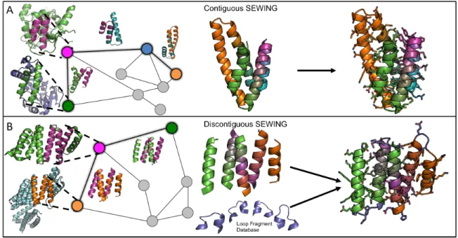
SEWING begins with the extraction of small structural motifs, or substructures, from
existing protein structures. These serve as the basic building block for all generated models.
We aimed to identify substructures that were both large enough to carry information regarding
structural preference, yet small enough to allow combinations that can generate novel globular
structures. Ultimately, we chose to extract two distinct types of substructures. The first
type of substructure is composed of contiguous stretches of protein structure that encompass
two secondary structural elements separated by a loop (Figure 2.1). These substructures
capture the relative orientation between adjacent secondary structure elements and maintain
local packing interactions. Additionally, there is evidence that substructures of this size adopt
a relatively limited number of conformations that have been sampled exhaustively in known
protein structures [12]. The second type of substructure is composed of groups of 3-5 secondary
structural elements, where each element makes make van der Waals contacts with every other
Non-adjacent, or discontiguous substructures maintain longer-range tertiary interactions that
provide valuable stability, and are often conserved during protein evolution [13].
Figure 2.1: Overview of the SEWING method. (A) Contiguous SEWING workflow for CA01. (B) Discontiguous SEWING workflow for DA05. Each panel, from left to right: parental PDBs with extracted substructures; Graph schematic colored nodes indicate substructures contained in final design model, superimposed structures show structural similarity indicated by adjacent edges; Design model before sequence optimization and loop design; Final design models.
The goal of SEWING is to combine and modify these extracted components in order to
develop new tertiary structures. In nature, homologous recombination guides the formation of
new protein chimeras, in which sequence similarity between DNA strands leads to combination
of the genetic material. This process enriches for proteins that are more likely to be well-folded
and functional, as sequence similarity filters for segments that are structurally compatible. In
the case of SEWING, we know the three dimensional structures of the building blocks, and
therefore, we can directly use structural information to probe which substructures are well
suited for combination. During SEWING, contiguous substructures are eligible for
combi-nation if the c-terminal region of one substructure shares high structural similarity with the
n-terminal region of another substructure, and superposition of the two regions does not create
any steric clashes between other regions in the two substructures. This type of superposition
adjacent in primary sequence is similar to that observed in the PDB. During discontiguous
SEWING, combinations are created by superimposing two elements (helices in this study)
from one substructure with two elements from another substructure. For both contiguous and
discontiguous SEWING, structure similarity is identified using a fast geometric hashing
ap-proach that ensures that the regions of interest can align with low-RMSD[14]. Once pairwise
structural similarity is calculated between all substructures, these data are used to generate
two large graphs: a contiguous graph and a discontiguous graph (Figure 2.1). The nodes in
each graph represent the substructures, and the edges indicate a level of structural similarity
that allows recombination. Novel structures are generated from each graph by traversing a
path, where each followed edge adds new structural elements to the design model. In this way,
many unique structures can be efficiently generated by randomly sampling paths. The
num-ber of edges included in the sampled paths can control the approximate size of the generated
structures. Importantly, unlike previously described methods of de novo backbone generation,
no target structure is required, and output structures span a diverse set of globular folds.
Sim-ilar to natural evolution, the design models created by recombination alone are suboptimal,
and require additional refinement through mutagenesis. This optimization step was achieved
using iterative steps of sidechain packing and backbone minimization available in the Rosetta
molecular modeling suite [15]. Preference for the amino acid sequence present in the parental
substructure was used to better preserve the structural interactions inherent to the parent
substructures. To test SEWING, we designed a diverse set of helical proteins using graphs
composed of contiguous substructures or discontiguous substructures. Contiguous
substruc-tures were extracted from a non-redundant dataset of approximately 8000 high-quality crystal
structures [16]. Discontiguous substructures were extracted from a non-redundant subset of
structures from the protein data bank (PDB), generated using the Pisces server [17]. In total,
33928 contiguous substructures, and 4584 discontiguous substructures were extracted.
2.2.2 Contiguous SEWING Design and Characterization
Design models from the contiguous graph were generated using 3-edge paths, and were
(Fig-ure 2.1). Initially, 10,000 alternative tertiary struct(Fig-ures were created and used as templates for
rotamer-based sequence optimization and energy minimization. These models were filtered and
sorted using metrics that evaluate predicted energy (normalized by sequence length), sidechain
packing, buried polar groups, and sequence/structure agreement[18]. The top 1,000 of design
models were then further refined with 3 rounds of the same sequence optimization protocol. In
total, 11 designs based on contiguous SEWING were selected for experimental characterization.
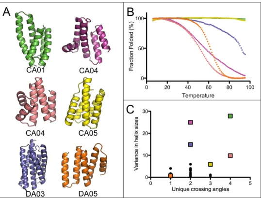
8 of these designs expressed well in E. coli and were readily purified from the soluble fraction
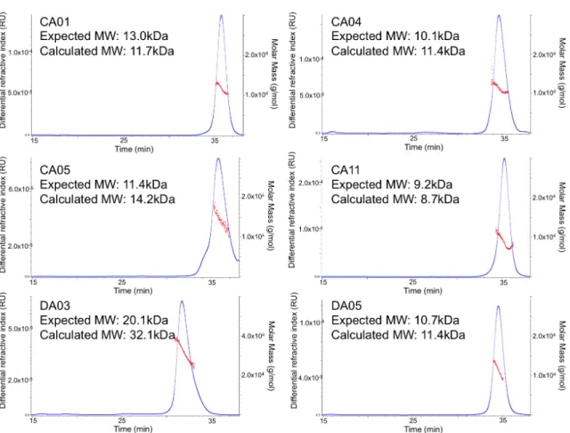
of 1L cultures. 4 of the 8 proteins were monomeric via Size Exclusion
Chromotagraphy/Multi-angle light scattering, had a circular dichroism (CD) spectra characteristic of a helical protein
and unfolded cooperatively upon thermal denaturation (Figure 2.2, S2.6). Two of the designed
proteins are hyperthermophiles and require high concentrations of chemical denaturant in
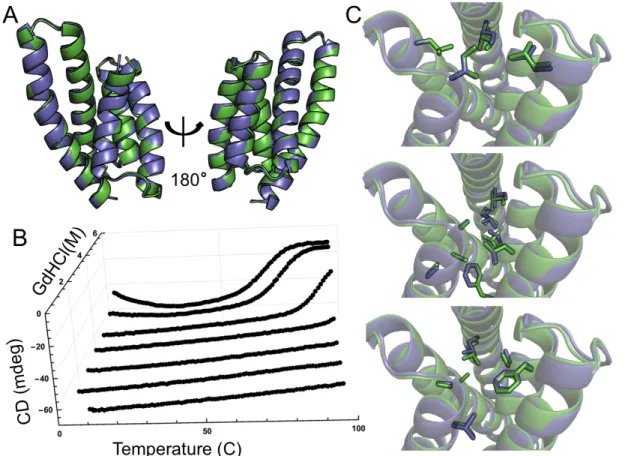
or-der to observe thermal unfolding (Figure 2.2B). For one design, CA01, several thermodynamic
parameters were determined by fitting a modified Gibbs-Hemholtz equation to the thermal and
chemical denaturation surface (Figure 2.3B, Supplemental Methods). The extrapolated
melt-ing temperature of 126◦C places it among the top .01% of values in the ProTherm database of
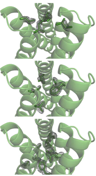
protein stabilities [19]. The crystal structure of CA01 was solved to 2.2˚Aand shows excellent
agreement with the design model, with a Cα RMSD of 0.7˚A. Similarly, the side-chain packing
of the protein core is nearly identical between the design model and experimental structure
(Figure 2.3).
The structural variety in the design models for the well-folded proteins is of particular note
(Figure 2.2). The SEWING generated models often include non-ideal elements such as kinked
or highly curved helices, long loops, and near perpendicular helix-crossing angles (Figure 2.2).
The topologies of SEWING models are also quite variable, particularly when compared to
previously designed alpha-helical proteins, which are restricted entirely to coiled-coils, repeat
proteins and up-down four helix bundles (Figure 2.2C). To further examine the structural
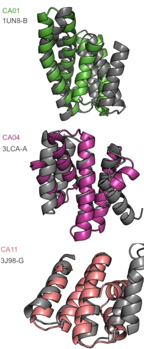
di-versity of SEWING models, we searched for structurally similar domains using the DALI server
[20]. Surprisingly, despite being derived from elements of existing structures, SEWING models
Figure 2.2: Well folded SEWING designs. (A) Final design models for contiguous and dis-contiguous SEWING. (B) Temperature denaturation curves for well-folded SEWING designs, colored to match design models. (C) A comparison of previously design helical structures (black dots), to SEWING models (colored squares) demonstrates the structural complexity
of SEWING designs. Crossing angles were counted as unique if they differed by>20 degrees
from each other.
serendipitous result further demonstrates the potential of recombination for the production of
novel protein structures.
2.2.3 Discontiguous SEWING Characterization
To test discontiguous SEWING, models were generated from 2-edge paths, and thus were
composed of structural elements from 3 parent structures. The variable number of helical
elements in the discontiguous substructures therefore allowed design models to be composed of
between 5 and 11 helices. Unlike models from the contiguous-substructure graph, discontiguous
models require the addition of loops between consecutive helices. Loops were designed using
a database of fixed-length fragments from the PDB, hashed on the geometric transform from
from the fragment-database based on the transform between the two disconnected helices. Each
fragment was then superimposed onto the design structure and optimized using gradient-based
minimization in Cartesian space. The best-scoring loop fragment was selected for use in the
final design model. Any path that created structures for which no loop-fragment could be
found was eliminated from the set of designs. Design models were filtered and optimized in
the same way as models from the contiguous graph. In total, 10 were selected for experimental
characterization.
Of these 10 designs, 2 expressed at levels sufficient for purification. Both purified proteins
were helical and folded as evidenced by CD (Figure 2.2). Similar to the results from the
con-tiguous designs, one disconcon-tiguous design demonstrated very high thermostability, requiring
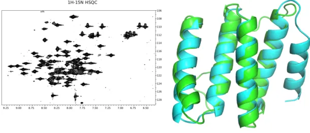
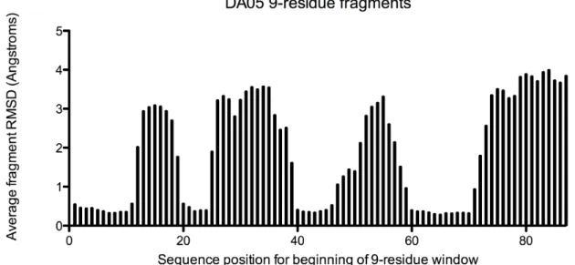
high levels of denaturant to completely unfold(Figure S2.6). The structure of DA05 was solved
using nuclear magnetic resonance (NMR) spectroscopy. The first 4 helices of the
experimen-tal structure match the design model very closely, with a Cα RMSD of 0.9˚A (Figure 2.4).
However, the NMR data indicate the final helix of the protein is disordered in solution. In
an effort to identify the errors in the design model that led to the unstructured region,
struc-tural preference for the designed sequence was evaluated with fragment analysis as described
previously [2]. Interestingly, the fragments extracted for the unstructured region showed
espe-cially poor preference for the designed helical structure (Figure S2.3). In light of this result,
fragment analysis was conducted on the remaining discontiguous design models. In nearly all
cases, the sequences for the designed loop regions showed poor agreement with the designed
structure. This discrepancy suggests that errors in the loop design protocol could account for
the relatively lower success rate of the discontiguous designs.
Figure 2.4: Structural analysis of discontiguous design DA05 via NMR reveals that the first four helices of the structure match well with the design model. However, the 5th helix of the structure is disordered in solution (Supplemental Methods).
2.2.4 Discussion
Our results show that computational adaptations of basic evolutionary principles, such
as recombination and mutation, can be used to accurately and rapidly design a diverse set of
structural elements that have proven difficult to incorporate into de novo designed structures.
A surprising result from this study is that despite the incorporation of these non-ideal protein
elements, several of the designed proteins exhibit high levels of thermostability, consistent with
proteins designed using solely ideal structures[1, 2]. This result suggests that proteins designed
for specific function have the potential to retain some of the remarkable thermodynamic
prop-erties achieved by modern protein design methods. The structure diversity displayed by the
SEWING designs demonstrates the power of recombination to generate novel structures. In
creating the designs, only a small portion of the SEWING graph was sampled. The graph
for contiguous SEWING contains over 30,000 nodes with 345 million edges, allowing an
esti-mated 7×10¹⁶ unique backbones that could be created by following three consecutive edges
(Supplementary Methods). The diversity will further increase when alternative types of
su-persecondary structure are included, such asβαmotifs andβ-hairpins, and discontiguous and
contiguous SEWING are merged. We anticipate that this structural diversity will be
advanta-geous for functional design, as every backbone generated with SEWING has unique surface and
pocket features that provide potential binding sites for ligands or macromolecules.
Addition-ally, SEWING offers a direct approach for stitching together functional motifs from naturally
occurring proteins, an approach frequently used by Nature to generate multi-functional
2.3 Supplemental Methods 2.3.1 Computational Modeling Extraction of Substructures
Contiguous and discontiguous substructures are composed of elements of secondary
struc-ture defined by the DSSP implementation in Rosetta[22]. The Rosetta implementation used
here simplifies secondary structure classification to helices (H), loops (L), andβ-strands (E).
Contiguous substructures are composed of 3 adjacent elements of secondary structure that
fol-low the pattern HLH. Discontiguous substructures are composed of 3-5 helical (H) elements, in
which each helix makes favorable Leonard Jones contacts with every other (a clique).
Contigu-ous substructures were extracted from the Top8000 dataset using the SmotifFeaturesReporter
built into the Rosetta Features framework. Discontiguous substructures were extracted using
the ModelFeaturesReporter on a non redundant set of the PDB generated from the Pisces
web server[17]. A complete set of inputs and commands can be found in the Supplemental
Material.
Geometric Hashing of Substructures
The geometric hashing algorithm used to calculate an all-against-all comparison of
ex-tracted substructures is an implementation of the algorithm described by Nussinov et. al[14].
The computational implementation is distributed as part of the Rosetta molecular modeling
suite. The current implementation differs from the described algorithm in that noncollinear
triplets (RS) were restricted to N/Cα/C backbone atoms of the same residue. For contiguous
substructures two RS that place 10 atoms of the same Rosetta AtomType from a single
sec-ondary structure element into identical or adjacent quarter-angstrom bins were considered
suf-ficient to infer structurally similarity. For discontiguous substructures, RS pairs were required
to place 10 atoms of the same Rosetta AtomType from two elements secondary structure. To
prevent clashes during recombination, any two RS pairs were rejected if they 1) placed any