Address for correspondence Dr. Nabeela Shahzadi
Dermatology Department, Unit I, King Edward Medical University/ Mayo Hospital, Lahore
Email: [email protected]
Review Article
Chronic urticaria: An approach towards
etiology and diagnosis. Part I
Introduction
Urticaria is characterized by the sudden appearance of wheals, which may be accompanied by angioedema. Superficial dermal edema gives rise to wheals, while edema in the deep dermis, hypodermis and gastrointestinal tract results in angioedema.1 Wheals may occur alone in about 50% of cases, wheals with angioedema in 40%, and angioedema without wheals in 10%, both occurring simultaneously or separately.2
CU is defined as urticaria that has been continuously or intermittently present for at least 6 weeks. The prevalence of CU in the general population has been estimated to range from 0.5% to 5%. The incidence of CU has been estimated at 1.4% per year. The duration of CU varies considerably and nearly 20% of patients remain symptomatic 20 years after onset,
especially, physical urticaria tend to persist the longest, often for many years.
Prevalence is two times more in females than in males. Although all age groups are prone to CU, the peak incidence is seen between 20 and 40 years, primarily the working years. However, the prevalence does not have any relationship with education, income, occupation, place of residence or ethnic background.
Severe itching, disfiguring wheals and disturbed sleep may be associated with impaired daily activities, emotional disturbances and poor quality of life.3
Terminology and classification
Exact definition of CIU/CSU differs between published guidelines,1,2 but the key defining
features are, a) no external physical triggers (non-inducible); and daily or episodic symptoms for >6 weeks.1,2 The European Academy of
Allergy and Clinical Immunology/ Global Allergy and Asthma European Network/ European Dermatology Foundation/ World Nabeela Shahzadi, Zahida Rani, Faria Asad, Ijaz Hussain
Department of Dermatology, Unit I, King Edward Medical University/ Mayo Hospital, Lahore
Abstract Chronic urticaria (CU) is one of the most frustrating and challenging dermatosis for patients and
physicians both. Apparently easy to diagnose, CU is still considered as a difficult to manage disease. Subtypes of CU include chronic idiopathic (spontaneous) urticaria, inducible urticaria, physical urticaria, autoimmune chronic urticaria and urticarial vasculitis. Physical urticaria may coexist with chronic idiopathic (spontaneous) urticaria. Evaluation of a patient with CU should involve consideration of various possible causes, although in most cases the cause is not identifiable. Investigation of CU should be guided by a thorough history and physical examination. Key words
Table 1 Definitions of chronic spontaneous urticaria and chronic idiopathic urticaria by different guidelines.
Associations Term used Definition
(EAACI/ GA(2) LEN/ EDF/ WAO)*
CSU Spontaneous (occurring without external stimuli) wheals and/or angioedema for >6 weeks
AAAAI† CIU Skin lesions persistent or recurring for >6 weeks (with or without angioedema). Persistent symptoms may be daily or episodic. CIU may be defined as idiopathic after exclusion of known etiologies. BSACI‡ CIU Daily/almost daily symptoms for >6 weeks and episodic acute
intermittent urticaria/angioedema lasting for hours or days and recurring over months or years with an unknown cause
CIU, chronic idiopathic urticaria; CSU, chronic spontaneous urticaria
*European Academy of Allergology and Clinical Immunology/EU-funded network of excellence, the Global Allergy and Asthma European Network/the European Dermatology Forum/World Allergy Organization
†American Academy of Allergy, Asthma & Immunology ‡British Society for Allergy and Clinical Immunology
Table 2 Classification of chronic urticaria according to cause[1,3].
I. Chronic spontaneous urticaria Spontaneous wheals and/or angioedema for more than 6 weeks without known cause
II. Chronic inducible urticaria
Dermographism Application of mechanical forces to the skin, wheals appear in 1 to 5 minutes.
Delayed pressure urticaria Vertical pressure wheals appear after 3 to 8 hours of latency. Urticaria secondary to cold Cold air/ water / wind
Urticaria secondary to heat Localized heat
Solar urticaria Ultraviolet and/or visible light
Urticaria/ vibratory angioedema Vibratory forces, usually pneumatic devices Aquagenic urticaria Contact with water, regardless of its temperature
Cholinergic urticaria Stress, perception of body temperature elevation by the hypothalamus Contact urticaria Allergic or pseudo-allergic
Figure 1 An overview of pathogenesis of urticaria [4,5].
Allergy Organization (EAACI/ GA(2) LEN/ EDF/ WAO) guidelines classify chronic urticaria
into chronic inducible urticaria (dermatographic, cold by contact, delayed pressure, heat by Cutaneous mast cells release
mediators in response to various factors including drugs, peptides and
vasoactive amines Trigger:'heat,' cold,'exercise' or'undefined' (CIU/CSU)' Symptom manifestation Symptom induction
via mediators, e.g. interleukins, histamine
Ur<caria'pathogenesis:'overview''
Mast'cells'are'the'key'effector'cells'' in'the'induc<on'of'ur<caria'symptoms' Ur<caria'and'Angioedema.'Zuberbier'T,'GraKan'C,'Maurer'M,'editors.'Berlin:'SpringerMVerlag,'2010' PRURITUS ERYTHEMA WHEAL INFILTRATE C A U S E Ac<va<on' Vasodila<on' Extravasa<on' Recruitment' MC' IgE' SCF' IgG' LPS' Complement' Anaphylatoxins' Neuropep<des' EndothelinM1' Bacteria' Interleukins' Chemokines' Oxytocine' Leukotriene' POMCs' Prostaglandins' Cannabinoids' Adenosine' Urokinase' Capsaicin' ?'FcεRI'
Kit' FcγR' TLRs' CR1/2,'CR3' C3aR,'C5aR' NK1' ETA/ETB'
CD48' ILM3,4,15R' CCR3' OTRs' CysLT1R' MG1/MCS' EP1/EP3' CB1/CB2' A2b/A3' uPAR' VR' PIR'A/PIR'B' ILM1,'ILM2,' ILM3,'ILM4,' ILM5,'ILM6,' ILM8,'ILM10,' ILM13,'TNF,' MIPs,'IFNMγ'
GMMCSF,' TGFMβ,' bFGF,' VPF/VEGF,'
contact, solar, aquagenic, cholinergic, contact, and vibratory) and chronic spontaneous urticaria (CSU), previously called chronic idiopathic urticaria. CSU may be due to autoantibodies (to IgE or FcεRI) or unknown cause.4-6A patient can
have more than one type of urticaria
Mechanisms and pathogenesis
Mast cell is the primary effector cell in chronic urticaria. Upon activation, mast cell degranulate and release histamine and other inflammatory mediators i.e. prostaglandins, leukotrienes, platelet growth factor etc. Consequently, there is vasodilatation, plasma extravasation, sensory nerve activation and a mixed inflammatory infiltrate comprising of CD4+ cells, monocytes, neutrophils, eosinophils and basophils (Figure 1).4,5 Cytokines profile shows increased
expression of interleukin (IL)-4, IL-5 and interferon-γ, suggestive of a mixed Th1/Th2 infiltrate. The incoming cells release more proinflammatory mediators, thus amplifying the inflammatory process. In the uninvolved skin of CU patients, there is upregulation of inflammatory cytokines, chemokines and adhesion molecules and higher number of T cells, which lowers the reactive threshold of mast cells thus maintaining the susceptibility to urticaria even during remission phase.
A number of heterogeneous pathomechanisms have been implicated in the degranulation of mast cells.
1. Autoimmunity and chronic urticaria
Over half of the patients with chronic idiopathic urticaria are thought to be caused by autoimmune mechanisms. This is supported by the following observations:5-8
• Autologous intradermal injection of sera
from some patients with CSU causes a wheal and flare reaction.
• Histology of urticarial lesions reveals
eosinophils, mast cells, and activated CD4+ T cells.
• IgG autoantibodies to the α subunit of
FcεRI or to IgE itself have been demonstrated in the serum of CU patients.
• A reduced percentage of blood basophils
(perhaps recruited to the skin) is found in patients with CSU and histamine releasing autoantibodies.
• HLA-DR alleles (HLA DR4 and DQ8)
that are generally associated with autoimmune disease are increasingly frequent in CSU.6
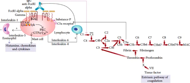
IgG autoantibodies (complement fixing IgG1 and IgG3 subclasses) against IgE (5-10%) or its high affinity receptor FcεRI (30-40%) are produced in 50% of patients with CSU/CIU. Binding of autoantibody to the α subunit of FcεRI induces degranulation of cutaneous mast cells. There is also complement activation and production of C5a, which augments the inflammatory process. Antibodies against the
low affinity IgE receptor (FcεRII),
antiendothelial antibodies, and complement C8
alpha-gamma (C8α-γ) deficiency also have
contributory role.
Numerous autoimmune conditions have been associated with chronic idiopathic urticaria,
including thyroid disease (especially
hypothyroidism), celiac disease, rheumatoid arthritis, Sjogren’s disease, SLE, and type 1
diabetes. Various autoimmune biomarkers of a
Nonimmunologic agonists
A number of other mediators e.g. substance P, endorphins, enkephalins, endogenous peptides and somatostatin can directly activate and degranulate mast cells independent of immunological reactions. Many drugs like aspirin, NSAIDs, Polymixin B, ACE inhibitors etc. cause urticaria by activation of nonimmunological pathways.1,9
Cellular abnormalities
The primarily abnormality in some patients of CU lies in cellular or subcellular system rather than immunologically mediated autoimmune mechanism. Various abnormalities in basophils of chronic urticaria patients have been reported like basopenia, paradoxical suppression of FcεRI-mediated release of histamine from basophils, imbalance between positive and negative regulators of signaling through FcεRI. Similarly, a direct role of mast cells is speculated. Vasoactive molecules may be released from mast cells on exposure to CU serum depleted of IgG showing that this process occurs independent of IgE receptor activation.5
Chronic urticaria an immune-mediated
inflammatory disorder
CU may represent an inflammatory disorder secondary to a defect of innate immunity. There is functional impairment of plasmacytoid dendritic cells (pDC) due to downregulation of toll-like receptor-9 and consequent reduced production of interferon-alpha by pDCs, altered cytokine-chemokine milieu and inflammatory process of CU.5
Chronic urticaria and clotting abnormalities
Severe exacerbations of urticaria are associated with a strong activation of coagulation cascade
that leads to fibrin formation and fibrinolysis as shown by elevated D-dimer plasma levels. Thrombin is a serine protease that enhances vascular permeability, activates and degranulates mast cells, and induces generation of anaphylatoxin C5a. The activation of extrinsic pathway of coagulation is thus proposed as yet another explanation.5,10
Figure 2 summarizes that pathologically CU may be considered as clinical manifestation of many diverse pathomechanisms which may act synergistically or sequentially to activate mast cells, the primary effector cell.5
Histology of chronic urticaria
Histologic examination of lesional biopsies in chronic urticaria shows edema of the upper and mid-dermis, along with dilatation of postcapillary venules and lymphatics of upper dermis. There is variable, but mixed perivascular inflammatory infiltrate comprising of neutrophils and/or eosinophils, macrophages, T cells, basophils, and mast cells but the vessel wall is usually not damaged. Occasionally, there may be leukocytoclastic vasculitis that does not leave any residual pigment or purpura.1-4
In angioedema, similar changes are observed in the lower dermis or subcutis.
Associations
Besides autoimmune diseases, various infectious, parasitic infestations and foods/food additives etc. are associated with chronic urticaria.
Helicobacter pyloriinfection
Figure 2 Pathogenesis of chronic urticaria: molecular intercommunication between autoimmune, complement, and coagulation cascade. ITAM: immunotyrosine activation motif, GTP: guanosine triphosphate, Lyn, Syk: cytoplasmic tyrosine kinase [5].
in patients with CU.7 It is suggested that H.
pylori can have an indirect involvement in the
etiology of CU, by reducing the immune tolerance and inducing the formation of autoantibodies, including the production of autoantibodies to anti-FcεRIα. However there is no consensus that the investigation of H. pylori
should be performed as a routine or, that when it is present, the treatment might influence the course of CU.1
Hepatitis B and C infection
Both hepatitis B and C can be the cause of acute or chronic urticaria. Patients with chronic urticaria living in areas of high prevalence of hepatitis C infection must be screened for it.1
Dental infections and urticaria
Lipopolysaccharides from oral flora Gram-negative bacteria e.g. Veilonella sp. can cause histamine release by mast cells, and, could be pathogenic factor in chronic urticaria in patients with odontogenic infection. It is suggested that patients with chronic urticaria should have their dental condition assessed.1
Helminthic parasites and infestations
The association of urticaria with the following parasites has been reported: Giardia lamblia,
Fasciola hepatica, Toxocara canis,
Echinococcus granulosus, Strongyloides
stercoralis, Hymenolepis nana, Blastocystis
hominis, Ascaris lumbricoides, Anisakis simplex,
Cimexlectularius (bedbug), Argas reflexus (bird
tick).1
The association between parasitism and urticaria has been better established with A. simplex and recently with B. hominis. Sensitization to A.
simplex can be investigated through specific
RAST test in peripheral blood.
The prevalence of B. hominis ranges from 10% in developed countries to 50% in less developed areas. Different genetic subtypes of B. hominis,
according to climate or seasonal changes and source of infection, have been identified to trigger CU in different regions of the world.11
Food as a cause of pseudo-allergic reactions
Gastrin, released by G cells in the gastric antrum and proximal duodenum immediately after feeding, may be involved in anaphylactic reactions and urticaria seen after the ingestion of certain foods.1 It is not always possible to
establish a direct correlation between clinical symptoms and the detection of antigen-specific IgE antibodies in cases of suspected food allergy. In recurring CU, it is assumed that there might be histamine intolerance caused by an excessive amount of histamine in the diet and/or by abnormal histamine metabolism (diamine oxidase deficiency). Diamine oxidase is the main enzyme involved in the degradation of histamine, acting predominantly in the intestinal mucosa. Alcohol and some medications may decrease the activity of this enzyme and lead to a higher sensitivity to rich or histamine-producing foods. Several experiments have demonstrated deficiency of diamine oxidase in enterocytes of patients with recurrent CU.
Certain fishes (tuna, sardines, anchovies), cheese, salami, sausage, certain fruits and vegetables (tomatoes), wine and beer are histamine-rich foods.
Food additives such as preservatives, dyes and natural salicylates may trigger or aggravate urticaria through pseudo-allergic non-IgE-dependent mechanisms. These additives are: sodium metabisulfite, sodium benzoate, monosodium glutamate, sodium nitrate, tartrazine, erythrosine, sorbic acid and butylated hydroxyanisole.
The general consensus is that food additives can aggravate chronic urticaria but they are rarely its sole cause.1-4
Impact on quality of life
CSU has been shown to cause significant morbidity and to have a negative impact on all aspects of a patient’s life. Fatigue, pain, and insomnia due to the constant itching of disease and visible lesions can lead to emotional upset and withdrawal from social activities. It can lead to psychological complaints such as anxiety, depression, or irritability. CSU symptoms can carry a high socioeconomic burden through a combination of direct healthcare costs and loss of work productivity.
Different tools like dermatology life quality index, urticaria-related quality of life index, medical outcomes study 12-item and 36-item short form health surveys, self-reported depression, anxiety, and sleep difficulties, the work productivity and activity impairment questionnaire, and health care resource have been used by different researchers to measure impact CU on quality of life.12-14
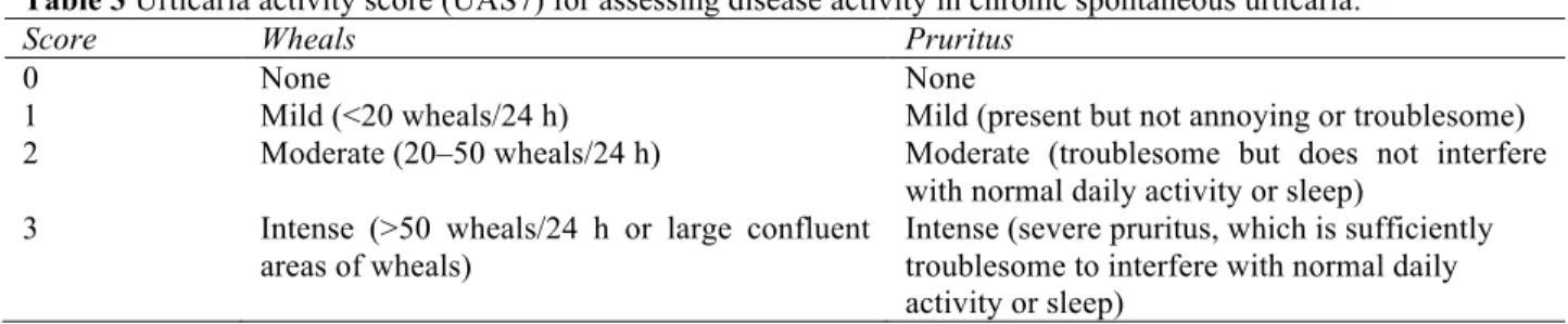
The UAS7 for assessing disease activity in CSU
Table 3 Urticaria activity score (UAS7) for assessing disease activity in chronic spontaneous urticaria.
Score Wheals Pruritus
0 None None
1 Mild (<20 wheals/24 h) Mild (present but not annoying or troublesome) 2 Moderate (20–50 wheals/24 h) Moderate (troublesome but does not interfere
with normal daily activity or sleep) 3 Intense (>50 wheals/24 h or large confluent
areas of wheals)
Intense (severe pruritus, which is sufficiently troublesome to interfere with normal daily activity or sleep)
UAS7 ranges from 0-42. A UAS7 score ≤ 6 is indicates well-controlled chronic urticaria.
Diagnosis of urticaria
Evaluation of a patient with chronic urticaria comprises of two steps i.e. confirmation of urticaria and second to find out the possible underlying etiology. All patients require a detailed history and complete physical examination, including visualization and confirmation of characteristic pruritic, raised erythematous lesions. Serial photographs are useful for documenting the extent and severity of the urticaria. The frequency, pattern, and duration of lesions should be recorded.
Clinical history
In the clinical history, following questions should be taken into consideration:
• Time of onset of disease
• Frequency/duration of and provoking
factors for wheals
• Diurnal variation
• Occurrence in relation to weekends,
holidays, and foreign travel
• Shape, size, and distribution of wheals • Associated angioedema
• Associated subjective symptoms of
lesions e.g. itch, pain
• Family and personal history regarding
urticaria, atopy
• Previous or current allergies, infections,
internal diseases, gastric/intestinal problems or other possible causes
• Psychosomatic and psychiatric diseases
• Surgical implantations and events
during surgery, e.g. after local anesthesia
• Induction by physical agents or exercise • Use of drugs e.g. non-steroidal
anti-inflammatory drugs (NSAIDs), injections, immunizations, hormones, laxatives, suppositories, ear and eye drops, and alternative remedies. Specific questioning about use of NSAIDs is important because up to 30% to 50% of CSU patients have exacerbations associated with NSAIDs ingestion
• Observed correlation to food • Relationship to the menstrual cycle • Smoking habits (especially use of
perfumed tobacco products or cannabis)
• Type of work • Hobbies
• Stress (eustress and distress)
• Quality of life related to urticaria and
emotional impact
• Previous therapy and response to
therapy
• Previous diagnostic procedures/results
Physical examination
targetoid plaques. They may coalesce to form large geographic patches. The surface remains smooth and unaltered. Lesions disappear usually within two to three hours without any residual pigmentation or change in the texture and never last longer than 24-48 hours; however, new lesions may develop simultaneously. Accompanying angioedema is seen in 40% of patients. It presents as nonpruritic, brawny, nonpitting edema, typically without well-defined margins and without erythema. Presentation of urticaria is similar in children and adults.
Further examination is guided by the clinical history. It may be pertinent to examine for any precipitating condition like focus of bacterial or fungal infection, autoimmune thyroid disease, collagen-vascular disease, chronic liver disease etc.
For inducible urticaria, diagnostic provocation tests should be performed. In case of symptomatic dermographism, elicit dermographism by stroking skin firmly with a tongue depressor or, if available, use a Fric test. Cold urticaria is diagnosed by cold provocation and threshold test by applying an ice cube to the skin for 5 minutes; urticaria appears on re-warming. For delayed pressure urticaria do pressure test, for heat urticaria, perform heat provocation and threshold test. Solar urticaria is diagnosed by challenge to UV and visible light of different wavelengths. Diagnosis of aquagenic urticaria is confirmed by wet cloth at body temperature applied for 20 min and cholinergic urticaria by exercise and hot bath provocation test.1-4,
Investigations
For patients with chronic urticaria who present with otherwise unremarkable history and physical examination findings, extensive laboratory work-up including skin or in vitro
testing for IgE to inhalants or foods is not recommended as it is neither cost-effective nor it improves patient care outcomes. Guidelines now recommend that the initial investigation of CSU should generally be limited to a complete blood count and measurement of inflammation markers such as erythrocyte sedimentation rate or C-reactive protein.1-4,12-14
Allergy skin tests generally have limited diagnostic value. The autologous serum skin test (ASST), performed by intradermal injection of autologous serum using careful sterile technique, is rarely used in practice. A positive ASST suggests the presence of autoantibodies to the high-affinity IgE receptor or to IgE; however, this test is not specific for CSU, hence not recommended by guidelines.16
Rest of laboratory testing should be based on clinical suspicion is appropriate.
A skin biopsy should be performed in patients with atypical urticaria i.e. those with burning or painful hives that persist for longer than 72 hours, to rule out urticarial vasculitis.
Patients with hyperpigmented lesions should have the skin stroked firmly to elicit Darier’s sign suggestive of cutaneous mastocytosis and a baseline serum tryptase level to rule out mastocytosis along with a lesional biopsy should be performed, if indicated.
Differential diagnosis of chronic urticaria
Many conditions can produce urticarial wheals, but they have a different underlying pathophysiology, prognosis and treatment. These conditions are diagnosed primarily by history and physical examination.1-4,12-14,17
• Papular urticaria (insect bite reaction) –
punctum after insect bites, common in children.
• Urticarial vasculitis (wheals last for > 24
hours, are painful, and leave residual hyperpigmentation or purpura).
• Henoch-Schonlein purpura - lower
extremity distribution, purpuric lesions, systemic symptoms
• Urticaria pigmentosa (orange to brown
hyperpigmentation of the lesions, wheals are of smaller diameters, and Darier’s sign is positive).
• Erythema multiforme – target like
lesions on face and distal limbs, acute and recurrent course.
• Fixed drug eruptions – tender,
well-defined, oval or round patches with central blistering and residual pigmentation, recur on same sites after taking offending drug.
• Atopic dermatitis - Maculopapular,
exudative, hyperkeratotic, scaly eruption in characteristic distribution.
• Morbilliform drug eruption – Pruritic,
maculopapular eruption, associated with drug intake
• Viral exanthema - Nonpruritic,
prodrome, fever, maculopapular lesions, individual lesions last for days.
• Autoinflammatory diseases -
cryopurin-associated periodic syndromes (familial cold autoinflammatory syndrome, Muckle–Wells syndrome, or neonatal onset multisystem inflammatory disease etc.),
• Schnitzler syndrome – monoclonal
gammopathy (IgM or IgG), symptoms of systemic inflammation, and by urticarial rashes
• Bullous pemphigoid – pre-bullous phase
of bullous pemphigoid may present as urticaria.
• Adult-onset Still’s disease, systemic-onset
juvenile idiopathic arthritis, may present as urticarial wheals.
A five-step algorithm help reach diagnosis in patients presenting with wheals or angioedema (Figure 3).
Course of the disease
Chronic urticaria runs an indolent course characterized by relapses. In most cases, the duration of CIU/CSU is estimated to be 1-5 years.1However, for some patients the disease
can last longer, sometimes up to 50 years.150%
of CU patients will resolve (with or without treatment) within 6 months of onset.2 Another
20% will resolve (with or without treatment) within 3 years of onset. Another 20% will resolve (with or without treatment) within 5 years of onset. Another <2% will resolve (with or without treatment) within 25 years.4
Factors associated with longer duration or more difficult to treat chronic urticaria include:1-4
• Failure of a single labeled dose of an H1
antihistamine to control chronic urticaria
• Long duration (6 months or more) at
time of presentation
• Angioedema • Physical urticaria
• Autoimmunity diseases/test results
(applies to adults but not children for thyroid pathology/autoantibodies)
• Positive autologous serum or plasma
intradermal skin test
• Serum IgG anti-IgE or IgG anti-FcεRI
antibodies
• Hypertension
• Subclinical activation of the extrinsic
coagulation pathway (Prothrombin fragments detected) or evidence of fibrinolysis (D-Dimer > 500 ng/mL)
Figure 3 Recommended diagnosis algorithm for patients presenting with wheals, angioedema, or both [5].
References
1. Criado PR, Criado R F J, Maruta C W et al. Chronic urticaria in adults: state-of-the-art in the new millennium. An Bras Dermatol. 2015;90:74-89.
2. Bernstein JA, Lang D M, Khan D A et al. The diagnosis and management of acute and chronic urticaria: 2014 update. J Allergy Clin Immunol. 2014;133:1270-72
3. Viegas LP, Ferreira MB, Kaplan AP. The maddening itch: an approach to chronic urticaria. J Investig Allergol Clin Immunol. 2014;24:1-5.
4. Zuberbier T, Aberer W, Asero R et al. The EAACI/GA(2) LEN/EDF/WAO Guideline for the definition, classification, diagnosis, and management of urticaria: the 2013
revision and update. Allergy. 2014;69:868-87.
5. Jain S. Pathogenesis of chronic urticaria: an overview. Dermatol Res Prac. 2014.
6. Sussman G, Hébert J, Gulliver W et al. Insights and advances in chronic urticaria: a Canadian perspective. Allergy Asthma Clin Immunol. 2015;11(1):7.
7. Confino-Cohen R, Chodick G, Shalev V et al. Chronic urticaria and autoimmunity: associations found in a large population study. J Allergy Clin Immunol. 2012;129:1307-13.
8. Fraser K, Robertson L. Chronic Urticaria and autoimmunity. Skin Ther Letter. 2013;18:7.
10. Cugno M, Marzano AV, Asero R et al. Activation of blood coagulation in chronic urticaria: pathophysiological and clinical implications. Intern Emerg Med. 2010;5:97-101.
11. Zuel-Fakkar NM, Abdel Hameed DM, Hassanin OM. Study of Blastocystis hominis isolates in urticaria: a case-control study. Clin Exp Dermatol. 2011;36:908-10. 12. Ferrer M, Bartra J, Giménez-‐‑Arnau A et al.
Management of urticaria: not too complicated, not too simple. Clin Exp Allergy. 2015;45:731-43.
13. Powell RJ, Du Toit GL, Siddique N et al. BSACI guidelines for the management of chronic urticaria and angio-‐‑oedema. Clin Exp Allergy. 2007;37:631-50.
14. Hide M, Hiragun T. Japanese guidelines for diagnosis and treatment of urticaria in comparison with other countries. Allergol Int. 2012;61:517-27.
15. Młynek A, Zalewska-Janowska A, Martus P et al. How to assess disease activity in patients with chronic urticaria? Allergy. 2008;63:777-80.
16. Konstantinou GN, Asero R, Maurer M et al. EAACI/GA(2)LEN task force consensus report: the autologous serum skin test in urticaria. Allergy. 2009;64:1256-68.
![Figure 1 An overview of pathogenesis of urticaria [4,5]. Allergy Organization (EAACI/ GA(2) LEN/ EDF/ WAO) guidelines classify chronic urticaria](https://thumb-us.123doks.com/thumbv2/123dok_us/7875335.2099480/2.918.131.808.625.976/overview-pathogenesis-urticaria-allergy-organization-guidelines-classify-urticaria.webp)
![Figure 3 Recommended diagnosis algorithm for patients presenting with wheals, angioedema, or both [5]](https://thumb-us.123doks.com/thumbv2/123dok_us/7875335.2099480/10.918.146.794.121.700/figure-recommended-diagnosis-algorithm-patients-presenting-wheals-angioedema.webp)
