Nijmegen
The following full text is a publisher's version.
For additional information about this publication click this link.
http://hdl.handle.net/2066/193558
Please be advised that this information was generated on 2020-09-10 and may be subject to
change.
Genome-Wide Association Study Reveals
Variants in
CFH
and
CFHR4
Associated with
Systemic Complement Activation
Implications in Age-Related Macular Degeneration
Laura Lorés-Motta, MSc,
1,*
Constantin C. Paun, MSc,
1,*
Jordi Corominas, PhD,
1,2Marc Pauper, MSc,
1,2Maartje J. Geerlings, MSc,
1Lebriz Altay, MD,
3Tina Schick, MD,
3Mohamed R. Daha, PhD,
4Sascha Fauser, MD, PhD,
3,5Carel B. Hoyng, MD, PhD,
1Anneke I. den Hollander, PhD,
1,2Eiko K. de Jong, PhD
1 Purpose: To identify genetic variants associated with complement activation, which may help to select age-related macular degeneration (AMD) patients for complement-inhibiting therapies.Design: Genome-wide association study (GWAS) followed by replication and meta-analysis. Participants: AMD patients and controls (n¼2245).
Methods: A GWAS on serum C3d-to-C3 ratio was performed in 1548 AMD patients and controls. For replication and meta-analysis, 697 additional individuals were genotyped. A model for complement activation including genetic and non-genetic factors was built, and the variance explained was estimated. Haplotype
analysis was performed for 8 SNPs across theCFH/CFHRlocus. Association with AMD was performed for the
variants and haplotypes found to influence complement activation.
Main Outcome Measures: Normalized C3d/C3 ratio as a measure of systemic complement activation. Results: Complement activation was associated independently with rs3753396 located in CFH(Pdiscovery¼
1.091015;Pmeta¼3.6610
21
;
b
¼0.141; standard error [SE]¼0.015) and rs6685931 located inCFHR4(Pdiscovery¼8.18 107;Pmeta¼6.32 108;
b
¼0.054; SE ¼0.010). A model including age, AMD diseasestatus, body mass index, triglycerides, rs3753396, rs6685931, and previously identified SNPs explained 18.7% of
the variability in complement activation. Haplotype analysis revealed 3 haplotypes (H1e2 and H6 containing
rs6685931 and H3 containing rs3753396) associated with complement activation. Haplotypes H3 and H6
conferred stronger effects on complement activation compared with the single variants (P¼2.531014;
b
¼0.183; SE¼0.024; andP¼4.28104;
b
¼0.144; SE¼0.041; respectively). Association analyses with AMDrevealed that SNP rs6685931 and haplotype H1e2 containing rs6685931 were associated with a risk for AMD
development, whereas SNP rs3753396 and haplotypes H3 and H6 were not.
Conclusions: The SNP rs3753396 in CFH and SNP rs6685931 in CFHR4 are associated with systemic
complement activation levels. The SNP rs6685931 inCFHR4and its linked haplotype H1e2 also conferred a risk
for AMD development, and therefore could be used to identify AMD patients who would benefit most
from complement-inhibiting therapies.Ophthalmology 2018;125:1064-1074ª2018 by the American Academy of
Ophthalmology. This is an open access article under the CC BY-NC-ND license (http://creativecommons.org/
licenses/by-nc-nd/4.0/).
See Editorial on page 962.
Supplemental material available atwww.aaojournal.org.
The complement system is an integral part of our innate immunity. Its best known physiologic functions are host defense against foreign intruders and homeostasis
mainte-nance.1 It consists of more than 30 plasma proteins and
cellular components that interact in proteolytic cascades
for an efficient and rapid activation leading to
inflammation, opsonization, and targeted cytolysis.2 The
complement system can be activated by 3 different pathways: the classical pathway, the lectin pathway, and
the alternative pathway (AP). The classical pathway is
activated by antibodyeantigen complexes and the lectin
pathway is activated by lectin or ficolin binding to
carbohydrates, both on the surfaces of pathogens. In contrast, the AP is activated constitutively at a low level
in a process known as tick-over.3
All 3 pathways lead to the formation of complement component 3 (C3) convertases that catalyze a proteolytic cleavage of complement C3 into the potent anaphylatoxin
C3a, and C3b, an opsonization molecule that can be further cleaved into C3d. Complement component 3b also can bind the cleaved form of factor B (Bb) to form the AP C3 con-vertase (C3bBb) that will cleave more C3, initiating an
amplification loop. Downstream in the cascade, complement
component 5 convertases are formed, initiating the terminal pathway with the subsequent formation of additional acti-vation products as well as the membrane-attack complex
that is responsible for cytolysis.4 The complement system
can be amplified rapidly, and therefore several inhibitory
proteins such as complement factor H (FH) and
complement factor I are in place regulating complement
activity.4
Deregulation and deficiencies of the complement system
have been reported to be associated with numerous
inflammatory, autoimmune, neurodegenerative, and
infec-tious disorders.5A prime example of a multifactorial disease
associated with a deregulation of the complement system is age-related macular degeneration (AMD). Age-related macular degeneration is characterized by a progressive degeneration of the central retina and is responsible for most cases of vision loss in the elderly with a pooled prevalence
of 8.9%.6,7 Age-related macular degeneration constitutes a
major health problem as by 2020, the number of people affected by a form of this disease is projected to be 196
million, rising to 288 million by 2040.8 Several lines of
evidence point toward an overactivation of the
complement system in AMD, mainly through a
dysregulation of the AP. Multiple genetic variants in or
near complement genes (CFH, C3, CFI, C2/CFB locus,
and C9) have been associated strongly with AMD.9,10
Moreover, complement components have been described
in drusen, the hallmark of the disease,11e14and complement
activation fragments in plasma or serum such as Ba, C3a, C3d, and component C5a have been found to be elevated
significantly in AMD patients compared with controls.15e21
Currently, there is no treatment available for most forms of AMD, nor is there an effective means to halt AMD pro-gression. Therefore, therapies for AMD, as well as for other diseases involving complement deregulation, are being
developed aiming to inhibit or lower complement
activation.22e24
Systemic complement activation levels demonstrate
considerable variation among individuals.16e20 As a
consequence, patients who have higher levels of
comple-ment activation may benefit more than others from the
up-coming therapies. A better understanding of the factors that
influence complement activation would facilitate the
selec-tion of the most suitable patients for complement-inhibiting therapies. Genetic markers are robust biomarkers that could be included in prediction models for complement activation. Several studies have previously evaluated the effect of genetic variation on complement activity; however, these studies were restricted to a limited number of single
nucleotide polymorphisms (SNPs).16e19,21,25
The aim of this study was to perform thefirst
genome-wide association study (GWAS) on systemic complement
activation levels. Identification of genetic variants
explain-ing complement activation levels will contribute to a better understanding of the molecular mechanisms of
complement-related diseases, will pinpoint potential drug targets, and will facilitate the selection of patients for complement-inhibiting therapies.
Methods
Study Population
This study included 2245 participants from the European Genetic Database (www.eugenda.org). The European Genetic Database is a multicenter database for the clinical and molecular analysis of AMD collected at the Radboud University medical center, Nijmegen, The Netherlands, and at the University Hospital of Cologne, Cologne, Germany. The study participants were sepa-rated into 2 cohorts: a discovery cohort comprising 1548 in-dividuals and a replication cohort of 697 inin-dividuals.
The study was performed in accordance with the tenets of the Declaration of Helsinki (seventh revision) and the Medical Research Involving Human Subjects Act. Approval of the local ethics committee of both University hospitals was obtained, and written informed consent was acquired from all participants. All the individuals included in the study agreed to the performed serum measurements and genotyping. All participants were of European descent and older than 50 years. Age-related macular degeneration and control status were assigned by multimodal image grading according to the standard protocol of the Cologne Image Reading Center by certified graders. Age, sex, height, and weight mea-surements were obtained by standardized interviewer-assisted questionnaires.
Serum Complement and Lipid Measurements
Serum was obtained by a standard coagulation and centrifugation protocol, and within 1 hour after collection, the samples were stored at 80C. Triglycerides and high-density lipoprotein cholesterol were measured using standard procedures by a clinical chemistry laboratory (Architect Analyzer; Abbott Diagnostics, Hoofddorp, The Netherlands). Complement component 3 was assessed by radial immunodiffusion (or Mancini method) using monospecific polyclonal rabbit antisera, and C3d was measured by rocket electrophoresis, as previously described.21 Complement
component 3d is a fragment of C3 generated upon activation of the system, and therefore a direct measurement of complement turnover.4Moreover, C3d has the longest half-life of all C3 split products.26The C3d-to-C3 ratio is a sensitive way of assessing the activation of the complement system independently of the baseline individual C3 concentration.27e29 The C3d-to-C3 ratio has been
described previously to be a robust biomarker for complement activation in AMD studies.19 The different measurements were performed for all samples in a single assay.
Genotyping
Genomic DNA was extracted from peripheral blood samples using standard procedures. The discovery cohort was genotyped with a custom-designed HumanCoreExome array by Illumina (Illumina Inc., San Diego, CA) within the International AMD Genetics Consortium. All the details regarding the design of the array, annotation, imputation, and quality control of the genotypic data have been described previously.9
Imputed lead variants in GWAS peaks that reached signifi -cance, rs6685931 and rs3130572, were confirmed by polymerase chain reaction and Sanger sequencing. The SNP rs6685931 was evaluated in 12 individuals representing the 3 genotypes, and a 100% of concordance with the imputed genotypes was achieved. The SNP rs3130572 (chromosome 6) was located in a highly
repetitive region and specific primers could not be designed; therefore, this SNP was excluded from further analysis. In the replication cohort, CFHrs3753396 andCFHR4 rs6685931 were genotyped using competitive allele-specific polymerase chain reaction assays according to the manufacturer instructions (KASP Genotyping Chemistry; LGC, Hoddesdon, UK).
Statistical Analysis
Natural log transformation was applied to normalize the skewed distribution of C3d/C3 measurements. A general linear model for ln(C3d/C3) including as independent variables the environmental factors collected was used to determine potential confounders. The
R2and adjustedR2statistics were estimated for the model. Addi-tionally, theR2statistic was estimated for each of the independent factors individually, performing separate models. Analyses were carried out using SPSS software version 20.0 (IBM Software and Systems, Armonk, NY).
A power calculation for the GWAS was performed using the Genetic power calculator.30 Association tests in the GWAS and replication analyses were performed by means of a linear Wald test from EPACTS software (http://genome.sph.umich.edu/wiki/ EPACTS) using allele dosages. Linear regression models adjusted for age, sex, body mass index (BMI), triglycerides, clinic site, and the first 2 ancestry principal components were used. Manhattan and Q-Q plots were generated using the
‘qqman’ R package (version 0.1.2; R Foundation for Statistical Computing, Vienna, Austria). The regional plots for chromosome 1 were generated using LocusZoom.31Meta-analysis offixed ef-fects based on effect size estimates and standard errors was per-formed using METAL software (version 2-11-03-25).32
Evaluation of an interaction between the identified SNPs and clinic or AMD status was performed including an interaction parameter on the general linear model and assessing nominal sig-nificance. Comparisons of systemic complement activation levels between the genotype groups were performed using a general linear model adjusted for age, BMI, triglycerides, and clinic sites including both the discovery and the replication cohorts. SPSS software version 20.0 (IBM Software and Systems, Armonk, NY) was used for these analyses.
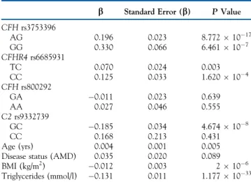
To estimate how much of the variation in systemic complement activation could be explained by the identified factors, general linear models for systemic complement activation were performed using SPSS software version 20.0. Only the 1548 individuals from the discovery cohort were included to accommodate the CFH
rs800292 andC2rs9332739 SNPs, which were not analyzed in the replication cohort. The adjustedR2statistic was estimated for the models.
Haplotype analysis was carried out for the 1548 patients geno-typed with exome-arrays using the haplo.glm function of the R library‘haplo.stats’(version 1.7.7). Analysis was performed based on a general linear model adjusted for age, sex, BMI, triglycerides, clinic site, and thefirst 2 ancestry principal components.
Single-variant and haplotype association analyses with AMD were performed for the 1548 individuals of the discovery cohort. Single-variant analyses were performed using a Firth bias-corrected likelihood-ratio test with EPACTS software. Haplotype analyses were based on chi-squared tests including haplotypes with a predicted probability of 0.75 or more using SPSS software version 20.0.
Risk scores for AMD-associated variants were calculated as a sum of the number of AMD risk-increasing alleles. Two risk scores were calculated: the first risk score included the 52 AMD-associated variants described in Fritsche et al,9 and the second risk score included the 19 variants located in or near complement
genes of these 52. The variants included in the complement risk score were: rs10922109, rs570618, rs121913059, rs148553336, rs187328863, rs61818925, rs35292876, and rs191281603 from the CFH locus; rs10033900 and rs141853578 from the CFI
locus; rs62358361 from the C9 locus; rs116503776, rs144629244, rs114254831, and rs181705462 from the C2/CFB/ SKIV2L locus; rs11080055 from the TMEM97/VTN locus; and rs2230199, rs147859257, and rs12019136 from the C3 locus. The risk scores were included in linear models for ln(C3d/C3) that included age, BMI, triglycerides, and clinic site as covariates, and the effect of the risk score was estimated. The 1548 individuals from the discovery phase, genotyped with the HumanCoreExome array, were included in these analyses. Figures including graphs were generated using Graphpad Prism version 5.03 (GraphPad Software, La Jolla, CA).
Results
Characteristics of the Study Cohorts
We evaluated the association of genetic variants with systemic complement activation levels through a GWAS in a discovery cohort of 1548 individuals, followed by replication in an inde-pendent cohort of 697 individuals. For both cohorts, demographics and information about AMD disease status, BMI, triglycerides, and high-density lipoprotein cholesterol was collected (Table 1).
Higher complement activation levels were associated indepen-dently with older age, AMD disease status, lower BMI, and lower triglyceride levels as previously described.21,33 Differences also were observed between the sample collection clinics (Table S2, available at www.aaojournal.org). Therefore, these factors were included as covariates in all consecutive analyses.
Genome-Wide Association Study Identifies 2 Independent Signals at theCFH/CFHRLocus to Be Associated with Systemic Complement Activation
We carried out a GWAS of normalized C3d/C3 levels as a measure of systemic complement activation. After quality control, a total of 1548 individuals and 9 972 920 variants were included in the analysis. The study had more than 80% of power to detect common variants (minor allele frequency5%), explaining2.6% or more of variance in complement activation levels.
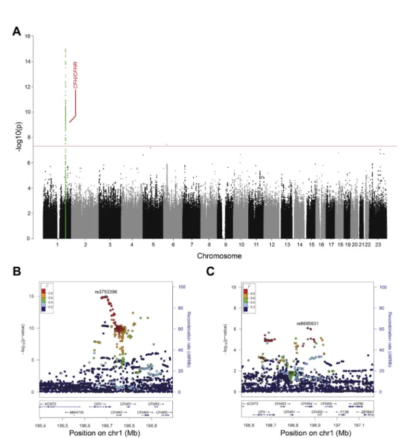
A total of 280 variants reached genome-wide significance (Manhattan plot, Fig 1A; QQplot, Fig S2, available at
www.aaojournal.org;
l
GC¼0.999). All variants, except for one,were located on chromosome 1 at the CFH/CFHR locus (chromosome 1, 196.643.724e197.061.086). The only variant
outside of this locus was located on chromosome 6 near the
PSORS1C1 gene, but could not be verified by Sanger sequencing. The SNP rs3753396 (c.2016A/G, p.Gln672Gln) located in exon 14 of the complement factor H (CFH) gene showed the strongest association with complement activation levels (P ¼ 1.09 1015;
b
¼ 0.145; standard error [SE], 0.018;Table 3; locus zoom depicted inFig 1B).Conditional analysis on the lead SNP revealed a second inde-pendent signal with aP value close to genome-wide significance for which the strongest associated variant was rs6685931. This SNP was also located at the CFH/CFHR locus, specifically in intron 1 (c.59e4315T/C) of the complement factor H related 4
(CFHR4) gene (P¼8.18107;
b
¼0.068; SE, 0.014;Table 3; locus zoom depicted inFig 1C).Figure 1. Graphs showing that the genome-wide association study identified 2 independent signals at theCFH/CFHRlocus associated with systemic complement activation levels.A, Manhattan plot illustrating thePvalues of each individual single nucleotide polymorphism (SNP) tested for association with systemic complement activation. The red horizontal line indicates the threshold considered for genome-wide significance (P¼5108).B, Locus zoom plot showing a detailed view of the chromosome 1 signal. The lead SNP rs3753396 is located in theCFHgene. The SNPs are colored based on their linkage disequilibrium estimate (r2) to the lead SNP.C, Locus zoom plot showing a detailed view of the signal on chromosome 1 (chr1) after conditioning the association analysis for rs3753396. Here, the lead SNP rs6685931 is located in theCFHR4gene. The SNPs are colored based on their linkage disequilibrium estimate (r2) to the lead SNP.
Table 1. Demographics and Other Characteristics of the Discovery and Replication Cohorts
Discovery Cohort (n[1548) Replication Cohort (n[697) Complement activation ln(c3d/c3), mean (SD) 1.459 (0.407) 1.464 (0.398)
Age (yrs), mean (SD) 73.2 (7.8) 73.3 (7.7)
Female sex (%) 60 58.8
AMD disease status, control (%) 53.7 37.4 BMI (kg/m2), median (quartiles) 25 (23
e28) 25 (23e28) Triglycerides (mmol/l), median (quartiles) 1.620 (1.170e2.220) 1.620 (1.165e2.210) HDL cholesterol (mmol/l), mean (SD) 1.489 (0.377) 1.478 (0.403) Clinic site - Radboud university medical center (%) 53.5 63 AMD¼age-related macular degeneration; BMI¼body mass index; HDL¼high-density lipoprotein; SD¼standard deviation.
Variants shown to be associated with complement activation fragments in previous studies were extracted from the GWAS re-sults.17,18The SNP rs800292 inCFHand the 2 SNPs in linkage disequilibrium rs4151667 in CFB and rs9332739 in C2 were associated nominally with systemic complement activation levels in the current study, showing the same direction of the effect. The SNP rs2230199 inC3and the SNP rs10490924 inARMS2could not be replicated (Table S4, available atwww.aaojournal.org).
Replication in an Independent Cohort Confirms the Effect of rs3753396 inCFHand rs6685931 in CFHR4 on Systemic Complement Activation
Replication analysis of rs3753396 in CFH and rs6685931 in
CFHR4 in an independent cohort of 697 study participants confirmed both variants to be associated significantly with systemic complement activation levels (rs3753396: P ¼ 1.39 106;
b
¼ 0.131; SE, 0.027; and rs6685931: P ¼ 8.62 103;b
¼0.038; SE, 0.014;Table 3). Subsequent meta-analysis showed associations for both rs3753396 (P¼3.661021;b
¼0.141; SE, 0.015) and rs6685931 (P ¼ 6.32 108;b
¼ 0.054; SE, 0.010), confirming that 2 independent signals at the CFH/CFHRlocus are associated with higher complement activation levels (Table 3). Sensitivity analyses adjusting for AMD disease status showed comparable results (Table S5, available at
www.aaojournal.org), and neither an interaction between clinic site and the identified SNPs (Prs3753396 clinic ¼ 0.436;
Prs6685931 clinic ¼ 0.676), nor an interaction between AMD
status and the identified SNPs (Prs3753396 AMD status ¼ 0.557;
Prs6685931AMD status¼0.658) was detected.
Next, mean complement activation levels in the genotype groups of rs373396 and rs6685931 were analyzed. For rs3753396 inCFH, the heterozygous AG genotype group showed higher complement activation levels compared with the reference AA genotype group (P¼6.231018;
b
¼0.152; SE, 0.018), and for the homozygousGG group, these levels were even higher (P ¼ 2.39 107;
b
¼0.267; SE, 0.052;Fig 3A). In the case of rs6685931 inCFHR4, a similar effect was observed: the heterozygous TC genotype group had higher complement activation levels than the reference TT genotype (P ¼ 103;b
¼ 0.063; SE, 0.019) and for the homozygous CC group, the levels were even higher (P¼3.62 107;b
¼0.118; SE, 0.023;Fig 3B). Analysis of the cumulative effect of both SNPs showed that the main effect on systemic complement activation levels is driven by rs3753396 inCFH, and rs6685931 in CFHR4 introduces additional variation to the rs3753396 genotypes (Fig 3C).A Model of Genetic and Nongenetic Variables Explains 18.7% of the Variability in Complement Activation
General linear models were built to determine how much of the variation could be explained by factors found to be associated with systemic complement activation. A model including only non-genetic factors (age, AMD disease status, BMI, and triglycerides) explained 12.6% of the variability in systemic complement acti-vation. With the addition of SNP rs3753396 to the model, 16.3% of the variability could be explained, and by including SNP rs6685931, a total of 17.3% was explained. We additionally incorporated SNPs associated with complement activation frag-ments in a previous study that replicated in our GWAS: rs800292 in CFH and rs9332739 in C2.18 Only rs9332739 remained associated independently with systemic complement activation levels, and the variance explained by the model rose to 18.7% (adjustedR2;Table 6). Table 3. Meta-analysis of Discovery and Replication Cohorts Identi fi es 2 Signals at the CFH/CFHR Locus Associated with Systemic Complement Activation Levels Lead Var iant (Minor Allele) Imputation Quality (R sq) * Chro mosome Positio n y Gene z Disc ove ry Coho rt (n [ 1548) R eplication C ohort (n [ 697) x Meta-analysis (n [ 2245) { Mino r A llele Freque ncy b (Sta ndard Error) P Value Mino r A llele Freque ncy b (Sta ndard Error) P Value b (Standard Erro r) P Value rs37533 96 (G) d 1:19 6 695 742 CF H 0.168 0.14 5 (0.018) 1.09 1 10 15 0.147 0.13 1 (0.027) 1.39 0 10 6 0.141 (0.01 5) 3.66 4 10 21 rs66859 31 (C) 0.99 1:19 6 867 233 C FHR4 0.439 0.06 8 (0.014) 8.18 4 10 7 0.493 0.03 8 (0.014) 8.62 0 10 3 0.054 (0.01 0) 6.32 0 10 8 *Not applica ble for genot yped var iants ( d ). yChromosome and chromoso mal posi tions des cribed ac cordin g to the reference sequenc e dat abase of th e Nation al Cen ter fo r Biotech nology Inf ormation (NCBI Re fSeq) hg19 hum an genome. zClosest ge ne to the lea d var iant. xReplication cohor t for rs6 685931 consis ted of 686 indi viduals. { Meta-analysis fo r rs66859 31 was perform ed in a tot al of 2234 indi viduals.
Haplotypes across theCFH/CFHRLocus Show Stronger Effects on Systemic Complement Activation Levels Compared with Individual Variants
To assess whether more variants at theCFH/CFHRlocus influence systemic complement activation and to determine the cumulative
effect of several variants on the same haplotype, we evaluated the effect of distinct haplotypes across theCFH/CFHRlocus on sys-temic complement activation. Haplotypes previously described for AMD already included rs3753396, the lead variant associated in the GWAS,34and were expanded by adding rs668593, the second independent signal. In total, 7 SNPs across theCFH/CFHRlocus yielded 9 different haplotypes with a predicted population
Figure 3. Graphs showing systemic complement activation levels stratified by rs3753396 and rs6685931 genotypes: rs6685931 introduced additional variation on the main effect of rs3753396. The y-axes represent the ln-transformed complement C3d/C3 ratio as a measure of systemic complement activation. Hor-izontal bars indicate the mean values for each genotype group. The complement-raising alleles for both single nucleotide polymorphisms are indicated in red. Association analyses included the 2245 individuals from the discovery and the replication cohorts.A, Distribution of complement activation levels for each genotype of rs3753396 inCFH.B, Distribution of complement activation levels for each genotype of rs6685931 inCFHR4.Pvalues were calculated adjusting the model for rs3753396.C, Distribution of complement activation levels over the genotype combinations of rs3753396 inCFHand rs6685931 inCFHR4.
frequency higher than 1% (Table 7; Table S8, available at
www.aaojournal.org).
Association with systemic complement activation levels revealed haplotypes with stronger effects on complement activation compared with the single SNPs identified in the GWAS. Haplo-types H1e2, H3, and H6 were associated with higher systemic
complement activation levels. Haplotype H3 carrying the complement-raising allele of rs3753396 (G) had a stronger effect on complement activation levels (P¼2.531014;
b
¼0.183; SE, 0.024) compared with the complement-raising allele of rs3753396 in the single variant analysis (b
¼0.141; SE, 0.015). Haplotypes H1e2 and H6 both carried the complement-raisingallele for rs6685931 (C). Haplotype H6 showed a stronger effect on complement activation levels (P¼4.82104;
b
¼0.144; SE, 0.041) compared with the single variant analysis for rs6685931 (b
¼ 0.054; SE, 0.010; Table 7; Table S8, available atwww.aaojournal.org).
The Single Nucleotide Polymorphism rs6685931 inCFHR4and Haplotype H1e2 Confer a Risk for Age-Related Macular Degeneration
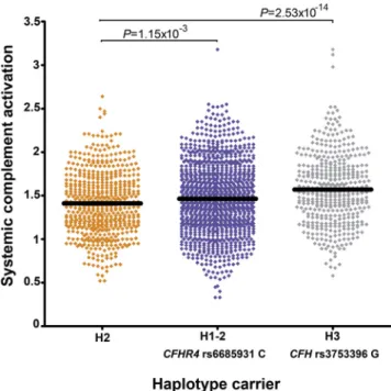
To identify genetic biomarkers that are relevant in the context of disease, we explored whether the SNPs and haplotypes associated with systemic complement activation levels also associate with AMD. The SNP rs3753396 inCFHwas not associated with AMD (P¼0.76). In contrast, the complement-raising allele of rs6685931 in CFHR4(C) was associated with an increased risk for AMD (P¼5.891012; odds ratio¼1.631; 95% confidence interval, 1.489e1.772;Table 9). These results are in concordance with the
largest GWAS on AMD reported to date (rs3753396, P ¼ 3 103; rs6685931,P¼1.0210495; odds ratio>1).9
In agreement with the single variant analysis of CFH
rs3753396, the haplotype H3 that gave the highest risk for higher systemic complement activation was not associated with AMD (P ¼ 0.80). Haplotype H6 carries the CFHR4 rs6685931 complement-raising allele (C), but did not reach significance in the association with AMD (P¼0.14); however, the frequency of H6 was relatively low (3%). Haplotype H1e2, the most common
haplotype carrying the CFHR4 rs6685931 complement-raising allele (C), showed a strong risk-conferring association with AMD (P ¼1.38 1012; odds ratio¼ 1.318; 95% confidence interval, 1.223e1.420;Table 9;Fig 4).
Finally, we determined whether other AMD-associated variants are associated with systemic complement activation levels. For this purpose, we extracted the 52 AMD-associated variants reported in the largest AMD study performed so far from the GWAS on complement activation levels.9However, no variants outside of the
CFH/CFHR locus were found to be associated with systemic complement activation levels at the genome-wide significance level, or at a significance level ofP<0.05/52¼0.001 (Table S10, available atwww.aaojournal.org). Interestingly, a risk score based on the 52 AMD risk-conferring alleles was associated with higher levels of complement activation (P¼0.043;
b
¼0.004; SE(b
)¼ 0.002). A similar risk score including only the variants located in or near complement genes was associated more strongly with higher levels of complement activation (P¼0.022;b
¼0.009; SE(b
)¼ 0.004). This complement risk score included 3 nominally associated variants: 2 common variants located in theCFHand theC2/CFB/ SKIV2Llocidrs10922109 and rs116503776, respectivelydand arare variant located in theCFIgene, rs141853578 or p.Gly119Arg. However, the effects of these genetic risk scores are smaller
Table 6. Model of Genetic and Nongenetic Variables Explaining 18.7% of the Variability in Systemic Complement Activation
b Standard Error (b) PValue
CFHrs3753396 AG 0.196 0.023 8.7721017 GG 0.330 0.066 6.461107 CFHR4rs6685931 TC 0.070 0.024 0.003 CC 0.125 0.033 1.620104 CFHrs800292 GA 0.011 0.023 0.639 AA 0.027 0.046 0.555 C2rs9332739 GC 0.185 0.034 4.674108 CC 0.168 0.213 0.431 Age (yrs) 0.004 0.001 0.005 Disease status (AMD) 0.035 0.020 0.089 BMI (kg/m2) 0.012 0.003 2106 Triglycerides (mmol/l) 0.131 0.011 1.1771033
AMD¼age-related macular degeneration; BMI¼body mass index.
R2 ¼ 0.193 (adjusted R2 ¼ 0.187). The model included the 1548 in-dividuals from the discovery phase.
Table 7. Association of Haplotypes across theCFH/CFHRLocus with Systemic Complement Activation Levels
Haplotype CFH
rs3753396 and
CFHR4rs6685931 Alleles Haplotype Frequency b Standard Error (b) PValue
H2 A-T 0.18 Reference Reference Reference
H1e2 A-C 0.36 0.062 0.019 1.148103 H3 G-T 0.14 0.183 0.024 2.5311014 H4 A-T 0.10 0.013 0.026 0.607 H5 A-T 0.04 0.058 0.038 0.128 H1e1 A-T 0.03 0.053 0.043 0.218 H6 A-C 0.03 0.144 0.041 4.823104 H7 A-C 0.03 0.060 0.046 0.192 H8 A-T 0.03 0.007 0.048 0.890
Haplotype association analyses with age-related macular degeneration were performed for the 1548 individuals in the discovery cohort. Haplotypes are coded as in Hageman et al.34If 2 different subhaplotypes based on the extra allele in single nucleotide polymorphism rs6685931 were found, the Hageman haplotypes were recoded as 1 or 2. Alleles associated with higher complement levels are underlined. The reference haplotype was set to the most common haplotype not carrying any complement-raising allele for rs3753396 or rs6685931.
compared with the single variant effects in the model for systemic complement activation described inTable 6.
Discussion
We conducted a GWAS on systemic complement activation levels, evaluating for an unbiased approach the genetic risk factors involved in the activation of this essential component
of the immune system. We identified and replicated 2
common variants, rs3753396 and rs6685931, that lead to higher systemic complement activation levels independently of age, sex, AMD disease status, triglycerides, and BMI. These 2 variants were included in a model for systemic complement activation, which explained 18.7% of its variability.
The SNP rs3753396 (c.2016A/G, p.Gln672Gln) is a
coding, synonymous variant located in exon 14 of theCFH
gene, and therefore this variant, or the linked causal vari-ant(s), may regulate complement activation levels through FH. Factor H is a key negative regulator of the AP and the
amplification loop of the complement cascade, which is
expressed constitutively in the liver and locally by other cell types, such as retinal pigment epithelial and endothelial
cells.35e37 Evidence to support the theory that rs3753396
exerts an effect on complement activation through FH comes from genetic studies on other diseases. The SNP rs3753396 has been reported to be associated with atypical hemolytic uremic syndrome, known to be caused by
muta-tions in CFH.38,39 Moreover, reduced susceptibility to
meningococcal disease also has been associated with
rs3753396. Meningococcal disease is caused by Neisseria
meningitides, which binds FH to avoid
complement-mediated killing.40 The SNP rs3753396 is in linkage
disequilibrium with rs1065489, also located in CFH
(c.2808G/T, p.Glu936Asp), which was proposed to be
the causal variant for meningococcal disease based on in
silico pathogenicity predictions.41
The SNP rs6685931 (c.59e4315T/C) is located in
intron 1 of theCFHR4gene. Factor H related 4 (FHR-4) is a
glycoprotein that, in contrast to the attenuating effects of FH, seems to promote complement activation. It binds the
complementfluid-phase C3b and forms an additional AP C3
convertase (FHR4-C3bBb), which is less susceptible to
FH-mediated decay.42 However, because rs6685931 is in high
linkage disequilibrium (r2 > 0.8) with several variants
located in the CFH gene, either FH or FHR-4 could be
responsible for the effects observed on complement activation.
We analyzed the association of genetic variants with systemic complement activation levels in a hypothesis-free manner. The results indicate that with our study design, the genetic variants with the largest effect on complement activation levels are rs3753396 and rs668593, located at the
CFH/CFHR locus. Moreover, other previously associated
variants in CFH and C2/CFB could be replicated.18
Haplotype analysis at the CFH/CFHR locus revealed 2
haplotypes with stronger effects on complement activation
levels compared with the individual SNPs. These findings
suggest that additional variants at the CFH/CFHR locus
play a role in the activation of the complement system.
Indeed, several rare coding variants in theCFHgene have
been shown to lead to increased complement activity.10
Genetic variants in other genes that influence systemic
complement activation levels may be uncovered with larger sample sizes that would allow for the detection of rarer variants and smaller effects. A compelling rare variant candidate that may merit further investigation is
CFI rs141853578 (p.Gly119Arg), which was found to be
nominally significant in our study. This variant has been
Figure 4. Graph showing complement activation levels stratified by common haplotypes across theCFH/CFHRlocus. The AMD risk haplo-type H1e2 shows high complement activation levels, and the none AMD-associated H3e1 haplotype shows the highest. Horizontal bars indicate the mean values for each haplotype carrier group. Haplotype carriers included in the graph had a posterior probability higher than 0.75. The haplotype group colors indicate the association with AMD: orange, protective; blue, risk conferring; grey, not associated. Association analyses were carried out for the 1548 patients genotyped with exome array.
Table 9. Association of Complement-Raising Single Nucleotide Polymorphism and Haplotypes with Age-Related Macular Degeneration: Single Nucleotide Polymorphism rs6685931 and
Haplotype H1e2 Confer a Risk for Age-Related Macular
Degeneration
Odds Ratio Confidence Interval PValue SNP rs3753396 1.031 0.839e1.223 0.756 SNP rs6685931 1.631 1.489e1.772 5.8891012 Haplotype H3 1.015 0.911e1.130 0.795 Haplotype H6 0.828 0.637e1.075 0.135 Haplotype H1e2 1.318 1.223e1.420 1.3821012 SNP¼single nucleotide polymorphism.
Single variant and haplotype association analyses with age-related macular degeneration were performed for the 1548 individuals from the discovery cohort. Haplotype analyses were based on chi-squared tests that compared the frequency of the analyzed haplotypes in patients versus controls.
associated previously with lower factor I levels in plasma and a lower ability to degrade C3d on the cell surface and
C3b in thefluid phase.43
In this study, AMD was associated with systemic com-plement activation, which is in agreement with previous
reports.15e18,20 In our analysis, rs6685931 in CFHR4 was
associated with both systemic complement activation and AMD. Haplotype analyses were in line with these results; we observed that the complement-raising allele of SNP
rs6685931 (C) was located mainly on the H1e2 haplotype,
which associated with a higher risk for AMD development. Thus, this SNP and its linked haplotype could serve as a robust biomarker for complement activation in the context of AMD and could be used to identify AMD patients who
would benefit most from complement-inhibiting therapies.
We noted that the rare haplotype H6 (with a frequency of 3%), also containing rs6685931, had a larger effect on complement activation levels compared with the single variant rs6685931. However, haplotype H6 was not
asso-ciated significantly with AMD, probably because of
statis-tical power limitations. Studies with larger cohort sizes may clarify the role of the H6 haplotype in AMD and may identify other rare haplotypes that associate with AMD and have larger effects on complement activation levels.
Strikingly, the genetic variant that was associated most strongly with systemic complement activation, rs3753396 in
CFH, and its main haplotype (H3) did not associate with
AMD. However, the SNP rs3753396 and haplotype H3 have been described to confer risk for atypical hemolytic uremic syndrome development. Atypical hemolytic uremic syndrome is a complement system-related disease that leads to systemic thrombotic microangiopathy and renal
endo-thelial injury.39,44Thisfinding suggests that the effect of the
haplotypes may be different systemically compared with the
AMD disease site, possibly through a tissue-specific effect
of the genetic variants. Consequently, systemic complement
activation may not always reflect complement activation in
the disease tissue, and therefore, it may not be the most appropriate measure for AMD studies. Genetic biomarkers
such as SNP rs6685931 and haplotype H1e2 are robust
markers that, together with the C3d-to-C3 ratio, could serve as biomarkers for complement activity studies in AMD. This is supported by a recent study demonstrating that complement activation levels in aqueous humor are higher
than in plasma samples of AMD patients.45 As a
consequence, the effect of rs6685931 and H1e2 on local
complement activation may be even larger than the effect seen on systemic levels.
Our results also could further the understanding of other complement-related diseases, as well as be used in the context of personalized medicine involving FH
supple-mentation therapy and other complement-targeting
therapies.46e48 Besides N. meningitidis, a number of
bac-teria, fungi, parasites, and viruses bind FH to avoid elimi-nation by the alternative pathway of the complement
system.49Also, some cancer cells express FH to avoid being
targeted by the immune system.50e52 Other FH-related
diseases for which our results may be of interest include hemolytic uremic syndrome, atypical hemolytic uremic
syndrome, encephalomyelitis, atherosclerosis, insulin
resistance, immunoglobulin A nephropathy, Alzheimer’s
disease, cisplatin nephropathy, as well as severe dengue, for
which variants in the CFH gene have been shown to be
protective.53
In conclusion, we identified 2 common variants located
at the CFH/CFHR locus, rs3753396 and rs668593, which
strongly influence systemic complement activation levels.
Moreover, our haplotype studies suggest that other genetic
variants in the CFH/CFHR locus influence systemic
com-plement activation. Genetic and nongenetic factors
identi-fied in this and other studies explain up to 18.7% of the
variability in systemic complement activation levels. The
common variant rs6685931 in CFHR4, and its associated
haplotype H1e2, could be used, together with other
envi-ronmental factors as well as rare genetic variants, to select
AMD patients who would benefit from
complement-inhibiting therapies. Acknowledgments
The authors thank Simon J. Clark, Lars G. Fritsche, and Valentina Cipiriani for their critical comments on the study design andfinal manuscript. We thank Alejandro Arias Vasquez and Joannes M. Groenewoud for their statistical assistance.
References
1. Ricklin D, Hajishengallis G, Yang K, et al. Complement: a key system for immune surveillance and homeostasis.Nat Immu-nol. 2010;11(9):785e797.
2. Dunkelberger JR, Song WC. Complement and its role in innate and adaptive immune responses.Cell Res. 2010;20(1):34e50.
3. Lachmann PJ, Halbwachs L. The influence of C3b inactivator (KAF) concentration on the ability of serum to support com-plement activation.Clin Exp Immunol. 1975;21(1):109e114.
4. Sarma JV, Ward PA. The complement system. Cell Tissue Res. 2011;343(1):227e235.
5. McGeer PL, Lee M, McGeer EG. A review of human diseases caused or exacerbated by aberrant complement activation. Neurobiol Aging. 2017;52:12e22.
6. Rudnicka AR, Jarrar Z, Wormald R, et al. Age and gender variations in age-related macular degeneration prevalence in populations of European ancestry: a meta-analysis. Ophthal-mology. 2012;119(3):571e580.
7. Chakravarthy U, Evans J, Rosenfeld PJ. Age related macular degeneration.BMJ (Clin Res ed.). 2010;340:c981.
8. Wong WL, Su X, Li X, et al. Global prevalence of age-related macular degeneration and disease burden projection for 2020 and 2040: a systematic review and meta-analysis.Lancet Glob Health. 2014;2(2):e106ee116.
9. Fritsche LG, Igl W, Bailey JN, et al. A large genome-wide association study of age-related macular degeneration high-lights contributions of rare and common variants. Nat Genet. 2016;48(2):134e143.
10. Geerlings MJ, de Jong EK, den Hollander AI. The comple-ment system in age-related macular degeneration: A review of rare genetic variants and implications for personalized treat-ment.Mol Immunol. 2017;84:65e76.
11. Hageman GS, Luthert PJ, Victor Chong NH, et al. An inte-grated hypothesis that considers drusen as biomarkers of immune-mediated processes at the RPE-Bruch’s membrane interface in aging and age-related macular degeneration.Prog Retin Eye Res. 2001;20(6):705e732.
12. Johnson LV, Leitner WP, Staples MK, et al. Complement activation and inflammatory processes in Drusen formation and age related macular degeneration. Exp Eye Res. 2001;73(6):887e896.
13. Wang L, Clark ME, Crossman DK, et al. Abundant lipid and protein components of drusen.PLoS One. 2010;5(4):e10329. 14. Fernandez-Godino R, Garland DL, Pierce EA. A local
com-plement response by RPE causes early-stage macular degen-eration.Hum Mol Genet. 2015;24(19):5555e5569.
15. Sivaprasad S, Adewoyin T, Bailey TA, et al. Estimation of systemic complement C3 activity in age-related macular degeneration.Arch Ophthalmol. 2007;125(4):515e519.
16. Scholl HP, Charbel Issa P, Walier M, et al. Systemic com-plement activation in age-related macular degeneration.PLoS One. 2008;3(7):e2593.
17. Reynolds R, Hartnett ME, Atkinson JP, et al. Plasma complement components and activation fragments: associa-tions with age-related macular degeneration genotypes and phenotypes. Invest Ophthalmol Vis Sci. 2009;50(12): 5818e5827.
18. Hecker LA, Edwards AO, Ryu E, et al. Genetic control of the alternative pathway of complement in humans and age-related macular degeneration.Hum Mol Genet. 2010;19(1):209e215.
19. Smailhodzic D, Klaver CC, Klevering BJ, et al. Risk alleles in CFHandARMS2are independently associated with systemic complement activation in age-related macular degeneration. Ophthalmology. 2012;119(2):339e346.
20. Lechner J, Chen M, Hogg RE, et al. Higher plasma levels of complement C3a, C4a and C5a increase the risk of subretinal fibrosis in neovascular age-related macular degeneration: complement activation in AMD.Immun Ageing. 2016;13:4. 21. Ristau T, Paun C, Ersoy L, et al. Impact of the common
genetic associations of age-related macular degeneration upon systemic complement component C3d levels. PLoS One. 2014;9(3):e93459.
22. Smailhodzic D, van Asten F, Blom AM, et al. Zinc supple-mentation inhibits complement activation in age-related mac-ular degeneration.PLoS One. 2014;9(11):e112682.
23. Yehoshua Z, de Amorim Garcia Filho CA, Nunes RP, et al. Systemic complement inhibition with eculizumab for geographic atrophy in age-related macular degeneration: the COMPLETE study.Ophthalmology. 2014;121(3):693e701.
24. Xu H, Chen M. Targeting the complement system for the management of retinal inflammatory and degenerative dis-eases.Eur J Pharmacol. 2016;787:94e104.
25. Paun CC, Lechanteur YT, Groenewoud JM, et al. A novel complotype combination associates with age-related macular degeneration and high complement activation levels in vivo. Sci Rep. 2016;6:26568.
26. Rother E, Lang B, Coldewey R, et al. Complement split product C3d as an indicator of disease activity in systemic lupus erythematosus.Clin Rheumatol. 1993;12(1):31e35.
27. Michel O, Sergysels R, Duchateau J. Complement activation in bronchial asthma evaluated by the C3d/C3 index.Ann Al-lergy. 1986;57(6):405e408.
28. Galle C, De Maertelaer V, Motte S, et al. Early inflammatory response after elective abdominal aortic aneurysm repair: a comparison between endovascular procedure and conventional surgery.J Vasc Surg. 2000;32(2):234e246.
29. Hempel JC, Poppelaars F, Gaya da Costa M, et al. Distinct in vitro complement activation by various intravenous iron preparations.Am J Nephrol. 2017;45(1):49e59.
30. Purcell S, Cherny SS, Sham PC. Genetic power calculator: design of linkage and association genetic mapping studies of complex traits.Bioinformatics. 2003;19(1):149e150.
31. Pruim RJ, Welch RP, Sanna S, et al. LocusZoom: regional visualization of genome-wide association scan results. Bioin-formatics. 2010;26(18):2336e2337.
32. Willer CJ, Li Y, Abecasis GR. METAL: fast and efficient meta-analysis of genomewide association scans. Bioinformat-ics. 2010;26(17):2190e2191.
33. Paun CC, Ersoy L, Schick T, et al. Genetic variants and sys-temic complement activation levels are associated with serum lipoprotein levels in age-related macular degeneration.Invest Ophthalmol Vis Sci. 2015;56(13):7766e7773.
34. Hageman GS, Anderson DH, Johnson LV, et al. A common haplotype in the complement regulatory gene factor H (HF1/CFH) predisposes individuals to age-related macular degeneration. Proc Natl Acad Sci U S A. 2005;102(20): 7227e7232.
35. Rodriguez de Cordoba S, Esparza-Gordillo J, Goicoechea de Jorge E, et al. The human complement factor H: functional roles, genetic variations and disease associations. Mol Immu-nol. 2004;41(4):355e367.
36. Chen M, Forrester JV, Xu H. Synthesis of complement factor H by retinal pigment epithelial cells is down-regulated by oxidized photoreceptor outer segments. Exp Eye Res. 2007;84(4):635e645.
37. Brooimans RA, van der Ark AA, Buurman WA, et al. Dif-ferential regulation of complement factor H and C3 production in human umbilical vein endothelial cells by IFN-gamma and IL-1.J Immunol. 1990;144(10):3835e3840.
38. Caprioli J, Castelletti F, Bucchioni S, et al. Complement factor H mutations and gene polymorphisms in haemolytic uraemic syndrome: the C-257T, the A2089G and the G2881T poly-morphisms are strongly associated with the disease.Hum Mol Genet. 2003;12(24):3385e3395.
39. Fremeaux-Bacchi V, Kemp EJ, Goodship JA, et al. The development of atypical haemolytic-uraemic syndrome is influenced by susceptibility factors in factor H and membrane cofactor protein: evidence from two independent cohorts. J Med Genet. 2005;42(11):852e856.
40. Kugelberg E, Gollan B, Tang CM. Mechanisms in Neisseria meningitidis for resistance against complement-mediated killing.Vaccine. 2008;26(suppl 8):I34eI39.
41. Martinon-Torres F, Png E, Khor CC, et al. Natural resistance to meningococcal disease related to CFH loci: meta-analysis of genome-wide association studies.Sci Rep. 2016;6:35842. 42. Hebecker M, Jozsi M. Factor H-related protein 4 activates
complement by serving as a platform for the assembly of alternative pathway C3 convertase via its interaction with C3b protein.J Biol Chem. 2012;287(23):19528e19536.
43. van de Ven JP, Nilsson SC, Tan PL, et al. A functional variant in the CFI gene confers a high risk of age-related macular degeneration.Nat Genet. 2013;45(7):813e817.
44. Pickering MC, de Jorge EG, Martinez-Barricarte R, et al. Spontaneous hemolytic uremic syndrome triggered by com-plement factor H lacking surface recognition domains.J Exp Med. 2007;204(6):1249e1256.
45. Schick T, Steinhauer M, Aslanidis A, et al. Local complement activation in aqueous humor in patients with age-related macular degeneration.Eye (Lond). 2017;31(5):810e813.
46. Buttner-Mainik A, Parsons J, Jerome H, et al. Production of biologically active recombinant human factor H in Phys-comitrella.Plant Biotechnol J. 2011;9(3):373e383.
47. Schmidt CQ, Slingsby FC, Richards A, et al. Production of biologically active complement factor H in therapeutically useful quantities.Protein Expr Purif. 2011;76(2):254e263.
48. Ricklin D, Lambris JD. New milestones ahead in complement-targeted therapy.Semin Immunol. 2016;28(3):208e222.
49. Ferreira VP, Pangburn MK, Cortes C. Complement control protein factor H: the good, the bad, and the inadequate.Mol Immunol. 2010;47(13):2187e2197.
50. Wilczek E, Rzepko R, Nowis D, et al. The possible role of factor H in colon cancer resistance to complement attack.Int J Cancer. 2008;122(9):2030e2037.
51. Junnikkala S, Hakulinen J, Jarva H, et al. Secretion of soluble complement inhibitors factor H and factor H-like protein (FHL-1) by ovarian tumour cells.Br J Cancer. 2002;87(10):1119e1127.
52. Ajona D, Castano Z, Garayoa M, et al. Expression of com-plement factor H by lung cancer cells: effects on the activation of the alternative pathway of complement. Cancer Res. 2004;64(17):6310e6318.
53. Pastor AF, Rodrigues Moura L, Neto JW, et al. Complement factor H gene (CFH) polymorphisms C-257T, G257A and haplotypes are associated with protection against severe dengue phenotype, possible related with high CFH expression. Hum Immunol. 2013;74(9):1225e1230.
Footnotes and Financial Disclosures
Originally received: August 9, 2017. Final revision: November 7, 2017. Accepted: December 13, 2017.
Available online: February 1, 2018. Manuscript no. 2017-1847. 1Department of Ophthalmology, Donders Institute for Brain, Cognition and Behaviour, Radboud University Medical Center, Nijmegen, The Netherlands.
2Department of Human Genetics, Donders Institute for Brain, Cognition and Behaviour, Radboud University Medical Center, Nijmegen, The Netherlands.
3
Department of Ophthalmology, University Hospital of Cologne, Cologne, Germany.
4
Department of Nephrology, Leiden University Medical Center, Leiden, The Netherlands.
5
Roche Pharma Research and Early Development, F. Hoffmann-La Roche Ltd., Basel, Switzerland.
Presented at: Association for Research in Vision and Ophthalmology Annual Meeting, Denver, CO, May 2017.
*Both authors contributed equally asfirst authors. Financial Disclosure(s):
The author(s) have made the following disclosure(s): T.S.: Lecturere
Bayer Health Care S.F.: EmployeeeRoche
Supported by: the European Research Council (the European Union’s Seventh Framework Programme [FP/2007-2013]/European Research Council grant agreement no. 310644 [MACULA]; the European Union’s Horizon 2020 research and innovation programme/European Research Council grant agreement no. 737607 [MACULA2]); Foundation Fighting Blindness, Columbia, Maryland (center grant no.: C-GE-0811-0548-RAD04 to Radboud University Medical Center); and the Stichting A. F. Deutman Oogheelkunde Researchfonds Nijmegen (Nijmegen, Gelderland,
The Netherlands). The sponsor or funding organization had no role in the design or conduct of this research. The authors acknowledge the contri-bution of the International AMD Genomics Consortium, supported by the National Institutes of Health, Bethesda, Maryland (grant no.: R01 EY022310).
HUMAN SUBJECTS: Human subjects were included in this study. No an-imal subjects were used in this study. The local ethics committees of Radboud University Medical Center and the University Hospital of Cologne approved the study, and written informed consent to participate in the study was ob-tained from all patients. The study was performed in accordance with the tenets of the Declaration of Helsinki (seventh revision) and the Medical Research Involving Human Subjects Act.
Author Contributions:
Conception and design: Lorés-Motta, Paun, den Hollander, de Jong Analysis and interpretation: Lorés-Motta, Paun, Corominas, Pauper, Geerlings, den Hollander, de Jong
Data collection: Lorés-Motta, Paun, Corominas, Pauper, Geerlings, Altay, Schick, Daha, Fauser, Hoyng, den Hollander
Obtained funding: den Hollander
Overall responsibility: Lorés-Motta, Paun, Corominas, Pauper, Geerlings, Altay, Schick, Daha, Fauser, Hoyng, den Hollander, de Jong
Abbreviations and Acronyms:
AMD ¼ age-related macular degeneration; AP¼ alternative pathway; BMI¼body mass index;C3¼component 3;GWAS¼ genome-wide association study; SE ¼ standard error; SNP ¼ single nucleotide polymorphism.
Correspondence:
Eiko K. de Jong, PhD, Department of Ophthalmology, Radboud University Medical Center, Route 409, Philips van Leydenlaan 15, 6525 EX Nijme-gen, The Netherlands. E-mail:[email protected].