Effect of Restricted Hip Flexors on Biomechanical Risk Factors for ACL Injury
Matthew Edward Mills
A thesis submitted to the faculty of the University of North Carolina at Chapel Hill in partial fulfillment of the requirements for the degree of Master of Arts in the Department of Exercise & Sport Science in the College of Arts & Sciences.
Chapel Hill 2013
Approved by:
Darin Padua, PhD, ATC Troy Blackburn, PhD, ATC Barnett Frank, MA, ATC
ii © 2013
Anterior Cruciate Ligament injuries are devastating on patients and society. It has been theorized that hip flexor restriction may affect muscles distally in the kinetic chain with no supporting literature. We hypothesized that those with hip flexor restriction will display biomechanics linked to ACL injury. Forty subjects completed functional tasks, range of motion measurements, and strength testing of gluteal muscles. Independent t-tests were run to determine the difference between control subjects and those with restricted hip flexors. Subjects with restricted hip flexors were observed to have less dorsiflexion, and less hip abduction and external rotation. There were no differences in gluteal strength. Restricted subjects were observed to have less anterior pelvic tilt at rest, less gluteus maximus activation during squatting, and exhibited differences in kinematics and kinetics during functional tasks. This indicates hip flexor restriction may lead to biomechanics identified as risk factors for ACL injury, and warrants further examination.
ABSTRACT
MATTHEW MILLS: Effect of Restricted Hip Flexors on Biomechanical Risk Factors for ACL Injury
iv
TABLE OF CONTENTS LIST OF FIGURES ... IX LIST OF TABLES ... X CHAPTER I ... 1
BACKGROUND ... 1
VARIABLES ... 5
RESEARCH QUESTION AND RESEARCH HYPOTHESIS ... 7
OPERATIONAL DEFINITIONS ... 14
ASSUMPTIONS ... 17
DELIMITATIONS: ... 17
LIMITATIONS: ... 18
SIGNIFICANCE OF THIS STUDY ... 18
CHAPTER II ... 19
INTRODUCTION ... 19
DEFINITION OF ACL INJURY ... 20
ANTERIOR CRUCIATE LIGAMENT INJURY PREVALENCE ... 21
NEGATIVE EFFECTS OF ACL INJURY ... 23
RISK FACTORS FOR ACL INJURY ... 24
Anatomical ... 24
Functional Task Biomechanics ... 28
LUMBO-PELVIC HIP COMPLEX CONTRIBUTIONS ... 31
Anatomy of Hip Flexor Group ... 31
Anterior Pelvic Tilt ... 33
Effects of Hip Flexor Tightness ... 33
SUMMARY ... 34
CHAPTER III ... 36
SUBJECTS ... 36
Inclusionary Criteria ... 36
Exclusionary Criteria: ... 37
INSTRUMENTATION ... 37
Electromagnetic Motion Capture System ... 37
Digital Inclinometer ... 37
Standard Goniometer: ... 38
Isometric Dynamometer: ... 38
Force Plate ... 39
Electromyography ... 39
PROCEDURES ... 39
Screening Session ... 39
Range of Motion Measurements ... 40
Electromyography ... 43
Motion Analysis ... 45
Overhead Squat ... 46
vi
Single Leg Squat ... 47
Isometric Strength Testing: ... 48
DATA PROCESSING AND ANALYSIS ... 49
Kinematics and Kinetics: ... 49
Jump Landing: ... 50
Single Leg Squat and Overhead Squat ... 50
Data Reduction ... 51
STATISTICAL ANALYSIS ... 51
CHAPTER IV ... 52
INTRODUCTION ... 52
METHODS ... 55
SUBJECTS ... 55
INSTRUMENTATION ... 56
PROCEDURES ... 58
RANGE OF MOTION MEASUREMENTS ... 58
ELECTROMYOGRAPHY ... 58
MOTION ANALYSIS ... 59
SQUATTING TRIALS ... 60
ISOMETRIC STRENGTH TESTING: ... 60
DATA ANALYSIS ... 60
STATISTICAL ANALYSIS ... 62
RESULTS ... 62
RANGE OF MOTION ... 62
GLUTEUS MAXIMUS ... 63
ELECTROMYOGRAPHY ... 63
KINEMATICS ... 64
DISCUSSION ... 65
FIGURES ... 70
FIGURE 1.1: SELECTION OF CONTROL GROUP ... 70
FIGURE 1.2: SELECTION OF RESTRICTED GROUP ... 70
FIGURE 3.1: MEASURE OF ILIOPSOAS TIGHTNESS ... 71
FIGURE 3.2: MEASURE OF RECTUS FEMORIS TIGHTNESS ... 71
FIGURE 3.3: MEASURE OF HIP EXTERNAL ROTATION ... 72
FIGURE 3.4: MEASURE OF HIP INTERNAL ROTATION ... 72
FIGURE 3.5: MEASURE OF HAMSTRINGS AT 90/90 ... 73
FIGURE 3.6: MEASURE OF HIP ABDUCTION ... 73
FIGURE 3.7: MEASURE OF PASSIVE DORSIFLEXION ... 74
FIGURE 3.8: MEASURE OF WEIGHT BEARING DORSIFLEXION ... 74
TABLES ... 75
TABLE 1: INTRACLASS CORRELATION COEFFICIENTS AND STANDARD ERROR OF THE MEASUREMENT FOR PASSIVE RANGE OF MOTION MEASUREMENTS ... 75
TABLE 2: INTRACLASS CORRELATION COEFFICIENTS AND STANDARD ERROR OF THE MEASUREMENT FOR ISOMETRIC STRENGTH MEASUREMENTS ... 75
viii
TABLE 5: MEANS AND STANDARD DEVIATIONS FOR EMG DATA OF HIP
EXTENSORS DURING AN OVERHEAD SQUATTING TASK ... 76 TABLE 6: MEANS AND STANDARD DEVIATIONS FOR PEAK KINEMATIC
VARIABLES DURING AN OVERHHEAD SQUATTING TASK ... 77 TABLE 7: MEANS AND STANDARD DEVIATIONS FOR DISPLACMENT
KINEMATIC VARIABLES DURING AN OVERHEAD SQUATTING TASK ... 78 APPENDIX ONE: ADDITIONAL RESULTS ... 79 Jump Landing Kinematics and Kinetics ... 79 Table A.1: Means and Standard Deviations for IGC Kinematic Variables
During a Jump Landing Task ... 80 Table A.2: Means and Standard Deviations for Peak Kinematic Variables
During a Jump Landing Task ... 80 Table A.3: Means and Standard Deviations for Displacement Kinematic
Variables During a Jump Landing Task ... 81 Table A.4: Means and Standard Deviations for Peak Kinetic Variables
During a Jump Landing Task ... 81 Single Leg Squat Kinematics and Kinetics ... 81 Table A.5: Means and Standard Deviations for Peak Kinematic Variables
During a Single Leg Squat ... 82 Table A.6: Means and Standard Deviations for Displacement Kinematic
Variables During a Single Leg Squat ... 82 Table A.7: Means and Standard Deviations for Peak Kinetic Variables
During a Single Leg Squat ... 83
LIST OF FIGURES
FIGURE 1.1: SELECTION OF CONTROL GROUP ... 70
FIGURE 1.2: SELECTION OF RESTRICTED GROUP ... 70
FIGURE 3.1: MEASURE OF RECTUS FEMORIS TIGHTNESS ... 71
FIGURE 3.2: MEASURE OF ILIOPSOAS TIGHTNESS ... 71
FIGURE 3.3: MEASURE OF HIP EXTERNAL ROTATION ... 72
FIGURE 3.4: MEASURE OF HIP INTERNAL ROATATION ... 72
FIGURE 3.5: MEASURE OF HAMSTRINGS AT 90/90 ... 73
FIGURE 3.6: MEASURE OF HIP ABDUCTION ... 73
FIGURE 3.7: MEASURE OF PASSIVE DORSIFLEXION ... 74
x
LIST OF TABLES
TABLE 1: INTRACLASS CORRELATION COEFFICIENTS
AND STANDARD ERROR OF THE MEASUREMENT FOR PASSIVE
RANGE OF MOTION MEASUREMENTS ... 75 TABLE 2: INTRACLASS CORRELATION COEFFICIENTS AND
STANDARD ERROR OF THE MEASUREMENT FOR ISOMETRIC
STRENGTH MEASUREMENTS ... 75 TABLE 3: MEANS AND STANDARD DEVIATIONS FOR RANGE
OF MOTION VARIABLES ... 76 TABLE 4: MEANS AND STANDARD DEVIATIONS FOR
STRENGTH VARIABLES ... 76 TABLE 5: MEANS AND STANDARD DEVIATIONS FOR EMG
DATA OF HIP EXTENSORS DURING AN OVERHEAD SQUAT ... 76 TABLE 6: MEANS AND STANDARD DEVIATIONS FOR PEAK
KINEMATIC VARIABLES DURING AN OVERHHEAD SQUAT ... 77 TABLE 7: MEANS AND STANDARD DEVIATIONS FOR
DISPLACMENT KINEMATIC VARIABLES DURING AN
OVERHEAD SQUAT ... 78 TABLE A.1: MEANS AND STANDARD DEVIATIONS FOR IGC
KINEMATIC VARIABLES DURING A JUMP LANDING TASK ... 80 TABLE A.2: MEANS AND STANDARD DEVIATIONS FOR PEAK
KINEMATIC VARIABLES DURING A JUMP LANDING TASK ... 80 TABLE A.3: MEANS AND STANDARD DEVIATIONS FOR
DISPLACMENT KINEMATIC VARIABLES DURING A JUMP
LANDING TASK ... 81 TABLE A.4: MEANS AND STANDARD DEVIATIONS FOR PEAK
KINETIC VARIABLES DURING A JUMP LANDING TASK ... 81 TABLE A.5: MEANS AND STANDARD DEVIATIONS FOR PEAK
KINEMATIC VARIABLES DURING A SINGLE LEG SQUAT ... 82 TABLE A.6: MEANS AND STANDARD DEVIATIONS FOR
DISPLACMENT KINEMATIC VARIABLES DURING A SINGLE
TABLE A.7: MEANS AND STANDARD DEVIATIONS FOR PEAK
CHAPTER I INTRODUCTION
BACKGROUND
Anterior Cruciate Ligament (ACL) injury is an extremely debilitating and commonly occurring injury within athletics worldwide. It is estimated that there are approximately 100,000 to 250,000 per year (Toth and Cordasco 2001; Marshall, Padua et al. 2007), with approximately 1 million physician visits annually in the United States (Marshall, Padua et al. 2007). Most commonly, the ACL is injured in a non-contact mechanism (Griffin, Agel et al. 2000; Agel, Arendt et al. 2005; Hewett T 2011), and frequently during a landing task (Shimokochi and Shultz 2008).
Soccer and basketball are frequently considered to be two major activities that have high risk of ACL injury (Ireland 1999; Agel, Arendt et al. 2005; Faunø P 2006; Yu and Garrett 2007). Soccer is widely thought to be the most popular sport in the world, with an estimated 17 million participates, and 7 million female participants (Lohmander, Ostenberg et al. 2004). Furthermore, soccer was observed to have a higher rate of ACL injury than basketball regardless of gender (Agel, Arendt et al. 2005).
old (Shea, Pfeiffer et al. 2004), with female soccer players observed to be 2.39 times more likely to sustain injury compared to their male counterparts (Ireland 1999). Furthermore, females were observed to have a prevalence of non-contact ACL injury of 0.17 per contact hour, compared to males rate of 0.05 per contact hour (Ireland 1999). Thus, it is evident that female soccer player are at a high risk of ACL injury, and the aim of research should be to understand and modify risk factors in this population.
ACL injury is devastating on not only the individual patient, but also on society as a whole. ACL injury in Denmark sees $5.4 million dollars billed to insurance companies(Gottlob C. A. 2000; Cumps, Verhagen et al. 2008). The estimated financial burden is approximately $11,500 per patient, which leads to an estimated $1.5 billion dollars in costs per year (Gottlob C. A. 2000). Not only is ACL injury financially devastating, but it is extremely physically disabling to patients. Thirty seven percent of patients never return to the same level of activity (Ardern, Webster et al.), with only 44% returning to competitive sports, and 18% never returning to any sport activity (Ardern, Webster et al.). The mean time from surgery to return to activity is 7.3 months (Ardern, Webster et al.). Furthermore, there is a serious psychological toll placed on those with ACL injury, as those who sustain ACL injury have a higher depression rate for a longer duration than those who suffer concussion (Mainwaring, Hutchison et al. 2010).
3
(Lohmander, Ostenberg et al. 2004; Meunier, Odensten et al. 2007). OA onset occurs 10-20 years earlier in those suffering ACL injury compared to a population with similar demographics, with 50% of patients suffering ACL injury presenting with OA developments (Lohmander, Ostenberg et al. 2004; Meunier, Odensten et al. 2007). In addition, 75% of patients have pain that interferes with their activities of daily life due to OA 12 years following ACL injury (Lohmander, Ostenberg et al. 2004).
While there are many biomechanical risk factors that predispose individuals to ACL pathology, lumbo-pelvic hip complex motion and medial knee displacement have both been linked to ACL injury and mechanism. Ireland identified the “position of no return” in which the hip abductors and extensors “shut down”, and fail to effectively control lumbo-pelvic-hip motion. This results in hip internal rotation, hip adduction, knee valgus, and tibial external rotation (Ireland 1999). Knee valgus, and by association, medial knee displacement has frequently been suggested as a biomechanical risk factor for ACL pathology (Hruska 1998; Delp, Hess et al. 1999; Boden, Griffin et al. 2000; Hertel J 2004; Agel, Arendt et al. 2005; Alentorn-Geli, Myer et al. 2009; Chiaia, Maschi et al. 2009). Hip musculature weakness has also been linked to greater anterior pelvic tilt (Popovich and Kulig). Greater anterior pelvic tilt has, in turn, been correlated with ACL injury (Hertel J 2004), and has been theorized to be caused by overactive/tight hip flexors.
co-activation ratios (Malinzak, Colby et al. 2001), and range of motion shifting from hip external rotation to hip internal rotation (Brophy, Chiaia et al. 2009). All of the above factors have been theorized to potentially increase ACL injury risk.
Hip flexor restriction has been theorized to affect lumbo-pelvic hip motion, as well as ACL injury risk factors, including medial knee displacement, excessive trunk forward flexion, and femoral internal rotation. Other studies have examined other aspects of hip and trunk biomechanics, and have linked it to ACL pathology risk factors, including lateral trunk motion, overactive hip adductors, and underactive or inhibited hip abductors (Hewett T 2011). Delp et al identified that greater hip flexion is associated with a shifting of the line of pull of the external rotators, which, in turn, forces these muscle to cause an internal rotation moment of the hip (Delp, Hess et al. 1999). Zeller et al identified that overactive rectus femoris musculature during a single leg squat increases knee valgus displacement (Zeller, McCrory et al. 2003). Furthermore, greater flexibility of the hamstrings and lesser activation has been suggested to decrease knee stiffness and increase knee valgus (Boden, Griffin et al. 2000; Zeller, McCrory et al. 2003). Female soccer players possess decreased hip flexor flexibility as measured by a “Thomas test” compared to normative values for a similar demographic that does not play soccer (Chiaia, Maschi et al. 2009). This “tightness” may be a result of either mechanical restriction, or a result of hyperactivity of the hip flexor musculature. As such, further examination of the effects of hip flexor tightness and its relationship to lower extremity biomechanics is indicated.
5
with restricted hip flexors and those with “normal” hip flexor length. Determining this relationship will allow for clinicians to screen for a potential risk factor for ACL pathology, and could potentially lead to prevention strategies through correction of muscle imbalances that characterize hip flexor muscle restriction.
VARIABLES
• Independent
o Hip Flexor Restriction (2 Groups)
§ Control: Those with normal hip flexor motion as measured by a modified “Thomas test” compared to normative values (Figure 1.1) § Restricted: Those who display limited hip flexor motion as
measured by a modified “Thomas test” compared to normative values (Figure 1.2)
• Dependent
o Kinematic Data
§ Sagittal Plane Hip Kinematics at initial ground contact § Frontal Plane Hip Kinematics at initial ground contact § Sagittal Plane Knee Kinematics at initial ground contact § Frontal Plane Knee Kinematics at initial ground contact § Peak Sagittal Plane Hip Kinematics
§ Peak Frontal Plane Knee Kinematics § Peak Trunk Forward Flexion Angle § Peak Anterior Pelvic Tilt
§ Sagittal Plane Hip Displacement § Frontal Plane Hip Displacement § Sagittal Plane Knee Displacement § Frontal Plane Knee Displacement § Medial Knee Displacement
o Range of Motion of Lower Extremity Musculature § Iliopsoas
§ Rectus Femoris § Hamstrings
§ Hip Internal Rotators § Hip External Rotators § Hip Adductors
§ Gastrocnemius/Soleus o Isometric Hip Strength
§ Gluteus Medius
• Measured at neutral and 20 degrees abduction
§ Gluteus Maximus
• Measured at neutral and 15 degrees extension
7 § Gluteus Maximus § Biceps Femoris
RESEARCH QUESTION AND RESEARCH HYPOTHESIS
1. Research Question 1: Is there a statistically significant difference between groups of female soccer players with restricted hip flexors as measured by a modified “Thomas test” and those with normal hip flexor length in trunk, hip, and knee kinematic data during three functional tasks.
a. Research Question 1A: Is there a statistically significant difference in sagittal plane hip kinematics at initial ground contact, peak values, and displacements between these groups during three functional tasks? b. Research Question 1B: Is there a statistically significant difference in
frontal plane hip kinematics at initial ground contact, peak values, and displacements between these groups during three functional tasks? c. Research Question 1C: Is there a statistically significant difference in
sagittal plane knee kinematics at initial ground contact, peak values, and displacements between these groups during three functional tasks? d. Research Question 1D: Is there a statistically significant difference in
frontal plane knee kinematics at initial ground contact, peak values, and displacements between these groups during three functional tasks? e. Research Question 1E: Is there a statistically significant difference in
f. Research Question 1F: Is there a statistically significant difference in peak hip extension between these groups during three functional tasks? g. Research Question 1G: Is there a statistically significant difference in
peak anterior pelvic tilt between these groups during three functional tasks?
• Research Hypothesis 1: The group with restricted hip flexors will have
statistically significantly differences in sagittal and frontal plane kinematics at initial ground contact, peak values, and displacements, peak anterior pelvic tilt, peak trunk forward flexion, peak hip extension and medial knee
displacement compared to a control group during three functional tasks. o
o Research Hypothesis 1A: The restricted hip flexor group will have
significantly more anterior pelvic tilt compared to the control group during three functional tasks.
o Research Hypothesis 1B: The restricted hip flexor group will have
significantly more trunk forward flexion compared to the control group during three functional tasks.
o Research Hypothesis 1C: The restricted hip flexor group will have
significantly more medial knee displacement compared to the control group during three functional tasks.
o Research Hypothesis 1D: The restricted hip flexor group will have
9
2. Research Question 2: Is there a statistically significant difference in passive hip range of motion between groups of female soccer players with restricted hip flexors as measured by a modified “Thomas Test” and those with normative hip flexor motion?
a. Research Question 2A: Is there a statistically significant difference in hamstring flexibility between these groups?
b. Research Question 2B: Is there a statistically significant difference in iliopsoas flexibility between these groups?
c. Research Question 2C: Is there a statistically significant difference in
rectus femoris flexibility between these groups?
d. Research Question 2D: Is there a statistically significant difference in hip
internal rotator flexibility between these groups?
e. Research Question 2E: Is there a statistically significant difference in hip
external rotator flexibility between these groups?
f. Research Question 2F: Is there a statistically significant difference in hip
adductor flexibility between these groups?
g. Research Question 2G: Is there a statistically significant difference in
ankle plantarflexor flexibility between these groups?
• Research Hypothesis 2: There will be a statistically significant difference in
o Research Hypothesis 2A: The restricted hip flexor length group will
have significantly more hamstring flexibility compared to the control group.
o Research Hypothesis 2B: The restricted hip flexor length group will
have significantly less iliopsoas flexibility compared to the control group.
o Research Hypothesis 2C: The restricted hip flexor length group will
have significantly less rectus femoris flexibility compared to the control group.
o Research Hypothesis 2D: The restricted hip flexor length group will
have significantly less hip internal rotator flexibility compared to the control group.
o Research Hypothesis 2E: The restricted hip flexor length group will
have significantly more hip external rotator flexibility compared to the control group.
o Research Hypothesis 2F: The restricted hip flexor length group will
have significantly less hip adductor flexibility compared to the control group.
o Research Hypothesis 2G: The restricted hip flexor length group will
have significantly less ankle plantarflexor flexibility compared to the control group.
11
flexor length as measured by a modified “Thomas test” and those with normative hip flexor length?
a. Research Question 3A: Is there a statistically significant difference in gluteus maximus isometric strength between these groups at neutral? b. Research Question 3B: Is there a statistically significant difference in
gluteus maximus isometric strength between these groups at 15 degrees of hip extension?
c. Research Question 3C: Is there a statistically significant difference in gluteus medius isometric strength between these groups at neutral? d. Research Question 3D: Is there a statistically significant difference in
gluteus medius isometric strength between these two groups at 20 degrees of hip abduction?
• Research Hypothesis 3: There will be a statistically significant difference in
hip isometric strength in a group of female soccer players with restricted hip flexor length and a control group.
o Research Hypothesis 3A: The restricted hip flexor length group will
have significantly less gluteus maximus isometric strength compared to the control group at neutral.
o Research Hypothesis 3B: The restricted hip flexor length group will
o Research Hypothesis 3C: The restricted hip flexor length group will
have significantly less gluteus medius isometric strength compared to the control group at neutral.
o Research Hypothesis 3D: The restricted hip flexor length group will
have significantly less gluteus medius isometric strength compared to the control group at 20 degrees of hip abduction.
4. Research Question 4: Is there a statistically significant difference between groups of female soccer players with restricted hip flexor length as measured by a
modified “Thomas test” and those with normal hip flexor length in trunk, hip, and knee kinetic data during three functional tasks.
a. Research Question 4A: Is there a statistically significant difference in peak hip extension moment between these groups during three functional tasks?
b. Research Question 4B: Is there a statistically significant difference in peak knee extension moment between these groups during three functional tasks?
c. Research Question 4C: Is there a statistically significant difference in peak hip adduction moment between these groups during three functional tasks?
13
• Research Hypothesis 4: The group with restricted hip flexor length will have
statistically significantly differences in peak anterior pelvic tilt, peak trunk forward flexion, peak hip extension and medial knee displacement compared to a control group during three functional tasks.
o Research Hypothesis 4A: The restricted hip flexor length group will
have significantly less peak hip extension moment compared to the control group during three functional tasks.
o Research Hypothesis 4B: The restricted hip flexor length group will
have significantly more knee extension moment compared to the control group during three functional tasks.
o Research Hypothesis 4C: The restricted hip flexor length group will
have significantly more peak hip adduction moment compared to the control group during three functional tasks.
o Research Hypothesis 4D: The restricted hip flexor length group will
have significantly more peak knee varus moment compared to the control group during three functional tasks.
• Research Question 5: Is there a statistically significant difference in groups of
female soccer players with restricted hip flexor length as measured by a modified “Thomas test” and those with normal hip flexor length in average hip extensor electromyographic activity during three functional tasks?
o Research Question 5A: Is there a statistically significant difference in
o Research Question 5B: Is there a statistically significant difference in
average Biceps Femoris activation between these groups during three functional tasks?
• Research Hypothesis 5: The group with restricted hip flexor length will have
statistically significantly differences in average electromyography of hip extensors compared to a control group during three functional tasks.
o Research Hypothesis 5A: The restricted hip flexor length group will
have significantly less average Gluteus Maximus activation during three functional tasks.
o Research Hypothesis 5B: The restricted hip flexor length group will
have significantly less average Biceps Femoris activation compared to the control group during three functional tasks.
Statistical Hypothesis
• HO: HFT=CG • HA: HFT≠CG
OPERATIONAL DEFINITIONS
• Hip Flexor Muscles
o Iliopsoas and Rectus Femoris as measured by a Modified “Thomas Test” (Harvey 1998)
• Modified “Thomas Test”:
15
contralateral leg into maximal hip flexion. The examiner then lowers the braced leg until the pelvis begins to anteriorly rotate or the first point of resistance. The angle is measured between the table and the thigh and recorded (Ferber, Kendall et al. 2010).
• Restricted Hip Flexor Tightness
o Subjects, when measured using a modified “Thomas test” have digital inclinometer readings of the ipsilateral thigh of greater than 0 degrees above the horizontal (Ferber, Kendall et al. 2010)
• Non-Contact Injury
o ACL failure (rupture) during a functional activity in the absence of any external force except ground reaction forces (Shimokochi and Shultz 2008)
• Lumbo-pelvic hip complex motion
o Any physiologic movement that occurs in the Lumbar Spine, Femoro-acetabular joint, Sacroilliac joint, or rotation of the innominate.
• Anterior Pelvic Tilt
o Motion (in degrees) of the ASIS in an anterior direction of the sagittal plane as measured through motion analysis.
• Forward Trunk Flexion
o Motion (in degrees) of the trunk rigid body segment angle relative to the world in the sagittal plane as measured through motion analysis.
o Movement of the joint center of the knee medially in the frontal plane over the great toe. (Bell, Padua et al. 2008)
• Female Soccer Player
o A player who participates in organized soccer activities at least 2 times per week for at least 45 minutes per occasion who plays under standard rules and regulations.
• Jump Landing Task
o A procedure in which the participant jumps forward off a 30-cm high box placed at a distance ½ of their body height from the leading edge of a right and left force plate. The participant is instructed to land with their right foot on the right force plate and their left foot on the left force plate. The participant lands and is instructed to immediately jump for maximal vertical height.
• Single Leg Squat Task
o A task in which a subject, while standing on their dominant leg, lowers their center of mass until the thigh is parallel to the ground, or the subject loses their balance and touches down with their unaffected side.
• Dominant Leg
17 ASSUMPTIONS
• A handheld dynamometer accurately depicts isometric strength at a specific joint
angle for a specific muscle
• Motion tracking is representative of physiologic motion compared to skin motion
with minimal motion artifact
• The use of a standard goniometer to measure active range of motion is indicative
of the antagonist’s length.
• The use of a digital inclinometer to measure active range of motion is indicative
of the antagonist’s length.
• EMG electrodes on the skin may not give a true reading of the underlying muscle
activity
DELIMITATIONS:
• Subjects will be Female Soccer Players from the University of North Carolina at
Chapel Hill Women’s Varsity, Club, Intramural, and Lifetime Fittness Soccer Programs
• Subjects will be ages 18 to 35 years old
• Subjects with history of lower extremity, abdominal, or spine injury in the past 3
months that limited activity for greater than three days will be excluded.
• Subjects with a history of any lower extremity, abdominal, or spine fracture or
LIMITATIONS:
• Selected athletes may not accurately represent all soccer players • Soccer players may not accurately represent other field sports
• Different subjects may have previous training in performing specified tasks • Laboratory environment may not accurately represent field environment • Motion artifact may limit the accuracy of motion capture devices.
• Footwear is not consistent among individuals and thus may lead to alterations
o Tennis Shoes may not affect the LE biomechanics in the same manner as soccer cleats during activity.
• This study did not measure the muscular activation of the other hip, thigh, and
lower leg muscles.
SIGNIFICANCE OF THIS STUDY
CHAPTER II
REVIEW OF THE LITERATURE
INTRODUCTION
Anterior Cruciate Ligament (ACL) injuries are debilitating, devastating, and costly injuries for patients and the healthcare system. Previous literature has looked at a wide array of various risk factors. These risk factors have included anatomical risk factors, as well as biomechanical risk factors. While anatomical risk factors are widely considered to be non-modifiable, biomechanical risk factors have been shown to be modifiable frequently (Hewett, Stroupe et al. 1996). Biomechanical risk factors have been identified proximally at the region of the lumbo-pelvic-hip complex (Ireland 1999; Boden, Griffin et al. 2000; Zeller, McCrory et al. 2003; Hewett T 2011), at the knee itself through kinetics and kinematics (Hruska 1998; Delp, Hess et al. 1999; Boden, Griffin et al. 2000; Hertel J 2004; Agel, Arendt et al. 2005; Alentorn-Geli, Myer et al. 2009; Chiaia, Maschi et al. 2009), as well as distally at the foot (Hertel J 2004).
kinetic chain, no methodology to date has substantiated this theory. Therefore, the purpose of this literature review is to examine the relationship between restricted hip flexor tightness and previously identified lower extremity biomechanical risk factors for ACL injury.
DEFINITION OF ACL INJURY
An ACL tear is commonly defined as a rupture of the Anterior Cruciate Ligament, an intracapsular ligament within the knee responsible for preventing anterior translation of the tibia on the femur (Neuman 2010). The ACL has two distinct bundles, the anterior-medial bundle, as well as the posterior-lateral bundle, which are named based on their relative attachments on the tibia (Neuman 2010). It is believed that the ACL provides approximately 85% of total passive resistance of anterior translation of the tibia(Neuman 2010). Tension within the cruciate ligaments in the knee is also responsible for assisting with the arthrokinematics of the knee, as well as proprioceptive feedback of the knee. Tearing of the anterior cruciate ligament, therefore, presents major complications, primarily knee instability (Noyes, Mooar et al. 1983).
21
Approximately 70% of ACL injuries are non-contact in nature (Agel, Arendt et al. 2005), suggesting that an individual’s movement itself was the cause of injury, thus, non-contact ACL injuries can be prevented through alterations of the athlete’s biomechanics. In cadaveric studies, the ACL was observed to be torn with anterior tibial shear forces, which results in anterior tibial translation, as well as increased stress on the ACL with valgus and knee flexion (Kennedy, Weinberg et al. 1974; Withrow, Huston et al. 2006). In human subjects, there were observed to be three main positions of risk. It was
observed that the greatest risk in a functional movement was knee internal rotation in full knee extension, with another position during varus loads in knee hyperextension and extension (Markolf, Burchfield et al. 1995). Lastly, valgus loads in knee flexion were observed to lead to increased risk of injury (Markolf, Burchfield et al. 1995). As such, the vast majority of ACL injuries may be preventable, which requires a better understanding of the underlying risk factors and how they may predispose an athlete for these
devastating injuries, as well as a better way to screen for these risk factors.
ANTERIOR CRUCIATE LIGAMENT INJURY PREVALENCE
cutting. (Boden, Griffin et al. 2000; Toth and Cordasco 2001; Alentorn-Geli, Myer et al. 2009; Boden, Torg et al. 2009; Hewett, Torg et al. 2009).
Activities with rapid accelerations and changes in direction, as described above, are common during athletic participation. One such sport that is widespread throughout the globe is soccer, in which athletes are required to have high velocity movements and changes in direction. It is currently estimated that there are 17 million players, with 7 million female athletes (Lohmander, Ostenberg et al. 2004). As such, soccer is considered to be a sport that places participants at high risk for ACL injury. This is validated through the finding that athletes were observed to have the highest rate of injury in soccer
compared to basketball per contact hour across genders (Agel, Arendt et al. 2005). The prevalence of ACL injury in soccer athletes has been repeatedly examined, and it was observed that there was the highest prevalence of injury in male athletes, yet the highest risk of injury in female athletes (Shea, Pfeiffer et al. 2004; Marshall, Padua et al. 2007). The higher prevalence has been attributed to the fact that they have higher exposure to activities likely to cause ACL pathology (Shea, Pfeiffer et al. 2004; Marshall, Padua et al. 2007). Further analysis of epidemiological data suggests there is a higher incidence of injury in females (Griffin, Agel et al. 2000; Shea, Pfeiffer et al. 2004), particularly those ages 16-18 (Shea, Pfeiffer et al. 2004). It was observed that female soccer players are 2.29 times more likely to sustain than male counterparts (Ireland 1999), as they have noncontact injury rates of 0.17 per contact hour in females, compared to 0.05 per contact hour in males (Ireland 1999). This finding was confirmed by
23
bias among those who suffer ACL injury, particularly within soccer athletes, which mandates that research examine potential risk factors in females.
NEGATIVE EFFECTS OF ACL INJURY
ACL injuries are also extremely debilitating for patients physically and emotionally. It was observed that 37% of patients never return to same level of
competition (Ardern, Webster et al.). In addition, only 44% return to competitive sports and 18% never return to sports at all (Ardern, Webster et al.). The mean time from surgery until the athlete returns to athletic activity was observed to be 7.3 months (Ardern, Webster et al.). Furthermore, it was observed that there was a greater level and duration of depression compared to those suffering from a concussion (Mainwaring, Hutchison et al. 2010).
There is also high risk of a second ACL tear, as after a patient tears their ACL, individuals with a previous ACL injury were observed to be at an increased risk of repeated injury, either on the affected side or contralateral side. It was observed that the risk of re-injury ranged from 33% (Dallalana, Brooks et al. 2007) to 27.2% (Pinczewski, Lyman et al. 2007). Furthermore, it was observed that there was an increase in risk for meniscus injury, as it was observed that there was a 45% risk for meniscus injury (Maletius and Messner 1999).
Furthermore, 10 to 20 years after diagnosis, 50% of patients were observed to have osteoarthritic development (Lohmander, Englund et al. 2007), with 75% of patients reporting pain that interferes with daily life due to osteoarthritis 12 years after ACL injury (Lohmander, Ostenberg et al. 2004).
ACL injuries are not only devastating on the individual, but also financially demanding on the healthcare system. It was observed that $5.4 million billed to insurance companies per year with $2.7 million covered by government in Denmark (Cumps, Verhagen et al. 2008). Furthermore, it is estimated that the average cost of ACL injury was $11,500 per patient (Gottlob C. A. 2000), which leads to an estimated cost of $1.5 billion per year (Gottlob C. A. 2000).
RISK FACTORS FOR ACL INJURY Anatomical
25
risk of ACL injury (Hertel J 2004; Pantano, White et al. 2005; Tillman, Bauer et al. 2005; Daneshmandi, Saki et al. 2011). Furthermore, increased Q angle has been linked with increased knee valgus angulation, which is the most commonly referenced risk factor for ACL injury (Powers 2003). With respect to the distal kinetic chain, foot pronation has also been linked to ACL injury risk (Hertel J 2004). While foot pronation can be modified through the use of orthotics, Q-angle is not considered to be a modifiable risk factor, and is frequently larger in females compared to males due to their wider pelvis, and is considered to be a potential contributor to the increased risk of ACL injury for females (Boden, Griffin et al. 2000; Hewett, Myer et al. 2006).
While anatomical factors proximally and distally in the kinetic chain have been linked with increased ACL risk, knee anatomical risk factors have consistently been linked to increased risk. One such risk factor that was identified was a decreased intercondyler notch size, which is frequently observed more in females than males (Boden, Griffin et al. 2000; McClay Davis and Ireland 2001; Tillman, Smith et al. 2002; Uhorchak, Scoville et al. 2003; Alentorn-Geli, Myer et al. 2009; Boden, Torg et al. 2009). ACL biomechanics have also been linked to ACL injury, as the specific
biomechanical properties vary depending upon gender (Chandrashekar, Slauterbeck et al. 2005; Chandrashekar, Mansouri et al. 2006). Furthermore, a decreased ACL size has been linked to ACL injury, and females have typically a smaller ACL compared to their male counterparts (Chandrashekar, Slauterbeck et al. 2005; Chandrashekar, Mansouri et al. 2006; Hashemi, Mansouri et al. 2011).
estrogen and progesterone. Several studies have observed that decreases in estrogen and progesterone in women have been linked to increased ligament laxity, and thus ACL injury (Boden, Griffin et al. 2000; Slauterbeck and Hardy 2001; Toth and Cordasco 2001; Slauterbeck, Fuzie et al. 2002; Wojtys, Huston et al. 2002). This is also substantiated as ACL injury frequencies are not uniform throughout menstrual cycle (Slauterbeck, Fuzie et al. 2002; Wojtys, Huston et al. 2002). However, other studies have refuted this claim, and observed no link between ACL rupture and hormone concentrations in females (Warden, Saxon et al. 2006).
Muscle Function
27
Decreased production of hamstring force has also been linked with ACL
pathology (More, Karras et al. 1993; Shimokochi and Shultz 2008; Alentorn-Geli, Myer et al. 2009). Hamstrings have been long considered to be vital in assisting the ACL in preventing anterior tibial translation and stabilizing the knee (Solomonow, Baratta et al. 1987; Boden, Griffin et al. 2000; Kwak, Ahmad et al. 2000). As such, muscle inhibitions have been observed to increase ACL risk factors. There has also been speculation
regarding overactive hip flexors affecting the hamstrings, however, there is no current literature supporting that hypothesis.
Muscle strength or hyperactivity has also been linked to ACL injury risk. Most commonly associated with ACL risk is a large unopposed quadriceps force (Boden, Dean et al. 2000; Boden, Griffin et al. 2000; Yu and Garrett 2007; Shimokochi and Shultz 2008; Kulas, Hortobagyi et al. 2010), as that provides an anterior translation of the tibia due to the attachment through the patellar tendon onto the anterior tibia via the tibial tuberosity. This was validated through the finding of increased reliance on the quadriceps for tibial stabilization in females compared to hamstrings in males (Huston and Wojtys 1996). The use of the quadriceps for stabilization may lead to increased anterior tibial shear force compared to male counterparts.
females (Blackburn, Padua et al. 2004; Blackburn, Bell et al. 2009). Furthermore,
hamstring neuromechanics were observed to be limited in females (Blackburn, Bell et al. 2009), as the rate of force production and time to 50% force were found to be slower in females compared to their male counterparts. This was correlated with decreased hamstring stiffness (Blackburn, Bell et al. 2009), which was linked to ACL injury risk. As such, determining potential causes of decreased hamstring stiffness, including reciprocal inhibition from hip flexor tightness, should be a point of emphasis for future research.
Functional Task Biomechanics
29
tightness or over-activity of the hip flexor group would lead to an increase in hip flexion at ground contact.
A second identified risk factor is landing with the knee flexed less than 30 degrees at initial ground contact (Boden, Dean et al. 2000; Lephart, Abt et al. 2002; Decker, Torry et al. 2003; Padua, Marshall et al. 2004; Salci, Kentel et al. 2004; Warden, Saxon et al. 2006). Landing with decreased knee flexion also increases the vertical ground reaction forces, which increases the load placed on the ACL, which accordingly would increase the risk of ACL injury (Nigg 1985). This compensation could be affected by the hip flexor group, due to the fact that over-activity and tightness of rectus femoris could cause decreased knee flexion through its attachment on the anterior tibia via the patellar tendon.
Medial knee displacement is a likely cause of ACL injury due to causing impingement of the ACL by the lateral femoral condyle, which leads to traumatic shearing and thus rupture (Kennedy, Weinberg et al. 1974).
However, knee valgus has been linked to other motions proximally in the kinetic chain. Hollis et. al (1991) first noted that knee valgus was linked to hip internal rotation as weight bearing knee flexion angles increase during a squatting task (Hollis, Takai et al. 1991). Shin et. al (2011) also observed that knee valgus in association with hip internal rotation increased the strain on the ACL more than either motion individually (Shin, Chaudhari et al. 2011). McLean et al (2005) also replicated these findings during a sidestepping task, as they observed that knee valgus was associated with increased hip flexion and femoral internal rotation (McLean, Huang et al. 2005), both of which have been theorized to be affected by the hip flexor group, as well as compensatory motions of synergistic muscles.
There also appears to be findings regarding hip internal rotation, which was correlated with functional knee valgus, and it’s relevance to ACL injury. Female soccer players were observed to have greater femoral internal rotation compared to normative values of similar demographics (Chiaia, Maschi et al. 2009), as well as when compared to males of similar demographics (Brophy, Chiaia et al. 2009). Femoral internal rotation has also been observed to be a risk factor for ACL pathology when combined with other rotational movements (Shimokochi and Shultz 2008; Alentorn-Geli, Myer et al. 2009).
31
which was defined by Ireland (1999), where she described the “position of no return”, a pattern in which the hip abductors and extensors “shut down”, which results in hip internal rotation, hip adduction, knee valgus, and tibial external rotation. This position is referred to as “no return”, as it is considered to be an extremely high risk for ACL injury (Ireland 1999).
This “position of no return” which Ireland described could be caused through a cascade of events stemming from the hip flexor group, as overactive hip flexors could result in the “shutting down” of the gluteal group and hamstrings, which Ireland
described as the hip extensors and abductors. As such, as Ireland described, it could lead to this position of extremely heightened risk for injury, and thus requires further
examination.
LUMBO-PELVIC HIP COMPLEX CONTRIBUTIONS
Anatomy of Hip Flexor Group
The primary hip flexor in the human body is considered to be the Illiopsoas muscle group, responsible for hip flexion, anterior tilting of the pelvis, as well as trunk flexion. It has also been observed to assist with hip external rotation with the hip
forgotten aspect is Psoas Minor, which runs from between twelfth thoracic and first lumbar vertebra and attaches to the pelvis near pectinial line. Unlike the other aspects, it is responsible for posterior pelvic tilting, but has been observed to be missing in up to 40% of people (Neuman 2010).
Another muscle that is primarily responsible for hip flexion is Rectus Femoris. It originates from the anterior inferior iliac spine and the superior rim of the acetabulum to the tibia via the patellar tendon. Its actions are acting as the primary knee extensor, as well as providing approximately 1/3 of the total isometric hip flexion torque (Neuman 2010)
There are several other muscles that assist with hip flexion, including Sartorius, Tensor Fascia Latte, Adductor Longus, and Pectineus. Sartorius originates on the anterior superior iliac spine, and inserts on the medial surface of the proximal tibia. It also is responsible for hip abduction and external rotation, which places the individual in the “figure four” position (Neuman 2010). Tensor Fascia Latte originates on the illium just lateral to Sartorius, and attaches on the proximal band of the Iliotibial Band, which extends to the lateral tubercle of the tibia. It is also responsible for hip abduction, hip internal rotation, and assists with stabilizing the lateral aspect of the knee (Neuman 2010)
33
pubis, and inserts on the linea aspera of the femur. It is also responsible for hip adduction and internal rotation along with hip flexion (Neuman 2010).
Anterior Pelvic Tilt
Along with providing the body with hip flexion, the hip flexor group also has been associated with assisting in anterior pelvic tilting. In particular, the hip flexors provide a strong anterior pelvic tilt unless the rectus abdominis is able to provide a counteracting posterior pelvic force strong enough to counterbalance that action (Hodges and Richardson 1997; Neuman 2010). Furthermore, anterior pelvic tilt has been linked to ACL pathology (Delp, Hess et al. 1999; Hertel J 2004; Alentorn-Geli, Myer et al. 2009; Chiaia, Maschi et al. 2009), as it can create hip internal rotation, which places the ACL in an increased risk position (Brophy, Chiaia et al. 2009). Furthermore, it was observed that hip weakness may lead to increased anterior pelvic tilt (Popovich and Kulig). As such, hip flexor tightness has been theorized as one potential cause for more hip flexion and anterior pelvic tilting in individuals, which could position the hip and pelvis in positions that may compromise the ACL.
Effects of Hip Flexor Tightness
Flexibility of hip flexors has been theorized to affect a wide array of
hip extension deficits were observed to lead to anterior pelvic tilting, which had
previously been established as a risk factor for ACL injury (Schache, Blanch et al. 2000). Furthremore, hip flexor tightness has been linked to an increased risk for low back pain (Kolber and Fiebert 2005), as well as increased injury incidence (Krivickas and Feinberg 1996).
Tightness in the hip flexor group has also been shown to affect the hamstrings (Chumanov, Heiderscheit et al. 2007; Riley, Franz et al. 2010) as it was linked to an increase in incidence of hamstring strains (Gabbe, Bennell et al. 2006). This is vital to ACL injury prevention, as Withrow et al (2008) observed that in cadaveric knees, increased hamstring force was associated with a greater than 70% reduction of force placed on the ACL (Withrow, Huston et al. 2008). Since the hamstrings are a synergist of the ACL, the effects of hip flexor tightness on the hamstrings could be detrimental to the stability of the knee and the ACL.
SUMMARY
35
CHAPTER III METHODOLOGY
SUBJECTS
A total of 40 females were selected from the women’s lifetime fitness, intramural, club, and varsity soccer teams at the University of North Carolina at Chapel Hill. The restricted hip flexor group consisted of 20 females, while the control group consisted of 20 females with no difference between groups for height, weight, or age. Group sizes were based on power calculations for an estimated power calculation of 0.80 based on effect size from previous studies (Ford, Myer et al. 2003; McLean, Huang et al. 2005; Pantano, White et al. 2005).
Each participant was assigned to either the “normal” group, or the “restricted” group based on her score of a modified Thomas test during a preseason screening. The modified Thomas test has been shown to have good reliability (Gabbe, Bennell et al. 2004; Peeler and Anderson 2007; Clapis, Davis et al. 2008).
Inclusionary Criteria
37 Exclusionary Criteria:
Participants were excluded from this study if they had any lower extremity, spine, or abdominal injury in the last 3 months that limited them for greater than 3 consecutive days. Participants with any lower extremity surgery or fracture were also excluded. Participants were also excluded if they had any current vestibular or mild traumatic brain injury. Furthermore, participants who fell between horizontal and 15 degrees below horizontal were excluded.
INSTRUMENTATION
Electromagnetic Motion Capture System
A TrackStar (Ascension Technologies, Inc, Burlington, VT) electromagnetic motion tracking system was used to track lower extremity kinematics. The device
consists of an extended range transmitter that emits an electromagnetic field and standard receivers (dimensions 25.4 X 25.4 X 20.3 mm) that detect the electromagnetic field. The TrackStar System tracked and recorded the positions and orientation of the receivers about the x, y, and z axes relative to the transmitter. The device was used to sample lower extremity kinematics at 140 Hz. Electromagnetic tracking systems have been observed to be reliable (An, Jacobsen et al. 1988), and accurate (An, Jacobsen et al. 1988; Milne, Chess et al. 1996) for three dimensional movement of body segments and joints in kinematic analysis.
Digital Inclinometer
measured using a digital inclinometer (Saunders Group, Inc., Chaska, MN). Intersession and intrarater reliability of the active range of motion testing procedure of the
investigator responsible for taking the measures in this study were calculated with intraclass coefficients (ICC) and standard errors of the measurement (SEM) for each range of motion measurement (ICC 3,k range 0.996-0.965, SEM Range 0.2502-2.024) (Table 2). A digital inclinometer has been found to be reliable and valid for these measurements (Bierma-Zeinstra, Bohnen et al. 1998).
Standard Goniometer:
Joint angles for measures of flexibility of the hip adductors and plantar flexors were measured using a standard 30.5 cm (12 in) plastic goniometer. Intersession and intrarater reliability of the passive range of motion testing procedure of the investigator responsible for taking the measures in this study was calculated with intraclass coefficients (ICC) and standard error of the measurement (SEM) for each range of motion measurement (ICC 3,k range, .909-.992; SEM range, 0.836-1.099) (Table 3.2). Isometric Dynamometer:
39 Force Plate
A non-conductive force plate (Bertec 4060-NC, Columbus, OH) was used to sample ground reaction force data at 1000 Hz during the jump-landing and squatting tasks.
Electromyography
A surface electromyography (EMG) system (Bagnoli-8; Delsys, Inc, Boston, MA) with an interelectrode distance of 10 mm, amplification factor of 1,000 (20 – 45 Hz), common-mode rejection ratio of 60 Hz (>80 dB), and input impedance > 1015//0.2 Ω//pF was used to record lower extremity muscle activity. Kollmitzer et al. (Kollmitzer,
Ebenbichler et al. 1999) showed EMG measures of lower extremity muscle activity to be reliable for short-term and long-term test-retest intervals.
PROCEDURES Screening Session
Participants underwent a screening process to determine group assignment. The screening protocol consisted of each participant completing a modified Thomas test and was performed with the subject lying supine on a table with both legs held tight to chest. The examiner stabilized the low back, sacrum, and pelvis, slowly lowering the
participant’s leg to the point of first resistance. A digital inclinometer was placed along anterior aspect of thigh between ASIS and patella halfway from the superior pole of patella to ASIS in order to determine hip flexor “tightness”.
The average of the two trials was recorded and subjects were classified based on normative values of greater than or equal to 0° above the horizontal axis for “restricted”, and greater than or equal to 15° below horizontal for “control”(Ferber, Kendall et al. 2010). Subjects who fell between the two classifications were disqualified. Selected participants were contacted at a later date to complete data collection.
On the day of data collection, previously selected subjects reported to the Sports Medicine Research Laboratory on one occasion for testing. Participants’ height and weight were recorded. Participants then completed a warm-up on a stationary cycle ergometer at a self-selected pace for five minutes at a rate of perceived exertion of 3/10.
Range of Motion Measurements
41
measurement and the arithmetic mean was calculated for each movement. The following procedures were used:
• Iliopsoas: The participant was positioned prone with her knee fully extended with
the hips stabilized to the table using webbing. The contralateral leg was stabilized
to the table using webbing. The subject was instructed to relax, and the examiner
moved the thigh posteriorly until the point of first resistance or pain. A digital
inclinometer was placed at the middle of the posterior thigh halfway between the
ishial tuberosity and the popliteal fossa and measured the angle formed from the
horizontal. (Figure 3.1)
• Rectus Femoris: The participant was positioned prone with her knee flexed to 90
degrees with the hips stabilized to the table using webbing. The contralateral leg
was stabilized to the table using webbing. The subject was instructed to relax, and
the examiner moved the thigh posteriorly until the point of first resistance or pain.
A digital inclinometer was placed at the middle of the posterior thigh halfway
between the ishial tuberosity and the popliteal fossa and measured the angle
formed from the horizontal. (Figure 3.2)
• Hip External Rotators: The participant was positioned in a prone position with
his/her knee bent to 90 degrees, so that the shank and foot were perpendicular to
the floor, and the femur was in line with the body; the other leg was flat on the
table. One researcher stabilized the participant’s pelvis by placing a hand on the
sacrum then grasp the shank of the leg to be measured with the opposite hand and
passively internally rotate the femur until the point of first resistance. Once this
horizontal, with a digital inclinometer placed perpendicular to the length of the
lateral fibula (Starkey and Ryan 2002). (Figure 3.3)
• Hip Internal Rotators: The participant was positioned in a prone position with
his/her knee bent to 90 degrees, so that the shank and foot was perpendicular to
the floor, and the femur was in line with the body; the other leg was flat on the
table. One researcher stabilized the participant’s pelvis by placing a hand on the
sacrum, then grasped the shank of the leg to be measured and passively internally
rotated the femur until the point of first resistance. Once this point was reached, a
second researcher measured the angle, with respect to the horizontal, with a
digital inclinometer placed perpendicular to the length of the lateral fibula
(Starkey and Ryan 2002). (Figure 3.4)
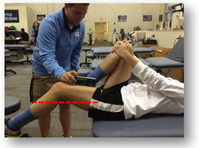
• Hamstrings at 90-90: The participant was positioned lying supine with the
dominant leg flexed to 90 degrees of hip and knee flexion, with the contralateral
leg flat on the table stabilized through the application of webbing. The researcher
instructed the participant to actively extend the knee to the point of first
compensation. A digital inclinometer was placed onto the mid anterior aspect of
tibia and measured the angle formed when compared to the horizontal (Magee
2006). (Figure 3.5)
• Hip Adductors: The participant was positioned supine with her legs in full
extension flat on the table. One clinician stabilized the contralateral anterior
superior iliac spine (ASIS) of the leg being tested and then grasped the medial
43
arm over the contralateral ASIS, the fulcrum over the ipsilateral ASIS, and the
movement arm over the femur in line with the middle of the patella (Starkey and
Ryan 2002). (Figure 3.6)
• Passive Dorsiflexion: The participant was positioned supine with the knee
straight. The subject was instructed to relax, and the examiner moved the plantar
aspect of the foot superiorly until the point of first resistance or pain. The ankle
dorsiflexion angle was measured using a goniometer as the angle formed by the
shaft of the fibula and the lateral midline of the foot (Piva, Fitzgerald et al. 2006).
(Figure 3.7)
• Weight Bearing Dorsiflexion: Participants were instructed to place their foot
perpendicular to the wall and lunge forward to touch the wall with their knee. The
foot was then be moved posteriorly until the maximum range of dorsiflexion is
reached, which will be identified by the heel lifting off the ground. A digital
inclinometer was placed distal to the tibial tuberosity to measure the angle of the
tibia relative to the vertical (Hart, Grindstaff et al. 2009). (Figure 3.8)
Electromyography
Prior to electrode application, each electrode site was identified and marked with a felt tip marker. Each site was shaved using an electric razor and cleaned with a 70% isopropyl alcohol solution to reduce skin impedance. The following muscles and electrode sites were utilized for the study:
• Gluteus Maximus: 20% of the distance from the second sacral vertebra to a point
• Biceps Femoris: 35% of the distance from the ischial tuberosity to the lateral side
of the popliteus cavity, starting from the ischial tuberosity (Rainoldi, Melchiorri et al. 2004)
Each electrode was placed parallel to the orientation of the muscle fibers; one reference electrode was placed over the tibial tuberosity of the ipsilateral tibia. Electrode placement was confirmed with manual muscle testing of each muscle and observation of the muscle activity on an oscilloscope. Once electrode placement was confirmed, the electrodes and leads were secured with omnifix tape. Each respective muscle group (gluteus maximus, biceps femoris) then underwent testing for maximal voluntary isometric contraction (MVIC); three, 5 second isometric holds, with one minute of rest between trials. The MVIC data were used to normalize all EMG activation amplitude data. This was done by dividing the average MVIC activation, averaged over a one second window during the period of greatest EMG activation, by the average EMG activation during the descent phase of the single leg squat. All EMG data were collected at 1000 Hz. The following positions were used for MVIC testing:
• Gluteus Maximus: The participant was placed in a prone position with the
dominant leg flexed at the knee to 90 degrees and the contralateral leg lying flat on the table. The researcher stabilized the pelvis by placing a hand on the subject’s sacrum. The researcher’s other hand was placed over the posterior aspect of the participant’s thigh, just proximal to the knee joint line. The
45
• Biceps Femoris: The participant was placed in a prone position with the dominant
leg flexed at the knee to 90 degrees, the tibia externally rotated, and the nondominant leg lying flat on the table. The researcher stabilized the leg to be tested by placing one hand on the distal 1/3 of the posterior aspect of the thigh. The researcher’s other hand grasped the posterior aspect of the dominant leg’s heel. The participant was instructed to attempt to pull his/her heel in towards his/her gluteal muscles as the researcher resisted the motion with pressure opposing the motion (Anderson and Hall 2005).
Motion Analysis
A MotionSTAR (Ascension Technologies, Burlington, Vermont) electromagnetic motion capture system was used to collect kinematic data during the squatting and jump-landing tasks. The sensors of the electromagnetic tracking system were placed on the subject’s lower extremity at the shank, and thigh, and sacrum. Additionally, a sensor was placed on the participant’s spine at the C7 vertebral level. The sensors were secured with athletic pre-wrap and white athletic tape. Following sensor placement, digitization of bony landmarks through a seventh electromagnetic sensor placed on a 5 cm stylus were completed in the following sequence; T12/L1 spineous process, medial femoral
was used to approximate the hip joint center (Bell, Brand et al. 1989). Three-dimensional coordinate data were collected at a sampling rate of 140 Hz. One minute of rest will be given between trials.
Overhead Squat
Participants were instructed to stand on a force plate holding her arms over her head and toes facing forward. The participant performed a double leg squat as she descended for one beat of a metronome and then return to the starting position in one beat. The metronome was set at a frequency of 60 beats/minute to control velocity. The participant was instructed as to what constitutes a successful trial, no additional coaching or instructions were given concerning technique. A trial was considered successful if the participant maintained proper form throughout the motion, the task was completed at the correct rate, the heels maintained contact with the ground, and the task was completed in a fluid motion. Subjects performed five practice trials of each task prior to testing. After five practice trials, motion analysis data were collected for five successful squats in succession.
Jump Landing
47
constitutes a successful trial, no additional coaching or instructions were given concerning technique. A trial was considered successful if the participant maintained proper form throughout the motion and the task was completed in a fluid motion. Subjects performed five practice trials of each task prior to testing. After five practice trials, motion analysis data were collected for five successful trials.
Single Leg Squat
Participants were instructed to stand on a force plate on their dominant leg, with the non-dominant leg flexed at the knee between 90° and 45°, with their hands placed on her hips, with their head, eyes and toes facing forward. The participant was instructed to flex the dominant knee as she descends for one beat of a metronome and then return to the starting position in one beat. The metronome was set at a frequency of 60
Isometric Strength Testing:
Participants were strength tested using a handheld dynamometer using standard manual muscle testing procedures commonplace to the sports medicine field. Intersession and intrarater reliability and precision will be established prior to data collection (Table 2).
• Gluteus Maximus: The participant was positioned prone with knee flexed to
90 degrees with thigh off table, and the contralateral leg straight. The researcher stabilized the participant’s hip by applying pressure on ipsilateral posterior superior iliac spine (PSIS). The participant was instructed to hold leg against applied inferior resistance of the researcher across midbelly of
hamstrings halfway between the gluteal fold and the knee joint line, and the peak isometric torque produced was measured with a handheld isometric dynamometer (Hislop and Montgomery 2007). This procedure was repeated with the subject’s leg placed into 15 degrees of hip extension verified by digital inclinometer.
• Gluteus Medius: The participant was positioned sidelying with the knee
49
2007). This procedure was repeated with the leg placed in 20 degrees of hip abduction, as verified by digital inclinometer.
DATA PROCESSING AND ANALYSIS Kinematics and Kinetics:
The Motion Monitor Software (Innovative Sports Training, Inc, Chicago, IL) was used to process the data. A global coordinate system was established where the x-axis corresponded with the antero-posterior axis, the y-axis corresponded to the medio-lateral axis, and the z-axis corresponded to the longitudinal axis. A local coordinate system for each segment was established and aligned with the world axis system after bony
landmark digitization. A right handed Euler angle sequence with rotation ordered (Y, X’, Z’’) was used to calculate joint angles in degrees. Trunk flexion(+)/extension(-) was defined as the trunk relative to vertical about the world y-axis. Trunk lateral flexion right (+)/left (-) occured about the world x-axis. Trunk rotation left (+)/right (-) occurred about the world z-axis. Pelvic tilt: anterior (+)/posterior (-) was defined relative to the trunk segment about the y-axis. Hip extension (+)/flexion (-), adduction (+)/abduction (-), internal rotation (+)/external rotation (-) occurred about the pelvis x, y, z-axes
Jump Landing:
Medial knee displacement was calculated as the maximum displacement along the y-axis during the loading phase. The loading phase was defined as the time between
initial ground contact (vertical ground reaction force >10 N) and peak knee flexion angle. Peak hip extension angle was calculated as the maximum value in the sagittal plane during the loading as well as takeoff stage, which will be defined as time from peak knee flexion until take off (vertical ground reaction force <10 N). Peak trunk forward flexion angle was calculated as the maximum value during the loading stage of the task. Peak anterior pelvic tilt was calculated as the maximum value of anterior superior iliac spine motion during the loading stage of the task.
Single Leg Squat and Overhead Squat
51 Data Reduction
All kinematic data was filtered using a fourth-order low-pass Butterworth filter at 10 Hz. Kinematic data was exported and reduced using a custom computer program. Isometric torque was calculated through force production measured with handheld dynamometry. Torques were normalized to body weight in kilograms.
STATISTICAL ANALYSIS