IDENTIFICATION AND CHARACTERIZATION OF TWO POTENTIAL NOVEL REGULATORS OF APOPTOSIS, MAGE-D2 AND SUMI-1
Hilary VanTassell Clegg
A dissertation submitted to the faculty of the University of North Carolina at Chapel Hill in partial fulfillment of the requirements for the degree of Doctor of Philosophy in Genetics and Molecular Biology in the Curriculum of Genetics and
Molecular Biology.
Chapel Hill 2011
© 2011
iii ABSTRACT
HILARY CLEGG: Identification and Characterization of Two Potential Novel Regulators of Apoptosis, MAGE-D2 and SUMI-1
(Under the direction of Yanping Zhang, Ph.D.)
separately from p32. Here, we show that MAGE-D2 is localized to the nucleolus, nucleus, and possibly the mitochondria, and preliminary data suggest potential roles for MAGE-D2 in apoptosis and cell cycle control. SUMI-1 is characterized more extensively in this work, and the data presented here establish SUMI-1 as a novel mitochondria-localized regulator of mitochondrial fission-fusion dynamics and BAX-mediated apoptosis. The data shown here support a model in which SUMI-1 resides at the mitochondrial outer membrane, where it regulates mitochondrial fusion and protects cells from apoptosis. Upon treatment with apoptosis-inducing stimuli, SUMI-1 translocates from the mitochondria, inhibiting mitochondrial fusion while fission continues unperturbed. This imbalance results in mitochondrial fragmentation, promoting BAX oligomerization on the mitochondrial outer membrane and ultimately leading to MOMP, cytochrome c
v
ACKNOWLEDGMENTS
vii
TABLE OF CONTENTS
LIST OF TABLES ...xii
LIST OF FIGURES ... xiii
LIST OF ABBREVIATIONS AND SYMBOLS ...xvi
CHAPTER I. INTRODUCTION ... 2
An Overview of Apoptosis ... 2
Apoptosis in Disease ... 3
Signaling Pathways and Regulatory Mechanisms in Apoptosis ... 6
Unknowns in Field of Apoptosis Research ... 16
p32/C1QBP: a Multifunctional Protein Recently Identified as a Regulator of Apoptosis ... 17
II. IDENTIFICATION OF MAGE-D2 AND SUMI-1 AS NOVEL INTERACTING PARTNERS FOR THE PRO-APOPTOTIC PROTEIN P32/C1QBP ... 23
Introduction ... 23
Screen for Novel p32-Interacting Proteins ... 24
Selection of MAGE-D2 and SUMI-1 for Further Study ... 25
Confirmation of Binding Between p32-SUMI-1 and p32-MAGE-D2 ... 26
Materials and Methods ... 27
Introduction and Background ... 36
Generation of Antibodies for Study of Endogenous MAGE-D2 ... 40
MAGE-D2 Localizes to the Mitochondria, Nucleus, and Nucleoli ... 41
Exploring a Potential Role for MAGE-D2 in Regulating Apoptotic Cell Death ... 43
Summary and Discussion... 45
Materials and Methods ... 49
IV. SUMI-1 IS A NOVEL REGULATOR OF MITOCHONDRIAL FISSION-FUSION DYNAMICS AND BAX-MEDIATED APOPTOSIS ... 62
Introduction ... 62
Background ... 63
SUMI-1 is a Highly-Conserved CHCH-Domain-Containing Protein ... 65
Generation of Antibodies for Study of Endogenous SUMI-1 ... 67
SUMI-1 Localizes to the Mitochondria ... 68
SUMI-1 Inhibits Apoptotic Cell Death ... 71
SUMI-1 Translocates From Mitochondria Prior to Cytochrome c ... 73
xi
V. REGULATION OF SUMI-1 DURING APOPTOSIS ... 118
Introduction ... 118
Apoptotic Stimuli Reduce Level of Endogenous SUMI-1 ... 118
SUMI-1 is Not Degraded by the Proteasome During Apoptosis ... 119
SUMI-1 is Potentially Regulated by Oligomerization ... 120
Discussion ... 124
Materials and Methods ... 128
VI. DISCUSSION ... 135
Identification of Novel Apoptosis-Regulating Proteins ... 135
MAGE-D2 Localization and Function ... 137
Regulation of Apoptosis by SUMI-1 ... 145
Roles for Interactions Among p32, SUMI-1, and MAGE-D2 ... 153
LIST OF TABLES TABLE
2-1. Potential p32-interacting proteins ... 33
xiii
LIST OF FIGURES FIGURE
1-1. Key signaling molecules in the intrinsic apoptosis pathway ... 19
1-2. Functional organization and general domain structure of BCL-2 family proteins. ... 20
1-3. Regulation of mitochondrial outer membrane permeabilization (MOMP) by BAX and BAK ... 21
1-4. Competing models of apoptosis regulation by BCL-2 family proteins ... 22
2-1. Clones stably expressing p32-Flag ... 30
2-2. U2OS cells stably expressing p32-Flag ... 31
2-3. Large-scale co-immunoprecipitation of p32 ... 32
2-4. Confirmation of p32-MAGE-D2 and p32-SUMI-1 binding ... 34
2-5. Endogenous binding between p32-MAGED2 and p32-SUMI-1 ... 35
3-1. Domain structure for MAGE-D2 ... 52
3-2. Antibody design for MAGE-D2 ... 53
3-3. MAGE-D2 antibody effectiveness and specificity... 54
3-4. Ectopically-expressed tagged MAGE-D2 localizes to nuclei and nucleoli ... 55
3-5. Expression of EGFP-tagged MAGE-D2 is toxic to cells and disrupts nuclear and nucleolar morphology ... 56
3-6. Endogenous MAGE-D2 localizes to the nucleoli, nuclei, and mitochondria ... 57
3-7. Localization of endogenous MAGE-D2 is heterogeneous ... 58
3-9. Flow cytometry analysis of effect of MAGE-D2 expression
on UV-induced apoptosis ... 60
3-10. Endogenous MAGE-D2 expression is absent in patches of confluent cells ... 61
4-1. Sequence alignment for SUMI-1 orthologs ... 91
4-2. Domain structure of SUMI-1 ... 92
4-3. Predicted intramolecular disulfide bond formation for SUMI-1 ... 93
4-4. Antibody design for SUMI-1 ... 94
4-5. SUMI-1 antibody effectiveness and specificity ... 95
4-6. Immunofluorescence imaging demonstrates mitochondrial localization of SUMI-1 ... 96
4-7. Subcellular fractionation of SUMI-1 ... 97
4-8. Immunogold-EM localization of SUMI-1 ... 98
4-9. SUMI-1 knockdown inhibits apoptotic cell death ... 99
4-10. SUMI-1 knockdown enhances cleavage of PARP and Caspase-3... 100
4-11. SUMI-1 knockdown inhibits apoptotic cell death ... 101
4-12. SUMI-1 overexpression protects against apoptotic cell death ... 102
4-13. SUMI-1 translocates from mitochondria during apoptosis ... 103
xv
4-19. SUMI-1 siRNA induces shift in proportion of 34 kD and
37 kD bands detected by BCL-xL S-18 antibody ... 109
4-20. Nonspecific band recognized by BCL-xL S-18 antibody is p32 ... 110
4-21. Region of homology between p32 and BH4 domain of BCL-xL ... 111
4-22. siRNA inhibition of SUMI-1 induces mitochondrial fragmentation ... 112
4-23. SUMI-1 regulates mitochondrial fusion ... 113
4-24. Model for regulation of MOMP by SUMI-1 ... 114
4-25. Increased expression of SUMI-1 in cancers ... 115
4-26. Increased expression of SUMI-1 in cancerous compared to non-cancerous cell lines ... 117
5-1. Apoptotic stimuli reduce the level of endogenous SUMI-1 ... 131
5-2. Proteasome inhibitor MG132 does not prevent apoptosis- associated reduction in SUMI-1 level ... 132
5-3. High molecular weight bands are detected by SUMI-1 antibody when cells are treated to undergo apoptosis ... 133
5-4. Transfection of untagged SUMI-1 increases HMW bands detected by SUMI-1 antibody ... 134
6-1. MAGE-D2 deletion mutants ... 158
LIST OF ABBREVIATIONS AND SYMBOLS BAK: BCL-2-antagonist/killer 1
BAX: BCL-2-associated X protein BCL-2: B-cell lymphoma 2
BCL-xL: BCL-2-like 1
BHD: BCL-2 homology domain
CHCH: Coiled-coil-helix-coiled-coil-helix
CHCHD2: Coiled-coil-helix-coiled-coil-helix domain containing 2
C1QBP: Complement component 1, q subcomponent binding protein EM: Electron microscopy
HMW: High molecular weight IP: Immunoprecipitation LUV: Large unilamellar vesicle MAGE: Melanoma antigen family MAGE-D2: Melanoma antigen family D2 MFN1: Mitofusin 1
MFN2: Mitofusin 2
2 CHAPTER I
INTRODUCTION
An Overview of Apoptosis
Apoptosis is a tightly-regulated form of programmed cell death that is critical for proper embryonic development, tissue homeostasis, and immune response, and aberrant regulation of apoptosis contributes to diseases such as autoimmune disorders, neurodegenerative disease, and cancer. Unlike necrosis, or “accidental” cell death, which is usually triggered by acute cellular injury, apoptosis is genetically programmed and is characterized by distinct morphological changes such as membrane blebbing, chromatin condensation, DNA fragmentation, and cell shrinkage (Kerr et al., 1972). In vertebrates, apoptosis usually occurs through one of two major pathways: extrinsic, or receptor-mediated apoptosis, and intrinsic, or mitochondria-mediated apoptosis. Both pathways result in the activation of executioner cysteine proteases (Caspases), which cleave downstream targets to carry out the execution phase of apoptosis.
falling) translates to the "dropping off" of petals or leaves from plants or trees (Kerr et al., 1972). The pronunciation of this word is still debated; Kerr et al. suggested that the second “p” be silent to reflect the word’s Greek roots, while others have pointed out that, while the “p” is often silent in Greek-derived words that begin with a “pt” combination, a “pt” found in the middle of a word is typically pronounced, as in “helicopter” and “cryptic.”
4
disorders. More than ten years ago, evasion of apoptosis was introduced as one of the six hallmarks of cancer that enable or promote tumor growth (Evan and Littlewood, 1998; Hanahan and Weinberg, 2000). The human body uses apoptosis as a defense against extensive DNA damage that may otherwise promote carcinogenesis; in other words, if a cell’s genetic material is sufficiently damaged, apoptosis is triggered in order to prevent the cell from progressing into a tumor (Kerr et al., 1994). Cells that escape this defense mechanism can potentially undergo additional changes and become transformed. Cancer cells employ a variety of strategies to disable induction of apoptosis. Most notable is inactivation of the tumor suppressor p53, which normally responds to DNA damage, abnormal proliferative signals, and other cellular stresses by triggering apoptosis, primarily through transactivation of apoptosis-promoting targets such as PUMA and BAX (Levine, 1997). Many chemotherapeutic agents, such as cisplatin, doxorubicin, and paclitaxel, fight tumors through induction of apoptosis, and identification of novel apoptosis-inducers and apoptosis-sensitizing agents is an area of ongoing investigation in cancer biology.
beta-cells in the pancreas. Similarly, inappropriate T-cell targeting of joints is presumed to occur in rheumatoid arthritis (Hayashi and Faustman, 2003), and the impaired apoptosis of these T-cells may result from induction of apoptosis-inhibiting BCL-2 and MCL-1 proteins (Liu and Pope, 2003). In systemic lupus erythematosus, reduced apoptosis leads to lymphoproliferation and general autoimmunity (Hayashi and Faustman, 2003).
6
mechanisms that induce neuronal apoptosis in Alzheimer’s Disease are still unclear.
Parkinson’s Disease is another progressive cognitive disorder involving neuronal death and is the second most common neurodegenerative disease, affecting six million people worldwide (Dauer and Przedborski, 2003). In this disorder, neuronal death occurs in dopamine-producing cells in the substantia nigra located in the midbrain (Levy et al., 2009). Apoptosis has been postulated as a mechanism for the neuronal death in Parkinson’s Disease, but controversy surrounds the issue because one marker of apoptosis—nuclear DNA cleavage— is not robustly present in brains of Parkinson’s Disease patients. However, a number of apoptotic signaling pathways have been implicated in the disorder, including involvement of BCL-2 family proteins, mitochondrial dysfunction, JNK (Jun Kinase) signaling, and activation of p53; furthermore, an apoptotic pathway involving BAX activation, cytochrome c release, and activation of Caspase-9 and Caspase-3 has been implicated in the development of the disease (Levy et al., 2009; Tatton et al., 2003). Understanding whether apoptosis regulates Parkinson’s Disease, and through what specific mechanisms and signaling pathways, will be essential for developing targeted therapies for this disorder.
Signaling Pathways and Regulatory Mechanisms in Apoptosis
downstream targets to induce the biochemical, biophysical, and morphological changes that constitute apoptosis (Slee et al., 2001). Caspase-3 activates the endonuclease CAD by cleaving its inhibitor, ICAD, leading to chromatin condensation and degradation of chromosomal DNA (Sakahira et al., 1998). The DNA repair enzyme poly (ADP-ribose) polymerase (PARP) is cleaved and inactivated in order to prevent ATP depletion (Simbulan-Rosenthal et al., 1998). The actin-binding protein Gelsolin is cleaved, and the resulting fragments subsequently cleave actin polymers, disrupting the cytoskeleton (Kothakota et al., 1997). The cell disintegrates into smaller, membrane-enclosed fragments called apoptotic bodies. In vivo, the final step of apoptosis occurs when phagocytic cells uptake these apoptotic bodies and recycle their contents. Cells undergoing apoptosis are recognized by phagocytes when phospholipids of the plasma membrane change orientation. Phosphatidylserine, for example, flips from the inner to the outer leaflet, providing a signal for disposal by noninflammatory phagocytes (Fadok et al., 2001; Ferraro-Peyret et al., 2002).
8
and activate adaptor proteins inside the cell (Grimm et al., 1996; Hsu et al., 1995), forming the death-inducing signaling complex (DISC). This complex induces auto-catalytic cleavage of pro-Caspase-8, leading to activation of Caspase-8 followed by subsequent cleavage and activation of executioner Caspases (Kischkel et al., 1995).
This research presented here will focus on the role of proteins that mediate the intrinsic apoptotic pathway (Figure 1-1), which can be activated by diverse stimuli such as UV radiation, ionizing radiation, and other forms of DNA damage as well as hypoxia, cytotoxic drugs, viral infections, and oxidative stress. During intrinsic apoptosis, cellular stressors such as DNA damage lead to mitochondrial outer membrane permeabilization (MOMP), releasing cytochrome
c and other factors from the mitochondrial intermembrane space. Upon its release into the cytosol, cytochrome c interacts with APAF-1 and Caspase-9 to form the apoptosome, leading to auto-activation and cleavage of Caspase-9 and subsequent activation of Caspase-3, Caspase-7, and downstream targets. This process can also be regulated by other proteins released from the mitochondria during MOMP, such as XIAP, DIABLO/SMAC, and HtrA2/OMI (Du et al., 2000; Duckett et al., 1996; Faccio et al., 2000; Tait and Green, 2010; Verhagen et al., 2000).
10
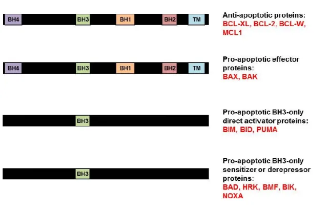
The BCL-2 family members’ functions and domains are summarized in Figure 1-2.
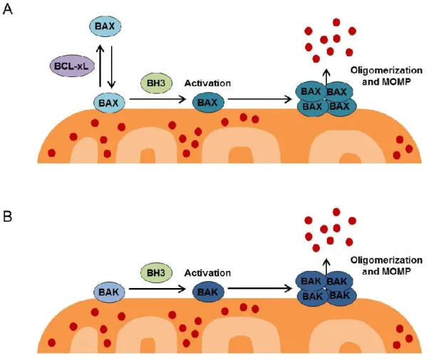
The mitochondrial outer membrane is permeabilized directly by effector proteins BAX and/or BAK (Figure 1-3). BAX continually cycles between the mitochondria and cytosol, while BAK is constitutively localized to the mitochondria. In healthy cells, BAX is retro-translocated from the mitochondria to the cytoplasm by BCL-XL, but during apoptosis, the interaction between BCL-XL and BAX is disrupted, and BAX accumulates at the mitochondria (Edlich et al., 2011). Upon induction of apoptosis, both BAX and BAK undergo conformational changes to become “activated” and oligomerize on the mitochondrial outer membrane (Wei et al., 2001), leading to membrane permeabilization through a mechanism that is not yet clear but that may occur through formation of a proteinaceous or lipid pore (Chipuk and Green, 2008). Rather than being evenly distributed around the mitochondria, active BAX accumulates in massive clusters at the ends of mitochondria or co-localized with mitochondrial fission and fusion sites (Karbowski et al., 2002), leading to the proposal that BAX may either hijack the fission/fusion machinery and/or take advantage of the altered mitochondrial membrane structure at these sites in order to permeabilize the membrane. The exact biochemical mechanism by which BAX and BAK permeabilize the membrane is currently an area of intense investigation and has been described as one of the “holy grails” of apoptosis research (Youle and Strasser, 2008).
Gavathiotis et al., 2010; Kim et al., 2006; Kim et al., 2009; Kuwana et al., 2005; Letai et al., 2002; Lovell et al., 2008; O'Connor et al., 1998; Ren et al., 2010; Wei et al., 2000). Conversely, the pro-apoptotic function of BAX and BAK can be inhibited by pro-survival BCL-2 family proteins such as BCL-xL, MCL-1, and BCL-2 itself (Boise et al., 1993; Chipuk et al., 2010; Kozopas et al., 1993; Oltvai et al., 1993). The precise mechanisms by which BCL-2 family members interact to mediate apoptosis are still under debate, and several models have been proposed that are not necessarily mutually exclusive (Chipuk et al., 2010) (Figure 1-4).
The direct activation model proposes that pro-apoptotic BH3-only proteins bind directly to BAX or BAK to induce their activation, and that the role of anti-apoptotic proteins in healthy cells is to bind and sequester BH3-only proteins to prevent them from activating BAX and BAK. Several BH3-only proteins are capable of directly activating BAX and/or BAK in isolated mitochondria or large unilamellar vesicles (LUVs) meant to mimic the mitochondrial outer membrane. It is difficult to detect an interaction between in BAX or BAK and BH3-only proteins
12
thereby deactivating, anti-apoptotic proteins such as BCL-2 and BCL-xL (Chen et al., 2005; Chipuk et al., 2008; Kuwana et al., 2005; Letai et al., 2002). These BH3-only proteins may act as either sensitizers or de-repressors for the effector proteins. The sensitization model purports that the interaction between BH3-only proteins and anti-apoptotic BCL-xL or BCL-2 would not induce apoptosis, but would sensitize cells to apoptotic stimuli. In this scenario, BAX or BAK would still require direct activation by BIM, BID, or PUMA, but the “sensitizer” BH3-only proteins would deactivate the anti-apoptotics (e.g. BCL-2), preventing inhibition of the direct activators. In this way, the sensitizer BH3-only proteins would lower the threshold for BAX and BAK activation (Chipuk et al., 2010).
mitochondrial outer membrane permeabilization was induced (Certo et al., 2006; Del Gaizo Moore et al., 2007).
Finally, a neutralization model has been proposed in which the effector proteins BAX and BAK are always active but are sequestered by anti-apoptotic proteins such as BCL-2 or BCL-xL. For apoptosis to occur, BH3-only proteins compete for binding with the anti-apoptotic proteins, releasing the already-active BAX or BAK. According to this model, direct activator BH3-only proteins are not required for apoptosis; release of the effector proteins from their inhibitors is sufficient to activate apoptosis.
One difficulty in reconciling these contradictory models is that many of the studies examining the interactions between BCL-2 family members are based on
14
intact cells, apoptosis is regulated by a combination of these models; sensitization and direct activation probably both contribute. As induction of MOMP is often a life-or-death decision for a cell, it is logical for it to be regulated by a complex mechanism. For example, one subset of BH3-only proteins may be required for direct activation of BAX and BAK, while another subset of sensitizing or derepressing BH3-only proteins must interact with anti-apoptotic proteins such as BCL-2 and BCL-xL to release their inhibition of the effector proteins.
PUMA, in which activation of BAX and BAK did not occur, suggests that these non-BCL-2 proteins may not always be sufficient in vivo to induce BAX- or BAK-mediated apoptosis (Ren et al., 2010). VDAC2 can regulate MOMP by binding to BAK, restraining it in an inactive monomer conformation. The following non-BCL-2 proteins are also reported to regulate MOMP through mechanisms that are unclear: Histone H1.2, 14-3-3θ , Ku70, and BRCC2 (reviewed by (Chipuk et al., 2010)).
16
Mitochondrial fragmentation occurs during apoptosis prior to cytochrome c
release (Brooks et al., 2007; Lee et al., 2004), and evidence has accumulated to support the idea that fragmentation plays an active role in MOMP. Inhibition of fusion-promoting proteins MFN1 or MFN2 causes mitochondria to become fragmented and sensitizes cells to MOMP and apoptosis (Sugioka et al., 2004), while overexpression of MFN1 or MFN2 reduces fragmentation and protects cells from apoptosis (Brooks et al., 2011). Additionally, a chemical inhibitor of fission, Mdivi-1, as well as fusion-inducing cysteine alkylators and N-ethyl-maleimide (NEM), partially inhibit fragmentation, MOMP, and apoptosis (Bowes and Gupta, 2005; Cassidy-Stone et al., 2008). Likewise, inhibition of fission-mediating DRP1 by RNAi or by introducing dominant-negative mutations in the GTPase domain partially inhibits mitochondrial fragmentation while delaying MOMP and apoptosis (Brooks et al., 2011; Frank et al., 2001). Furthermore, a recent study showed compelling evidence that DRP1 modulates apoptosis by altering mitochondrial dynamics, stimulating BAX oligomerization (Montessuit et al., 2010).
Unknowns in Field of Apoptosis Research
this process is often the point of no return for deciding a cell’s fate. More specifically, controversy still exists regarding the competing models (direct activation, sensitization, derepression, and neutralization) for interactions among BCL-2 family members in regulating MOMP. There is also a high level of interest in elucidating the precise biochemical mechanisms by which BAX and BAK permeabilize the mitochondrial outer membrane (i.e. through formation of a protein or lipid pore, or an alternate mechanism). Gaining a better understanding of these processes will contribute to our knowledge of organismal development and homeostasis and may aid in the design of targeted therapeutic drugs to treat diseases affected by aberrant regulation of apoptosis, such as autoimmune disorders, neurodegenerative disease, and cancer.
p32/C1QBP: a Multifunctional Protein Recently Identified as a Regulator of
Apoptosis
18
reported to mediate apoptotic response (Chowdhury et al., 2008; Kamal and Datta, 2006).
20
Figure 1-2. Functional organization and general domain structure of BCL-2 family proteins.
22
Figure 1-4. Competing models of apoptosis regulation by BCL-2 family proteins.
CHAPTER II
IDENTIFICATION OF MAGE-D2 AND SUMI-1 AS NOVEL INTERACTING PARTNERS FOR THE PRO-APOPTOTIC PROTEIN P32/C1QBP
Introduction
Recently, our lab uncovered novel roles for the multifunctional protein p32/C1QBP in regulating apoptosis by two distinct mechanisms. First, p32 was found to be required for apoptosis induced by the tumor suppressor ARF (Itahana and Zhang, 2008). Later, p32 was found to be a critical mediator for apoptosis induced by a broad range of stimuli (unpublished data). Thus, p32 regulates apoptosis by two mechanisms: 1) It recruits ARF to the mitochondria, where ARF induces a change in mitochondrial membrane potential (∆Ψm),
24 Screen for Novel p32-Interacting Proteins
In order to identify potential novel interacting partners for p32, a large-scale co-immunoprecipitation was carried out for Flag-tagged p32. First, cell lines were generated to express p32-Flag in a stable manner. U2OS (human osteosarcoma) cells were transfected with a pcDNA3-p32-Flag plasmid, and clones expressing the plasmid were selected for with the antibiotic G418. Clones stably-expressing p32-Flag were obtained, and expression of p32-Flag was confirmed by western blotting (Figure 2-1). Correct mitochondrial localization of p32-Flag was confirmed by immunofluorescence imaging, as indicated by co-localization with the mitochondria-labeling dye MitoTracker™ Red (Figure 2-2).
Selection of MAGE-D2 and SUMI-1 for Further Study
After careful examination of the eight putative p32-interacting proteins identified, two were selected for further study: MAGE-D2, an uncharacterized melanoma antigen family protein, and SUMI-1, an uncharacterized protein predicted to localize to the mitochondria.
26
inhibitor of apoptosis) and appears to accelerate degradation and inactivation of XIAP during apoptosis (Jordan et al., 2001).
SUMI-1 is a small, CHCH domain-containing protein, and no studies regarding its functions were published when we began our research on this protein. SUMI-1 was selected for further study primarily because another protein reported to be a mouse ortholog of SUMI-1 (NDG1; Nur77 downstream gene 1) was shown to regulate apoptosis after induction by the nuclear steroid orphan receptor Nur77 (Rajpal et al., 2003). However, upon further investigation, this protein was found not to be a direct ortholog of SUMI-1 as reported. In addition, unlike the majority of putative p32-interacting proteins identified, SUMI-1 was predicted to localize to the mitochondrion (Claros and Vincens, 1996), an organelle with strong ties to apoptotic cell death.
Confirmation of Binding Between p32-SUMI-1 and p32-MAGE-D2
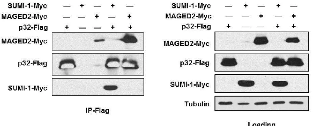
was detected in the negative control lane, which likely represents spillover from another lane and/or incomplete washing of beads; however, a dramatically higher signal was detected in cells transfected with p32-Flag, despite lower expression of MAGE-D2-Myc (see loading panel), indicating that MAGE-D2-Myc does interact nonspecifically with the Flag antibody (Figure 2-4). Thus, we detected binding of ectopically expressed p32 with both SUMI-1 and MAGE-D2
28
DMEM supplemented with 10% FBS, 100 U/ml penicillin, and 100 g/ml streptomycin. DNA transfections were carried out with Effectene® (Qiagen) according to the manufacturer’s instructions. Cells were transfected with pcDNA3-p32-Flag or pcDNA3 vector and subject to selection with G418. Individual clones were isolated and cultured, and expression of p32-Flag was examined by immunofluorescence staining and/or western blotting.
DNA Plasmids. C-terminally Flag-tagged p32, N-terminally Myc-tagged SUMI-1, and N-terminally Myc tagged MAGE-D2 were cloned into a pcDNA3.1 vector (Invitrogen) and confirmed by restriction digest and DNA sequencing.
30
Figure 2-1. Clones stably expressing p32-Flag.
Figure 2-2. U2OS cells stably expressing p32-Flag.
32
Figure 2-3. Large-scale co-immunoprecipitation of p32.
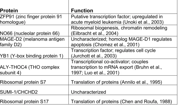
Table 2-1. Potential p32-interacting proteins.
Bands identified by mass spectrometry following a large-scale co-immunoprecipitation of p32-Flag in stably-transfected U2OS cells.
Protein Function
ZFP91 (zinc finger protein 91 homologue)
Putative transcription factor; upregulated in acute myeloid leukemia (Unoki et al., 2003) NO66 (nucleolar protein 66)
Ribosomal biogenesis, chromatin remodeling (Eilbracht et al., 2004)
MAGE-D2 (melanoma antigen family D2)
Uncharacterized; homolog MAGE-D1 regulates apoptosis (Chomez et al., 2001)
YB1 (Y-box binding protein 1)
Transcription factor; regulates cell cycle (Jurchott et al., 2003)
ALY-THOC4 (THO complex subunit 4)
Transcriptional co-activator; couples transcription to mRNA export (Bruhn et al., 1997; Luo et al., 2001)
Ribosomal protein S7 Translation of proteins (Annilo et al., 1995) SUMI-1/CHCHD2 Uncharacterized
34
Figure 2-4. Confirmation of p32-MAGE-D2 and p32-SUMI-1 binding.
CHAPTER III
LOCALIZATION AND FUNCTIONAL STUDIES OF THE UNCHARACTERIZED MELANOMA ANTIGEN FAMILY PROTEIN, MAGE-D2
Introduction and Background
well-known, although roles have been uncovered for Type II MAGE proteins in cell survival, cell cycle progression, and apoptosis (Barker and Salehi, 2002).
38
The exact mechanism is unclear but appears to involve formation of a transient complex between MAGE-D1 and XIAP that may lead to XIAP’s degradation and inactivation (Jordan et al., 2001). MAGE-D1-induced apoptosis and cell cycle arrest have both been shown to require activation of the tumor suppressor p53 (Kendall et al., 2005; Wen et al., 2004), which is contrary to the roles of several anti-apoptotic Type I MAGE proteins that have been shown to inhibit p53’s transcriptional activity (Doyle et al., 2010; Marcar et al., 2010; Monte et al., 2006; Yang et al., 2007).
adult tissues tested; its expression increases steadily during embryonic development, reaching a maximum just before birth; and mouse embryonic tissues contain a higher level of MAGE-D2 expression than do adult tissues (Chomez et al., 2001). The expression of human MAGE-D2 is elevated in a number of cancers including those derived from the breast (fresh breast cancer tissue, but not breast cancer cell lines) (Hudelist et al., 2006), appendix (Modlin et al., 2006), stomach (Kidd et al., 2006b), and small intestine (Kidd et al., 2006a). Its overexpression is also associated with cancer progression and metastasis—It is elevated 5-10 fold in Type III/IV gastrointestinal cancers but not in Type I/II tumors (Kidd et al., 2006b), it is associated with progression of appendiceal tumor malignancy (Modlin et al., 2006), and it is expressed to a significantly higher degree in colon cancer tumors with liver metastasis than those that have not metastasized (Li et al., 2004).
40
study, we sought to characterize the subcellular localization of MAGE-D2 and investigate whether it plays a role in apoptotic cell death.
Generation of Antibodies for Study of Endogenous MAGE-D2
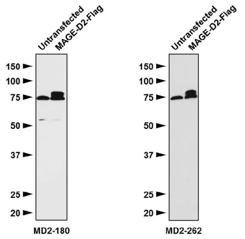
antigens was shipped to our laboratory for purification. Antibodies were affinity-purified in columns conjugated with the associated peptide antigen and tested in our lab by western blotting for effectiveness and specificity. Each antibody detected both endogenous and ectopically-expressed MAGE-D2 and was specific to MAGE-D2 (Figure 3-3).
MAGE-D2 Localizes to the Mitochondria, Nucleus, and Nucleoli
42
immunofluorescence stained with MAGE-D2 (MD2-262) antibody. As observed with the ectopically expressed proteins, endogenous MAGE-D2 localized primarily to the nucleoli, followed by the nucleus (Figure 3-6). However, a small amount of cytoplasmic staining was also detected in a pattern reminiscent of mitochondria. To determine whether this indeed represented mitochondrial staining, we co-stained cells with MitoTracker™ dye and observed co-localization (Figure 3-6), suggesting that a portion of endogenous MAGE-D2 localizes to the mitochondria. Interestingly, the localization of endogenous MAGE-D2 appears quite heterogeneous, with some cells exhibiting primarily nucleolar and nuclear staining, while other cells have stronger staining in the mitochondria, and others have staining that appears diffuse throughout the nucleus and cytoplasm (Figure 3-7).
enrichment of certain types of residues, number of acidic residues, hydrophobicity, and isoelectric point (Claros and Vincens, 1996). According to MitoProt II, a prediction program for mitochondrial localization that takes into account 47 sequence parameters affecting mitochondrial targeting, the probability that MAGE-D2 is exported to mitochondria is low (0.0452) (Claros and Vincens, 1996). It is possible, however, that the association of MAGE-D2 with mitochondria is based on interaction with another mitochondria-localized protein. This scenario seems plausible, as MAGE-D2 interacts with the mitochondrial protein p32. Immunogold-electron microscopy data shows that a portion of p32 localizes to the mitochondrial outer membrane (data not shown), where it could interact with MAGE-D2 following translation of the protein in the cytoplasm.
44
Summary and Discussion
MAGE-D2 is an uncharacterized MAGE (melanoma antigen) family protein that, unlike the majority of MAGE family members, is ubiquitously expressed in normal adult tissues. MAGE-D2 is a Type II MAGE protein, meaning that in addition to sharing the MAGE homology domain (MHD I) with all MAGE proteins, it also shares a MAGE homology domain II (MHD II) with a subset of MAGE proteins, including the MAGE-D subfamily. The physiological functions of MAGE proteins are mostly unknown, but roles have been reported in the regulation of cell survival, cell proliferation, ubiquitin ligase activity, and apoptosis (Barker and Salehi, 2002; Doyle et al., 2010). The closest homolog of D2, MAGE-D1/NRAGE, regulates apoptosis through several distinct mechanisms, but the functions of MAGE-D2 are unknown. In order to explore a potential function for MAGE-D2 in regulation of apoptosis, we generated several tools for its study, including tagged proteins for ectopic expression and antibodies for biochemical analyses of endogenous MAGE-D2 in cells.
46
experimental artifact due to recognition of a nonspecific protein by the MAGE-D2 antibody. One method for answering this question is through knockdown of MAGE-D2 with RNAi followed by immunofluorescence staining with MAGE-D2 antibody to determine whether the mitochondrial signal disappears. If RNAi reduces the mitochondrial staining, we can conclude that the apparent mitochondrial localization is genuine. Another means to confirm the localization of MAGE-D2 is through subcellular fractionation, such as the differential detergent mechanism that was utilized in Chapter IV (Figure 4-7) to assess the localization of SUMI-1.
interaction has not been determined. The nucleolus is a sub-nuclear body whose main functions are to carry out synthesis of ribosomal RNA (rRNA) and assembly of ribosomes—complexes consisting of proteins and RNA that translate proteins from mRNA sequences. Many proteins found in the nucleolus are components of the ribosomes themselves, and others regulate ribosomal biogenesis or rRNA synthesis, but a number of proteins in this compartment have no known ties to nucleolar functions. It has been proposed that the nucleolus might act as a storage area for proteins, keeping them inactive until they are required elsewhere in the cell (Pederson and Tsai, 2009). Immunofluorescence staining of endogenous MAGE-D2 suggests that it may also reside in the mitochondria. Inside this organelle, ATP is produced by oxidative phosphorylation, providing most of the energy supply for the cell. Mitochondria also play an important role in the intrinsic apoptotic pathway. During apoptosis, the mitochondrial outer membrane is permeabilized, releasing cytochrome c and other contents of the mitochondrial intermembrane space, leading to Caspase activation and execution of apoptosis (Chipuk et al., 2010). Thus, it is possible that MAGE-D2 mediates apoptosis through its association with the mitochondria.
48
example, it can be determined whether knockdown of MAGE-D2, such as with siRNA, affects apoptosis. One complication is that extensive redundancy is thought to exist among MAGE family members, so knockdown of one MAGE protein may be compensated for by another. It may be necessary to inhibit more than one MAGE protein (e.g. MAGE-D2 and MAGE-D1) to see an effect. Mechanistic studies can also help determine whether MAGE-D2 has a bona-fide role in regulating apoptosis. In addition, we have not demonstrated that overexpression of MAGE-D2 increases apoptosis, per se, rather than another form of cell death. While quantification of apoptosis by assessing the percentage of cells with sub-G1 content is highly precise for cells that are not actively cycling, such as thymocytes, this method is not flawless for analysis of cycling cells, as it does not discriminate between apoptotic cells, fragmented cells, and other debris—a clear peak representing apoptotic cells cannot be observed in cycling cells. Other assays must be carried out to examine other markers of apoptosis, such as western blotting for PARP cleavage or Caspase-3 activation.
immunofluorescence, confluent regions contained patches of cells expressing little to no endogenous MAGE-D2. It would be interesting to determine whether expression of MAGE-D2 is linked to cell proliferation and/or cell cycle control.
Materials and Methods
Cell Culture, transfection, and apoptotic treatments. U2OS cells were obtained from ATCC and cultured in a 37°C incubator with 5% CO2 in DMEM
supplemented with 10% FBS, 100 U/ml penicillin, and 100 g/ml streptomycin. DNA transfections were carried out with Effectene® (Qiagen) according to the manufacturer’s instructions. Apoptosis treatments included UVC radiation (dosage as indicated) using a Stratalinker® UV Crosslinker (Stratagene) and cisplatin (10 µg/ml).
50
DNA plasmids. N-terminally Myc tagged and C-terminally Flag-tagged MAGE-D2 were cloned into a pcDNA3.1 vector (Invitrogen). MAGE-MAGE-D2 was cloned into a pEGFP-N1 vector (Clontech) to generate a MAGE-D2-EGFP fusion protein. All plasmids were confirmed by restriction digest and DNA sequencing.
Generation of antibodies. Rabbit anti-MAGE-D2 antibodies were produced by immunizing rabbits (performed by PRF&L, Canadensis, PA) with KLH-conjugated peptide antigens corresponding to amino acids 180-194 (MD2-180) and 262-278 (MD2-262) of MAGE-D2. Sera were affinity purified, and antibody specificity was tested by western blotting (Figure 3-2).
Flow cytometry analysis. Cells were trypsinized, washed in PBS, and fixed in cold 70% ethanol. Cells were treated with RNAse A and stained with propidium iodide. Cells were analyzed using a FACSCalibur flow cytometer (BD Biosciences) at the UNC Flow Cytometry Core Facility, and data were analyzed with Dako software.
52 Figure 3-1. Domain structure for MAGE-D2.
Figure 3-2. Antibody design for MAGE-D2.
54
Figure 3-3. MAGE-D2 antibody effectiveness and specificity.
Figure 3-4. Ectopically-expressed tagged MAGE-D2 localizes to nuclei and nucleoli.
56
Figure 3-5. Expression of EGFP-tagged MAGE-D2 is toxic to cells and disrupts nuclear and nucleolar morphology.
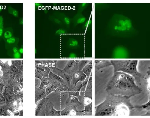
A) Live-cell fluorescence imaging of U2OS cells transfected with N-terminally-EGFP-tagged MAGE-D2 shows that expression of this construct is toxic to cells, with many transfected cells dying (observed as shrunken, white floating cells in phase-contrast image).
Figure 3-6. Endogenous MAGE-D2 localizes to the nucleoli, nuclei, and mitochondria.
58
Figure 3-7. Localization of endogenous MAGE-D2 is heterogeneous.
Figure 3-8. Overexpression of MAGE-D2 sensitizes cells to UV-induced cell death.
60
Figure 3-9. Flow cytometry analysis of effect of MAGE-D2 expression on UV-induced apoptosis.
A) Flow-cytometry analysis shows that ectopic expression of MAGE-D2 sensitizes cells to UV-induced cell death. U2OS cells were transfected with MAGED2-Flag DNA, treated 24 h later cells with UV (6 mJ/cm2) where indicated to induce apoptosis, and were fixed 18 h later in ethanol, stained with propidium iodide, and analyzed by flow cytometry for DNA content.
Figure 3-10. Endogenous MAGE-D2 expression is absent in patches of confluent cells.
1This chapter is based, with modifications, on a manuscript submitted for publication (Clegg et al.) and includes data generated by Yong Liu, Jiehui Di, Laura A. Tollini, Yizhou He, Aiwen Jin, and Paula Miliani de Marval.
CHAPTER IV
SUMI-1 IS A NOVEL REGULATOR OF MITOCHONDRIAL FISSION-FUSION DYNAMICS AND BAX-MEDIATED APOPTOSIS1
Introduction
Background
Apoptosis, or programmed cell death, is a critical process during vertebrate development and is abnormally regulated in a diverse array of diseases. Apoptosis can occur through two pathways—receptor-mediated (extrinsic) apoptosis, and mitochondria-mediated (intrinsic) apoptosis. The extrinsic pathway is activated by interaction of extracellular ligands with death receptors, while the intrinsic pathway is triggered by intracellular stresses such as DNA damage and is modulated by the cell’s mitochondria. Both pathways converge upon activation of cysteine proteases (Caspases), which cleave downstream substrates to induce the physiological and morphological changes associated with apoptosis such as membrane blebbing, DNA fragmentation, and cell shrinkage.
64
to an increase in fission and/or a decrease in fusion. Overexpression of fusion-promoting proteins MFN1 or MFN2 protects cells from apoptosis (Brooks et al., 2011), while inhibition of fission-mediating DRP1 partially inhibits mitochondrial fragmentation while delaying MOMP and apoptosis (Brooks et al., 2011). Recently, it was shown that DRP1-induced mitochondrial fragmentation promotes apoptosis by stimulating BAX oligomerization (Montessuit et al., 2010). Despite advances that have been made in our understanding of MOMP, much remains unknown about the mechanisms leading to BAX activation and oligomerization, especially the roles that mitochondria and their associated proteins play in this process. Identification of novel proteins that regulate MOMP can significantly aid in our understanding of this process.
regulates mitochondrial fusion in order to maintain a balance between fusion and fission, providing a mechanism for SUMI-1’s regulation of BAX and MOMP. Furthermore, most of these experiments were carried out in intact cells using endogenous rather than overexpressed proteins, and our results are therefore likely to be physiologically relevant. Finally, we observed that SUMI-1 expression is upregulated in cancer cell lines, and data obtained from Oncomine™ demonstrates that SUMI-1 is indeed overexpressed in multiple cancers, consistent with SUMI-1’s anti-apoptotic function. This study identifies a novel regulator of apoptosis, sheds light on previously-unexplained mechanisms governing mitochondrial dynamics, MOMP, and BAX activation in regulating apoptotic cell death, and provides a potential therapeutic target for chemosensitization in cancer treatment.
SUMI-1 is a Highly-Conserved CHCH-Domain-Containing Protein
66
coiled-coil helix) domain. SUMI-1 also contains a consensus sequence for import into the mitochondrial intermembrane space (ITS) mediated by another CHCH-domain-containing protein, Mia40 (Figure 4-2).
The CHCH domain is characterized primarily by four cysteine residues spaced exactly 10 amino acids apart (a C-X9-C motif) (Schultz et al., 1998). Pairs of cysteines in this domain typically form covalent disulfide bonds, eliciting changes in tertiary protein structure or facilitating oligomerization and/or interactions with other CHCH domain-containing proteins (Arnesano et al., 2005). Analysis of the SUMI-1 sequence with an intramolecular disulfide connectivity prediction program, DiANNA 1.1, predicts that a bond may form between cysteines 1-3 and another bond between cysteines 2-4 in the CHCH domain to create a loop structure at the C-terminus (Figure 4-3) (Ferre and Clote, 2005a, b).
COX assembly has recently been shown to be mediated by its interaction with SCO-1 (Banci et al., 2008a). Mia40/Tim40 assists in transporting proteins into the mitochondrial intermembrane space by forming temporary disulfide bonds with cysteine-containing proteins as they are imported (Chacinska et al., 2004; Hofmann et al., 2005; Sideris et al., 2009). C2360 is expressed only in human proliferative cytotrophoblasts and not in adult tissues; its function is unknown (Westerman et al., 2004). Finally, the yeast protein MRP10 is a component of the mitochondrial ribosomal 37S subunit, which mediates translation of proteins (Jin et al., 1997). The function of the CHCH domain has been studied most extensively in Cox17, where the domain was shown to change configuration based on oxidative-reductive status, and is thought to regulate homo-oligomerization (Arnesano et al., 2005). The CHCH domain in SUMI-1 is well-conserved across species: The four cysteines comprising the domain are present in 8 of the 9 orthologs shown in Figure 4-1.
Generation of Antibodies for Study of Endogenous SUMI-1
68
and CHCH domain comprised of four cysteines spaced 10 amino acids apart at the C-terminus. These features made the majority of the sequence unsuitable or undesirable because the mitochondrial targeting region could potentially be cleaved off the protein upon mitochondrial translocation, the transmembrane region is hydrophobic (meaning it is not likely to be exposed at the protein surface), and the closely-spaced cysteines in the CHCH domain precluded any antigens longer than 10 amino acids due to the need to have either zero or one terminally-located cysteine and no internal cysteines in the antigen sequence. The regions selected were SU-94 (amino acids 94-108; RPDITYQEPQGTQPA), located adjacent to the central transmembrane domain, and SU-124 (amino acids 124-133; CAQNQGDIKL), located within the CHCH domain (Figure 4-4). The second antigen was shorter than desired (10 amino acids) in order to ensure that no internal cysteines were present in the sequence. The peptides were used to immunize rabbits, and the resulting serum containing polyclonal antibodies was affinity-purified as described in Chapter III. The purified antibodies were then tested by western blotting and immunofluorescence for effectiveness and specificity. The antibody generated with the SU-94 antigen is highly specific to SUMI-1 and can be used for western blotting, immunofluorescence, and co-immunoprecipitation (Figures 4-5 and 2-5).
SUMI-1 Localizes to the Mitochondria
and Vincens, 1996). To determine whether SUMI-1 indeed localizes to the mitochondria, we examined its subcellular localization by immunofluorescence. We transfected U2OS cells with plasmids expressing C-terminal Flag-tagged SUMI-1, fixed the cells after 24 hours, and carried out immunofluorescence staining for Flag. SUMI-1-Flag localized primarily to mitochondria, as indicated by co-staining with MitoTracker™ dye (Figure 4-6A).
formaldehyde-70
membrane and could also be observed inside the mitochondria, although the sub-mitochondrial localization (i.e. matrix, inner membrane, or intermembrane space) was not clear (Figure 4-8). Thus, SUMI-1 appears to reside both inside the mitochondria and at the outer mitochondrial membrane. SUMI-1 contains a consensus ITS (intermembrane targeting sequence) that directs Mia40-mediated import of proteins into the mitochondrial intermembrane space. It is possible that upon import, a subset of SUMI-1 is retained in the mitochondria (either in the intermembrane space or embedded in the inner membrane) while another subset becomes anchored via the transmembrane domain into the mitochondrial outer membrane.
Another study that identified SUMI-1 in a screen for novel mediators of cell migration also examined the subcellular localization of SUMI-1 and reported that SUMI-1 resides in the cytoplasm. However, the authors examined only the sub-cellular localization of N-terminally tagged protein, and an N-terminal tag often disrupts mitochondrial localization in proteins containing an N-terminal MTS. We also examined the localization of N-terminally tagged Myc-SUMI-1 by immunofluorescence, and as expected, the mitochondrial localization was lost, with the N-terminally-tagged protein appearing primarily in the cytoplasm and nucleus (Figure 4-6E).
mitochondria, with a subset of SUMI-1 located within the mitochondria and another portion associated with the mitochondrial outer membrane.
SUMI-1 Inhibits Apoptotic Cell Death
72
stimuli (Chipuk et al., 2010). To confirm whether the decrease in cell number resulting from these treatments was indeed due to apoptosis, we carried out western blotting to examine the level of cleaved poly (ADP-ribose) polymerase (PARP) and Caspase-3, both indicators of apoptosis (Simbulan-Rosenthal et al., 1998). Cells treated with UV exhibited PARP and Caspase-3 cleavage, and this was augmented by pre-treatment with SUMI-1 siRNA (Figure 4-10). In addition, apoptosis-associated morphological changes (cell shrinkage, detachment, rounding, and membrane blebbing) were observed in cells treated with apoptotic agents, and these features were enhanced upon SUMI-1 knockdown (example shown in Figure 4-11). U2OS cells were treated with the indicated siRNA for 48 hours followed by UV radiation (6 mJ/cm2) where indicated, and images were taken at 10x magnification. Membrane blebbing can be observed clearly in a higher-magnification (40x) image of U2OS cells treated with SUMI-1 siRNA and UV (Figure 4-11). Together, these data show that SUMI-1 knockdown sensitizes cells to apoptotic cell death, implicating SUMI-1 as a negative regulator of apoptosis.
SUMI-1 overexpression protected UV-treated cells from apoptosis (89% survival compared to 51% for control) (Figure 4-12A). SUMI-1 overexpression also reduced PARP cleavage during UV-induced apoptosis as determined by western blotting (Figure 4-12B). Together with the SUMI-1 knockdown experiments, these data strongly suggest that SUMI-1 is an inhibitor of apoptosis.
SUMI-1 Translocates From Mitochondria Prior to Cytochrome c
74
mJ/cm2) and fixed for immunofluorescence staining at the indicated time points. At each time point, we observed a greater percentage of cells with SUMI-1 release than with cytochrome c release, indicating that SUMI-1 translocates from the mitochondria prior to cytochrome c. Four hours post-UV, 12% of cells had released SUMI-1, while 7.6% had released cytochrome c; eight hours post-UV, 40% and 23% of cells had released SUMI-1 or cytochrome c, respectively; and 16 hours post-UV, 87% and 78% of cells had released SUMI-1 or cytochrome c, respectively (Figure 4-14B). These data indicate that SUMI-1 translocates from the mitochondria prior to cytochrome c.
SUMI-1 Regulates Mitochondrial Outer Membrane Permeabilization (MOMP)
and BAX Activation
release. Cells in which cytochrome c was released (staining appeared diffuse throughout the cell) were quantified. SUMI-1 knockdown significantly increased the kinetics with which cytochrome c was released, with a greater percentage of cells observed with released cytochrome c in si-SUMI-1-treated cells compared to si-NS-treated cells at 1 hour (3.9% and 1.2%, respectively), 2 hours (14% and 5%, respectively), and 4 hours post-UV (35% and 7.5%, respectively) (Figure 4-15), indicating that endogenous SUMI-1 inhibits MOMP.
76
To determine whether SUMI-1 inhibits BAX activation, we treated cells with SUMI-1 siRNA for 48 hours, induced apoptosis with UV, and examined BAX activation by immunofluorescence staining with BAX 6A7 antibody. Cells were pre-treated with QVD to inhibit apoptotic events downstream of cytochrome c
SUMI-1 Does Not Regulate BCL-xL Deamidation
co-78
shRNA as a more stringent control, and two different SUMI-1 antibodies were utilized for the IP. Following incubation with each antibody, an interaction was observed between BCL-xL and SUMI-1. In sh-SUMI-1-expressing cells, less SUMI-1 was immunoprecipitated, and a corresponding decrease was observed in the BCL-xL band (Figure 4-18B).
UV-induced change (Figure 4-19). By comparing the migration of these bands with those observed in other publications showing BCL-xL deamidation, it appeared that the 34 kD band might correspond to singly-deamidated BCL-xL, with the 37 kD band corresponding to deamidation at both asparagine residues. To examine this, we generated BCL-xL point mutants to partially mimic deamidation at one or both sites (asparagine is replaced with aspartate: N52D and/or N66D), and other mutants that cannot be deamidated (asparagine is replaced with alanine: N52A and/or N66A). However, the deamidation mimics, while migrating higher than the 26 kD predicted size for BCL-xL, migrated at a lower apparent size than the 34 kD and 37 kD bands that were modulated by RNAi of SUMI-1 (data not shown).
80
SUMI-1 Regulates Mitochondrial Fission-Fusion Dynamics
As we did not detect any confirmed interactions between BCL-2 family proteins and SUMI-1, we considered alternative mechanisms by which SUMI-1 might regulate BAX. BAX-mediated MOMP is reported to be regulated by mitochondrial fusion-fission dynamics (reviewed recently by (Martinou and Youle, 2011)). To determine whether SUMI-1 affects these processes, we treated cells with SUMI-1 siRNA and examined the resulting mitochondrial morphology by immunofluorescence staining with the mitochondrial marker Tim23. As shown in Figure 4-22A, SUMI-1 knockdown led to mitochondrial fragmentation, with cells exhibiting smaller, more punctate mitochondria than found in control cells.
82
respectively. The two groups of cells were then seeded together in the same plate, and PEG was applied for 5 minutes to induce fusion of cells. This process creates hybrid cells in which a cell with red-labeled mitochondria is merged with a cell with green-labeled mitochondria, allowing the two separately-labeled mitochondrial groups to intermingle in an intact cell. Under conditions of normal mitochondrial fusion, red and green mitochondria fuse, resulting in yellow mitochondria (Figure 4-23B, left panel). Without fusion, red and green mitochondria remain distinct (Figure 4-23B, right panel). The numbers of hybrid cells with and without mitochondrial fusion were tallied and the results plotted. SUMI-1 knockdown impaired fusion in the absence of apoptotic stimuli (66% of hybrids exhibited fusion compared to 97% for control cells). Consistent with previous reports (Lee et al., 2004), we observed a decrease in fusion when cells were treated with UV to induce apoptosis (82% of hybrids with fusion), and SUMI-1 knockdown augmented UV-induced inhibition of fusion (19% of hybrids displayed fusion) (Figure 4-23C). These results suggest that SUMI-1 regulates mitochondrial dynamics by inhibiting mitochondrial fusion, providing a plausible mechanism for SUMI-1’s regulation of BAX-mediated MOMP and apoptosis.
Discussion
intense research, with particular interest in the mechanisms leading to BAX and BAK activation and oligomerization. Elucidating these mechanisms may aid in our discovery of therapeutics for apoptosis-associated diseases such as immune disorders, neurodegenerative disease, and cancer. Identifying novel proteins regulating MOMP can enhance our understanding of this process.
BAX-84
translocation. Non-mitochondrial SUMI-1 is not required for apoptosis, however, as demonstrated by two pieces of evidence. First, transfection with N-terminally-tagged SUMI-1, which lacks mitochondrial localization (Figure 4-6E), does not appear to influence apoptosis (data not shown). More importantly, knockdown of SUMI-1 with siRNA promotes rather than inhibits apoptosis, indicating that cytoplasmic SUMI-1 is not required for this process. Instead, it is possible that non-mitochondrial SUMI-1 may carry out a pro-apoptotic, rather than anti-apoptotic, function in the cytoplasm and/or nucleus, or it may contribute to biochemical or morphological changes observed during apoptosis such as phosphatidylserine flipping.
upregulation of SUMI-1 in cancers, along with our data showing that SUMI-1 knockdown sensitizes cancer cells to chemotherapeutic agents, suggests that further research may be warranted to investigate SUMI-1 as a diagnostic marker in cancers or as a potential chemosensitizing drug target to enhance current cancer therapies.
86
supplemented with 10% FBS, 100 U/ml penicillin, and 100 g/ml streptomycin. DNA transfections were carried out with Fugene-6 or Fugene-HD (Roche), and siRNA transfections were performed with Oligofectamine (Invitrogen), according to the manufacturers’ instructions. Apoptosis treatments included UVC radiation (dosages as indicated) using a Stratalinker® UV Crosslinker (Stratagene), cisplatin (10 µg/ml for U2OS and HeLa), doxorubicin (7.5 µM for U2OS), and staurosporine (20 µM for U2OS). Where indicated, cells were pretreated for 1 hour with pan-Caspase inhibitor Q-VD-OPh (R&D Systems, 10 µM for U2OS and 30 µM for HeLa cells). Subcellular fractionation was carried out as described previously (Itahana and Zhang, 2008).
SDS-PAGE, co-immunoprecipitation, and western blotting. Cells were lysed in 0.5% NP-40 buffer for straight westerns and either 0.1% NP-40 or 1% CHAPS lysis buffer, where indicated, for immunoprecipitation (IP). Lysates for co-IP were pre-cleared for 30 minutes with CL-4 beads and incubated in primary antibody for 4 hours to overnight followed by incubation in Protein A beads for 1 hour. Beads were washed 3x, and protein complexes were eluted using 1x SDS-PAGE sample buffer. Samples were resolved by SDS-SDS-PAGE on a 15% polyacrylamide gel and transferred onto a 0.2 µM nitrocellulose membrane. Membranes were blocked for a minimum of 30 minutes in phosphate-buffered saline blocking buffer with 0.1% Tween-20 (PBST) and 5% nonfat dried milk. Membranes were incubated for 2 hours to overnight in primary antibody, incubated for 1-2 hours in secondary HRP-conjugated antibody, and exposed with Supersignal West Pico or Dura (Pierce).
88
in fluorescence mounting medium (Dako) and analyzed using an Olympus IX81 inverted microscope combined with a SPOT™ digital microscope camera and imaging software (SPOT™ Imaging Solutions).
Antibodies. Rabbit anti-SUMI-1 antibody was produced by immunizing rabbits (performed by PRF&L, Canadensis, PA) with a KLH-conjugated peptide antigen corresponding to amino acids 94-108 of SUMI-1. Serum was affinity purified, and antibody specificity was tested by western blotting and immunofluorescence staining with SUMI-1 knockdown (Figure 4-5). The following antibodies were purchased commercially: actin (Chemicon), PARP (C2-10, BD Pharmingen), HSP70/GRP75 (H-155, Santa Cruz), cytochrome c (6H2.B4, BD Pharmingen), Tim23 (BD Transduction Laboratories), Active BAX (6A7, BD Biosciences), BIM (C34C5, Cell Signaling), BCL-2 (50E3, Cell Signaling), BAX (Cell Signaling, D2E11), and BCL-xL (S-18, Santa Cruz sc-634; 54H6, Cell Signaling).
90
Table 4-1. Conservation among SUMI-1 orthologs.
Figure 4-1. Sequence alignment for SUMI-1 orthologs.
92 Figure 4-2. Domain structure of SUMI-1.
A) Schematic representation of the SUMI-1 protein, with putative mitochondrial targeting signal (MitoProt), transmembrane domain (TMpred), CHCH (coiled-coil-helix coiled-coil-(coiled-coil-helix) domain, and intermembrane space targeting sequence (ITS) shown. The transmembrane domain is highly conserved across SUMI-1 orthologs. The CHCH domain is comprised of four evenly-spaced cysteines, with positions indicated. The predicted transmembrane domain is located in a highly-conserved region of the SUMI-1 protein.
94 Figure 4-4. Antibody design for SUMI-1.
A) Schematic of SUMI-1 protein showing approximate location of the peptide antigens (SU-94 and SU-124) selected for production of SUMI-1 rabbit polyclonal antibodies. Red triangles indicate the approximate positions of the selected peptides.
Figure 4-5. SUMI-1 antibody effectiveness and specificity
A) Whole-membrane western blot showing specificity and effectiveness for purified rabbit polyclonal antibody targeting SUMI-1 (SU-94).
96
Figure 4-6. Immunofluorescence imaging demonstrates mitochondrial localization of SUMI-1.
A) C-terminally-tagged SUMI-1-Flag localizes to the mitochondria, as indicated by costaining with MitoTracker™ dye. SUMI-1-Flag was detected by immunofluorescence with Flag antibody.
B) Mitochondrial localization of endogenous SUMI-1 in U2OS cells detected by immunofluorescence with SUMI-1 antibody, demonstrated by costaining for mitochondrial marker cytochrome c.
C) Mitochondrial localization of SUMI-1 in HeLa cells detected as in (B).
D) Mitochondrial localization of SUMI-1 in MEF (mouse embryo fibroblast) cells detected as in (B).
Figure 4-7. Subcellular fractionation of SUMI-1.
98
Figure 4-8. Immunogold-EM localization of SUMI-1.
Figure 4-9. SUMI-1 knockdown promotes apoptotic cell death.
100
Figure 4-10. SUMI-1 knockdown enhances cleavage of PARP and Caspase-3.
Figure 4-11. SUMI-1 knockdown promotes apoptotic cell death.
(A) SUMI-1 knockdown sensitizes U2OS cells to UV-induced cell death. Phase-contrast images (10x) are shown of cells treated for 48 hours with nonspecific siRNA (si-NS) or SUMI-1 siRNA (si-SUMI-1), with or without UV treatment (25 mJ/cm2 for 3 hours). Floating (dead) cells appear rounded and white.
102
Figure 4-12. SUMI-1 overexpression protects against apoptotic cell death. A) Overexpression of untagged SUMI-1 protects U2OS cells from apoptosis. Cells were treated for 24 h with control adenovirus (expressing GFP) or adenovirus expressing untagged SUMI-1 and then treated with UV to induce apoptosis (25 mJ/cm2 for 8 hours). The number of cells surviving is shown as a percentage of Ad-GFP control.
Figure 4-13. SUMI-1 translocates from mitochondria during apoptosis.
A) SUMI-1 translocates from mitochondria during apoptosis. U2OS cells were pretreated
with QVD for 1 h and treated with UV to induce apoptosis (6 mJ/cm2 for 24 h), where
indicated. Endogenous SUMI-1 was detected by immunofluorescence staining.
B) A higher-magnification immunofluorescence image of endogenous SUMI-1 (green)
and endogenous cytochrome c (red) together in UV-treated (6 mJ/cm2 for 12 h) U2OS
cells reveals that cytochrome c can be observed in mitochondria from which SUMI-1 has