Original Research Article
A clinico-epidemiological study of acute encephalitis syndrome with
multi organ dysfunction
Mahima Mittal
1*, Komal P. Kushwaha
1, Ashok K. Pandey
2, Milind M. Gore
2INTRODUCTION
Encephalitis is a leading cause of morbidity and mortality not only in India, but worldwide. Of the pathogens reported to cause encephalitis, the majority are viruses. However in a substantial proportion of cases the etiology remains unknown.1 Seasonal outbreaks of Japanese
Encephalitis/Acute Encephalitis Syndrome (AES) that have been occurring with striking regularity in this and adjoining areas lead to substantial mortality.2-4 Our
hospital is the sole tertiary care hospital for a population
of approximately 50 million and the annual admission rate for AES in this hospital is 2000-2500 patients/year, which is nearly 95% of the reported cases of AES from this region. Although Japanese encephalitis virus (JEV) was the key etiological agent for AES in this region, during 2008-2012 the positivity of IgM ELISA for JE was less than 10%.5 Etiology for most of the remaining
cases, labeled broadly as non JE AES, is unknown. The manifestations in these are also different from that of JE. Evidence of myocarditis and enterovirus infection has been found in recent studies in few of this case.6-9 The
ABSTRACT
Background: Acute Encephalitis Syndrome (AES) is a pressing public health problem in Eastern Uttar Pradesh, India. Japanese Encephalitis (JE), which has been endemic in this region, has shown a declining trend. AES not due to JE (Non JE) constitutes more than 75% of all cases and is associated with non-neurological clinical manifestations also. The etiology of Non JE AES is still unknown. We studied the clinic-epidemiological profile of AES, and compared Non JE AES to JE.
Methods: This study was done in a tertiary care hospital in children 1-15 years. Clinical features, CSF analysis, biochemical tests, radiological features and outcome were studied in AES cases.
Results: Out of 505 patients, 27 had an identifiable non-viral etiology. JE was a cause in 31/478(6.48%) patients. The remaining 447 patients were labeled as Non JE AES. Headache at presentation (25.8% vs. 10.7%) and hypertonia (22.6% vs. 9.8%) were significantly higher in JE patients. Swelling over the body (21.25%vs 3.2%) and hypotonia (25.7% vs. 3.2%) were significantly more common in Non JE AES. Although cardiac involvement and multi-organ involvement was more in Non JE AES, the difference was not statistically significant. Overall mortality was 32.42%.
Conclusions: JE is no longer an important cause of AES in this region. Swelling over the body, floppiness and multi organ involvement are predominant features of Non JE AES. Although many infectious diseases can present with similar features, a viral etiology seems most likely. Further efforts are needed to identify etiology.
Keywords: Acute encephalitis syndrome, Encephalitis, Myocarditis, Multi-organ dysfunction
1Department of Pediatrics, Baba Raghav Das Medical College, Gorakhpur, Uttar Pradesh, India
2National Institute of Virology, Gorakhpur Unit, Gorakhpur, Uttar Pradesh, India
Received: 04 March 2017
Revised: 08 March 2017
Accepted: 14 March 2017
*Correspondence:
Dr. Mahima Mittal,
E-mail: [email protected]
Copyright: © the author(s), publisher and licensee Medip Academy. This is an open-access article distributed under the terms of the Creative Commons Attribution Non-Commercial License, which permits unrestricted non-commercial use, distribution, and reproduction in any medium, provided the original work is properly cited.
present study was conducted to describe the clinico-epidemological patterns of Non JE AES in comparison to AES due to JE. The clinic epidemiological pattern will be helpful in planning a testing algorithm to look for possible etiologies.
METHODS
AES cases between 1-15 years of age, admitted in the pediatric wards were included. Acute encephalitis syndrome was defined as a person with the acute onset of fever (<15days) and a change in mental status (including symptoms such as confusion, disorientation coma, or inability to talk) and/or new onset seizures (excluding simple febrile seizures). Patients showing an increase in irritability, somnolence or abnormal behavior greater than that seen with usual febrile illness were also included as per WHO definition.9 All patients with a known
neurological illness, any congenital anomaly or patients with a previous diagnosis of AES were excluded.
Study procedure
Out of all cases of AES admitted to a pediatric ward on alternate days (thrice a week), every third patient fulfilling the inclusion criteria was enrolled. The study was done from April 2012 to March 2014. The Institutional ethics committee approved the study and informed consent was obtained before enrollment by the patients who were investigated and treated as per institutional protocols by their physician.
Investigations undertaken
Apart from a detailed history and examination, the patients were tested for Typhoid (Typhi Check Standard Diagnostic USA), Malaria (MERISCREEN Malaria Pf/Pv Ab: Meril Diagnostics) and Dengue (Standard Diagnostic USA). Other investigations performed in all cases were hematological (haemoglobin, total and differential leucocyte count) random blood sugar, arterial blood gases, renal function test (blood urea, serum creatinine), liver function test (SGOT, SGPT, serum bilirubin, PT and APTT) and CPK-MB in the institutional lab. Troponin I was done using QDx Trop I (DiaSys system- detection limit >0.3ng/ml). CSF examination was performed on the day of admission in all patients wherever patient’s general condition permitted a lumbar puncture. CSF for biochemistry and microbiological examination was sent to the Departmental side lab as soon as possible. A part of the CSF and sera samples were tested for presence of IgM antibodies against JE (anti JE IgM MAC ELISA). ECG and Echocardiography was performed in cases with suspected myocarditis. CT scan was performed in cases stable for transportation and MRI was performed at discharge or follow-up.
Data analysis
The JE and Non JE groups were compared in terms of their clinical features and investigations. Categorical data
in both groups was compared using Fisher exact test (2 tailed t test). Variables were considered significant if the two-tailed p value was <0.05. Mean and standard deviations were used to depict continuous data.
RESULTS
Out of total 505 patients enrolled, 12 were positive for malaria, 7 were positive for IgM antibodies against typhoid, and 8 had positive bacterial blood cultures. Further analysis was done on the remaining 478 patients. Out of these 31 were IgM positive for JE and the rest were termed non-JE AES (n=447). IgM for Dengue was done in 150 cases and all were negative.
Majority of the cases belonged to a low socioeconomic status (n=445, 88%), went for open field defecation (n=418, 83%) and gave history of living in close proximity to animals (n=201, 40%). Shallow (n=280, 55%) and deep (n=114, 23%) hand pumps were the commonest source of drinking water. Parents of 90 (18.8%) children gave a definite history of JE vaccination, 133 (27.8%) were not vaccinated while the vaccine status was unknown in the remaining 53.3% children. Of the 505 children, 145 (28%) were referred from primary or secondary public health facilities and 198 (39%) referred by private practitioners. There was no prior medical contact in 162 (32%) who came directly to our institute. Only 18% availed the ambulance facility and the rest came in private vehicles.
Clinical presentation
The mean duration of illness before hospitalization was 7.59 days (SD=4.2 days). Besides fever, other complaints with which patient presented are listed in Table 1. Headache was a significant complaint among JE positive patients whereas edema was significant in Non JE patients. Edema was mainly periorbital. The rash seen in these patients was reddish, macular, all over the body including palms and soles.
Clinical examination and investigations
54 no abnormality was detected. The common abnormalities detected on MRI were: meningitis in 19,
encephalitis in 15, leuco-encephalitis in Nine and acute demyelinating encephalomyelitis in Three patients.
Table 1: Demographic details of AES patients and clinical history on admission.
Variables AES patients (n=478) JE (n=31) Non JE AES (n=447)
Median Age (IQR) 5 (3,8) 5 (3,8) 5 (3,8)
Male to female ratio 1.16:1 1:1 1.18:1
Symptoms
Fever(>390C) 335 (70.1) 22 (71.0) 313 (70.0)
Fever <3 days 68 (14.2) 2 (6.5) 66 (14.8)
Seizures/convulsion 257 (53.8) 20 (64.5) 237 (53.0) Altered sensorium 235 (49.2) 15 (48.4) 220 (49.2)
Irritability 76 (15.9) 4 (12.9) 72 (16.1)
Headache 56 (11.7) 8 (25.8) 48 (10.74)*
Increased somnolence 44 (9.2) 3 (9.7) 41 (9.2) Behavioural abnormality 17 (3.6) 1 (3.2) 16 (3.6) Psychosis /hallucinations 12 (2.5) 0 (0.0) 12 (2.7)
Coma 38 (8.0) 2 (6.5) 36 (8.1)
Focal Neurological deficit 10 (2.1) 0 (0.0) 10 (2.2)
Vomiting 187 (39.1) 14 (45.2) 173 (38.7)
Swelling all over the body 96 (20.1) 1 (3.2) 95 (21.25)* Loose motion / abdominal pain 70 (14.6) 6 (19.4) 64 (14.3)
Jaundice 9 (1.9) 0 (0.0) 9 (2.0)
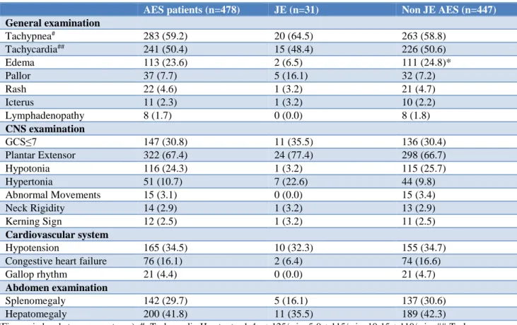
Table 2: Findings on general and systemic examination among AES patients.
AES patients(n=478) JE (n=31) Non JE AES (n=447)
General examination
Tachypnea# 283 (59.2) 20 (64.5) 263 (58.8)
Tachycardia## 241 (50.4) 15 (48.4) 226 (50.6)
Edema 113 (23.6) 2 (6.5) 111 (24.8)*
Pallor 37 (7.7) 5 (16.1) 32 (7.2)
Rash 22 (4.6) 1 (3.2) 21 (4.7)
Icterus 11 (2.3) 1 (3.2) 10 (2.2)
Lymphadenopathy 8 (1.7) 0 (0.0) 8 (1.8)
CNS examination
GCS≤7 147 (30.8) 11 (35.5) 136 (30.4)
Plantar Extensor 322 (67.4) 24 (77.4) 298 (66.7)
Hypotonia 116 (24.3) 1 (3.2) 115 (25.7)
Hypertonia 51 (10.7) 7 (22.6) 44 (9.8)
Abnormal Movements 15 (3.1) 0 (0.0) 15 (3.4)
Neck Rigidity 14 (2.9) 1 (3.2) 13 (2.9)
Kerning Sign 12 (2.5) 1 (3.2) 11 (2.5)
Cardiovascular system
Hypotension 165 (34.5) 10 (32.3) 155 (34.7)
Congestive heart failure 76 (16.1) 2 (6.4) 74 (16.6)
Gallop rhythm 21 (4.4) 0 (0.0) 21 (4.7)
Abdomen examination
Splenomegaly 142 (29.7) 5 (16.1) 137 (30.6)
Hepatomegaly 200 (41.8) 11 (35.5) 189 (42.3)
Encephalitis was evidenced by the presence of hyperintensities in the cortical and subcortical areas involving the brain stem, cerebellar areas, deep nuclei and early hydrocephalus. Cardiovascular: Congestive heart failure was clinically defined by the triad of
tachycardia, tachypnea and hepatomegaly. Although not significant, there was a trend of CHF being more common in non JE AES. CPK-MB elevation (>24IU) was seen in nearly 40% cases.
Table 3: Abnormalities in haematological and biochemical parameters among AES patients.
AES patients JE JE negative AES
Hematology n= 469 n=31 n=438
HB < 10mg/dl 258 (55.0) 13 (41.9) 153 (34.9)
TLC > 15000cell/mm3 166 (35.4) 13 (41.9) 153 (34.9)
Platelets <50000 58 (12.4) 4 (12.9) 54 (12.3)
CSF n=377 n=23 n=354
Color (Clear) 377 (100.0) 23 (100.0) 354 (100.0)
Protein (40-100mg/dl) 216 (57.3) 13 (56.5) 203 (57.3) Cells (5-300cell/mm3) 344 (91.3) 20 (87.0) 324 (91.5)
Serum No positive/No. tested No. positive/ No. tested No. positive/No. tested RBS <40 mg/dl 16/355 (4.5) 1/25 (4.0) 15/330 (4.6)
Serum Na <120meq/dl 23/391 (5.9) 1/24 (4.2) 22/367 (6.0) Serum K <2.5meq/dl 8/391 (2.1) 1/24 (4.2) 7/367 (1.9) Azotemia (Urea>40mg/dl) 97/394 (24.6) 5/23 (21.7) 92/371 (24.8) Inc. Creatinine mg/dl# 174/394 (44.2) 9/23 (39.1) 165//371 (44.5)
SGOT > 40IU/dl 288/450 (64.0) 18/30 (60.0) 270/420 (64.3) SGPT > 40 IU/dl 312//450 (69.3) 20/30 (66.7) 292/420 (69.5) Serum Bil ↑ 83/276 (30.1) 5/21 (23.8) 78/255 (30.6) PT INR >1.5 76/351 (21.7) 8/20 (40.0) 68/331 (20.5) Trop I positive 32/141 (22.7) 1/15 (6.7) 31/126 (24.6) CPK-MB >24U/L 141/348 (40.5) 7/18 (38.9) 134/330 (40.6)
Serum creatinine = 1-5 years: >0.5mg/dl; 5-10 years >1mg/dl; 10 years> 1.4 mg/dl
ECG, done in 141 cases revealed sinus tachycardia (29/141, 20.5%), low voltages (10/141, 7.6%), conduction defects (3/141, 2.1%) and T wave inversion in various leads (32/141, 22.7%) (Low voltage complex was taken when the QRS amplitude is <5mm in all limb leads and <10mm in all precordial leads).
2D Echocardiogram was done in 113 non JE AES cases with either features of CHF, raised CPK-MB or persistent hypotension. The common findings were decreased ejection fraction (17, 14.5%), increased left ventricular thickness (23, 20.4%) decreased fractional shortening (10, 8.5%) and regional wall motion abnormalities (30, 26.5%). Cardiac MRI was performed in five patients at follow up (i.e after one month) and patchy centromyocardial delayed enhancement in the mid- inferior segment suggestive of myocarditis was reported in one case and pericarditis reported in one case.
Others
Thrombocytopenia was not commonly seen. Deranged renal functions were not uncommon and renal biopsy done in one patient showed lymphocytic infiltration.
Transaminitis was not associated with a significant rise in serum bilirubin levels.
Out of the total, 131 (27.4%) patients had respiratory failure either at admission or during their hospital stay and required ventilatory support. Chest X-ray showed cardiomegaly (57/297, 19.2%) and pulmonary edema in 15.2% (45/297) cases. Interstitial pneumonia was also a feature in 26% (78/297) cases and resolved by the 3rd week. These were seen equally in both the groups.
Outcome
Full recovery was seen in 298 (62.3%) patients, 155 (32.4%) expired and outcome could not be ascertained in 25 (5.2%) as they refused continued hospital stay. In the JE group mortality was slightly higher (35.48%) as compared to Non JE (32.21%) but difference was non-significant.
DISCUSSION
AES in India, individual studies have shown varying positivity for JE, ranging from 31% to 80%.10 most
studies were done before JE vaccination was included in the routine immunization by the Government of India. Mass JE vaccination was done in the year 2010 in this and surrounding districts. Along with this there have been intense public education programs, residual mosquito spray and free distribution of insecticide-impregnated mosquito nets. There has also been introduction and breeding of Gambusia fish, a larvicidal fish, in water logged fields. The decrease in positivity for JE in this area could be a collective influence of all these efforts, because vaccination status in our study population is very low.11 a similar low positivity has been shown in a study
in Tamil Nadu.12 A mutation in the JE virus and
decreased virulence has been postulated as one of the possible reasons.
We had aimed at finding differences in clinic epidemiological patterns of JE and non JE encephalitis. The epidemiological features were similar in both the groups with no geographical difference in the distribution of cases. Both groups also had near similar neurological presentations with headache and hypertonia being more common in JE and hypotonia in non JE. JE virus has a predilection for the basal ganglia and hypertonia is a documented feature.13 Hypotonia has been shown to be a
feature of brain stem involvement, and has been documented in Nipah and Enterovirus.14,15 Neurological
signs in acute encephalitis do not usually help in reliably identifying the underlying etiology.11 Neuroimaging has
not been helpful in differentiating between the two, both in the acute phase as well as at follow up. Localizing features like involvement of basal ganglia, thalamus etc which have been documented in Japanese encephalitis in previous studies, were not found in the study population.13 Non JE encephalitis patients had more
edema and a multi organ involvement characterised by mild transaminitis, deranged renal functions and evidence of myocarditis.
Myocarditis was suggested by presence of increased CPK MB, Troponin I positivity, ECG and Echocardiac findings. CPK MB and troponin I have been considered to be sensitive markers of myocarditis in many studies in the past.16 Echocardiographic findings similar to our
study have also been documented by others.17 Myocardial
biopsy is the gold standard for diagnosis of myocarditis but has its limitations. Cardiac MRI has emerged as a sensitive and specific noninvasive tool for diagnosis of myocarditis.18 Cardiac MRI suggestive of myocarditis
was present in two of our cases. The use of cardiac MRI as a diagnostic tool is limited by financial constraints in our study and could be done only in five patients. Edema could be attributed to either heart failure due to myocarditis or to an abnormal cytokine activation that produces a severe inflammatory response, leading to leaky capillaries.19 Most patients in the study group had
features of a multi-organ involvement. This kind of
multi-organ involvement where patients are affected in large numbers have been reported in many infections like Rickettsial infections, scrub typhus, leptospirosis and viral illnesses like enterovirus, Dengue, etc.19-23 In India,
scrub typhus is an emerging infection and has been reported from many states.24 Although patients have a
similar presentation but absence of an eschar and non-clustering of cases do not support the possibility. Leptospirosis is another emerging infection with multisystem involvement. However, there is a significant hepatic involvement with high Serum Bilirubin levels, albuminuria, conjunctival suffusion and meningism. These features, which are used in a score for diagnosis of leptospira, were absent in the study population.25 Apart
from the epidemiological profile, the widespread use of antibiotics for fever in the periphery and non-responsiveness of these rather treatable conditions makes their possibility less likely.
Other common viruses having similar presentations like HSV, mumps, measles usually are sporadic and epidemics of this volume have not been reported with any of these. Dengue outbreaks in India have been recently on the surge and might have a multisystem involvement but none of those tested for the illness were positive.
Enteroviruses have been postulated as potential causative agents of AES.10 In the year 2006, National Institute of
Virology demonstrated enterovirus (EV) in CSF of 66/306 (21.6%) of AES patients by RTPCR.3 Sequencing
and phylogenetic analyses of PCR products from 59 (89.3%) of 66 specimens showed similarity with EV-89 and EV-76 sequences. A similar study published from a tertiary care teaching hospital of Lucknow revealed the predominance of various enteroviruses. Enterovirus infections worldwide have shown a presentation with multiorgan dysfunction.13 the poor sanitation and lack of
safe drinking water in our study population, epidemiologically favours water borne infection. Enterovirus has however not been demonstrated consistently and because of its ubiquitous nature and widespread prevalence even in healthy individuals, its etiological role has been questioned. A predominantly multi organ dysfunction can be caused by both viral and non-viral etiologies. It may also result as a manifestation of severe systemic inflammatory response syndrome. Studies aimed at identifying etiologies causing MODS may be able to identify the causative agent and help in planning preventive measures, as mortality in AES still remains high.
CONCLUSION
form of myocarditis is feature of this encephalitis. These features may help in identifying etiology.
ACKNOWLEDGEMENTS
Author would like to thank to Dr. Manoj Murhekar for reviewing the manuscript and valuable suggestions.
Funding: This work was supported by The Indian Council of Medical Research [Grant No: VIR/36/2010-ECD-1] Conflict of interest: None declared
Ethical approval: The study was approved by the Institutional Ethics Committee
REFERENCES
1. Kneen R, Michael BD, Menson E, Mehta B, Easton A, Hemingway C, et al. Management of suspected viral encephalitis in children - Association of British Neurologists and British Pediatric Allergy immunology and Infection Group National Guidelines. J Infect. 2012;64:449-77
2. Saxena SK, Singh M, Pathak AK, Mathur A. Reply to 'Encephalitis outbreak finds Indian officials unprepared'. Nat Med. 2006;12:269-70.
3. Parida M, Dash PK, Tripathi NK, Ambuj, Sannarangaiah S, Saxena P. Japanese Encephalitis Outbreak, India, 2005. Emerg Infect Dis. 2006; 12(9):1427-30.
4. Mittal M, Kushwaha KP. AES: Clinical Presentation and Dilemmas in Critical Care Management. J Commun Dis. 2014;46(1):50-65.
5. Ranjan P, Gore M, Selvaraju S, Kushwaha KP, Murhekar M: Changes in acute encephalitis syndrome after introduction of Japanese encephalitis vaccine in a region of India. J Infect. 2014;69(2):200-2.
6. Bhatt GC, Bondre VP, Sapkal GN, Sharma T, Kumar S, Gore MM et al. Changing clinico-laboratory profiles of encephalitis patients in the eastern Uttar Pradesh region of India. Tropical Doctor. 2012;42:106-08. 7. Sapkal GN, Bondre VP, Fulmali PV, Patil P,
Gopalkrishna V, Dhadania V, et al. Enteroviruses in Patients with Acute encephalitis, Uttar Pradesh, India; Emerg Infect Dis. 2009;15(2):295.
8. Kumar A, Shukla D, Kumar R, Idris MI, Usha K. Misra, and Dhole T. Molecular Epidemiological Study of Enteroviruses Associated with Encephalitis in Children. Indian J Clini Microbiol. 2012;50(11):3509-12.
9. World Health Oraganisation. Acute Encephalitis Syndrome.Japanese encephalitis surveillance standards. From WHO-recommended standards for surveillance of selected vaccine-preventable diseases; 2006.
10. Joshi R, Kalantri SP, Reingold A, Colford JM Jr. Changing landscape of acute encephalitis syndrome in India: a systematic review: Natl Med J India. 2012;25(4):212-20.
11. Ranjan P, Gore M, Selvaraju S, Kushwaha KP, Srivastava DK, Murhekar M. Changes in acute
encephalitis syndrome incidence after introduction of Japanese encephalitis vaccine in a region of India. J Infect. 2014;69:200-2.
12. Gunasekaran P, Kaveri K, Arunagiri K, Mohana S, Kiruba R, Kumar VS, et al. Japanese encephalitis in Tamil Nadu (2007-2009). Indian J Med Res. 2012;135(5):680-82.
13. Kalita JJ, Misra UK, Pandey SS, Dhole TN. A comparison of clinical and radiological findings in adults and children with japanese encephalitis. Arch Neurol. 2003;60(12):1760-64.
14. Khean JG, Chong TT, Nee KC, Patrick SKT, Adeeba K, Sazilah AS, et al. Clinical features of nipah virus encephalitis among pig farmers in Malaysia. The New England J Medic. 2000;342(17):1229-35.
15. Chaudhuri A, Kennedy PGE. Diagnosis and treatment of viral encephalitis. Postgrad Med J. 2002;78:575-58. 16. Lauer B, Niederau C, Kuhl U, Schannwell M,
Pauschinger M, Strauer BE, et al. Cardiac troponin T in patients with clinically suspected myocarditis. J Am Coll Cardiol. 1997;30:1354-9.
17. Felker GM, Boehmer JP, Hruban RH, Hutchins GM, Kasper EK, Baughman KL, et al. Echocardiographic findings in fulminant and acute myocarditis. J Am Coll Cardiol. 2000;36:227-232.
18. Friedrich MG, Sechtem U, Schulz-Menger J, Holmvang G, Alakija P, Cooper LT, et al. International Consensus Group on Cardiovascular Magnetic Resonance in Myocarditis. Cardiovascular magnetic resonance in myocarditis: A JACC White Paper. J Am Coll Cardioll .2009;53:1475-87.
19. Mong HO, Wong SC, Lewthwaite P, Cardosa MJ, Solomon T. Clinical features, diagnosis, and management of enterovirus 71. Lancet Neurol. 2010;9:1097-110.
20. Vaz LS, Gupta NK. Outbreak of scrub typhus in Jammu-a report. MJAFI. 2006;62:342-3.
21. Vivekanandan M, Mani A, Priya YS, Singh AP, Jayakumar S and Purty S . Outbreak of scrub typhus in Pondicherry. J Assoc Physicians India. 2010;58:24-8. 22. Chauhan V, Mahesh DM, Panda P, et al. Profile of
Patients of Leptospirosis in Sub-Himalayan Region of North India. J Assoc Phys India. 2010;58:354-6. 23. Kumar R, Tripathi S, Tambe JJ, Arora V, Srivastava A,
Nag VL. Dengue encephalopathy in children in Northern India: Clinical features and comparison with non-dengue. J Neurol Sci. 2008;269:41-8.
24. Mathai E, Rolain JM, Verghese GM, Abraham OC, Mathai D, Mathai M et al. Outbreak of scrub typhus in southern India during the cooler months. Ann N Y Acad Sci. 2003;990:359-64.
25. Faine S. Guidelines for the control of Leptospirosis. WHO offset publication. 1982;67.