FUNCTIONAL SELECTIVITY AT THE D1 DOPAMINE RECEPTOR: STUDIES USING SKF83959
by
Andrew Charles Kant
A thesis submitted to the faculty of The University of North Carolina at Chapel Hill in partial fulfillment of the requirements for the degree of Master of Science in the Curriculum in Toxicology.
Chapel Hill 2008
ABSTRACT
FUNCTIONAL SELECTIVITY AT THE D1 DOPAMINE RECEPTOR: STUDIES USING SKF83959
(Under the direction of Richard B. Mailman, Ph.D.)
PREFACE
I have prepared my thesis in accordance with the guidelines set forth by the Graduate School of the University of North Carolina. This thesis consists of a general introduction, two research chapters of original data, and an overall conclusion chapter. Each data chapter includes an abstract, introduction, methods and materials, results and discussion sections. A complete list of the literature cited throughout the thesis has been appended. References are listed in alphabetical order and follow the format of The Journal of Pharmacology and Experimental Therapeutics.
Available publications from this work
Brown JT, Kant AC, Mailman RB. Rapid, semi-automated, and inexpensive radioimmunoassay of cAMP: Application in GPCR-mediated adenylate cyclase assays. J Neurosci Methods 2008 (in press).
ACKNOWLEDGMENTS
TABLE OF CONTENTS
ABSTRACT...II
PREFACE...III
ACKNOWLEDGMENTS...IV
LIST OF TABLES...IX
LIST OF FIGURES...X
LIST OF ABBREVIATIONS...XII
CHAPTER 1.SCOPE OF THIS WORK...1
Dopamine systems ...1
Dopamine Receptors...2
Parkinson’s Disease ...3
Toxicology and Parkinson’s disease ...4
Genetics of Parkinson’s ...5
Current pharmacotherapy of Parkinson’s ...6
Functional selectivity...10
Goals of this thesis...14
Aim 1. Develop a streamlined, highly sensitive and high-throughput method for measuring adenylate cyclase activity...15
Aim 2. Investigate the functional selective signaling properties of various classes of D1 agonists. ...15
CHAPTER 2: RAPID, SEMI-AUTOMATED, AND INEXPENSIVE RADIOIMMUNOASSAY OF CAMP:APPLICATION IN GPCR-MEDIATED ADENYLATE CYCLASE ASSAYS...16
Abstract...16
Introduction...17
Experimental procedures and Results...19
Materials and reagents ...19
Sample Generation and storage ...19
cAMP Radioimmunoassay ...20
Iodination reaction...20
Purification of iodinated product...22
Preparation of primary antibody conjugation to amine-terminated beads ...23
Radioimmunoassay...24
Discussion...25
Elimination of secondary antibody allows direct detection ...26
Optimization of cAMP antiserum conditions ...27
Assay precision and accuracy...28
Cost issues and alternative technology...30
CHAPTER 3. D1 RECEPTOR STIMULATION ACTIVATES ADENYLATE CYCLASE BUT NOT PHOSPHOLIPASE C...32
Abstract...32
Introduction: ...33
Materials and Methods ...35
Cell Culture ...35
Competition Binding ...36
cAMP accumulation assay and RIA...36
PLC assay ...37
Data Analysis...38
Results...38
Discussion...44
CHAPTER 4:SUMMARY OF FINDINGS AND FUTURE DIRECTIONS...50
Summary of findings ...50
Future Directions ...51
LIST OF TABLES
Table 3.1: Competition binding of high affinity D1 compounds ... 39
LIST OF FIGURES
Figure 1.1. Schematic of brain dopamine pathways ... 2
Figure 1.2. Functional selectivity and its implications in therapeutic and toxic actions of drugs... 12
Figure 2.1: Reaction scheme for synthesis of 2′-O-[4-monosuccinyladenosine 3′:5′-cyclic monophosphate-3-iodotyrosyl methyl ester... 21
Figure 2.2: Chromatogram of radioiodination ... 23
Figure 2.3. cAMP standard curves generated under varying assay conditions ... 28
Figure 2.4. Measurement of D1 dopamine receptor-mediated cAMP accumulation ... 29
Figure 2.5. Precision profile demonstrates the Coefficient of Variation as a function of the concentration of cAMP standards ... 30
Figure 2.6. Schematic flowchart of the described method... 31
Figure 3.1: Structures of the phenylbenzazepine SKF83959 and other ligands used in this study... 34
Figure 3.2: Competition binding of compounds in hD1-HEK293 cells ... 40
Figure 3.3: Adenylate cyclase activation in stable HA-hD1-HEK293 cells... 40
Figure 3.4: Adenylate cyclase activity in caudate tissue from Sprague-Dawley male rats ... 41
Figure 3.6: D1 specific antagonist treatment has no effect on PLC activity in
LIST OF ABBREVIATIONS 6-OHDA 6-hydroxydopamine
AA arachidonic acid
AC adenylate cyclase
BRET bioluminescence resonance energy transfer
cAMP cyclic AMP; adenosine 3',5'-cyclic monophosphate
CSP cysteine-string-protein-alpha
DREADD designer receptors exclusively activated by designer drugs
DA dopamine
DARPP-32 dopamine- and cyclic AMP-regulated phosphoprotein
EPS extrapyramidal symptoms
GPCR G Protein coupled receptor
GRK G-protein coupled receptor kinases
HEPES 4-(2-hydroxyethyl)-1-piperazine-ethane-sulfonic acid
IP1 inositol 1-phosphate
IP3 inositol 1,4,5-trisphosphate
IP3 inositol 1,4,5-trisphosphate
K0.5 concentration corrected IC50 (apparent affinity constant) when nH≠ 1.0.
L-DOPA 3,4-dihydroxy-L-phenylalanine, levodopa
MPTP 1-methyl 4-phenyl 1,2,3,6-tetrahydropyridine
PD Parkinson’s disease
PKA protein kinase A
PLC phospholipase C
PP2A protein phosphatase 2A
PSD postsynaptic density
RGS regulators of G protein signaling
SKF38393 2,3,4,5-tetrahydro-7-8-dihydroxy-1-phenyl-1-H-3-benzazepine
SKF83959 6-Chloro-7,8-dihydroxy-3-methyl-1-(3-methylphenyl)-2,3,4,5-tetrahydro-1H-3-benzazepine
SNpc substantia nigra pars compacta
CHAPTER 1. SCOPE OF THIS WORK DOPAMINE SYSTEMS
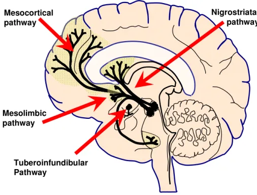
Nigrostriatal pathway Mesocortical
pathway
Mesolimbic pathway
Tuberoinfundibular Pathway
Figure 1.1. Schematic of brain dopamine pathways.
DOPAMINE RECEPTORS
Although the two D1-like receptors are structurally very similar, there are some key variations in localization. D5 receptors are scarcely expressed, if at all, in the human striatum, whereas D1 receptors are highly expressed. In other areas like the hippocampus and prefrontal cortex, both receptors play important roles, including in working memory (Bergson et al., 1995; De Keyser, 1993). D1-like receptors are exclusively located post-synaptically.
In addition to localized expression differences, there is also interesting evidence of distinct signaling between the two subtypes. Studies using HEK293 cells demonstrated that when the γ7 subunit of the heterotrimeric G protein is degraded, D1-mediated adenylate cyclase signaling is abrogated, whereas D5 receptor signaling remains in tact (Wang et al., 2001). There also may be differences in DARPP-32, ERK1/2 and GluR1-AMPA signaling between the D1-like receptors. D1 receptors, and to a much lesser extent D5, were found in rats to be differentially modulated with SKF 83822 (O'Sullivan et al., 2008). Interestingly, this particular compound is purported to signal exclusively through phospholipase C rather than via adenylate cyclase (O'Sullivan et al., 2004). These data are based on behavioral assessments alone, however, and have not yet been replicated in an in vitro system. Aspects of this thesis explore some of these relevant mechanisms.
PARKINSON’S DISEASE
Parkinson’s disease is characterized by three types of symptoms: primary motor, secondary motor and non-motor. Primary motor signs are characterized by resting tremor, bradykinesia, rigidity and postural instability, and the diagnosis of the disease is made on the basis of the occurrence of several of these cardinal signs. These are the most common and notable signs of Parkinson’s disease, however many patients experience secondary motor symptoms that can include fatigue, speech control, loss of facial expression, micrographia and difficulty swallowing. Additional non-motor symptoms that can be particularly difficult for a patient include depression, constipation, pain, dementia, and memory difficulties. Although currently therapies address the motor domain well, the non-motor symptoms are more difficult to treat.
Toxicology and Parkinson’s disease
Exogenous toxicants have been implicated as a playing a role in a subset of Parkinson’s disease cases (Ascherio et al., 2006). The first example of this toxicant-induced neurodegeneration came from 1-methyl-4-phenyl-1,2,3,6-tetrahydropyridine (MPTP) (Davis et al., 1979; Langston et al., 1983). MPTP itself is relatively non-toxic, but when taken up by the brain monoamine oxidase B located in astrocytes form the toxic metabolite 1-methylphenylpyridium (MPP+). As with Parkinson’s disease, MPP+ kills dopamine neurons located in the pars compacta of the substantia nigra (SNpc). Parkinson’s-like symptoms such as tremor, bradykinesia, and postural instability rapidly occur.
have suggested positive associations with long-term exposure to pesticides such as paraquat (Liou, 1997), rotenone (Kamel et al., 2001), and organophosphates (Firestone et al., 2005; Wechsler et al., 1991). Rat studies using rotenone and MPTP have shown clear causation of parkinsonism-like symptoms (Caboni et al., 2004; Helmuth, 2000). Nonetheless, it remains unclear to what degree, if any, pesticides facilitate or plays a causal role in the development of Parkinson’s disease.
In addition to pesticides, a recent study by Stepens et al. (2008) has suggested a potential role for manganese. The study found that people using methcathinone, a psychoactive drug of abuse, exhibited extrapyramidal syndrome (EPS). The methcathinone preparations were found to be tainted with high levels of manganese and MRI results showed hyperintensity in the globus pallidus and substantia nigra (Stepens et al., 2008). The patients did not recover from EPS following discontinuation of methcathinone, suggesting irreversible damage to the globus pallidus and substantia nigra. This hypothesis is supported by work in a rodent model studying inhalation of divalent and trivalent. Mice exhibited signs of akinesia, postural instability and action tremor, while post-mortem analysis showed roughly a 70% decrease in tyrosine hydroxylase positive neurons in the SNpc (Ordoez-Librado et al., 2008).
Genetics of Parkinson’s
provided the first clue as to its function. Using transgenic mice, expression of α-synuclein restores neuroprotection following the deletion of cysteine-string protein-α (CSPα). The function of CSPα is not replaced by α-synuclein, instead it acts as a downstream effector in the pathway by binding to phospholipids in an α-helical conformation. These data provide specific evidence of in vivo function of α-synuclein which prevents neurodegeneration of presynaptic neuronal processes (Chandra et al., 2005). These data are complicated by the inability of endogenous α-synuclein to prevent lethality in CSPα knockout mice, but the specific rescue of neurodegeneration and known association between CSPα and α-synuclein reinforces the necessity of further mechanistic studies.
An additional genetic role has been implicated for E3 ligase parkin, involved with the ubiquitin-proteosome system, which when omitted causes autosomal recessive juvenile parkinsonism (Kitada et al., 1998). It has been posited that mutations in this protein lead to misfolding and aggregation of proteins which facilitate the death of SNpc dopaminergic neurons (Hattori and Mizuno, 2004). It is also interesting to note that Lewy bodies (commonly found in neurons of Parkinson’s disease and Alzheimer’s patients) are immunoreactive to ubiquitin. This perhaps reinforces the role of parkin and more generally dysfunction of protein degradation in the pathology of Parkinson’s disease. Other studies have investigated roles for other parkin isoforms, specifically park7, however their incidence is significantly less than that of park2, the main known culprit in parkin-related Parkinson’s disease (Hedrich et al., 2004).
Current pharmacotherapy of Parkinson’s
amount of dopamine in terminal fields. Levodopa is always used with a peripheral decarboxylase inhibitor (carbidopa or benserazide) to decrease peripheral adrenergic and dopaminergic side effects. Although dramatically effective for some years, long-term treatment with L-DOPA eventually leads to a decline in therapeutic benefit, although this may be attenuated somewhat by the use of adjuvants such as MAO-B (monoamine oxidase) inhibitors (selegiline or rasagiline), COMT (catechol-o-methyltransferase) inhibitors (entacapone or tolcapone) and other agents like NMDA (N-methyl-D-aspartic acid) antagonists (amantadine). Despite its effectiveness, levodopa therapy tends to eventually lead to debilitating side effects, especially dyskinesias, as well as loss of effectiveness expressed as “on-off” effects. There have been many direct dopamine agonists that have been used in PD, and two currently are widely used: pramipexole (Mirapex) and ropinirole (Requip). These target the D2-like receptors, and although having a useful place, they are only fractionally better than placebo relative to levodopa. The use of these D2 agonists is thought to smooth out the pattern of target neuron effects, and as an adjunct therapy to levodopa decrease side effects like dyskinesia. They have a narrow dose-response relationship, however, and can cause nausea and vomiting, as well as psychosis. This occurs largely via the same mechanism by which they act therapeutically (i.e., activation of D2-like receptors).
D1 agonists and Parkinson’s disease
(Mailman and Huang, 2007). This pulsatility, and the resulting dyskinesias, is probably the result of the short half-life of levodopa, its indirect mechanism of action, and the continuingly fewer dopamine neurons left to process the levodopa. In theory, therefore, the use of dopamine agonists should be beneficial because one could better control the degree and timing of receptor activation.
Currently, no centrally active D1-selective agonist is approved for human clinical use, but there has been much experimental work addressing this paradox. Early data suggested that D1 agonists were not involved in dopamine-induced amelioration of PD symptoms. These studies were, however, problematic, principally due to the fact that the available D1 agonists like SKF38393 were only partial agonists (Barone et al., 1986). Thus, an early study showed little to no effect on tremor while inducing bradykinesia in the marmoset animal model (Close et al., 1985). While SKF38393 has excellent potency for the D1 receptor, its intrinsic activity for adenylate cyclase activation is less than half that of dopamine. For these reasons, the common view in the field for many years was that all therapeutic actions of levodopa and agonists were mediated via D2 receptors (Cederbaum and Schleifer, 1990).
deliver high bolus doses, was limited by hypotension, its primary side effect (Blanchet et al., 1998).
Subsequent to these studies, a second D1 full agonist A-77636 was discovered and also shown to have dramatic acute antiparkinson effects in a marmoset MPTP-induced model of parkinsonism (Kebabian et al., 1992). It, however, was limited by a rapid tolerance (Asin et al., 1994) that might be a result of prolonged internalization of the D1 receptor caused by this drug (Ryman-Rasmussen et al., 2007). After abandoning A77636, Abbott developed a new compound A-86929 that is a structural analog of dihydrexidine (Michaelides et al., 1995) with very similar pharmacological properties. It was shown not to cause the tolerance seen with A-77636 (Asin et al., 1997). Like dihydrexidine, it was also very short-acting and not orally available, so a prodrug form, ABT-431 was developed to overcome these pharmacokinetic problems (Shiosaki et al., 1996). ABT-431 was tested in humans, and is the only drug to have efficacy that equals levodopa (Rascol et al., 1999; Rascol et al., 2001), and possibly a better side effect profile (Rascol et al., 1999). These and other data have made the potential of D1 agonists in treating Parkinson’s disease clear, although a variety of pharmacological (seizures, hypotension) and pharmaceutical (chemical instability, oral availability, short duration of action) problems have yet prevented the approval of a drug. Promisingly, these problems seem to be idiosyncratic to specific drugs, leaving open the possibility of discovery of a successful drug. This summary underscores the pharmacological and toxicological importance of understanding D1-related mechanisms.
Other potential beneficial actions of D1 agonists
to 10 fold higher than that of D2 receptors, pointing to their role in the mesocortical pathway (Hall et al., 1994; Lidow et al., 1991). More specifically, the prefrontal cortex has been shown to play a critical role in working memory, a subcategory of short-term memory (Goldman-Rakic, 1998; Goldman-Rakic et al., 2000). In young human subjects, pergolide was shown to improve delayed matching performance when compared with placebo, further proof for the role D1 agonists have in improving working memory (Muller et al., 1998). Indeed, as the first full D1 agonist, dihydrexidine was a major tool in showing the importance of D1 activation (Arnsten et al., 1994; Johnson et al., 1995; Schneider et al., 1994; Steele et al., 1996; Steele et al., 1997). This again underscores the importance of understanding the relevant mechanisms of D1 agonists.
FUNCTIONAL SELECTIVITY
difficult, as reviewers often assumed the data were experimental artifacts caused by off-site receptor or non-receptor actions within a functional assay.
Figure 1.2. Functional selectivity and its implications in therapeutic and toxic actions of drugs. Adapted from Mailman (2007)
Although the overlying theory and data support the notion that drugs can have relative intrinsic efficacy, the mechanisms by which this occurs remain elusive. There are two schools of thought that seek to describe functional selectivity more precisely: conformational induction and drug-active state selection. The importance of this distinction lies with applying the theory of functional selectivity to the drug development process. A greater understanding of these mechanisms would certainly lead to better therapeutic exploitation of functional pathways.
complex, when compared to another ligand of a different chemical family, may be quite different due to the unique mode of agonist binding. In very simplistic terms, the theory of induction is dependent on specific ligand properties to lead to a functionally selective outcome.
Testing of the sub-theories of functional selectivity remains difficult due to problems with GPCR crystallization. Bioluminescence resonance energy transfer (BRET) has proven to be an important technique when studying conformation changes in living cells. A powerful study by Swaminath et al. (2004) used the β2 adrenergic receptor to label Cys265 with a fluorescent dye which resides in the third intracellular loop, an area thought to be important for G protein coupling. The authors demonstrated that different catecholamines induce a sequence of two different conformational changes, a rapid and a slow change in the receptor. Moreover, dopamine was found to induce only a rapid change in conformation leading to adenylate cyclase activation, but not receptor internalization. This compared to norepinephrine and epinephrine, which induced both rapid and slow responses, leading to both adenylate cyclase and receptor internalization. These data strongly support the notion of agonist-induced conformational changes, but these a great deal of research remains with other receptor models and families to allow understanding of the intricacies of functional selectivity.
evidence was initially derived from the observation that GPCRs couple to G proteins in the absence of agonist, accounting for basal activity of the receptor. This, together with studies demonstrating coupling of multiple G proteins to a given receptor has strengthened the logical possibility of sub-populations of receptors that may be more apt to bind a given ligand (Kenakin, 1995). Moreover, the use of over-expression in vitro models may lead to G protein promiscuity and thus the creation of an artifact of active-state receptors. It should be noted however that these two theories for the underlying modality of functional selectivity need not be mutually exclusive, although one may predominate.
The ultimate goal of studying functionally selective drugs is to learn how to decrease unwanted side effects while retaining clinical efficacy. As noted earlier, aripiprazole has interesting functionally selective properties and is currently used in patients with schizophrenia, bipolar disorder and depression. To this end, a major effort is underway to study in vivo mechanisms. The use of animal models engineered to express designer receptors called DREADDs (designer receptors exclusively activated by designer drugs) has begun (Nawaratne et al., 2008), in addition researchers are using more extensive screening of compounds and neuronally derived cell lines. Given the complex nature of cellular signaling, particularly neuronal, the constant improvement of techniques and theories will surely unravel the puzzle.
GOALS OF THIS THESIS
Aim 1. Develop a streamlined, highly sensitive and high-throughput method for measuring adenylate cyclase activity.
Because my proposed work involved adenylate cyclase assays as a major signaling pathway, improvements were made to existing assays that then accelerated my work on Aim 2. The improved method cut costs and assay time, but preserved the sensitivity and reproducibility of the assay.
Aim 2. Investigate the functional selective signaling properties of various classes of D1 agonists.
CHAPTER 2:
RAPID, SEMI-AUTOMATED, AND INEXPENSIVE RADIOIMMUNOASSAY OF CAMP: APPLICATION IN GPCR-MEDIATED ADENYLATE CYCLASE
ASSAYS ABSTRACT
Cyclic AMP (cAMP) is an important signal transduction second messenger that is commonly used as a functional mirror on the actions of G protein-coupled receptors that can activate or inhibit adenylate cyclases. A radioimmunoassay for cAMP with femtomole sensitivity was first reported by Steiner more than 30 years ago, and there have been several subsequent modifications that have improved this assay in various ways. Here we describe additional improvement to existing methods that markedly improve speed and reduce cost without sacrificing sensitivity, and is also adaptable to analysis of cGMP. The primary antibody is coupled directly to magnetic beads that are then separated from unbound marker using filtration on microplates. This eliminates the need for a secondary antibody, and markedly increases throughput. In addition, we report a simple, reproducible, and inexpensive method to make the radiomarker used for this assay. Although still requiring the use of radioactivity, the resulting method retains a high degree of accuracy and precision, and is suitable for low-cost high-throughput screening. Use of aspects of this method can also improve throughput in other radioimmunoassays.
INTRODUCTION
Cyclic AMP (3’,5’-cyclic adenosine monophosphate; cAMP) is a key second messenger involved in numerous intracellular signaling pathways (Antoni, 2000; McPhee et al., 2005). Production of cAMP is controlled by the membrane-bound family of adenylate cyclases (ACs) that convert adenosine triphosphate to cAMP. The activity of most of the ACs is regulated by heterotrimeric GTP-binding proteins (e.g., Gαs/olf, Gαi/o) that directly interact with the intracellular region of GPCRs and can both increase or decrease enzyme activity (Hanoune and Defer, 2001). In addition, phosphodiesterases can catalyze the degradation of cAMP (Weishaar, 1986).
The measurement of adenylate cyclase activity can be accomplished using radiometric assays that follow the incorporation of a radioactive precursor into cAMP (Salomon, 1979; Schulz and Blum, 1985). More commonly, however, a variety of methods that quantify cAMP have been used both for assessment of adenylate cyclase activity, as well as for measuring tissue content of cAMP or breakdown of this second messenger. A major advance for the field was the development by Steiner et al. (1972) of a radioimmunoassay (RIA) for cAMP that offered a high degree of sensitivity and specificity that was soon improved by Harper and Brooker (1975). Attempts at automating this assay actually led to a commercial instrument (Brooker et al., 1976), but this proved unwieldy.
to a solid scintillant surface. These assays are convenient and reproducible, but are often more expensive than traditional radiometric methods and generally speaking less sensitive. Reporter-gene assays utilize cell lines expressing reporter enzymes such as luciferase, green fluorescent protein (GFP), and β-lactamase. Levels of intracellular cAMP are detected via the expression level of a reporter gene that is modulated by transcription factor binding to upstream cAMP response elements (CRE). Reporter-gene assay are generally less expensive than the radiometric assays discussed above, however, they are often plagued by high false-positive hit rates. Several novel, non-radiometric methods to quantify cAMP also have recently become available. These assays involve the use of luminescent proximity (ALPHAScreen®) (Ullman et al., 1994), enzyme complementation technology (DiscoveRx, HitHunterTM EFC), or electrochemiluminescence (Meso Scale Discovery) to detect receptor-mediated changes in intracellular cAMP. Each method is readily compatible with automated high throughput screening (HTS), and often demonstrates a high level of sensitivity, but requires a high degree of instrumentation to maximize throughput putting it beyond the reach of most academic labs.
rapid method for the routine production of the [125I]-labeled cAMP derivative that is used as the radiomarker in this RIA.
EXPERIMENTAL PROCEDURES AND RESULTS Materials and reagents
Dihydrexidine was synthesized according to procedures previously published (Brewster et al., 1990). Acetic anhydride, dopamine, IBMX, pargyline, propranolol, SKF38393, and triethyleneamine, and 2’-O-monosuccinyladenosine 3’:5’ monophosphate tyrosyl methyl ester (ScAMP-TME) were purchased from Sigma-Aldrich (St. Louis MO). HEPES was obtained from Research Organics, Inc. (Cleveland OH). Dulbecco’s modified eagle’s media (DMEM), penicillin/streptomycin, and fetal bovine serum (FBS) were purchased from Gibco/Invitrogen. UniFilter-96 GF/B RIA filter plates, Microscint™ 20, and Na125I were purchased from Perkin-Elmer (Waltham, MA, USA). Donkey anti-goat antibody was purchased from Jackson ImmunoResearch (West Grove, PA, USA). Amine terminated BioMag® beads were purchased from Polysciences, Inc. (Warrington, PA, USA), and pre-conjugated Biomagnetic Particles (BMP) to donkey anti-goat secondary was obtained from Rockland, Inc (Gilbertsville, PA, USA).
Sample Generation and storage
cAMP is a relatively heat and acid stable compound that does not require special storage. The following procedure illustrates a common way that samples are generated from a GPCR-based cellular system, but the assay that follows can be used for almost any matrix.
streptomycin (Gibco), and supplemented with 10% fetal bovine serum at 37°C, 5% CO2. Saturation binding experiments with the D1-selective antagonist [3H]SCH23390 using membrane homogenates provided a Bmax of approximately 4.5 pmol/mg protein.
Cell membrane adenylate cyclase assay: Assay buffer was prepared containing 100 mM HEPES, 4 mM MgCl2, 2 mM EDTA, 100 mM NaCl 10 µM pargyline, 500 µM IBMX, 0.1% ascorbic acid, pH 7.4. Drug dilutions were prepared at a range of 10-4 to 10-10 M with three replicates per drug treatment. Diluted drugs, ATP (2 mM), GTP (5 µM), phosphocreatine (20 mM), creatine phosphokinase (185 U/tube) and propranolol (100 µM to block endogenous β1-adrenergic receptors) were added in a total volume of 100 µL in each well of a 48-well plate. The reaction was initiated by addition of HEK-hD1 cell membranes. Plates then were vortexed briefly, and incubated at 30°C for 15 min. The reaction was terminated with 500 µL 0.1 M HCl, and stored at 4°C. Prior to transferring samples for the RIA, plates are centrifuged for 5 min at 2,500 g using a RC-3B centrifuge from Sorvall Instruments (H2000B rotor) to pellet cellular debris. Plates will keep indefinitely at 4°C following the assay.
cAMP Radioimmunoassay Iodination reaction
NH2
HO
H2N
NH2
HO
H2N
N N N
N
O
O P O O O O O O O O N N N N O
O P O O O O O O O O I I I– Chloramine-T N N N N O O O P O O O O O O O N N N N O O O P O O O O O O O
Figure 2.1: Reaction scheme for synthesis of 2′-O-[4-monosuccinyladenosine 3′:5′-cyclic monophosphate-3-iodotyrosyl methyl ester. Conditions described in Methods (molar excess of precursor) favor the formation of the monoiodinated product (see Figure 2.2).
The following reagents and buffers are required:
• 0.5 M phosphate buffer, pH 7.6. We usually make this by titrating 15 mL of 0.5 M K2HPO4 with ca. 1.5 mL of NaH2PO4 to pH 7.6.
• 0.05 M phosphate buffer (pH 7.6). This is prepared by adding 10 mL of the 0.5 M phosphate to 90 mL H2O.
• Carrier-free Na125I. We usually use 2 or 5 mCi. If more than 5 mCi is used, the amount of precursor should be increased proportionally,
• Precursor ScAMP-TME [2’-O-monosuccinyladenosine 3’:5’ monophosphate tyrosyl methyl ester; Sigma M2257]. From the 1 mg commercial size, we make 1-1.5 mL of a stock solution containing 0.1 mg/mL of distilled water. Aliquots (50 µL) are added to microfuge tubes, labeled, and frozen at -20 C. A single aliquot is used for each radioiodination. The frozen precursor appears stable for several years.
• Chloramine-T: (20 mg/10 mL 0.05 M PO4).
• Sodium metabisulfite: (24 mg/10 mL 0.05 M PO4).
thawed aliquots of ScAMP-TME (5 µg/50 µL H2O) is added, the cap screwed back on, and the vial mixed on a vortexer for 15 sec. Following this, Chloramine T (100 µL of 2 mg/mL solution) is added, and timing begun as the mixture is vortexed. After ~45 sec, the reaction is terminated by addition of sodium metabisulfite (200 µL of 2.4 mg/mL solution). [Safety note: Unreacted 125I is potentially volatile, and a potential health hazard. The use of concentrated (0.5 M) phosphate buffer insures that the reaction solution does not become acidic, a condition favoring the liberation of molecular iodine. In addition, this reaction is done in a chemical hood.]
Purification of iodinated product
0
10
20
30
40
0
10
20
30
40
R
T(min)
0
.0
1
A
U
F
S
Figure 2.2: Chromatogram of radioiodination. [Bottom tracing] shows injection of cAMP-Sc-TME precursor alone using conditions as described in Methods using 254 nm UV detection. The solvent front emerges at ~ 2 min, and the precursor elutes at ~ 6min. The signal in the solvent front and a detectable shoulder on the major peak is consistent with the 95% purity estimated by the supplier. [Top tracing] Actual results from a radioiodination. The monoiodinated product that is
immunologically recognized elutes at ~28 min, and is the fraction to be collected and used for the RIA. This fraction contains from 60-70% of the radioactivity in a typical reaction. The fraction eluting at ~40 min also contains significant radioactivity (10-20%), and is presumably the diiodinated form. These two peaks account for ~80% of the total radioactivity injected, with the remainder of the radioactivity largely eluting in the solvent front (representing unreacted iodine or highly polar reaction by-products).
The radioactivity is estimated using a hand-held radioactivity detector (or one can count 1 µL aliquots), and the tubes with the highest radioactivity (usually 3-4 tubes) are pooled together, diluted with 1.5 volumes of methanol, and then divided into two or more aliquots for storage at -20°C. Under these conditions, the marker is usable for a minimum of four months, although there is a significant loss of material due to decay.
Preparation of primary antibody conjugation to amine-terminated beads
Inc). Lyophilized antibody was reconstituted in distilled water to a final concentration of 0.5 mg/mL, and dialyzed in coupling buffer (0.01 M pyridine in distilled water, pH 6.0), changing the buffer three-times over a 9 hr period. The beads then were prepared by washing with coupling buffer, and magnetically separating three times. Glutaraldehyde solution (5% glutaraldehyde in coupling buffer) was mixed with the BioMag® beads, and reacted for three hours with rotation. The beads were washed four times with coupling buffer, and antibody was added to the beads with rotation for 16-24 hrs. Glycine quenching solution (1 M glycine, pH 8.0) was combined with beads and rotated for 30 min. Primary α-cAMP-beads were mixed a volume of 20 mL of storage buffer (0.01 M Tris, 0.1% NaN3, 0.1% w/v BSA, 0.15 M NaCl, 1 mM EDTA, pH 7.4), and stored at 4°C. The antibody-bead conjugate was used for up to three months with no appreciable sign of degradation. Fidelity of the conjugate was assessed by determining the ratio of binding between two sets of tubes, one containing radiolabeled cAMP bound to primary antibody and the other containing only radiolabeled cAMP. A ratio of 0.2-0.3 was found to be ideal while less than 0.2 led to inconsistent replicates.
Radioimmunoassay
resembles the original hapten. 125I-cAMP was then added within 30 minutes of acetylation. Optimal ranges for radioactivity were determined to be between 280 cpm/µL to 320 cpm/µL for iodinated 125I-cAMP-scTME. An aliquot (20 µL) of conjugated-primary antibody then was added to bind labeled and unlabeled cAMP (in 50 mM sodium acetate, 0.1% BSA, pH 4.75). Plates were incubated overnight at 4°C. Radioimmunoassay reactions were terminated by filtration with UniFilter-96 plates (Perkin-Elmer) with dH2O. Plates were washed three times and then dried at 50°C for 1 hour. Microscint™ 20 fluid (50 µL) was added to the wells, and counted on a TopCount NXT (Perkin-Elmer) for 2 min or 2σ = 5%.
Data Analysis
Standard data were fit to a one-site binding competition model using Prism 4 (GraphPad Inc, San Diego CA USA). Sample data were fit by interpolation using standard data to obtain fmol cAMP values. A sigmoidal regression model was used to fit the data to obtain EC50 and maximal efficacy values over the complete dose range (10-4-10-10 M).
DISCUSSION
Elimination of secondary antibody allows direct detection
All prior procedures have used secondary antibodies to separate free and antibody-bound 125I-cAMP-ScTME after the incubation of the analytical samples with the primary antibody. Techniques have included ammonium sulfate precipitation (Steiner et al., 1972), charcoal-albumin (Harper and Brooker, 1975), and more recently, polyethylene glycol-assisted secondary separation of bound and unbound 125I-cAMP (Amersham Biosciences) in which samples are pelleted by centrifugation, excess fluid in each tube decanted or aspirated, and bound radioactivity quantified. Subsequent modifications of this method have used secondary antibody conjugated to magnetic beads for detection of cAMP. All of these procedures are relatively laborious and we therefore examined whether both cost and time savings might result from elimination of the use of secondary antibody. We hypothesized that the primary antibody could be conjugated directly to Biomag® amine-terminated beads (see Materials and Methods), and then used in a one-step assay. We therefore used the beads prepared as described above.
with 30 µL of primary antibody (1:40 dilution), samples were harvested using Filtermate Harvester (Packard) and plates were dried for ~1 h. Scintillation fluid (50 µL) was added to each well, and plates were counted on a TopCount NXT. Cross-well variation was corrected for following the manufacturer’s protocol. Not only does this result in useful standard curves, but application to a well-characterized system (the dopamine D1 receptor) results in EC50 values consistent with earlier literature. My results demonstrate that cAMP antiserum conjugated to beads can be used to separate bound and free 125I-cAMP with the method of separation utilized in this study.
Optimization of cAMP antiserum conditions
0.0 0.5 1.0 1.5 2.0 2.5 3.0 0.0 0.5 1.0 1.5 2.0 2.5 3.0
0 500 1,000 1,500 0 500 1,000 1,500
log [cAMP] (fmol)
R a d io a c ti v it y ( c p m )
0.0 0.5 1.0 1.5 2.0 2.5 3.0 0.0 0.5 1.0 1.5 2.0 2.5 3.0
0 250 500 750 0 250 500 750
log [cAMP] (fmol)
0 0.5 1.0 1.5 2.0 2.5 3.0 0 0.5 1.0 1.5 2.0 2.5 3.0
0 200 400 600 800 1,000 0 200 400 600 800 1,000
log [cAMP] (fmol) 0 0.5 1.0 1.5 2.0 2.5 3.0
0 0.5 1.0 1.5 2.0 2.5 3.0
0 500 1,000 1,500 2,000 2,500 0 500 1,000 1,500 2,000 2,500
log[ cAMP] (fmol)
R a d io a c ti v it y ( c p m ) A B C D
Figure 2.3. cAMP standard curves generated under varying assay conditions. Standards were incubated for 2 hrs. at room temperature with 50 µL (A) and 10 µL (B) primary antibody and overnight at 4 C [50 µL (C), 10 µL (D)]. Each assay condition yielded a viable standard curve, indicating that the conditions can be tailored according to the user’s needs.
Assay precision and accuracy
-10.0 -7.5 -5.0 -2.5
-10.0 -7.5 -5.0 -2.5
0 25 50 75 100 125 0 25 50 75 100 125 EC50 DA 6.0 µM
SKF38393 3.7 µM
log [drug] (M)
c A M P a c c u m u la ti o n (% m a x D A )
-10.0 -7.5 -5.0 -2.5
-10.0 -7.5 -5.0 -2.5
0 25 50 75 100 125 0 25 50 75 100 125 EC50 DA 0.98 µM
SKF38393 6.2µM
log [drug] (M)
DA SKF38393 DA SKF38393 DA SKF38393 DA SKF38393
Figure 2.4. Measurement of D1 dopamine receptor-mediated cAMP accumulation utilizing [Left panel] secondary antibody-PEG assisted RIA method, and [right panel] our new RIA method (primary antibody conjugated to beads. cAMP production was measured using HEK293 cell
membranes transiently expressing human D1 dopamine receptors. Data are expressed as % maximal
cAMP stimulation caused by dopamine. The curves shown represent mean + SEM for quadruplicate determinations of cAMP accumulation from four separate experiments.
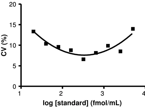
1
2
3
4
1
2
3
4
0
5
10
15
20
0
5
10
15
20
log [standard] (fmol/mL)
C
V
(
%
)
Figure 2.5. Precision profile demonstrates the Coefficient of Variation as a function of the concentration of cAMP standards.
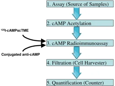
Cost issues and alternative technology
In this study we have demonstrated an improved method of cAMP detection that allows for the quick, accurate measurement of femtomole levels of cAMP. A flowchart of this method is shown in Figure 2.6. We have eliminated the need for secondary antibody and time-consuming separation techniques. By altering the mode of detection and assay format, we have increased throughput and excluded laborious steps inherent to the previous method. Although our research focus is on whole-cell and membrane assays of Gαs/OLF, Gαi/o and Gαq/11 coupled GPCRs, the method is applicable to any measurement of cAMP and can be easily adapted for cGMP.
$1,517 for 50 µCi (Perkin Elmer; NEX130050). Reagents suitable for dozens of radioiodinations cost less than $200, and 5 mCi of Na125I can be purchased from Perkin Elmer for $155 yielding a total cost of finished product for a single iodination of < $100/mCi, several-hundred-fold less than the commercial cost.
1. Assay (Source of Samples)
2. cAMP Acetylation
3. cAMP Radioimmunoassay
4. Filtration (Cell Harvester)
5. Quantification (Counter)
125I-cAMPscTME
Conjugated anti-cAMP
Figure 2.6. Schematic flowchart of the described method.
CHAPTER 3.
D1 RECEPTOR STIMULATION ACTIVATES ADENYLATE CYCLASE BUT
NOT PHOSPHOLIPASE C ABSTRACT
INTRODUCTION:
D1-like dopamine receptors, part of a family of proteins termed G protein coupled receptors (GPCRs), are characterized by their ability to stimulate adenylate cyclase via Gαs/olf coupling (Brown and Makman, 1972; Herve et al., 1993). Several studies have suggested, however, that D1 receptors may also couple to Gαq/11, and thus activate phospholipase C (Felder et al., 1989a; Jose et al., 1995; Wang et al., 1995; Yu et al., 1996; Zhen et al., 2005). The possibility of concurrent coupling of the D1 receptor to Gαq/11 and Gαs/olf, together with prior evidence of agonist-induced preferential G protein coupling, opens the door to studying important mechanistic questions about functional selectivity (Urban et al., 2007a).
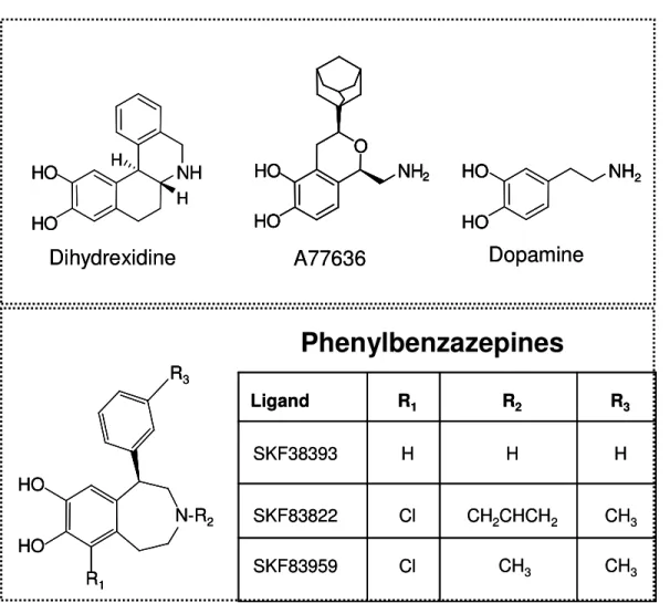
Early studies used the 6-OHDA lesioned rat model utilized SKF83959 to assess both behavioral endpoints and cAMP generation in striatal tissue. Gnanalingham et al. showed that SKF83959 (see Figure 3.1) stimulated adenylate cyclase in a seemingly dose-independent fashion. Significant stimulation at 10 µM compared to the intact unlesioned side was observed, however, at increased concentrations no dose-dependent increase was observed. Lesioned rats exhibited pronounced contralateral turning with treatment of SKF83959 at the highest concentration of the drugs tested but again, dose-independence confounded the interpretation of results (Gnanalingham et al., 1995).
to the D1 selective activation of phospholipase C. In the 6-OHDA rat model, SKF83959 by itself induced contralateral turning that was inhibited by SCH23390 (Cools et al., 2003).
R3 R1 HO HO N-R2 R3 R1 HO HO N-R2 R3 R1 HO HO N-R2 NH2 HO HO Dopamine NH2 HO HO NH2 HO HO NH2 HO HO Dopamine A77636 O HO HO NH2 A77636 O HO HO NH2 O HO HO NH2 O HO HO NH2 NH HO HO H H Dihydrexidine NH HO HO H H NH HO HO H H Dihydrexidine R3 R2 R1 Ligand
CH2CHCH2 CH3
SKF83822 CH3 CH3 SKF83959 Cl Cl H H H SKF38393 R3 R2 R1 Ligand
CH2CHCH2 CH3
SKF83822 CH3 CH3 SKF83959 Cl Cl H H H SKF38393
Phenylbenzazepines
Figure 3.1: Structures of the phenylbenzazepine SKF83959 and other ligands used in this study.
stimulates the production of inositol 1-phosphate (IP1), which was used as a downstream marker of activation for our studies.
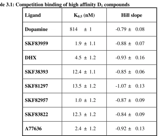
In this study, we demonstrate high affinity binding of ligands from several different structural families of D1 compounds. We also show that SKF83959 has partial agonist activity at adenylate cyclase using both hD1-HEK293 cell model and caudate rat tissue. A significantly greater potency was found in caudate rat tissue, whereas maximal efficacy was roughly half that in the HEK293 cell model. In addition, a lack of phospholipase C activation was observed for all D1 agonists tested.
MATERIALS AND METHODS
Dihydrexidine was synthesized according to procedures published previously (Brewster et al., 1990). The D1-selective antagonist [3H]SCH23390 was synthesized according to procedures published previously (Wyrick and Mailman, 1985). The compounds dopamine, A77636, SKF83822, SKF83959 and SKF38393 and all other reagents were purchased from Sigma-Aldrich (St. Louis MO) or Tocris Bioscience (Ellisville MO).
Cell Culture
penicillin, 50 µg/mL streptomycin (Gibco/Invitrogen), and supplemented with 10% fetal bovine serum at 37°C, 5% CO2. Following selection, the HA-hD1-pcDNA5/FRT HEK293 cells were maintained with 7.5µg/mL blasticidin and 100µg/mL hygromycin. The expression of hD1 receptor was titrated with tetracycline (0.05 µg/mL) to controllably induce a receptor Bmax of ~5 pmol/mg protein.
Competition Binding
Competition binding experiments were performed to assess the affinity (K0.5) of SKF83959 and other reference compounds for the hD1 receptor. HA-hD1-pcDNA5/FRT HEK293 cell membranes expressing wild-type receptor were incubated with [3H]SCH23390 (final concentration of 1µM) and varying concentrations of ligand in buffer (50mM HEPES, 4 mM MgCl2, 0.01% ascorbic acid, pH 7.4 with KOH). Total binding was defined as the amount of [3H]SCH23390 bound in the absence of a competing ligand. Non-specific binding was determined by binding in the presence of 1µM non-tritiated SCH23390. Experiments were setup in triplicate for each assay condition in 96 well plates. Reactions were terminated by filtration using a Packard 96 Filtermate Harvester (Packard BioScience Company; Meridian, Connecticut). Upon drying the plates at 50ºC for 1 hr, 35 µL of Packard MicroScint 20 scintillation cocktail was added to each well. Total CPM was assessed using a Packard TopCount NXT Microplate scintillation counter (Packard, Downers Grove, IL).
cAMP accumulation assay and RIA
and 100 µM propranolol (to block endogenous β1-adrenergic receptors) were added in a total volume of 100 µL in each well of a 48-well plate. The reaction was initiated by addition of hD1-HEK293 cell membranes. Plates were then vortexed briefly an incubated at 30°C for 15 min. The reaction was terminated with 500 µL 0.1 M HCl and stored at 4°C. Prior to transferring samples for the RIA, plates are spun down for 5 min at 3200 rpm using a RC-3B refrigerated centrifuge from Sorvall Instruments (H2000B rotor) to pellet cellular debris. Plates will keep indefinitely at 4°C following the assay. Where rat caudate tissue was needed, the protocol was the same as above with the addition of adenosine deaminase (1 U/mL) and specific antagonists to block off-target receptor activation: ketanserin (100 µM), yohimbine (10 µM), haloperidol (10 µM), prazosin (10 µM) and propranolol (10 µM).
Sample aliquots were taken from the cAMP accumulation plates, in addition, cAMP standards were added to the 96-well RIA plates. Sodium acetate (50 mM, pH: 4.75) was added, followed by a mixture of triethylamine/acetic anhydride (2:1). Plates were vortexed and [125I]-cAMP-ScTME added (28-30,000 DPM/50 µL) followed by anti-cAMP primary antibody diluted at 1:40 (in 1% BSA solution). Plates were incubated overnight at 4ºC. Biomagnetic particle (BMP) anti-Goat IgG [H&L] (Rockland, Gilbertsville, PA) were added and incubated for 1 hr at 4ºC. Plates were filtered using a 96-well plate harvester (Packard Filtermate 196) with ice cold acetate buffer (Harper and Brooker, 1975).
PLC assay
146 mM NaCl, 5.5 mM Glucose, 50 mM LiCl pH: 7.4) and added to the plates, which were incubated for 1 hr at 37ºC, 5% CO2. Cells were then incubated in lysis buffer for 30 min. Samples were transferred to the IP-One ELISA plate in addition to IP-1 standards, α-IP-1 mAb, and competitive IP1-HRP conjugate. The plate was incubated for three hours at room temperature followed by three washing steps. The colorimetric reaction was completed by addition of TMB (tetramethylbenzidine), incubated for 30 min, and terminated with stop solution. The 96-well plate was read at 450 nm/620 nm using a Vmax plate reader (Molecular Devices, Sunnyvale, CA).
Data Analysis
Competition binding and cAMP accumulation data were analyzed using sigmoidal nonlinear regression with a variable slope dose-response fit, yielding IC50 or EC50 values and Hill slopes values. Values for K0.5 of the compounds were calculated from the IC50 derived from regression analysis (Cheng and Prusoff, 1973). Data are expressed as means ± SEM. One way ANOVA was used to assess significance for potency and intrinsic efficacy between rat tissue and human cell models. Adenylate cyclase data were expressed relative to the percentage of cAMP produced by 10 µM dopamine.
RESULTS
Table 3.1: Competition binding of high affinity D1 compounds
Ligand K0.5 (nM) Hill slope
Dopamine 814 ± 1 -0.79 ± 0.08 SKF83959 1.9 ± 1.1 -0.88 ± 0.07
DHX 4.5 ± 1.2 -0.93 ± 0.16
SKF38393 12.4 ± 1.1 -0.85 ± 0.06 SKF81297 13.5 ± 1.2 -1.07 ± 0.13 SKF82957 1.0 ± 1.2 -0.87 ± 0.09 SKF83822 12.3 ± 1.2 -0.84 ± 0.09 A77636 2.4 ± 1.2 -0.92 ± 0.13
HEK-D1-wt Binding
-11 -10 -9 -8 -7 -6 -5 -4
-10 0 10 20 30 40 50 60 70 80 90 100 110 120 DA (n=3) SKF83959 (n=3) DHX (n=3) SKF38393 (n=3) SKF 81297 (n=3) SKF 82957 (n=3) SKF 83822 (n=3) A77636 (n=3) log [DRUG] % T o ta l B in d in g
Figure 3.2: Competition binding of compounds in hD1-HEK293 cells. Results are from three independent experiments performed in triplicate with ± SEM shown.
hD1-wt HEK293 cells (partial agonists)
-12 -11 -10 -9 -8 -7 -6 -5 -4 0 10 20 30 40 50 60 70 80 90 100 110 120 SKF83959 SKF38393 SKF 83822 Log [DRUG] % M a x D A
hD1-wt HEK293 cells (full agonists)
-12 -11 -10 -9 -8 -7 -6 -5 -4 0 10 20 30 40 50 60 70 80 90 100 110 120 DA A77636 DHX Log [DRUG] % M a x D A
Caudate rD1 (partial agonists)
-12 -11 -10 -9 -8 -7 -6 -5 -4 0 10 20 30 40 50 60 70 80 90 100 110 120 SKF83959 SKF38393 SKF83822 Log [DRUG] % M a x D A Caudate rD1 (full agonists)
-12 -11 -10 -9 -8 -7 -6 -5 -4 0 10 20 30 40 50 60 70 80 90 100 110 120 DA A77636 DHX Log [DRUG] % M a x D A
Figure 3.4: Adenylate cyclase activity in caudate tissue from Sprague-Dawley male rats. Non-linear regression curves were used for best fit to obtain potency (EC50) and intrinsic activity value. Data are expressed as % maximal stimulation by 10 µM DA Data are representative of 3 independent assays run in triplicate and each value represents the mean + S.E.M.
Table 3.2. Adenylate cyclase activation by key ligands.
hD1-HEK293 cells Rat caudate tissue Compound
Potency (nM) Efficacy (% DA) Potency (nM) Efficacy (% DA)
SKF83959 3.4 ± 0.33 47.1 ± 4.7 0.26 ± 0.56 27.7 ± 2.3 SKF83822 85.6 ± 0.25 63.2 ± 3.0 8.3 ± 0.13 67.2 ± 3.2 A77636 8.0 ± 0.16 96.8 ± 4.6 6.0 ± 0.17 81.7 ± 3.4 SKF38393 15.8 ± 0.26 42.7 ± 4.7 15.2 ± 0.23 43.5 ± 3.0
DHX 8.5 ± 0.18 81.9 ± 4.9 18.4 ± 0.23 76.6 ± 5.5
Data represent the means ± SEM (in nM and % maximal dopamine stimulation) from three independent experiments performed in triplicate.
IP-1 Stimulation
hD1-HEK-293 cells
-12 -11 -10 -9 -8 -7 -6 -5 -4 -3
0 10 20 30 40 50 60 70 80 90 100 110 120 130 Carbachol SKF 83959 SKF 83822 DHX A77636 Log [DRUG] % B /B O
Figure 3.5: IP1 stimulation via phospholipase C in HA-hD1-HEK293 cells. IP1 was used as a
surrogate for IP3 generation. Carbachol was used as a positive control and indicated the PLC
pathway was intact. Dose-response curves were generated using 7-8 concentrations of test compounds. Non-linear regression curves were used for best fit to obtain potency (EC50) and intrinsic activity value. Data are expressed as % maximal stimulation by 10 µM DA. Data are representative of 3 independent assays run in triplicate and each value represents the mean + S.E.M.
D1 receptors do not stimulate IP-3 formation in vitro
compared with stimulation of endogenous muscarinic receptors with carbachol. To insure that PLC machinery is intact within the hD1-HEK293 cells, we used ATP stimulate endogenous adenosine receptors in order to produce a positive response (data not shown). IP1, a downstream metabolite of IP3, is used as a surrogate marker of activity for IP3 stimulation. Activation of PLC is mediated via Gαq/11 and has been putatively linked to the D1 receptor, however, these results suggest no PLC activity mediated by the D1 receptor. Figure 3.6 shows treatment with dopamine alone and dopamine blocked with the specific D1 antagonist SCH23390. This reinforces the inability of D1 receptors in mediating PLC activity.
hD1 Specific blocking
-12 -11 -10 -9 -8 -7 -6 -5 -4 -3
0 10 20 30 40 50 60 70 80 90 100 110 120
DA
DA (W/SCH)
Log [DRUG]
%
B
/B
O
DISCUSSION
My data show that the D1 receptor appears not to signal through phospholipase C, and indeed, that SKF83959 specifically has no such activity in contrast to previous studies (Jin et al., 2003; Yu et al., 1996). Other studies of phenylbenzazepine compounds led to the hypothesis, alternatively, of the existence of a novel “D1-like” phospholipase C-coupled receptor (Friedman et al., 1997; Undie et al., 1994). These reports found potency values for phosphoinositide hydrolysis to be in the 1 mM-100 µM ranges. With such low potency, the possibility of an off-target action is a clear possibility that has not been adequately addressed. These earlier reports also noted that some phenylbenzazepine compounds that stimulated PLC, but not cyclase, as well as vice versa (Jin et al., 2003; Yu et al., 1996). Experiments have shown that knockout of D1 receptors abolishes the cyclase activity of the partial agonist SKF38393, yet not affecting SKF38393-induced inositol phosphate (IP) accumulation (Friedman et al., 1997). The generation of IP was found only at very high concentrations of agonist, suggesting to us that this effect was likely due to off-target effects. The hypothesis that there is an new “D1-like” PLC-coupled receptor is further weakened by any of the orphan GPCRs in the human or mouse genome have characteristics compatible with a D1 -like identity.
human D1 and rat D1 receptor show 94% homology (Li et al., 2004), and in addition a thorough examination of NIMH Psychoactive Drug Screening Program database shows minimal differences in binding profiles between the two species’ receptors (Roth, 2008). It is interesting that increased potency is found in rat caudate tissue where D1 receptor expression is at least one order of magnitude lower than in HEK293 cells (data not shown). One would expect increases in potency in populations with higher receptor expression, assuming the receptor:G Protein:effector stoichiometry remains the same. The possibility remains, however, that drug-specific differences in potency and efficacy exist, but this remains to be proven.
Evidence for a cAMP/PKA-independent signaling pathway can be found in studies with adenylate cyclase V deficient mice (Iwamoto et al., 2003; Lee et al., 2002). While 85-90% of cyclase activity is abrogated, locomotion is enhanced. It is not clear from this study, however, whether the behavioral effects are due to a non-cyclase dependent PLC pathway. My results clearly show intact cAMP signaling and hence PKA activation but no PLC response. Thus, in the hD1-HEK293 cell model, it seems clear that PLC activation is not dependent on PKA.
that SKF 83822 indeed has no PLC activity, but is a partial agonist for AC, albeit with lessened potency.
A potential limitation of my work is the use of IP1 as a surrogate marker for IP3. The short-lived existence of IP3 (~ 30 sec) makes reliable quantification difficult. On the other hand, previous work with Gαq-coupled receptors demonstrated that IP1 is an excellent surrogate for IP3 formation (Trinquet et al., 2006). Consistent with this, PLC activation by carbachol and ATP lead to IP1 generation that is proportional to the formation of IP3. Thus, this is unlikely a confound in the current experiments.
Several studies have shown concurrent Gαq/11 and Gαs/olf coupling to the D1 receptor (Mannoury la Cour et al., 2007; Panchalingam and Undie, 2000; Wang et al., 1995), and some reports indicate differential coupling upon addition of SKF83959 (Rashid et al., 2007). All four members of the Gαq/11 family (αq, α11, α14, α16) have been shown to activate PLC-β isoforms, but not PLC-γ, PLC-δ or PLC-ε (Hepler et al., 1993; Kozasa et al., 1993; Lee et al., 1992). There is clear evidence from other receptor systems, namely the A1 adenosine receptor, that agonists can induce specific receptor conformations leading to selective activation of Gs, Gi, or Gq proteins (Cordeaux et al., 2004). Other examples of such functional selectivity are plentiful (Berg et al., 1998; Bonhaus et al., 1998; Brink et al., 2000; Gazi et al., 2003; Harikumar and Chattopadhyay, 1999) as has been recently reviewed (Urban et al., 2007a).
with the intent of changing membrane dynamics (and thus the receptor conformation) in such a way that Gq/PLC was favored over Gs/adenylate cyclase signaling (Panchalingam and Undie, 2005). The use of deoxycholate, while useful in demonstrating the possibility of selective Gq-coupling, does not suggest probable G protein coupling to the D1 receptor in vitro or in vivo.
Early evidence has suggested SKF82526, a D1 full agonist, stimulates PLC in rat renal cortical membranes (Felder et al., 1989b). This effect was blocked by the selective D1 antagonist SCH23390, whereas the α-adrenergic antagonists prazosin and phentolamine had no effect. Additionally, the D2 agonist LY 171555 did not induce IP release. The signaling properties of renal cortical membranes may be similar to my HEK293 cell model, yet we find no evidence of IP release.
noted that the measurement of intracellular Ca2+ currents is not necessarily directly translatable from PLC stimulation. Similar data have been found in other studies. Thus, there is a strong possibility that a non-PLC Ca2+ mechanism exists (Lin et al., 1995).
Intracellular calcium release is a critical and seemingly ubiquitous effector, which is released upon activation of the IP3 receptor at the ER. Studies in primary cultures of neocortical and hippocampal cells have demonstrated that intracellular calcium may be released with treatment of D1/D5 agonists, however, this was found to be dependent on priming from other Gαq/11-linked receptors such as glutamine, serotonin, muscarinic and adrenergic receptors. Not surprisingly, forskolin stimulation of cAMP alone was not sufficient to induce intracellular calcium release (Lezcano and Bergson, 2002). Additionally, the authors found that D1/D5 mediated calcium release was not observed in striatal tissue.
Phospholipase C may be activated by both Gαq and Gβγ; while in vitro studies indicate high potency activation of PLC by Gαq its clear that Gβγ still plays a role.PLC-β has distinct sites for Gαq and Gβγ activation and thus can be synergistically activated (Runnels and Scarlata, 1999). The composition of specific Gβγ subunits appears also to affect its potency for PLC-β (Boyer et al., 1994). This potential Gβγ-mediated regulation by not only D1-like receptors, but among GPCR signaling tone, further complicates interpretation.
facilitating D1 receptor signaling. One example is β-arrestin 2; traditionally thought of as playing a role in GPCR desensitization, it has been found to signal through Akt and PP2A, bypassing Gα signaling mechanisms (Beaulieu et al., 2005). This G protein-independent mode of signaling provides a new way of understanding the complex interactions among signaling partners, its potential role in prolonged stimulation of receptors, warrants further analysis.
CHAPTER 4: SUMMARY OF FINDINGS AND FUTURE DIRECTIONS SUMMARY OF FINDINGS
Parkinson’s disease is thought to affect anywhere from 500,000 to 1 million people every year in the US, assuredly more throughout the world. The etiology of the disease remains elusive for the vast majority of non-familial cases although we gain better understanding everyday of dopaminergic signaling. The studies in this thesis offer a better understanding of D1 receptor effectors in two ways, by showing negative evidence of PLC activation via D1 mechanisms and also by demonstrating specific differences in signaling between rat striatal tissue and the hD1-HEK293 cell model.
Critical to the understanding of functional selectivity is that multiple functional pathways couple to the receptor, allowing both differential potencies and intrinsic efficacies. The D1 dopamine receptor has of course been long known to couple to adenylate cyclase, however, other than GPCR internalization mechanisms, there is a dearth of other independent functional pathways known to couple to it.
My phospholipase C results show little to no activation by dopamine and select D1 agonists. If these results hold true in future studies under in vivo conditions, this would sharpen understanding toward the development of D1 agonists; my findings limit PLC-based hypotheses that sought to explain therapeutic side effects, or potential exploitation of this functional pathway for therapeutic gain. It seems clear that in this model PLC is not dependent on the cAMP/PKA pathway.
Given that Gαq coupling to D1 has only been demonstrated in rat neuronal tissues and that PLC signaling is intact in HEK293 cells, it seems likely that two possibilities exist. Firstly, species specific differences within the third intracellular loop of the D1 receptor lead to inefficient Gαq coupling in the human form, however this remains to be studied. The second possibility is that Gαq activation requires priming of Gαq-coupled receptors. This observation has been seen in several studies; however, the protein(s) involved have yet to be resolved. One report indicated direct interaction between the D1 receptor and calcyon and its role in potentiating calcium release, yet this report was retracted when the authors found no direct interaction in addition to minimal increases in calcium release when calcyon was present (Lezcano et al., 2000).
FUTURE DIRECTIONS
(postsynaptic density) proteins, and other chaperones, while being dependent on coupling to Na+, K+ and Ca+ ion channels to relay action potentials to neighboring post-synaptic neurons. This would appear to be the necessary next step in studying PLC dependence on D1 receptor activation. Transgenic mouse models are also an important tool to be considered, as in vivo D1-mediated PLC activation has not yielded a clear result.
An important step in determining the mechanisms involved with D1 signaling is a better understanding of which G proteins are coupled to the receptor. As noted in Chapter 2, previous studies have shown Gαq coupling in rat striatal tissue (Mannoury la Cour et al., 2007; Panchalingam and Undie, 2000; Wang et al., 1995), however, this has not been observed in HEK293 cells. This particular approach has been historically problematic due to difficulties in developing epitopes against specific G proteins, leading to high rates of false positives.
Thus calcium does not seem to be an appropriate candidate for the study of functional selectivity, due to its PKA/PKC dependence.
G protein-receptor kinases (GRKs) are another viable pathway for study of functionally selective compounds due to direct interaction with the receptor. These proteins act to phosphorylate GPCRs upon their activation, in turn this leads to desensitization of the receptor, preventing further interaction with G proteins. This is the first in a complex series of steps leading to GPCR internalization, which may involve arrestins, lipid rafts and caveolae. The D1 receptor has been shown to couple to GRK2, GRK3, GRK4, GRK5 and GRK6 (Fraga et al., 2006; Tiberi et al., 1996; Watanabe et al., 2002). The theoretical possibility remains that different classes of agonists may differentially influence GRK interaction with the receptor. Indirect evidence of this phenomenon has been demonstrated previously (Ryman-Rasmussen et al., 2007).
pathway shows promise but further studies must be carried out to gain a clearer understanding of signaling partners.


![Figure 2.2: Chromatogram of radioiodination. [Bottom tracing] shows injection of cAMP-Sc-TME precursor alone using conditions as described in Methods using 254 nm UV detection](https://thumb-us.123doks.com/thumbv2/123dok_us/8322980.2206425/36.918.227.715.98.503/chromatogram-radioiodination-injection-precursor-conditions-described-methods-detection.webp)

![Figure 2.4. Measurement of D 1 dopamine receptor-mediated cAMP accumulation utilizing [Left panel] secondary antibody-PEG assisted RIA method, and [right panel] our new RIA method (primary antibody conjugated to beads](https://thumb-us.123doks.com/thumbv2/123dok_us/8322980.2206425/42.918.153.796.110.355/measurement-dopamine-receptor-mediated-accumulation-utilizing-secondary-conjugated.webp)