CRANIOFACIAL CHARACTERIZATION OF PATIENTS WITH MARFAN SYNDROME
Christian M. Johnson
A thesis submitted to the faculty at the University of North Carolina at Chapel Hill in partial fulfillment of the requirements for the degree of Master of Science in the School of Dentistry
(Orthodontics).
Chapel Hill 2017
Approved by:
Sylvia Frazier-Bowers Luiz Pimenta
ABSTRACT
Christian M. Johnson: Craniofacial Characterization of Patients with Marfan Syndrome (Under the direction of Sylvia Frazier-Bowers)
Background: Marfan Syndrome (MFS) is a life-threatening connective tissue disorder with an often elusive diagnosis. Diagnosis is based on clinical findings outlined in the Ghent criteria which define hallmark features of the syndrome in the cardiovascular, ocular, and skeletal systems. The morbidity and mortality associated with MFS warrant timely diagnosis and intervention that can improve long-term prognosis. Previous research has highlighted the diagnostic value of craniofacial features in diagnosis; accordingly the aim of this study was to investigate craniofacial and dentoalveolar features in child, adolescent, and young adult patients with MFS. We hypothesized that a distinct craniofacial morphology exists for patients with MFS that can be described quantitatively and qualitatively. Methods: Twenty subjects with a positive diagnosis of MFS were recruited for this study (N=20). Craniofacial anthropometric measurements were made on each subject and compared to established norms of age- and sex-matched controls. The test measurements were compared to the control measurements by
ACKNOWLEDGEMENTS
TABLE OF CONTENTS
LIST OF TABLES ... viii
LIST OF FIGURES ... ix
LIST OF ABBREVIATIONS ...x
LIST OF SYMBOLS ... xi
REVIEW OF THE LITERATURE ...1
Marfan Syndrome (MFS) ...1
Etiology of MFS ...2
Epidemiology of MFS...2
Diagnostic Criteria for MFS ...3
Challenges to Diagnosis ...5
Diagnostic Criteria in Clinical Practice ...7
Diagnosis and Phenotype in Children ...9
Benefits of Early Diagnosis ...11
Consequences of Misdiagnosis ...12
Management of MFS ...12
Craniofacial Characterization of MFS ...13
Craniofacial Characterization of other Syndromes ...15
Conclusion ...16
References ...18
Introduction ...20
Methods...22
Sample...22
Procedures ...23
Statistical Analysis ...24
Results ...25
Demographic and Biometric Results ...25
Intraoral Examination Results ...26
Assessment of Photographs ...26
Craniofacial Anthropometric Measurements ...26
Z-score Distribution by Group ...27
Discussion ...27
Conclusions ...33
References ...35
Tables ...37
Figures...38
LIST OF TABLES
LIST OF FIGURES
Figure 1 – Frequency of z-score distribution for sample population vs.
normal population for facial width ...38
Figure 2 – Frequency of z-score distribution probands vs. non-probands for facial width ...38
Figure 3 – Cardiovascular Anomalies...38
Figure 4 – Ocular Anomalies ...39
Figure 5 – MFS Craniofacial Features ...39
Figure 6 – Morphological Height of Face...39
Figure 7 – Physiognomical Height of the Upper Face ...40
Figure 8 – Mandible Height ...40
Figure 9 – Lower Face Height ...40
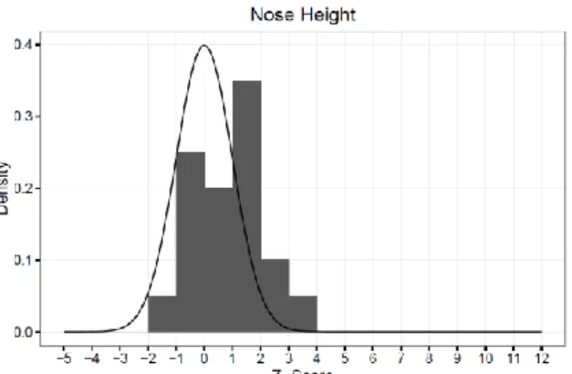
Figure 10 – Nose Height ...41
Figure 11 – Width of Nose...41
Figure 12 – Biocular Width ...41
Figure 13 – Intercanthal Width ...42
Figure 14 – Width of the Head ...42
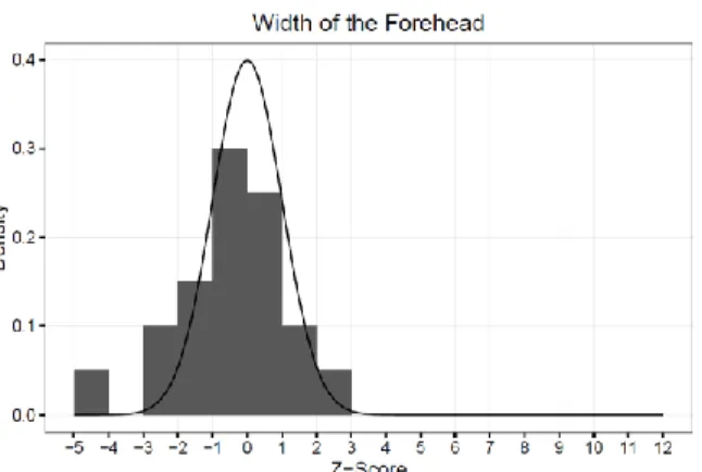
Figure 15 – Width of the Forehead ...42
Figure 16 – Width of the Face ...43
LIST OF ABBREVIATIONS MFS Marfan Syndrome
FBN1 Fibrillin-1
LIST OF SYMBOLS
© Copyright Symbol
A REVIEW OF THE LITERATURE
Marfan Syndrome
Marfan Syndrome (MFS) is a life-threatening connective tissue disorder characterized by multi-system organ involvement. The syndrome was first described by Dr. Antoine Marfan, a pediatrician, in 1896 at a meeting of the Medical Society of Paris1. He presented a five-year-old girl with disproportionately long limbs that he described as arachnid-like1. It wasn’t until almost fifty years later that the other hallmark cardiovascular and ocular manifestations of the
syndrome were described2.
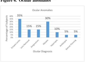
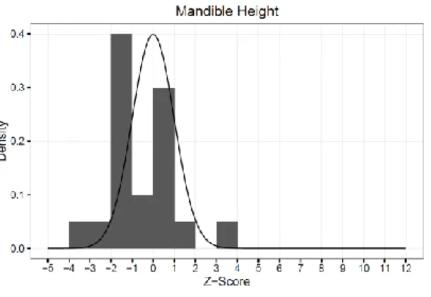
The cardinal features of MFS occur in the cardiovascular, ocular, and skeletal systems. The morbidity and mortality associated with the clinical presentation of MFS highlight the importance of early diagnosis and management3. By far, the characteristics found within the cardiovascular system are the primary source of morbidity and early mortality and include dilatation of the aorta which can progress to aortic dissection/rupture, mitral valve prolapse with/without regurgitation, tricuspid valve prolapse, and enlargement of the proximal pulmonary artery3. The hallmark feature within the ocular system is ectopia lentis, dislocation of the lens of the eye, and it is observed in approximately 60% of affected individuals3. Of increasing concern and morbidity, patients with this syndrome are at increased risk for retinal detachment,
extremities being disproportionately long for the size of the trunk, and patients being abnormally tall for their age3. Other skeletal features are observed in the craniofacial region and include a long and narrow face, downward slanting of the palpebral fissures, malar hypoplasia, and a retruded chin (retrognathia); intraorally, a highly-arched palate and dental crowding have been described3. While features of MFS are present in other organ systems, the highly diagnostic and discriminating features are found within the cardiovascular, skeletal, and ocular systems.
Etiology of MFS
MFS is caused by a mutation in the gene coding for the protein fibrillin-1 (FBN1) resulting in reduced amounts of functional FBN13, 4. Fibrillin-1 binds to other proteins to form microfibrils which are one of the fibers that provide flexibility and strength to connective tissue4. Microfibrils also store growth factors responsible for controlling growth and repair of tissues and organs throughout the body4. Decreased amounts of functional FBN1 lead to a decreased
formation of microfibrils, resulting in the release of excess growth factors, a decrease in tissue elasticity, tissue overgrowth, and tissue instability4. The sequela of these events give rise to the clinical presentation of MFS, as the structural integrity of connective tissue is compromised2.
MFS is an autosomal dominant disorder with 75% of new cases being the direct result of inheritance from a parent, and approximately 25% of new cases result from a new mutation3, 4.
Epidemiology
Diagnostic Criteria for Marfan Syndrome
The diagnosis of MFS is a clinical diagnosis made by the observation of cardinal features in various organ systems, the presence of a family history of MFS, and/or genetic testing for a mutation of the FBN1 gene or other genes. The most recent and revised diagnostic criteria, The Revised Ghent Nosology for the Marfan Syndrome, were published in 2010 by Loeys et al.6. The need for revision from the previous criteria7 arose from the observation more emphasis needed to be placed on hallmark features of MFS in the cardiovascular and ocular systems, and more consideration needed to be given to alternative diagnoses6. The alternative diagnoses have clinical features that overlap with MFS but may also include features with a higher morbidity and/or mortality than MFS6. In the revised criteria, more weight is given to two distinct features of MFS, aortic root aneurysm/dissection and ectopia lentis6. Diagnostic criteria exist for other organ systems, as discussed below, but the diagnostic finding of both aortic root
enlargement/dissection and ectopia lentis confers the diagnosis of MFS in the absence of discriminating features for other syndromes6. While this is not the only pathway for being diagnosed, it highlights an important change to the diagnostic criteria. The other pathways are outlined below, and include clinical findings in other organ systems, a family history of MFS and a positive mutation in the FBN1 gene6. Additionally, there are features that are commonly observed in MFS that do not independently discriminate for MFS6. If these features exist in combination, the diagnosis of systemic involvement for MFS is given; if there is systemic involvement and another hallmark feature, the diagnosis of MFS can be inferred6. Summary of the Diagnostic Algorithm for MFS below6:
Aortic diameter Z≥2 or dissection and ectopia lentis
Aortic diameter Z≥2 or dissection and mutation of FBN1
Aortic diameter Z≥2 or dissection and a systemic score ≥7
Ectopia lentis, mutation of FBN1, and known aortic dilatation (Z≤2)
In the presence of a family history of MFS, a patient can be diagnosed with MFS if the patient has:
Ectopia lentis
Systemic score ≥7
Aortic diameter z≥2 or dissection if ≥20 years old
Aortic diameter z≥3 or dissection if ≤20 years old
Aortic root enlargement/dilatation is defined for MFS as having an aortic root diameter measurement, at the level of the sinuses of Valsalva, with a Z-score (Z) ≥2 when
standardized for age and body size6. There are caveats to the above diagnostic criteria to rule out other syndromes. See the revised criteria for these caveats6.
The systemic score is calculated by summation of points, and a score ≥7 denotes systemic involvement:
Wrist and thumb sign—3 points (wrist or thumb sign 1 point)
Pectus carinatum—2 points (pectus excavatum or chest asymmetry 1 point)
Hindfoot deformity—2 points (plain pes planus 1 point)
Pneumothorax—2 points
Dural ectasia—2 points
Reduced upper segment:lower segment ratio and increased arm:height ratio and no severe scoliosis—1 point
Scoliosis or thoracolumbar kyphosis—1 point
Facial features (must have 3/5): dolichocephaly, enophthalmos, downslanting palpebral fissures, malar hypoplasia, retrognathia—1 point
Skin striae—1 point
Myopia > 3 diopters—1 point
Mitral valve prolapse—1 point
As can be seen from the diagnostic criteria, emphasis is placed on those clinical features that carry the highest morbidity and mortality. All skeletal features fall under systemic
involvement even though they are some of the most striking physical features. However, they don’t yield high diagnostic value independently. Craniofacial features, the focus of this research, are included in the diagnostic criteria, but only reach diagnostic significance when multiple facial features are observed in combination.
Challenges to Diagnosis
As previously discussed, the diagnosis of MFS is primarily a clinical diagnosis and relies on the ability of a clinician to observe/identify features specific to the syndrome. Diagnosis can by highly elusive due to the high degree of variability in clinical presentation, the age-dependent nature of some manifestations, and the host of differential diagnoses that exist3, 6, 8.
of MFS. As an example, a family in this study had several members who were known carriers of a FBN1 mutation9. Some had been diagnosed with ectopia lentis and some children within the family required surgical repair of mitral valve prolapse9. These clinical findings would give clinical suspicion for a possible diagnosis of MFS9. However, expressivity was found to be different within the family and there was inability to predict who would be more severely
affected solely on the basis of a known FBN1 mutation9. This finding is confirmed by Judge and others who noted that even when members of a family share the same mutation, phenotypic variation is prominent5, 8. A different family in the Summers’ study demonstrated an occurrence of a de novo mutation in the FBN1 gene in a male child resulting in MFS9. The sibling of this proband had musculoskeletal features consistent with MFS, but the features alone did not qualify him for a positive diagnosis of MFS9. Genetic testing identified the de novo mutation in the proband and confirmed the absence of the mutation in the proband’s parents and sibling9. Identification of the mutation as being spontaneous rather than inherited provided reassurance the sibling of the proband likely did not have MFS9.
to allow for timely surgical intervention to slow the progression of cardiovascular defects10. If a patient with MFS does not exhibit enough physical features to be diagnosed in the absence of genetic testing, the abnormalities associated with the syndrome can progress undetected until the abnormality reaches clinical significance10. Within the skeletal system, the wrist/ thumb sign and arm span/height ratio >1.05 were more prevalent in the Asian patients10. However, in the ocular system, ectopia lentis was more common in Caucasian patients10. This research not only demonstrated the phenotypic variability that exists between ethnicities, it also highlighted the importance of assessing the applicability of the diagnostic criteria to patients of different ethnicities.
Diagnostic Criteria in Clinical Practice
Researchers have evaluated the application of the diagnostic criteria for MFS to populations with and without MFS to determine how well the criteria discriminates between affected and non-affected individuals. This is important as to lessen the incidences of false positives and false negatives in the diagnosis of MFS.
Sponsellar and colleagues investigated ways in which patients were recognized as
needing a referral for evaluation of MFS and the prevalence of current diagnostic features of in a population of subjects with and without MFS11. They examined a population of patients
attending care at a pediatric orthopedics clinic and a pediatric sports medicine clinic11. The population consisted of patients with a confirmed diagnosis of MFS (n=183) and those without MFS (n=1257)11. They examined the prevalence of physical, mostly skeletal, diagnostic features for MFS in both cases and controls11. Overall, the physical diagnostic features were more
physical features of MFS11. When evaluating sensitivity and specificity of physical features, craniofacial features were the most sensitive for MFS and when combined with the thumb and wrist sign, the most specific11. This demonstrates singular physical features of MFS are relatively prevalent within the general population, but that observed in isolation do not infer a diagnosis of MFS. However, when multiple physical features are observed in combination, the suspicion for MFS increases. Included in their research was identifying the individual
responsible for initial suspicion for MFS resulting in referral for evaluation. In 26% of cases it was a pediatrician, a family member in 21%, ophthalmologist in 14%, family practitioner in 8.4%, orthopedist in 7.7%, and another contact (undefined) in 23% 11. As can be seen from these findings, it is prudent practitioners from different specialties be knowledgeable about MFS.
increased the odds of correctly identifying a patient with MFS12. They concluded the facial features were more specific than sensitive and could be used as a tool to prioritize patients for referral for evaluation for MFS; however, they should not be relied upon as the sole metric for initial screening12.
Diagnosis and Phenotype in Children
As mentioned, the diagnosis of MFS can be challenging due to the age-dependent nature of some physical features; these features do not manifest or become distinct for MFS until after significant growth has occurred. Research emphasis has been placed on ways to diagnose MFS sooner in life as this leads to earlier management and treatment. Several researchers have studied MFS in children/adolescents specifically to understand the phenotypic presentation in this population.
Lipscomb and colleagues evaluated 40 subjects with MFS less than 16 years of age8. Of the 40 evaluated, 10 were index cases with no prior family history of MFS8. This percentage of index cases, 25%, is in agreement with the global description of all patients, children and adults, with MFS8. For the index cases, the average age at diagnosis was 11.4±3.95 years and for non-index cases, 7.31±5.23 years8. In regard to craniofacial features of MFS, 36 of 40 subjects were described as having a highly-arched palate and 33 of 40 were noted to have mid-face hypoplasia, micrognathia, and down-sloping palpebral fissures8. When evaluating the cardiovascular system, aortic root dilatation was observed in 17 patients (42.5%) but none of the subjects had
monitor aortic root dilatation; a delay in detection can potentially lead to adverse clinical outcomes8. As can be seen, index cases tended to be diagnosed later in childhood resulting in missed years of echocardiographic examination and delayed prophylactic intervention to manage aortic root dilatation. It highlights the need for diagnostic criteria to help identify index cases earlier in life.
presence of ectopia lentis14. The two most discriminating features leading to a diagnosis of MFS were aortic diameter >3SD above the mean and presence of ectopia lentis, both of which were given more weight in the most recent diagnostic criteria for MFS14. The findings in this study are in agreement with those of the Lipscomb study which showed probands are diagnosed later in life; they also demonstrated probands were more severely affected clinically.
Benefits of Early Diagnosis
Consequences of Misdiagnosis
There are several consequences of misdiagnosis of MFS for a patient who indeed does not have MFS. Included are inappropriate/improper surveillance and or treatment that can be costly, discrimination by employers, or discrimination and/or difficulty with obtaining
insurance9. Alternatively, a missed diagnosis of MFS infers more severe consequences as premature death can result from an aortic dissection.
Management of Marfan Syndrome
The multi-system nature of MFS dictates a multidisciplinary approach to comprehensive management. For most patients, this requires a team including a cardiologist, cardiothoracic surgeon, ophthalmologist, orthopedist, and geneticist3. Annually, these patients are monitored with an echocardiogram to monitor aortic dilatation and an ophthalmologic exam to assess for ectopia lentis, cataracts, glaucoma, or retinal detachment3.
When medical professionals encounter these patients outside of the specialties outlined above, it is common for patients with MFS to present with eyeglasses for correction of myopia 3, 6. Aortic dilatation is usually managed by medications to reduce the hemodynamic stress placed on the aortic wall such as beta blockers 3, 6. For dental professionals, these patients may require antibiotic prophylaxis prior to invasive dental treatment in the presence of mitral valve or aortic valve regurgitation3.
patients are cautioned to avoid substances that stimulate the cardiovascular system such as caffeine or decongestants3.
Although MFS is considered a rare disease, it is likely a medical professional will encounter a patient with MFS during his/her professional career. Awareness of MFS and its clinical presentation is of importance as it may have implications for the medical treatment being provided. In addition, the findings of medical practitioners from different specialties may be instrumental in enhancing the diagnostic criteria for MFS, increasing awareness, and developing new research avenues.
Craniofacial Characterization of Marfan Syndrome
Research continues to focus on isolating and describing clinical/physical features that discriminate for MFS. One such area of research devoted to this is qualitatively and
quantitatively describing craniofacial and dental features of patients with MFS.
base17. A measurement greater than 80° is indicative of a prognathic mandible and less than 80° indicative of a retrognathic mandible17. For the ANB measurement, a normal Class I relationship between the maxilla and mandible exists when the angular measurement is between 2°-4°17. A measurement greater than 4° is indicative of a Class II jaw relationship and less than 2° of a Class III jaw relationship17. They also evaluated vertical skeletal relationships by assessing the S-N/Go-Gn angle; 32°=normal; greater than 32°=long face; and less than 32°=short face17. Of their population of 26 subjects with MFS, 84% were maxillary retrognathic, 88% were mandibular retrognathic, and 81% were both maxillary and mandibular retrognathic16. With respect to ANB, 44% had a normal ANB measurement while 48% had a measurement indicative of a Class II jaw relationship16. When evaluating the vertical skeletal relationship, 72% of the subjects fell into the long face category16. These findings are in agreement with the skeletal diagnostic features of MFS previously described which include dolichocephaly (long-face) and mandibular retrognathia. Their research was novel because is quantitatively described
temporomandibular disorders and previous orthodontic treatment18. Disorders of the
temporomandibular joint were reported by 39.2% of the MFS subjects and 62% had previous orthodontic treatment18. It is expected that all patients with MFS would be encouraged to receive routine dental care with a primary dentist, but it is also likely these patients will be evaluated and/or treated by dental specialists for either temporomandibular disorders or for orthodontic treatment. As such, dental professionals may be in a unique position to identify patients with MFS and aid in further research on craniofacial and dental characterization of patients with MFS.
Craniofacial Characterization of other Syndromes
There are several medical syndromes that include craniofacial anomalies. The facial anomalies associated with each syndrome can be highly discriminating for the syndrome and as such, are used in diagnosis. Leslie Farkas recognized this, and sought to develop a method for evaluating and quantifying craniofacial characteristics of syndromes with distinct facial features such as Down’s Syndrome and Apert’s Syndrome19, 20. He found the facial features observed were described qualitatively in the literature but without quantification and/or comparison to controls/normative data19, 20.
His quantitative investigation of the facial anatomy of patients with Down’s Syndrome included making select craniofacial anthropometric measurements on a test population of subjects with Down’s Syndrome19. He selected the measurements based on previous research that identified specific facial features commonly observed in patients with Down’s Syndrome22. The measurements from the subjects with Down’s Syndrome were then compared to normative data by calculating a z-score for each measurement19. He categorized the measurements based on z-score as listed below19:
z score±1: optimal facial measurement
z score <-1 but >-2 or >1 but <2: normal facial measurement
z score <-2 or >2: subnormal or supernormal facial measurement
z score <-3 or >3: severe facial abnormality
The results from this study provided quantitative evidence for qualitatively described differences in craniofacial morphology of patients with Down’s Syndrome. This method of research provided a framework for future research in quantitatively describing facial features of syndromes with known craniofacial anomalies19.
Conclusion
clinical features of MFS in the child/adolescent population. As such, this research project aims to quantitatively and qualitatively describe craniofacial features in child/adolescent patients with MFS by, 1) making select craniofacial anthropometric measurements on each subject and
comparing these measurements to normative data and 2) evaluating clinical photographs of subjects with MFS to identify discriminating features.
REFERENCES
1. Marfan, A.B., Un cas de deformation congenitale des quatre membres, plus prononcee aux extremities,
caracterisee par l"allongement des os avec un certain degre d'amincissement. Bul Soc Chir Paris,
1896. 13: p. 220-225.
2. Gott, V.L., Antoine Marfan and his syndrome: one hundred years later. Md Med J, 1998. 47(5): p.
247-52.
3. Dietz, H.C., Marfan Syndrome, in GeneReviews(R), R.A. Pagon, et al., Editors. 1993: Seattle
(WA).
4. Marfan Syndrome. April 30, 2015 [cited 2015 May 2]; Available from:
http://ghr.nlm.nih.gov/condition/marfan-syndrome.
5. Judge, D.P. and H.C. Dietz, Marfan's syndrome. Lancet, 2005. 366(9501): p. 1965-76.
6. Loeys, B.L., et al., The revised Ghent nosology for the Marfan syndrome. J Med Genet, 2010.
47(7): p. 476-85.
7. De Paepe, A., et al., Revised diagnostic criteria for the Marfan syndrome. Am J Med Genet, 1996.
62(4): p. 417-26.
8. Lipscomb, K.J., J. Clayton-Smith, and R. Harris, Evolving phenotype of Marfan's syndrome.
Arch Dis Child, 1997. 76(1): p. 41-6.
9. Summers, K.M., et al., Challenges in the diagnosis of Marfan syndrome. Med J Aust, 2006.
184(12): p. 627-31.
10. Franken, R., et al., Clinical features differ substantially between Caucasian and Asian populations of
Marfan syndrome. Circ J, 2013. 77(11): p. 2793-8.
11. Sponseller, P.D., et al., Improving clinical recognition of Marfan syndrome. J Bone Joint Surg
Am, 2010. 92(9): p. 1868-75.
12. Ting, B.L., et al., The diagnostic value of the facial features of Marfan syndrome. J Child Orthop, 2010. 4(6): p. 545-51.
13. Mueller, G.C., et al., Impact of age and gender on cardiac pathology in children and adolescents with
Marfan syndrome. Pediatr Cardiol, 2013. 34(4): p. 991-8.
14. Stheneur, C., et al., Study of phenotype evolution during childhood in Marfan syndrome to improve
clinical recognition. Genet Med, 2014. 16(3): p. 246-50.
15. Willis, L., G.E. Roosevelt, and A.T. Yetman, Comparison of clinical characteristics and frequency
of adverse outcomes in patients with Marfan syndrome diagnosed in adulthood versus childhood.
Pediatr Cardiol, 2009. 30(3): p. 289-92.
16. De Coster, P., et al., Craniofacial structure in Marfan syndrome: a cephalometric study. Am J Med
17. Steiner, C.C., [Importance of cephalometry in orthodontic treatment]. Inf Orthod Kieferorthop, 1969. 1(2): p. 3-12 passim.
18. Staufenbiel, I., et al., Periodontal conditions in patients with Marfan syndrome - a multicenter case
control study. BMC Oral Health, 2013. 13: p. 59.
19. Farkas, L.G., et al., Surface anatomy of the face in Down's syndrome: linear and angular
measurements in the craniofacial regions. J Craniofac Surg, 2001. 12(4): p. 373-9; discussion 380.
20. Farkas, L.G., J.C. Kolar, and I.R. Munro, Craniofacial disproportions in Apert's syndrome: an
anthropometric study. Cleft Palate J, 1985. 22(4): p. 253-65.
21. Farkas, L.G., Anthropometry of the head and face in medicine. 1981, New York: Elsevier. xxii,
293 p.
22. Farkas, L.G., J.C. Posnick, and T. Hreczko, Anthropometry of the head and face in 95 Down
CRANIOFACIAL CHARACTERIZATION OF MARFAN SYNDROME
Introduction
Marfan Syndrome (MFS) is a life-threatening connective tissue disorder characterized by multi-system organ involvement with cardinal features occurring in the cardiovascular, ocular, and skeletal systems1. It is caused by a mutation in the gene coding for the protein fibrillin-1 (FBN1)2. The morbidity and mortality associated with the clinical presentation of MFS highlight the importance of early diagnosis and management. The characteristics found within the
cardiovascular system (aortic dilatation and/or aortic dissection) are the primary source of morbidity and early mortality1, and it has been noted that undiagnosed adult patients have well-established cardiovascular pathology and suboptimal clinical outcomes3. Within the ocular system, the hallmark feature is ectopia lentis (dislocation of the lens of the eye), a condition seen in approximately 60% of patients1. While the features in the skeletal system are not a major source of morbidity/mortality and do not account for sudden or premature death, they are fundamental in diagnosis4. Skeletal features are the most striking physical features of MFS and may lead to a suspicion for this syndrome in undiagnosed patients. These features include excessive linear growth of long bones, increased arm span to height ratio, and distinct
craniofacial features (dolichocephaly, malar hypoplasia, enophthalmos, retrognathia, and down-slanting palpebral fissures)1. While features of MFS occur in other organ systems, the
The diagnosis of MFS is a clinical diagnosis, and the diagnostic criteria are outlined in
The Revised Ghent Nosology for the Marfan Syndrome published by Loeys et al. in 2010 (see Appendix 1). The criteria set forth objective clinical requirements that must be observed to infer the diagnosis of MFS; consideration is given to clinical findings in various organ systems, a family history of MFS, and a positive mutation of the FBN1 gene 5.
Prior research has highlighted the value of early diagnosis as a prerequisite for improved clinical outcomes 3; however, diagnosis can be highly elusive due the high degree of variability in clinical presentation and the age-dependent nature of some features 1, 5, 6. Additionally, the hallmark features, aortic dilatation/aneurysm and ectopia lentis, require more advanced
diagnostic tests than are routinely prescribed for the general population7. This underscores the need for a method to objectively assess other readily observable features as a screening tool for MFS. Studies have evaluated the application of the diagnostic criteria in populations consisting of MFS subjects and non-MFS subjects to determine how well the criteria discriminate between affected and non-affected individuals; these studies found that facial features, as outlined in the MFS diagnostic criteria, are valuable in diagnosis. Ting and colleagues utilized frontal and lateral photographs to evaluate facial features of patients with a confirmed diagnosis of MFS and age- and sex-matched controls8. Three physicians with extensive experience treating patients with MFS evaluated all photographs for the presence of the five diagnostic craniofacial features8. The physicians were able to discriminate between subjects and controls with an accuracy of 72.6% solely on the basis of assessing facial features8. Similarly, Sponseller and colleagues evaluated the diagnostic sensitivity and accuracy of the presence of not only craniofacial
craniofacial features with a positive thumb sign provided the highest diagnostic accuracy 7. Of equal importance, was their finding that 19% of MFS patients had zero or one skeletal feature and that in very young patients, it may take time for the full phenotype to evolve 7. The authors concluded that further studies are needed to objectively define and describe craniofacial features as currently, they are only subjectively defined 7. In summary, current literature demonstrates the pivotal role craniofacial features may play in the diagnosis of MFS, especially in the absence of a family history of MFS, genetic testing, or other discriminating features.
Few studies have attempted to quantify the facial features described for MFS in the Ghent Nosology as most have been qualitative studies. The quantitative studies that have been
conducted have used lateral cephalometric radiographs to compare craniofacial findings from MFS patients to control subjects 9, 10. We report here the first anthropometric study of patients affected with MFS in order to: 1) quantify craniofacial features in a cohort of child, adolescent, and young adult patients with a confirmed diagnosis of MFS and compare them to a control population, 2) qualitatively examine subjects for the presence of diagnostic facial features, 3) document occlusal relationships, and 4) collect relevant demographic and biometric information.
Methods:
This cross-sectional study was approved by the Institutional Review Board at the University of North Carolina at Chapel Hill.
Sample:
Subjects were recruited during the Marfan Foundation Annual Conference in Chicago, IL in 2015 and in Rochester, MN in 2016. The study population included twenty (N=20)
10.7±6.0years (age range 4-25 years). The subjects were primarily female (60%) and white (70%) (see Table 1 for complete demographic data).
Inclusion Criteria:
Confirmed diagnosis of MFS and subject age between 4-25 years.
Procedures:
Craniofacial Assessment:
Twelve craniofacial anthropometric measurements (see Table 2 for craniofacial
measurements) were obtained on each subject using spreading and sliding calipers as described by Farkas 11. The test measurements were compared to age- and sex-matched controls from previously published normative data by calculating a z-score for each measurement11. Z-scores were categorized using a classification system describing facial dimensions that was developed by Farkas and colleagues12.
Categorization of z-scores was as follows12: z-score±1: optimal facial measurement
z-score: <-1 but >-2 or >1 but <2: normal facial measurement z-score: <-2 or >2: subnormal or supernormal facial measurement z-score: <-3 or >3: severe facial abnormality
Photographs including a lateral, frontal in repose, and frontal smiling were obtained to document and qualitatively describe facial features. Each subject was assessed for the
hypoplasia, enophthalmos, retrognathia, and down-slanting palpebral fissures 5. The definition of these facial features, as outlined by the National Human Genome Research Institute, was used as a rubric to complete this part of our analysis 13, 14. Two examiners evaluated all photographs independently and recorded his/her clinical judgment of the presence/absence of each facial feature. When disagreement occurred between the examiners, a consensus diagnosis was reached after discussion.
Intraoral Examination:
A clinical exam was completed to document occlusal relationships. For those subjects with erupted first permanent molars, the Angle Classification System, Class I, Class II, and Class III dental malocclusion, was used to document molar and canine relationships15. For those in the primary dentition, molar classification was recorded as mesial step, distal step, or flush terminal plane 15.
Questionnaire:
Each subject or subject’s guardian completed a questionnaire to obtain biometric and demographic information.
Statistical Analysis:
Statistical analysis were undertaken using Microsoft Excel 2013 (version
15.0.4885.1000) and Statistical Analysis System (SAS) version 9.3 (Cary, NC). Individual z-scores were calculated in Microsoft Excel for each test measurement using the mean
The z-scores were grouped as described by Farkas12. In SAS, a Fisher’s exact test, using a level of significance of p=.05, was completed for each craniofacial anthropometric
measurement to compare the observed frequency of z-score distribution to an expected frequency of z-score distribution of a normal population (see Figure 1).
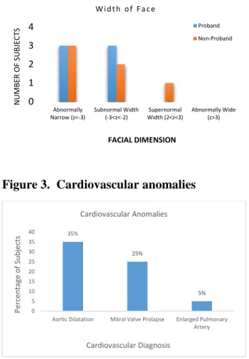
Data was stratified by family history (proband vs non-proband), cardiovascular diagnosis (cardiovascular anomaly vs no cardiovascular anomaly) and by age (5-9; 10-14; 15-18; and 19-25). A Fisher’s exact test, (p=.05), was completed to compare frequency of z-score distribution between the groups. For these groups, we evaluated only the frequency of z-score distribution for z-scores outside of the normal range (z<-2 or z>2) (see Figure 2).
The remaining data from the clinical exam, photographic exam and questionnaire are presented as percentages.
Results:
Demographic and Biometric Results:
Our analysis of the demographic and biometric data revealed fifty percent of subjects had no family history of MFS (probands) and fifty percent had a family history of MFS
Eighty percent of subjects reported one or more ocular anomalies (see Figure 4), and twenty percent reported no ocular anomalies. The most prevalent ocular anomaly was ectopia lentis (35%).
Thirty-five percent of subjects reported current or a past history of orthodontic treatment, while 55% reported no history of orthodontic treatment. Two subjects/guardians did not provide an answer to this question.
Intraoral Examination Results:
Fifty percent of subjects had a Class I molar relationship, 40% Class II, and 5% Class III. Assessment of Lateral and Frontal Photographs:
All facial features described in the diagnostic criteria were observed in our subjects (see Figure 5). The most prevalent facial features observed were retrognathia (54%) and down-slanting palpebral fissures (62%).
Craniofacial Anthropometric Measurements:
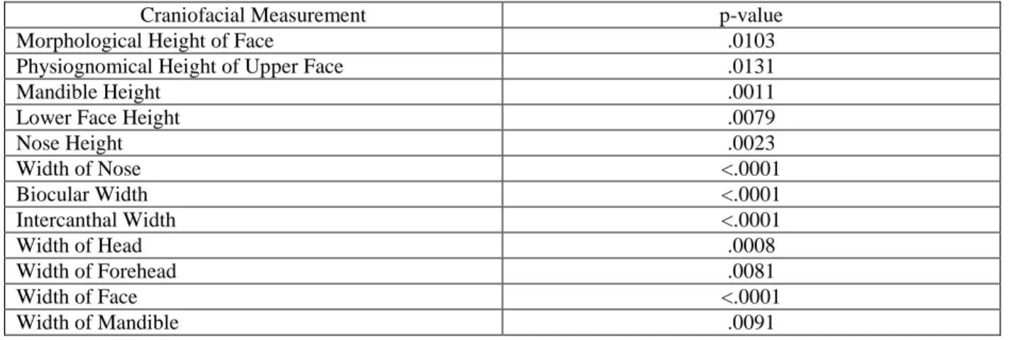
For all 12 craniofacial anthropometric measurements, the frequency of z-score
distribution for our MFS sample was significantly different from the distribution expected in a normal population, p=.05 (see Table 3). Our subjects tended to be under-represented in the optimal/normal categories and over-represented in the subnormal, supernormal, and abnormal categories.
width, facial width, and width of the nose, the majority of subjects fell outside the normal range for facial dimension (see Figures 6-17).
Frequency of z-score Distribution by Groups
There was no statistically significant difference in frequency of z-score distribution for craniofacial measurements for probands vs. non-probands, those with or without a cardiovascular anomaly, or by age (p=.05).
Discussion:
In this cross-sectional study, we evaluated biometric and demographic markers, occlusal relationships and craniofacial morphology in child, adolescent and young adult patients with MFS. We sought to discover if quantifiable differences in facial morphology existed in our subjects when compared to a control population, and to determine if biometric markers could be correlated to facial morphology. Our aim was to enhance the current body of literature related to the diagnostic value of craniofacial findings in patients with MFS by quantifying differences in facial features which may aid in earlier recognition and diagnosis.
radar to be evaluated for MFS, as it is known this syndrome is inherited in an autosomal
dominant pattern. Whereas for probands, there must be a compelling clinical finding or adverse clinical event for a diagnostic evaluation for MFS to be pursued. The recommended annual echocardiographic and ocular examination for patients with MFS or suspected MFS should be implemented as early as possible to assess for the presence/progression of aortic dilatation and ectopia lentis1. This yearly surveillance is delayed in implementation for undiagnosed probands and has the potential to lead to adverse clinical outcomes. There are several reported incidences of the diagnosis of MFS being made post-mortem in individuals who died suddenly as a direct result of cardiovascular complications related to MFS17, 18. To avoid these types of outcomes, diagnostic strategies have to focus on identifying probands at an earlier age to allow for prophylactic medical intervention.
was placed on aortic dilatation as a hallmark feature as it is not observed frequently in the general population 5. It has been noted that many manifestations of MFS are age-dependent which may explain the lower prevalence found in our younger study population 1, 5. It is possible that aortic dilatation had not yet manifested in our subjects resulting in a lower reported
prevalence.
When evaluating ocular morbidity, 80% of subjects reported at least one ocular anomaly and some reported multiple anomalies. The ocular anomaly that is highly specific for MFS is ectopia lentis and is reported to occur in 60% of patients with MFS 1, 16. In our population, 35% of subjects reported ectopia lentis and 10% lens removal which is a treatment modality for severe cases of ectopia lentis 20. Once again, our decreased prevalence could be due to our younger sample population and this condition not yet manifesting.
In our questionnaire, we inquired about each subject’s history of orthodontic treatment. Thirty-five percent of subjects reported a prior history of orthodontic treatment. While no clinical dental findings are included in the most recent diagnostic criteria for MFS, they were included in the previous diagnostic criteria published in 199621. Several studies and case reports have reported on dental findings in patients with MFS focusing on palatal vault height, palatal width, and dental crowding 10, 22, 23. Docimo et al. evaluated pediatric patients with MFS (N=32) and found that 56% of subjects had a crossbite (mono- or bilateral) and 69% had evidence of an ogival (high and arched) palate; they estimated the prevalence of crossbite in their MFS
reported on the prevalence of orthodontic treatment among subjects with MFS and found that 62% (N=51) had a previous history of orthodontic treatment. Their study population was older (mean age 40.2± 15.4years) than our population which could account for the discrepancy in orthodontic treatment prevalence 24. Orthodontic treatment is rarely indicated in the primary dentition and is not routinely recommended in the mixed dentition stage. It is likely some subjects in our sample were not at a developmental stage to warrant orthodontic treatment at the time they were included in our study.
In this report, we also evaluated occlusal relationships to determine if certain features were characteristic of MFS patients. We found that 40% of our subjects had a Class II or mesial step molar occlusion. Previous studies have reported on Class II molar relationships and excess overjet in patients with MFS 22, 25, 26. However, these features are not highly discriminate for MFS and occur in the general population. The recommended treatment to address this
malocclusion would not differ significantly for patients with MFS when compared to the general orthodontic population.
Clinical photographs were taken on thirteen subjects and assessed for the facial features outlined in the most recent diagnostic criteria. Lateral photographs taken on six subjects were undiagnostic for assessment for dolichocephaly, and therefore, the assessment for the presence of dolichocephaly was completed on only 7 subjects. We found that 43% of subjects presented with dolichocephaly which is slightly less than reported in a study by Docimo et al. who found that 47% of pediatric patients with MFS had dolichocephaly; their sample size (N=32) was much larger than ours10. Retrognathia and down-slanting palpebral fissures were the two most
presence of retrognathia was reported as 56%10. Ting and colleagues evaluated facial features in an older population of MFS patients (N=76; mean age=18 years, age range 1-55 years), and found dolichocephaly in 60%8. It could be speculated the decreased prevalence of facial features in pediatric populations exists because the subjects have not “grown into” these features 1, 6, 9. A longitudinal assessment of facial features of pediatric MFS patients would allow for investigation to determine if facial features become more prevalent with age.
For 9 of the 12 craniofacial anthropometric measurements, the majority of subjects (≥65%) fell within the normal range (z-score ±2) for facial dimension. When compared to age- and sex-matched controls, the facial features of our sample population did not vary significantly from the control mean. De Paepe et al. noted the nature of the phenotype of MFS is a continuum that at the mild end of the spectrum, merges with the normal population21. The historical
literature for MFS reports that some musculoskeletal features are absent or less evident during growth, and diagnosis in children or teenagers can be difficult4 which likely accounts for our findings. For three of the measurements, biocular width, facial width, and width of the nose, the majority of our subjects fell outside of the normal range. For biocular width, our subjects had a super normal/abnormally wide biocular width. Down-slanting palpebral fissures has been well documented in patients with MFS and is a part of the current diagnostic criteria 5. Down-slanting of the palpebral fissures appears as a downward drop of the lateral aspect of the eye fold; this results in a more pronounced elliptical shape of the eye which may account for this finding. For facial width, our subjects had a subnormal/abnormally narrow facial width; malar hypoplasia is prevalent in MFS 5 and likely explains this finding. When considering width of the nose, we found that 25% had a subnormal/abnormally narrow nose and 30% had a
width of the face. However, there have been not previous reports of increased nasal width in MFS patients. The only explanation we can provide is normal variation that exists within a population. We could not find any other studies that utilized craniofacial anthropometric measurements to assess facial features in MFS and therefore cannot compare our findings to other studies.
We evaluated frequency of z-score distribution for all craniofacial measurements between probands and non-probands, those with and without cardiovascular anomalies, and by age. Our purpose in doing so was to determine if probands showed more deviation from the norm than non-probands which could have potentially led to a suspicion for MFS. For cardiovascular anomalies, we aimed to determine if those who reported a cardiovascular anomaly were more severely affected globally in other organ systems such as the skeletal system. By stratifying our sample by age, we sought to find out if craniofacial morphology became more abnormal as age increased. For all of our analyses, we found there was no statistically significant difference in frequency of z-score distribution between the groups. Studies have reported the phenotypic expression of MFS is unpredictable 27, 28 which possibly explains our finding of no difference between groups. It has also been noted that phenotypic variation is prevalent in families with MFS carrying the same genetic mutation, and attempts to find genotype-phenotype correlations have been met with limited success16, 28-30. While this may partly explain our findings, our small sample size is likely a contributing factor too; we simply may not have recruited enough subjects to detect differences.
demonstrate a quantifiable difference in craniofacial morphology. This does not mean a difference does not exist, but that continued research with larger sample sizes needs to be pursued. It is also likely that the use of published norms, with large standard deviations, was a less sensitive tool to detect meaningful differences. We could not find any other studies that utilized our method of analysis to which our findings could have been compared, underscoring that this study represents the first time craniofacial features of MFS have been assessed with anthropometric analysis.
Conclusions:
1.) Probands tended to be diagnosed later in life than non-probands.
2.) Aortic dilatation and ectopia lentis were present in our study population but not as prevalent as reported in previous studies.
3.) Retrognathia and down-slanting palpebral fissures were the two most prevalent diagnostic facial features in our study population.
4.) Our hypothesis of quantifiable, distinct craniofacial features for MFS was rejected for 9 of 12 craniofacial measurements, as the majority of subjects fell within the normal range for facial morphology. However it was accepted for 3 of the 12 measurements (binocular width, facial width, and width of the nose), as the majority of subjects fell outside the normal range.
5.) There was no statistically significant difference in frequency of z-score distribution for craniofacial anthropometric measurements between probands vs. non-probands, subjects with or without a cardiovascular anomaly, or by age.
The age- and sex-matched controls were historical controls being established nearly 20-30 years ago. It is unknown if facial dimensions have evolved over time. Also, the control sample consisted of only North American whites. Our sample included Hispanic and Asian subjects and facial morphology may be different between races.
The craniofacial anthropometric measurements made on each subject require correct landmark identification and accuracy when making the measurements. There could have been error introduced into the measurements due to incorrect landmark identification or due to inaccurate measurements.
The biometric data obtained using the questionnaire was not verified by referencing the subject’s medical chart. This could have resulted in under- or over-reporting.
Future Directions:
REFERENCES
1. Dietz, H.C., Marfan Syndrome, in GeneReviews(R), R.A. Pagon, et al., Editors. 1993: Seattle (WA).
2. Marfan Syndrome. April 30, 2015 [cited 2015 May 2]; Available from:
http://ghr.nlm.nih.gov/condition/marfan-syndrome.
3. Willis, L., G.E. Roosevelt, and A.T. Yetman, Comparison of clinical characteristics and frequency of adverse outcomes in patients with Marfan syndrome diagnosed in adulthood versus childhood. Pediatr Cardiol, 2009. 30(3): p. 289-92.
4. De Maio, F., et al., Orthopaedic Aspects of Marfan Syndrome: The Experience of a Referral Center for Diagnosis of Rare Diseases. Adv Orthop, 2016. 2016: p. 8275391. 5. Loeys, B.L., et al., The revised Ghent nosology for the Marfan syndrome. J Med Genet,
2010. 47(7): p. 476-85.
6. Lipscomb, K.J., J. Clayton-Smith, and R. Harris, Evolving phenotype of Marfan's syndrome. Arch Dis Child, 1997. 76(1): p. 41-6.
7. Sponseller, P.D., et al., Improving clinical recognition of Marfan syndrome. J Bone Joint Surg Am, 2010. 92(9): p. 1868-75.
8. Ting, B.L., et al., The diagnostic value of the facial features of Marfan syndrome. J Child Orthop, 2010. 4(6): p. 545-51.
9. De Coster, P., et al., Craniofacial structure in Marfan syndrome: a cephalometric study.
Am J Med Genet A, 2004. 131(3): p. 240-8.
10. Docimo, R., et al., Association between Oro-Facial Defects and Systemic Alterations in Children Affected by Marfan Syndrome. J Clin Diagn Res, 2013. 7(4): p. 700-3.
11. Farkas, L.G., Anthropometry of the head and face. 2nd ed. 1994, New York: Raven Press. xix, 405 p.
12. Farkas, L.G. and I.R. Munro, Anthropometric facial proportions in medicine. 1987, Springfield, Ill., USA: Thomas. xxiv, 344 p.
13. Hall, B.D., et al., Elements of morphology: standard terminology for the periorbital region. Am J Med Genet A, 2009. 149A(1): p. 29-39.
15. Proffit, W.R., Contemporary orthodontics, ed. H.W. Fields, et al. 2013, St. Louis, Mo.: Elsevier/Mosby.
16. Stheneur, C., et al., Study of phenotype evolution during childhood in Marfan syndrome to improve clinical recognition. Genet Med, 2014. 16(3): p. 246-50.
17. Wang, Y., et al., Postmortem diagnosis of Marfan syndrome in a case of sudden death due to aortic rupture: Detection of a novel FBN1 frameshift mutation. Forensic Sci Int, 2016. 261: p. e1-4.
18. Demak, R., Marfan Syndrome: A Silent Killer, in Sports Illustrated. 1986. p. 30-35. 19. Mueller, G.C., et al., Impact of age and gender on cardiac pathology in children and
adolescents with Marfan syndrome. Pediatr Cardiol, 2013. 34(4): p. 991-8. 20. Ectopia Lentis Syndrome. [cited 2017 February 12]; Available from:
http://www.heartworksgala.com/resource/fact-sheet/ectopia-lentis-syndrome#.WKEcwVUrK6I.
21. De Paepe, A., et al., Revised diagnostic criteria for the Marfan syndrome. Am J Med Genet, 1996. 62(4): p. 417-26.
22. Utreja, A. and C.A. Evans, Marfan syndrome-an orthodontic perspective. Angle Orthod, 2009. 79(2): p. 394-400.
23. Westling, L., B. Mohlin, and A. Bresin, Craniofacial manifestations in the Marfan syndrome: palatal dimensions and a comparative cephalometric analysis. J Craniofac Genet Dev Biol, 1998. 18(4): p. 211-8.
24. Staufenbiel, I., et al., Periodontal conditions in patients with Marfan syndrome - a multicenter case control study. BMC Oral Health, 2013. 13: p. 59.
25. Tsang, A.K., A. Taverne, and T. Holcombe, Marfan syndrome: a review of the literature and case report. Spec Care Dentist, 2013. 33(5): p. 248-54.
26. Jain, E. and R.K. Pandey, Marfan syndrome. BMJ Case Rep, 2013. 2013.
27. Summers, K.M., et al., Challenges in the diagnosis of Marfan syndrome. Med J Aust, 2006. 184(12): p. 627-31.
28. Pyeritz, R.E., The Marfan syndrome. Annu Rev Med, 2000. 51: p. 481-510.
29. Judge, D.P. and H.C. Dietz, Marfan's syndrome. Lancet, 2005. 366(9501): p. 1965-76. 30. Dietz, H.C., Potential Phenotype-Genotype Correlation in Marfan Syndrome: When Less
Table 1. Sample demographic data
N=20
Males 8 (40%)
Females 12 (60%)
White 14 (70%)
Hispanic 4 (20%)
Asian 2 (10%)
Table 2. Craniofacial measurements
Craniofacial Measurement Landmarks
Morphological Height of Face N-Gn
Physiognomical Height of Upper Face N-Sto
Mandible Height Sto-Gn
Lower Face Height Sn-Gn
Nose Height N-Sn
Width of Nose Al-Al
Biocular Width Ex-Ex
Intercanthal Width En-En
Width of Head Eu-Eu
Width of Forehead Ft-Ft
Width of Face Zy-Zy
Width of Mandible Go-Go
Table 3: p-values for frequency of z-score distribution MFS population vs. normal population
Craniofacial Measurement p-value
Morphological Height of Face .0103
Physiognomical Height of Upper Face .0131
Mandible Height .0011
Lower Face Height .0079
Nose Height .0023
Width of Nose <.0001
Biocular Width <.0001
Intercanthal Width <.0001
Width of Head .0008
Width of Forehead .0081
Width of Face <.0001
Figure 1. Frequency of z-score distribution for sample population vs. normal population for facial width
Abnormal (z<-3) Subnormal (-3<z<-2) Normal (-2<z<-1) Optimal (-1<z<1) Normal (1<z<2) Supernormal (2<z<3) Abnormal (z>3) Null 0.03 (0.13%) 0.43 (2.14%) 2.72 (13.59%) 13.65 (68.27%) 2.72 (13.59%) 0.43 (2.14%) 0.03 (0.13%) MFS Subjects 6 (30%) 5 (25%) 5 (25%) 3 (15%) 0 (0%) 1 (5%) 0 (0%)
Figure 2. Frequency of z-score distribution probands vs. non-probands for facial width
Figure 3. Cardiovascular anomalies 0 1 2 3 4 Abnormally Narrow (z<-3) Subnormal Width (-3<z<-2) Supernormal Width (2<z<3) Abnormally Wide (z>3) N UM B ER O F SUB JE C TS FACIAL DIMENSION
W i d t h o f F a c e
Proband Non-Proband 35% 25% 5% 0 5 10 15 20 25 30 35 40
Aortic Dilatation Mitral Valve Prolapse Enlarged Pulmonary
Figure 4. Ocular anomalies
Figure 5. MFS craniofacial features
Figure 7. Physiognomical height of the upper face
Figure 8. Mandible height
Figure 10. Nose height
Figure 11. Width of nose
Figure 13. Intercanthal width
Figure 14. Width of the head
Figure 16. Width of the face
APPENDIX 1. REVISED GHENT NOSOLOGY FOR THE MARFAN SYNDROME