The role of gut microbiota and diet in lipid metabolism
Ivana Semova
A dissertation submitted to the faculty of the University of North Carolina at Chapel Hill in partial fulfillment of the requirements for the degree of Doctor of Philosophy in the
Department of Cell and Molecular Physiology
Chapel Hill 2012
Approved by:
John Rawls, PhD
Kay Lund, PhD
Manzoor Bhat, PhD
Michael Goy, PhD
ABSTRACT
IVANA SEMOVA: The role of microbiota and diet in lipid metabolism (Under the direction of Dr. John Rawls)
The large community of microorganisms in the intestine (microbiota) has been
identified as an additional metabolic organ in our body. However, the microbial role in dietary
lipid absorption in the intestine is unclear. Improved understanding of the trialogue between
dietary nutrients, microbiota and host lipid metabolism can help develop strategies for
decreased or increased absorptive capacity as respective treatments for obesity or
malnutrition.
Here, we use in vivo imaging of fluorescent fatty acid analogues to investigate the
effect of microbiota and nutrients on dietary fatty acid absorption in a zebrafish host. Our
results demonstrate that the microbiota stimulates fatty acid uptake and lipid droplet
accumulation in the intestinal epithelium. The microbiota promotes an increase in enterocyte
lipid droplet number, but not size in a diet-dependent manner. We show that the presence of
microbial community also results in enhanced dietary fatty acid absorption into the liver and
non-gastrointestinal tissues. These findings show that the microbiota stimulates intestinal
and extra-intestinal fatty acid absorption in a diet-dependent manner.
To determine whether diet affects the zebrafish microbial community composition,
we performed sequencing of the bacterial 16S ribosomal RNA gene in gut and water
samples from colonized zebrafish under different dietary conditions. Our analysis of bacterial
community composition is the first one to show that dietary nutrients promote intestinal
the Firmicutes primary isolate Exiguobacterium sp. canʼt survive and colonize the intestine
under starved conditions. These findings indicate that Firmicutes survival and growth may
require a nutrient-rich environment.
Finally, we tested whether the diet-induced increase in intestinal Firmicutes
abundance could be partially responsible for the observed increase in lipid droplet number in
fed zebrafish colonized with the microbiota. Colonization of germ-free zebrafish with single
bacterial strains revealed that lipid droplet number is increased by Firmicutes and lipid
droplet size is increased by other bacterial types. These results indicate that the microbiota
stimulates intestinal and extra-intestinal fatty acid absorption and that different microbial
members mediate intestinal fatty acid absorption via distinct mechanisms. This work
demonstrates novel interactions between nutrients and microbiota that enhance dietary fat
ACKNOWLEDGEMENTS
I would like to take this opportunity and acknowledge many of the people that have
contributed to my life journey and success in graduate school. First, I would like to
acknowledge my academic mentors over the years that have provided not only valuable
education but also inspiration. I would like to thank Dr. John Rawls for giving me the
opportunity to join his lab and guiding my research accomplishments over the last five years.
John has been instrumental for my growth as a scientist and researcher and I am extremely
grateful for his influence on my career decision. I would also like to thank the rest of my
committee, Drs. Manzoor Bhat, Michael Goy, Kay Lund and Matthew Wolfgang for all of
their insightful comments, experimental suggestions and care for the quality of my education
during graduate school. I would also like to thank my previous academic advisors Dr. Mark
Levandoski at Grinnell College and Dr. Phanis Antonoglou at The Anatolia College in
Thessaloniki for inspiring me to pursue my dreams and become a researcher.
I have also been fortunate to interact with great labmates who have made the entire
graduate school experience more enjoyable. I specifically would like to thank Jordan
Cocchiaro for taking the time to edit several chapters of my dissertation and provide me with
extremely helpful comments and encouraging words.
I want to thank the entire Department of Cell and Molecular Physiology for providing
the supportive environment for my personal and professional growth. I was fortunate to work
closely with Dr. Ann Stuart and benefit from all of her extremely useful advice on how to
Vicki Morgan, Yvonne Cooper and Jan McCormick for answering all of my departmental
administrative questions.
I want to thank my family and friends for their unconditional love and support. My
college and graduate school friends have had a tremendous impact on the person I have
become as an adult, since I left my home to pursue my academic dreams when I was only
16. I want to thank my dear friend Pavitra Kannan for keeping in close touch even after
many years and miles away from each other. Of course, I have made many, many wonderful
friends while at UNC so many thanks to them for opening their hearts and homes to me. I
want to thank Stephen Jones for sharing all the laughs and tears for the last eight years of
my life. Heʼs been instrumental in my personal and scientific growth and I thank him for that.
Finally, I want to thank my parents Mirjana and Trajche Shemovi, my brother Riste Shemov
and my grandmother Todorka Nikolova because I am certain that I could not have
accomplished everything I did so far if it wasnʼt for them. I cannot imagine having a more
supportive and loving family that has made many sacrifices over the years to ensure my
happiness. I try to show my love and appreciation of their support every day and I am
TABLE OF CONTENTS
LIST OF TABLES...ix
LIST OF FIGURES...x
LIST OF ABBREVIATIONS... xii
CHAPTER I. INTRODUCTION... 1
II. THE ROLE OF INTESTINAL MICROBIOTA IN LIPID METABOLISM... 8
SUMMARY ... 8
INTRODUCTION... 8
FUTURE DIRECTIONS... 33
III. MICROBIOTA AND DIET REGULATE FATTY ACID ABSORPTION IN THE ZEBRAFISH INTESTINE... 37
SUMMARY ... 37
INTRODUCTION... 38
MATERIALS AND METHODS ... 41
RESULTS... 44
DISCUSSION ... 52
SUPPLEMENTAL INFORMATION ... 67
SUPPLEMENTAL EXPERIMENTAL PROCEDURES... 69
IV. CONCLUSIONS AND FUTURE DIRECTIONS ... 93
APPENDIX: THE ROLE OF PSEUDOMONAS AERUGINOSA AUTOINDUCERS
IN HOST METABOLIC AND IMMUNE GENE EXPRESSION ... 104
LIST OF TABLES
IV. MICROBIOTA AND DIET REGULATE FATTY ACID ABSORPTION IN THE ZEBRAFISH INTESTINE
3.S1 Calculated proximate composition of custom Zeigler
zebrafish larval diets ... 86
3.S2 Description of samples subjected to 16S rRNA gene
pyrosequencing... 87
3.S3 Bacteria taxon abundance as defined by 16S rRNA
gene sequences... 88
3.S4 Alpha diversity of 6 dpf zebrafish gut and water
samples... 91
LIST OF FIGURES II. INTESTINAL MICROBIOTA IN LIPID METABOLISM
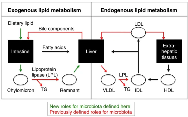
2.1 Novel and known roles of microbiota on lipid metabolism ... 36
III. MICROBIOTA AND DIET REGULATE FATTY ACID ABSORPTION IN THE ZEBRAFISH INTESTINE
3.1 Liposome delivery assay shows fatty acids accumulate in the
intestinal epithelium in the presence of microbiota and diet ... 57
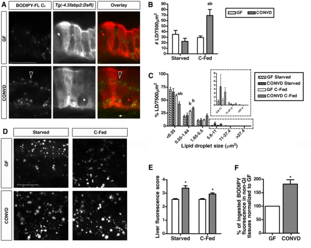
3.2 The microbiota stimulates lipid absorption into intestinal epithelial
lipid droplets and extra-intestinal tissues ... 59
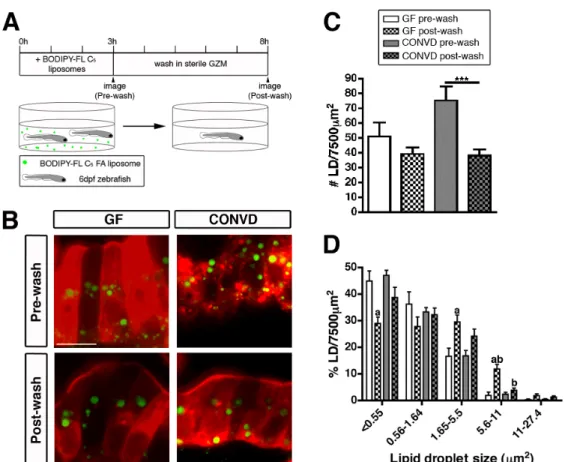
3.3 Lipid droplet clearance is more efficient in the presence
of microbiota ... 61
3.4 16S rRNA gene sequencing reveals distinct bacterial communities in the zebrafish gut and water that are strongly influenced by
dietary status... 62
3.5 Monoassociation with individual community members reveals diet-dependent colonization of a representative Firmicutes
species ... 64
3.6 Monoassociations reveal distinct bacterial mechanisms for
inducing fatty acid absorption in the intestinal epithelium ... 65
3.7 Model for diet-dependent microbial regulation of intestinal
fatty acid absorption ... 66
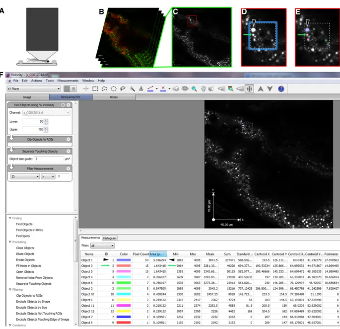
3.S1 Development of a lipid droplet quantification assay for the zebrafish intestine ... 77
3.S2 A short (3 hr) incubation with BODIPY-FL C5 liposomes results in
lipid droplet formation in 6 dpf GF and CONVD zebrafish... 79
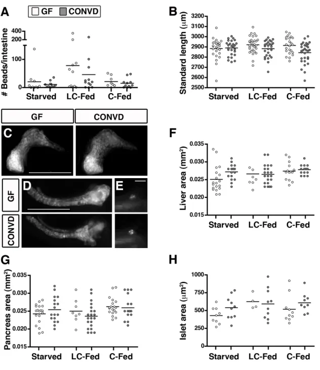
3.S3 GF and CONVD zebrafish have similar feeding behavior, growth
rates and development of the GI tract ... 81
3.S4 The presence of microbiota and host diet does not stimulate
digestive function in 6 dpf zebrafish ... 83
3.S5 Schematic depiction of experimental design for 16S rRNA gene sequence-based evaluations of gut and water bacterial
communities under different diet conditions ... 84
samples ... 85
IV. CONCLUSIONS AND FUTURE DIRECTIONS
4.1 Transcript levels of FA metabolic genes are reduced in the GI
tracts of 6 dpf CONVD vs GF zebrafish ... 103
APPENDIX: THE ROLE OF PSEUDOMONAS AERUGINOSA AUTOINDUCERS IN METABOLIC AND IMMUNE GENE EXPRESSION
A.1 Schematic representation of microbial regulation of fat storage
and the two major P. aeruginosa quorum sensing systems ... 120
A.2 P. aeruginosa bacterial mechanisms virulence factor regulator (vfr) and quorum sensing are not required for regulation of fiaf, fabp2
and mpo transcript levels in 6 dpf zebrafish ... 121
A.3 Treatment with P. aeruginosa autoinducers C4- and
C12-homoserine lactone regulate fiaf, fabp2 and mpo expression in 6 dpf GF and zebrafish colonized with wild-type PAK
and quorum sensing mutants ... 122
A.4 Colonization with wild-type PAK and QS receptor mutant results in
intestine-specific recruitment of mpo-positive neutrophils ... 123
A.5 Monoassociation with Pseudomonas zebrafish isolate ZWU0006 regulates digestive enzyme expression and activity in a
LIST OF ABBREVIATIONS
16S rRNA 16 Svedberg unit of ribosomal ribonucleic acid
AMPK Adenosine monophosphate-activated protein kinase
Angpt4 Angiopoietin-like protein 4 (also called Fiaf)
ANOVA Analysis of variance
apoB ApolipoproteinB
ATP Adenosine triphosphate
BODIPY-FL Boron-dipyrromethene fluorescent dye
C diet Control diet
CFU Colony forming units
CM Chylomicron
CoA Coenzyme A
CONVD Conventionalized
DGAT Diacylglycerol acyltransferase
dpf days post fertilization
EnzChek Boron-dipyrromethene TexasRed casein
ER Endoplasmic reticulum
FA Fatty acid
FABP Fatty acid binding protein
FABPpm Fatty acid binding protein at plasma membrane
FAT/CD36 Fatty acid translocase
FATP Fatty acid transport protein
FFA Free fatty acid
GF Germ-free
GFP Green fluorescent protein
GI Gastrointestinal
GZM Gnotobiotic zebrafish media
HDL High density lipoprotein
hrs hours
HSL Homoserine lactone
IAP Intestinal alkaline phosphatase
IDL Intermediate density lipoprotein
KEGG Kyoto Encyclopedia of Genes and Genomes
LC diet Low-calorie diet
LCFA Long-chain fatty acid
LD Lipid droplet
LDL Low density lipoprotein
LPL Lipopolysaccharide
MCFA Medium-chain fatty acid
MG Monoacylglycerol
mL Mililiter
µm Micrometer
µM Micromolar
mpo Myeloperoxidase
mRNA Messenger ribonucleic acid
MTP Microsomal triglyceride transfer protein
nm Nanometer
OTU Operational taxonomic unit
PBS Phosphate buffered saline
PCoA Principal coordinates analysis
PED6 N-((6-(2,4-dinitrophenyl)amino)hexanoyl)-1-palmitoyl-2-BODIPY-FL-pentanoyl-sn-glycero-3-phosphoethanolamine
PFA Paraformaldehyde
PLA2 Phospholipase A2
PPAR Peroxisome proliferator-activated receptor
PUFA polyunsaturated fatty acid
QS Quorum sensing
RT-PCR Reverse transcriptase polymerase chain reaction
SCFA Short-chain fatty acid
SD Standard deviation
SEM Standard error of the mean
sp species
TG Triglycerides
TMAO trimethylamine-N-oxide
CHAPTER ONE
INTRODUCTION
Dietary effect on microbial community composition
Numerous studies in hosts from worm to human showed that different nutrients (fats, proteins, carbohydrates) affect bacterial abundance of certain microbial species (Ley et al., 2008; Wu et al., 2011; Muegge et al., 2011). In zebrafish (Figure 3.4), pythons (Secor et al., 2008) and mammals (Ley et al., 2005; Turnbaugh et al., 2009) we and others have shown that an increase in caloric input promotes the abundance of a major bacterial phylum called Firmicutes in the intestine. In rodents fed a “Western diet” (high-fat/high-carbohydrate diet), increase in Firmicutes levels has been associated with increased energy harvest from polysaccharides and fat storage in the body (Turnbaugh et al., 2006). Our zebrafish studies suggest similar effects of Firmicutes on intestinal lipid absorption, as a single Firmicutes strain induced fatty acid uptake in the intestinal epithelium (Figure 3.6). Increased Firmicutes abundance has been associated with both diet and genetic mouse models of obesity
(Turnbaugh et al., 2008; Ley et al., 2005). Collectively, these studies suggest that Firmicutes may affect the development of obesity; however, more direct evidence is needed to
determine the contribution of this bacterial phylum to energy harvest from different types of nutrients.
species. High levels of dietary protein also had an impact on the microbiota of domesticated cats (Lubbs et al., 2009). Surprisingly, reduced dietary protein levels did not have a
significant impact on the zebrafish gut or water microbiota (Figure 3.4). In addition, studies have shown that dietary fiber can alter the microbial community of mice (Neyrinck et al., 2010), dogs (Middelbos et al., 2010), and humans (De Filippo et al., 2010). Together, these findings suggest that both caloric value and nutrient content of diet affect the microbial community composition in the intestine.
Microbiota impacts intestinal metabolic processes
The microbiota can impact nutrient metabolism along the intestinal length. In humans, the microbiota has been shown to modify bile acids and stimulate lipid
emulsification in the small intestine (Ridlon et al., 2006; Martin et al., 2007). Members of the microbial community in the distal intestine, on the other hand, possess genes that encode enzymes involved in digestion of complex carbohydrates (Xu et al., 2003; Xu et al., 2007). Therefore, the distal gut microbiota may complement some of the human digestive functions by degrading carbohydrates that cannot be digested otherwise due to lack of human
hydrolytic enzymes (Flint et al., 2008; Louis et al., 2007). In mice, the microbiota affects gut permeability which is important in fatty acid diffusion and lipopolysaccharide-mediated inflammatory response (Cani et al., 2009). Another study in germ-free (GF; animals without any microbes) mice suggested that increased excretion of fat in the intestine could
contribute to the observed obesity-resistance phenotype of these animals (Rabot et al., 2010). Altogether, these findings clearly demonstrate the microbial impact on nutrient digestive and absorptive processes in the intestinal lumen and epithelium.
zebrafish and mice (Rawls et al., 2004; Rawls et al., 2006; Larsson et al., 2012). One of these microbially-regulated genes, called Angiopoietin-like protein 4/Fasting-induced adipose factor (Angptl4/Fiaf), is an important mediator of host lipid metabolism (Yoshida et al., 2002). The microbiota regulates energy storage in adipose tissue via suppression of intestinal Fiaf transcript levels in a mouse host (Bäckhed et al., 2004). Therefore, understanding the mechanisms involved in microbial regulation of Fiaf expression in the intestine is of great importance for obesity prevention and treatment strategies. My work presented in the appendix shows preliminary results of bacterial factors that regulate transcript levels of fiaf as well as another biomarker of lipid metabolism (fatty acid binding protein 2) and innate immune function (myeloperoxidase) in a zebrafish host.
Microbiota impacts metabolism in non-intestinal tissues
microbial roles in regulation of lipid metabolite levels systemically; however, it is not always clear whether the studied metabolites are derived from exogenous (dietary) or endogenous lipid sources. In addition, there is lack of in vivo evidence for microbial stimulation of dietary lipid absorption in the intestine. In order to distinguish between the microbial effects on exogenous versus endogenous lipids, I summarize our current understanding of the metabolic fates of these two types of lipid in the body as well as the microbial
contribution to some of these processes in chapter 2 of my dissertation. In chapter 3, I elucidate the role of the microbiota and individual bacterial species on dietary fatty acid absorption in zebrafish intestinal and extra-intestinal tissues.
Zebrafish as a model organism to study host-microbe interactions and lipid metabolism
intestine is fully functional by 5 days post fertilization (dpf) when lipid and protein digestion and absorption commence (Farber et al., 2001; Hama et al., 2009). Dietary lipid
composition, digestive physiology and lipid metabolic processes are generally conserved among zebrafish and humans (Carten and Farber, 2009). As a result, the zebrafish is an attractive model organism for human diseases including cardiovascular disease, fatty liver disease and obesity (Carten and Farber, 2009; Hölttä-Vuori et al., 2010). Zebrafish have a large advantage over mammalian models for in vivo studies of the hostʼs metabolic
processes since the optical transparency of the larvae allows for observation of digestive and absorptive function using fluorescent protein and lipid probes (Farber et al., 2001; Hama et al., 2009; Carten et al., 2011; Chapter 3). We took advantage of available fluorescent lipid and protein substrates to show that the microbiota can regulate some of these digestive and absorptive processes in the intestine (Chapter 3).
In addition, the zebrafish rapid growth and body transparency allow for high throughput screening for bacterial and host factors involved in metabolic function. In the appendix, I show that Pseudomonas aeruginosa products regulate transcript levels of several fatty acid metabolic and innate immune genes. In our future studies, we intend to utilize a P. aeruginosa PA14 strain mutant library (Liberati et al., 2006) to screen for bacterial genes and mechanisms required for regulation of metabolic activity using quantitative RT-PCR and in vivo assays of digestive and absorptive function.
These results have broad implications for the field of host-microbe interactions because they provide the foundation for future characterization of bacterial and host metabolic
CHAPTER TWO
INTESTINAL MICROBIOTA IN LIPID METABOLISM
SUMMARY
The metabolic fates of exogenous (dietary) and endogenous (de novo synthesized)
lipids are affected by environmental challenges such as dietary nutrients and the large
intestinal community of microorganisms (gut microbiota). Despite previous research efforts,
our understanding of the trialogue between the microbiota, dietary lipids and host lipid
metabolism is incomplete. This review summarizes our current knowledge of the molecular
mechanisms involved in dietary lipid processing in the small intestine as well as the
generation of lipoprotein particles from exogenous and endogenous lipid substrates in
peripheral tissues. Later in the review, we discuss the impact of the microbiota on host lipid
metabolism and the effect of dietary nutrient composition on microbial community structure
and metabolic function.
INTRODUCTION
Fat is a major macronutrient in animal diet. Dietary fat metabolism is affected by host
digestive physiology and anatomy, as well as by environmental factors such as ingested
dietary nutrients and the gut microbiota. In mammals, dietary lipid digestion occurs primarily
in the stomach and the anterior region of the small intestine (duodenum) by gastric and
pancreatic lipases, respectively. Emulsification of fat by bile acids is important for efficient
get activated as acyl-CoA derivatives to be used as energy sources, get reassembled into
triglycerides and stored in cytosolic lipid droplets or packaged in lipoprotein particles
(chylomicrons) for exocytosis and extra-intestinal metabolism. Once transported out of the
intestine, chylomicrons (CMs) circulate through the lymphatics and enter the vasculature via
the portal vein. In circulation, CMs interact with lipoprotein lipase (LPL) that hydrolyzes
triglycerides and releases free fatty acids (FFAs) for uptake into peripheral tissues
(Borensztajn, 1979). After LPL hydrolysis, the smaller, cholesteryl ester-rich chylomicron
remnant circulates to the liver where it enters the pool of endogenous lipid to be used for
hepatic very low density lipoprotein (VLDL) formation (see Figure 2.1). Secreted VLDLs
circulate and undergo hydrolysis by LPL resulting in generation of intermediate density
lipoprotein (IDL) and low density lipoprotein (LDL) particles. Another lipoprotein (high density
lipoprotein, HDL) is responsible for cholesterol transport from peripheral tissues to the liver
for excretion (Fava et al., 2006). These metabolic processes are summarized in Figure 2.1
and described in more depth throughout this review, with emphasis on their regulation by
environmental factors such as dietary nutrients and the gut microbiota.
The microbiota can contribute to lipid metabolism by affecting both intestinal and
extra-intestinal metabolic processes. In the intestinal lumen, the microbiota is thought to
contribute to increased lipid digestion (Kosa and Ragauskas, 2011) and bile salt modification
that improves fatty acid emulsification and absorption (Martin et al., 2008). In the intestinal
epithelium, the microbiota affects the absorptive capacity via changes in intestinal
permeability (Cani et al., 2008). The microbiota also provides bacterial or nutrient-derived
ligands that can regulate the hostʼs transcriptional machinery by interaction and activation of
host transcription factors in intestinal and extra-intestinal tissues (Sanderson et al., 2009).
Finally, on a systemic level, the presence of microbiota regulates circulating levels of lipid
system development and response (Sharma et al., 2010). However, currently there is very
little distinction between the microbial impact on exogenous versus endogenous lipid
sources. Improving our understanding of how the microbiota mediates dietary lipid
absorption in the intestine can provide us with new preventative and treatment strategies for
diet-induced obesity and malnutrition. The purpose of this review is to summarize the most
current findings on lipid metabolism with a more in-depth coverage of dietary lipids and the
role of gut microbiota in mediating these metabolic processes on a systemic level.
Fat metabolism in various animals
Lipid is a major macronutrient for humans that provides 40-55% of the caloric value
in Western diet (Binder and Reuben, 2009). At birth, human infants switch from glucose-rich
to lipid-rich food source as almost half of the energy content in human milk comes from fat
(Lindquist and Hernell, 2010). It has been hypothesized that the high fat content in our diet is
partially due to our high level of encephalization (large brain:body mass). In comparison to
other primates and mammals of similar size, we allocate a larger portion of our daily calories
as energy for the brain. As a result, we have increased our demands for energy-dense food
which is typically rich in fats. This dietary change may have also altered our gastrointestinal
tract, resulting in expansion of our small intestine and reduction of the colon as well as
enhanced capacity to digest and metabolize higher fat diets in comparison to chimpanzees
and gorillas (Leonard et al., 2010).
Despite these differences in GI anatomy and dietary fat preferences between us and
our closest relatives, most of our knowledge of the lipid metabolic fates in humans is based
on discoveries made in cell culture and animal studies. It is important to note that each
culture studies have allowed for an important initial characterization of cellular metabolic
processes; however, most cell lines do not express many of the metabolic genes found in
vivo (Shulzhenko et al., 2011) and fail to recapitulate the effect of the surrounding luminal
environment that includes bile, mucus and microbiota. Research in worms, flies, fish, mice,
pigs and many other animals has generated a more complete picture of the mechanisms
involved in lipid metabolism at a systemic level. It is important to note that interspecies
differences in gastrointestinal (GI) physiology and diet preferences affect lipid metabolic
processes, which is the reason why animal hosts used in the summarized studies are
highlighted throughout this review. Despite innate differences in GI physiology and nutrition,
animal models have provided us with important discoveries of evolutionarily conserved
molecular factors involved in lipid metabolism such as fatty acid binding proteins and
transporters. These findings emphasize the importance of lipid metabolic processes for the
survival of different species.
Metabolic routes of exogenous (dietary) lipids in the intestine
Types of dietary lipids: Dietary lipid composition factors (such as saturation and length of the FA acyl chain) are important for determining physiological outcomes and
disease development in humans. Cholesterol, saturated and trans-fatty acid levels in diet
have been linked to coronary heart disease (Austin, 1991) and atherosclerosis (Katan, 2000).
On the other hand, (ω-3) polyunsaturated fatty acids eicosapentaenoic and
docosahexaenoic acid protect against macrophage-induced inflammation and stimulate
systemic insulin sensitivity (Oh et al., 2010). Dietary fat content consists primarily of
triglycerides (TGs), phospholipids and cholesterol. In energetic terms, the typical Western
monounsaturated fatty acids and 6% of polyunsaturated fatty acids (Nassir and Abumrad,
2009).
In addition to saturation, the acyl chain length also has an impact on fatty acid
metabolic fate. Short-chain fatty acids (SCFA; C2-C5) get absorbed via sodium-coupled
transporter SLC5A8 in the ileum and colon to mediate many beneficial functions in epithelial
biology and metabolism (Cresci et al., 2010). Medium-chain fatty acids (MCFA; C6-C16), the
predominant fat form in infant diet, are thought to enter enterocytes via diffusion where they
get used primarily as energy source (Papamandjaris et al., 1998). Long-chain fatty acids
(LCFA; C17-C22), representing the majority of dietary fatty acids found in human diet, are
thought to be actively transported by fatty acid translocases and binding proteins discussed
below. MCFA, which are saturated, are rarely found in human food; however, in the past
decade there has been an effort to synthesize structured triglycerides with lower
fat-producing value by chemical interesterification of medium- and long-chain FAs onto the
same glycerol backbone (Phan and Tso, 2001). Previous studies in rodents and humans
have reported reduced body weight, fat storage and increase in plasma triglyceride levels on
a medium-chain vs long-chain triglyceride diet which may be a result from a shift towards
increased use of medium-chain fatty acids for energy and de novo lipogenesis in the liver
rather than storage in the body (Geliebter et al., 1983; Hill et al., 1989). It has been argued
that this increase in lipogenesis on medium-chain triglyceride diet is due to faster intestinal
absorption, hepatic portal transport, carnitine-independent mitochondrial metabolism
(peroxisomal and omega-oxidation) and a low affinity for esterification, all of which lead to a
faster and greater oxidation of MCFA in comparison to LCFA (Papamandjaris et al., 1998).
Together, these findings indicate that differences in fatty acyl chain length and saturation
impact the metabolic fates of FAs in the body, and that MCFA could be useful as agents for
Dietary fat digestion: In humans, fat digestion begins in the mouth with lingual lipase, produced by salivary glands in the tongue, and continues in the stomach with the
activity of both lingual and gastric lipase produced by chief cells. However, the majority of fat
digestion occurs in the small intestine, as only 15% of digestion occurs prior gastric
emptying (Binder and Reuben, 2009). Triglyceride digestion in the stomach plays an
important role especially for neonates. Human milk fat contains a considerable amount of
medium-chain triglycerides and acid lipases (lingual and gastric) are more efficient at
digestion of medium- vs long-chain triglycerides. In addition, the lingual and gastric lipases
are the primary enzymes involved in neonatal fat digestion as the pancreatic lipase system
is still not fully functional (Hamosh, 1996). The stomach is also the location where most of
the fat emulsification aided by dietary phospholipids occurs, which is required for efficient
digestion by the pancreatic lipase.
Upon entry into the duodenum, pancreatic lipase hydrolyzes the fatty acids on the
sn-1 and sn-3 positions of a triglyceride and produces two free fatty acids (FFA) and a
2-monoacylglycerol (2-MG). Further hydrolysis results in a release of another FFA and a
glycerol backbone. Phospholipid and cholesteryl ester digestion also occurs in the small
intestine by the activity of pancreatic phospholipase A2 (PLA2) and cholesterol esterase,
respectively. Released FFA and mostly 2-MG get taken up by enterocytes, a process which
is enhanced by micellar solubilization by bile salts (Westergaard and Dietschy, 1976). In
order for single molecules to reach the epithelial brush border, they need to diffuse across
an unstirred water layer, which is a challenge for hydrophobic FA and MG molecules. Mixed
micelles, which result from bile salt emulsification of FFA, 2-MG, cholesterol and
phospholipids, are important vesicles that get sensed by enterocytes via scavenger receptor
SR-BI/CLA-1 (Béaslas et al., 2009). A study in pig intestinal explants incubated with lipid
SR-BI/CLA-1 was endocytosed from the brush border and accumulated in cytosolic lipid droplets
but not secreted lipoproteins (Hansen et al., 2003). The role of these lipid droplet structures
is discussed below.
Lipid uptake in enterocytes: Intestinal intracellular trafficking and processing of fatty acids is not completely understood and controversial. Once fatty acids in mixed
micelles reach the brush border, they become protonated and leave the micelle to be taken
up via passive diffusion or active transport by binding proteins. The exact fatty acid uptake
mechanisms are unresolved. Studies in rats suggest that active transport is the major route
for linoleate (derived from ω-6 linoleic acid) uptake at low concentrations, while passive
diffusion predominates at high concentrations (Chow and Hollander, 1979). Furthermore, the
length of the acyl chain affects FA solubility and transport across the epithelium. MCFA are
less hydrophobic than LCFA and are therefore, thought to diffuse more freely across the
epithelium and enter the portal venous blood that transports it directly to the liver. On the
other hand, longer FA need active transport by proteins such as fatty acid translocase
(FAT/CD36) and fatty acid binding protein at the plasma membrane (FABPpm). Fatty acid
transport proteins (FATP2-FATP4) were originally thought to contribute to FA transport,
however FATP4 was later shown to have a CoA acylase function that serves to activate FA
prior storage or use as energy via FA oxidation (Jia et al., 2009; Milger et al., 2006).
Cholesterol uptake is mediated by adenosine-triphosphate binding cassettes A1, G5 and G8
and Niemann-Pick C1 like 1 transporter (Plösch et al., 2005).
Regulation of transport proteins is dependent on dietary fat levels. As a consequence,
one of the frequent problems in obesity is intestinal adaptation to high-fat diet and increased
absorptive capacity (Lynes and Widmaier, 2011). However, the mechanisms that mediate
epithelial transport capacity in response to dietary fat levels are only partially understood. A
transcription factor peroxisome proliferator-activated receptor γ (PPARγ) and CD36
(Bassaganya-Riera et al., 2004). In humans, however, CD36 mRNA levels did not correlate
with protein levels along the length of the intestine since the ileum showed the highest
protein expression, with lower mRNA levels than in the duodenum, jejunum and colon
(Masson et al., 2010). Therefore, transcript levels of fatty acid transporters do not
necessarily correlate with protein expression at least in human subjects.
Intestinal fatty acid fates: Free fatty acids that get taken up have several metabolic fates, which are partially dependent on their site of entry (apical or basolateral side of the
enterocyte). FAs bind the intestinal FA binding protein (FABP2) and liver FA binding protein
(FABP1). The two proteins are expressed in the intestine, and are thought to be
transcriptionally regulated by C/EBP (Cohn et al., 1992), although their expression might be
regulated by genetic and not dietary factors. These proteins belong to a larger family of
intracellular binding proteins that evolved after the animal kingdom separated from plants
and fungi and is present in both invertebrates and vertebrates (Haunerland and Spener,
2004). Their known roles include protecting cells from cellular damage induced by excess
FA and increasing the FA concentration gradient by binding FFAs in the cell (Haunerland
and Spener, 2004). However, FABP2 overexpression and knock-out in both cells and mice
have lead to inconclusive results about the function of this binding protein in fat absorption,
suggesting that there might be compensatory mechanisms involved in such a critical
process. On the other hand, FABP1 KO mice on a western diet were resistant to obesity
(Newberry et al., 2006), suggesting that this protein influences fat storage in the body.
An additional intestinal enzyme has been implicated in nutrient absorption in the
small intestine. Intestinal alkaline phosphatases (IAP) are metalloenzymes that hydrolyze a
spectrum of phosphomonoesters such as the 5ʼ terminal phosphate group of DNA and RNA,
(Millán, 2006). IAP is expressed at the brush border (Xie et al., 1997; Bates et al., 2007), the
membranes of particles secreted by enterocytes basolaterally and at the apical membrane
after their passage through tight junctions (Mahmood et al., 2003), and around lipid droplets
in enterocytes (Moss, 1982; Narisawa et al., 2007; Warnes, 1972). Furthermore, IAP
expression increases in the duodenum and jejunum of rats (Kaur et al., 2007), piglets
(Dudley et al., 1994) and mice (Millán, 2006; Nakano et al., 2007) in response to increased
dietary fat levels. IAP dephosphorylates the fatty acid translocase CD36 in murine 3T3
fibroblasts, which suggests functional interaction between CD36 and IAP (Ho et al., 2005).
CD36 phosphorylation is thought to be important for its function, since Luiken and
colleagues (2002) showed that cAMP phosphodiesterase inhibitors increase cAMP levels
and the CD36 activity potentially via increased phosphorylation in skeletal muscle cells.
Based on these findings, it is predicted that CD36 physically interacts with IAP to potentially
regulate LCFA uptake in the small intestine. This interaction between CD36 and IAP is
thought to be responsible for the adaptation of the distal intestine and increased absorptive
capacity in mammals in response to high-fat diet (Lynes and Widmaier, 2011).
Bound fatty acids get trafficked to the endoplasmic reticulum (ER), which is the site
of triglyceride re-esterification and prechylomicron formation. The transfer mechanism is
unresolved. At the ER, FFA and MG are used as substrates to re-synthesize TG primarily
via the MG pathway and with the activity of MG acyltransferases (MGAT1, 2 and 3) and DG
acyltransferases (DGAT1 and 2) (Buhman et al., 2002). A DGAT1 knockout mouse model
showed that there might be compensatory mechanisms that involve DGAT2 and DG
transacylase activity in FA uptake and TG formation in enterocytes (Buhman et al., 2002).
The FAs used in TG formation usually reflect the ones found in the diet (Redgrave and
Dunne, 1975; Parks et al., 1981), and TG synthesized from dietary FA is preferentially
synthetases (Mansbach and Nevin, 1998). The exception was observed during fasting when
very low density lipoprotein (VLDL)-size particles are formed in enterocytes consisting of
lipids derived from bile, sloughed enterocytes and plasma FAs (Hussain, 2000).
Another metabolic fate of fatty acyl-CoA is its use as an energy source via fatty acid
oxidation. Even though only a small portion of the dietary FA pool is oxidized in enterocytes,
it becomes an important energy source during fasting. This process is compartmentalized to
the mitochondrion during β-oxidation and requires a membrane transport system facilitated
by carnitine palmityl transferase-1α (CPT-1α) for LCFA substrates. Medium-chain FA on the
other hand, can cross the mitochondrial membrane freely to be oxidized (Friedman et al.,
1990).
FA metabolic fate in the enterocyte depends on the uptake route from the lumen.
Studies in humans and rodents have shown that the site of cellular entry can impact the
intracellular metabolic fate of FA and MG (Gangl and Renner, 1978; Storch et al., 2008).
Both palmitate and oleate substrates undergo oxidation at higher rates when taken up via
the basolateral than apical surface of enterocytes, which the authors speculate is due to the
subcellular localization of mitochondria (even distribution at the apical and basolateral side)
and ER (primarily at the apical side). Furthermore, FAs taken up on the apical side get
incorporated into TG, DG or MG more often than into phospholipids in comparison to FAs
that get taken up basolaterally (Storch et al., 2008).
Intestinal lipid droplet formation: Lipid droplets (LDs) have been recognized as important organelles in the body that serve as temporary TG stores that can get depleted or
replenished depending on dietary fat levels. In addition to their storage function, LDs also
play a role as building elements for organelles by providing phospholipids and sterols
in the ER lumen independent of synthesis of CM lipoprotein apolipoproteinB (apoB)
(Hamilton et al., 1998). In fact, lack of apoB synthesis resulted in larger LD formation in the
ER (Mak and Trier, 1975). A study in suckling mice identified cytosolic LDs that lacked cell
membrane (Young et al., 1995), which suggests of two separate lipid pools – an
ER-independent TG stored in cytosolic LD and an ER-dependent TG used in CM formation.
Both lipid droplet pools (cytosolic LD and CM) have various size distribution, which is
thought to increase with fat feeding and independent of apoB levels (Davidson et al., 1987;
Davidson et al., 1988).
Cytosolic LDs get coated with proteins of the PAT (perilipin, adipophilin, TIP47)
family that play various functions in LD biogenesis (Bickel et al., 2009). For example, TIP47
mediates the incorporation of newly-synthesized TG into LDs, while perilipin and adipophilin
associate only with pre-existing LDs so they get degraded through a proteosome-depedent
pathway when LDs are absent from cells (Masuda et al., 2006). Furthermore, LD-associated
PAT protein levels are dependent on the duration of the dietary fat challenge in mice, as
TIP47 expression is higher after an acute than chronic challenge, while adipophilin
expression follows the opposite trend. Localization was also affected, with TIP47 but not
adipophilin coating LDs after an acute high-fat feeding, while adipophilin but not TIP47 is
observed on LDs after a chronic exposure to high-fat diet (Lee et al., 2009). Therefore, these
proteins are unique to cytosolic LDs and get regulated by the amount and the duration of
dietary fat feedings.
Chylomicron assembly: The process of CM formation begins at the ER where re-synthesized TG binds the microsomal triglyceride transfer protein (MTP) and gets
transferred to newly-synthesized ApoB protein and cholesterol esters to form a primordial
chylomicron. MTP is a protein complex localized to the ER that consists of protein disulfide
MTP large subunit are the cause of the metabolic disease abetalipoproteinemia in humans
(Wetterau et al., 1992), mice (Raabe et al., 1998) and zebrafish (Schlegel et al., 2006).
These studies demonstrate that MTP is an essential protein in the initial assembly of
intestinal lipoproteins in several different vertebrate species.
ApoB is another important protein involved in CM formation. It exists in two forms,
ApoB100 and ApoB48. The larger protein, ApoB100 is enriched in the liver and the prenatal
small intestine and has a low density lipoprotein-binding domain (Black, 2007). The other
form, ApoB48 is enriched in the small intestine and lacks the low density-binding domain.
Both forms are produced by the same gene with ApoB48 being generated by
posttranscriptional mRNA editing (Davidson and Shelness, 2000). While ApoB48 is required
for primordial CM assembly in the ER, ApoB100 is required for VLDL assembly in
hepatocytes. Lipidation of ApoB48 by MTP activity is important to prevent nascent ApoB
degradation and lipoprotein assembly (Hussain et al., 2012). In addition to MTP and ApoB48,
another protein Apo A-IV is also added to the surface of the forming particle (van
Greevenbroek and de Bruin, 1998). Additional factors that regulate CM formation include
rate of TG synthesis, the size of the intracellular lipid pool and lipid trafficking and
translocation in cells (van Greevenbroek and de Bruin, 1998), all of which are dependent on
the amount of dietary FA taken up from the lumen.
The primordial CM gets transported from the ER to the cis-Golgi in a prechylomicron
transport vesicle, which is the rate-limiting step in lipid absorption in a rat model (Siddiqi et
al., 2006). This process is mediated by FABP2 that assists the budding off of the ER, while
the trafficking and fusion with the Golgi is mediated by the coating protein II (COPII) family
members Sar1 and Sec23/24 and the soluble N-ethylmaleimide-sensitive factor attachment
protein receptor (SNARE) fusion complex which consists of vesicle-associated membrane
process is well-conserved even in human newborns that adapt to large dietary lipid loads by
adjusting gene expression levels.
The final step of mature CM formation in the Golgi is followed by basolateral
secretion of these lipoprotein particles into the lymphatic lacteals of humans and suckling
pigs whose GI tracts are closely related to ours. In contrast, in suckling rodents the portal
vein is the primary lipid transport route, since suckling rats exhibit inefficient dietary lipid
transport in lymphatic chylomicrons (Black, 2007). Once chylomicrons exocytose into the
lymphatics, they enter circulation through the thoracic duct, circulate in the vasculature and
interact with lipases such as lipoprotein lipase (LPL) for TG hydrolysis and uptake of FA into
tissues such as the muscle and adipose tissue.
Endogenous lipid synthesis (de novo lipogenesis) in the liver
The liver supplies both exogenous- and endogenous-derived lipid substrates to
peripheral tissues via uptake of FFA from chylomicron remnants or de novo fatty acid
synthesis, respectively. Both lipid substrates get packaged into VLDL particles and released
into the circulation. Hepatic lipid levels are affected by metabolic imbalances in insulin
resistance, type 2 diabetes (Adiels et al., 2007; Adiels et al., 2008), obesity and fatty liver
diseases (Dumas et al., 2006). In hepatocytes, like in enterocytes, there are several different
lipid pools: cytosolic LDs, ER-luminal apoB-free LDs and apoB-containing VLDL precursors
(Olofsson et al., 2000; Shelness and Sellers, 2001). These lipid pools serve as substrates
during lipolysis and TG generation for VLDL particles.
Hepatic enzymes involved in lipolysis: The well-studied lipases such as hormone-sensitive lipase and adipose triglyceride lipase are absent (Holm et al., 1987) or expressed
ER-associated lipase that is expressed at high levels in the liver is the triacylglycerol hydrolase
(TGH). Knockout of this enzyme in mice resulted in smaller cytosolic LDs in primary
hepatocytes. Furthermore, absence of TGH had no effect on nascent LD formation which
colocalized with TIP47, while ADPR colocalized with the surface of pre-formed LDs that
were larger in size. When the authors looked at the LD maturation process, they observed
that colocalization and lipid transfer from nascent into preformed LDs was delayed but not
absent in TGH KO hepatocytes. Therefore, these findings suggest that TGH is important in
determining the dynamics of lipid transfer from newly synthesized to preformed LDs in
hepatocytes (Wang et al., 2010). In addition to its role in lipid transfer, lack of TGH also
resulted in a shift in the lipid metabolite pool with increased DG levels in the ER, which is
thought to recruit CTP:phosphocholine cytidyltransferase to the active
membrane-associated lipid pool and increase phosphotidylcholine (PC) synthesis (Jamil et al., 1993).
The increase in PC levels is correlated with increased surface area and reduced size of LDs,
which may be due to failure to form new LDs or defective fusion process as suggested by a
study in Drosophila S2 cells (Guo et al., 2008).
Hepatic VLDL production: The process of VLDL formation is similar to CM
production in the intestine, and occurs in the ER where lipids are transferred from cytosolic
LD pools in an MTP-dependent manner. The lipid substrates used in VLDL formation are
dependent on the nutritional status and hormonal factors. Under fed conditions, VLDL lipids
are derived from TG hydrolysis in LDs or CM remnants. Under fasted conditions, the
substrates include circulating FFA from adipose tissue lipolysis, de novo TG synthesis and
phospholipids hydrolysis (Xiao et al., 2011). These conditions also regulate the intracellular
FA fates, with elevated FFA flux to the liver and elevated de novo lipogenesis priming the
liver to store and secrete lipids as VLDL particles. This phenotype is observed in
scope of this review (for further reading, please refer to Xiao et al, 2011). Circulating FFA
are taken up in hepatocytes and re-esterified by DGAT1 to contribute to the cytosolic LD
pool. Lipid transfer from LDs to the site of VLDL formation is mediated by TGH.
Apolipoproteins involved in VLDL formation: Like in the intestine, ApoB is involved in VLDL in its longer form, ApoB100. This protein is exclusively expressed in the
liver of humans, while it is found in both the liver and intestine of rodents. Unlike intestinal
apoB formation, hepatic apoB molecule quality is regulated by several mechanisms that
involve ER-associated degradation (ERAD) and post-ER proteolysis pathway (PERPP)
mechanisms (Xiao et al., 2011). Additional apolipoproteins associated with VLDL particles
include apoC-III which helps recruit TG to nascent apoB and promotes larger VLDL
formation (Sundaram et al., 2010).
Upon generation of a nascent particle in the ER, VLDL exits in an ER-derived vesicle,
which fuses with the Golgi in a SNARE complex-dependent manner (Siddiqi et al., 2010).
After lipidation is completed in the Golgi, VLDL is secreted into the vasculature and
circulates to provide lipid substrates to peripheral tissues via LPL-mediated lipolysis. Since
both CM and VLDL particles require the same removal machinery from circulation and
therefore compete for LPL activity in humans (Brunzell et al., 1973) and rats (Karpe and
Hultin, 1995).
Lipid metabolism in peripheral tissues
Lipid is an energy-rich nutrient whose metabolism is dependent on the overall energy
status and homeostasis of the organism. Secreted exogenous lipids from the intestine
(chylomicrons) and endogenous lipids from the liver (VLDL) circulate in the vasculature
peripheral tissues based on energy demands. For example, intestinal chylomicron
production is stimulated not only by an increase in intestinal luminal fat content but also by
circulating FFA levels (Xiao et al., 2011). Similarly, circulating FFA can also stimulate VLDL
production in the liver, while prolonged exposure to FFA in the plasma leads to hepatic
insulin resistance which causes increased VLDL production (Lewis et al., 2002).
Fed vs fasted states: Lipid metabolism is sensitive not only to dietary lipid but also carbohydrate and protein levels. During overnight fasting, circulating FFA released from
adipose tissue fat hydrolysis lead to increased VLDL production, while prolonged fasting
results in an increase in adipose tissue hydrolysis that provides more than 90% of FFA in
VLDL particles (Barrows and Parks, 2006). Circulating VLDL particles get hydrolyzed to
provide FFA as energy source in peripheral tissues resulting in the formation of intermediate
density lipoproteins (IDL) and low density lipoproteins (LDL). After feeding, chylomicrons are
the predominant lipoprotein particles secreted from the intestine into the lymphatics (Tso et
al., 1984). VLDL production is decreased in comparison to fasted state, while circulating
lipoproteins get hydrolyzed and FFA get taken up for storage primarily in adipose tissue.
Therefore, dietary nutrient availability mediates the metabolic fates of exogenous and
endogenous lipids in the body.
The intestinal microbiota as a metabolic partner
Our microbial self: Our mammalian ancestors evolved ~160 million years ago in a microbe-dominated world. In order to ensure our survival, we learned to co-exist with this
large microbial community that outnumbers us. In particular, we form a very intimate bond
with the group of microorganisms that reside on and within our body, including the large and
represents the largest microbial community on our body, and outnumbers the total number
of human cells in an adult human. The biological impact of this microbial community has
been demonstrated in biological events such as intestinal cell proliferation (Cheesman et al.,
2011), vascular remodeling (Reinhardt et al., 2012), nutrient metabolism (Claus et al., 2011),
and immune function (Round et al., 2011) in numerous hosts.
The role of intestinal microbiota in human health and disease
The role of the intestinal community of microorganisms in metabolism has been
established to the point where the gut microbiota is now accepted as an additional metabolic
organ in our body. Studies in vertebrates and invertebrates have identified the importance of
gut microbiota in host physiology (Sekirov et al., 2010) as well as pathologies related to
metabolism such as the ones presented in obesity (DiBaise et al., 2008), diabetes (Musso et
al., 2010), metabolic syndrome (Tilg, 2010), fatty liver disease (Abu-Shanab and Quigley,
2010). Part of the culprit for the recent high prevalence of these metabolic diseases in
humans is the increased consumption of high-calorie diets such as the Western diet
(30-40% of its caloric value comes from fat) that has high levels of saturated and trans fatty
acids and low levels of the anti-inflammatory n-3 PUFAs (Kris-Etherton et al., 2002;
Simopoulos, 2002). As a result of these recent dietary changes, diseases of energy
imbalance have become a major health concern in developed and a growing concern in
developing countries. The effects of diet and microbiota on the development and
The effect of diet on microbiota
Microbial ecology: Evolutionary studies show that changes in environmental conditions (particularly dietary habits) caused our microbial partners to co-evolve with us.
Ley and colleagues used bacterial 16S ribosomal RNA gene analysis of 60 mammalian
species from several different locations around the globe to show that the host diet and
phylogeny both influence the gut microbial community composition and diversity (2008).
Herbivores showed the highest genus-level richness in comparison to omnivores and
carnivores. The authors suggested that this dietary effect on microbial diversity is due to
functional and anatomical adjustments in the intestine of these dietary groups. In order to
digest dietary nutrients from plants, herbivores adapted by extending their gut retention time
to allow for microbial fermentation in the foregut or the hindgut. Microbial communities in
omnivores on the other hand, cluster separately into hindgut fermenters and ones with
simple guts (Ley et al., 2008).
Another study sampled fecal DNA from 33 mammalian species and used shotgun
and targeted sequencing of bacterial 16S ribosomal RNA genes to compare the gut
microbial communities. Principal coordinates analysis plots of microbial communities
showed that bacterial communities associated with herbivores clustered separately from
those in omnivores and carnivores, further emphasizing the importance of plant-based
versus meat-based diet in mediating microbial community composition (Muegge et al., 2011).
However, the lack of correlation between mammalian phylogeny and overall distribution of
microbial species and function suggests that there may not be a significant co-evolution
between mammals and their gut microbiota and microbiome. The authors argue that this
discrepancy between microbial composition and function may be due to differences in
enzymes involved in amino acid biosynthesis, while the carnivore samples have increased
levels of enzymes involved in amino acid degradation.
Microbial community composition and function has also been extensively studied in
lean versus obese hosts in order to identify which bacterial phyla are enriched under excess
nutrient availability. Both diet (high-fat, high-carbohydrate) and genetic (ob/ob) models of
obesity show increased abundance of Firmicutes vs Bacteroidetes, which represent the two
major phyla in human and mouse gut microbiota (Ley et al., 2005; Ley et al., 2006;
Turnbaugh et al., 2008). Microbiota transplantation from obese mouse donors into ex-GF
mice showed that the obese microbiota increases adiposity in a lean host, which suggested
that the microbiota from obese hosts has increased energy harvesting capacity relative to
the microbiota from lean mouse hosts (Turnbaugh et al., 2008). Another study showed that
the distal microbiome of obese twins is enriched in pathways involved in fatty acid
biosynthesis and phosphotransferase system (Turnbaugh et al., 2009), which could alter the
levels and composition of carbohydrate and fatty acid metabolites that become available to
the host. Therefore, host dietary habits can modify nutrient metabolism via direct transfer of
calories from nutrients and indirect effects on host energy storage via alterations of microbial
community structure and function.
The effect of microbiota on metabolism of exogenous lipids
The microbial role in complex carbohydrate metabolism in the colon has been
studied extensively (Turnbaugh et al., 2008; Turnbaugh et al., 2009; Flint et al., 2012) and
will not be covered in detail here. Briefly, bacterial genes, enzymes and metabolites are
thought to complement the function of host digestive enzymes that cannot digest complex
of short-chain fatty acids (acetate, propionate and butyrate) that serve as an energy source
in the intestine and other organs. The microbial enhancement of polysaccharide digestion
and increase in nutrient availability is thought to be partially responsible for larger fat storage
observed in mouse and human hosts (Bäckhed et al., 2004; Turnbaugh et al., 2008).
However, a comparative study in children on a modern Western diet or on a rural African
diet showed that the children on African diet had higher levels of SCFA than the children on
Western diet (De Filipo et al., 2010). Furthermore, SCFA have beneficial effects on intestinal
health as these nutrients contribute to differentiation of epithelial cells, provide fuel for
colonocytes, modulate ion and water transport and decrease colorectal cancer incidence
(Topping and Clifton, 2001; Wong et al., 2006; Sellin, 1999). Propionate specifically has
been shown to have beneficial effects on a systemic level by lowering liver and plasma fatty
acid levels and inflammation and increasing satiety, therefore potentially contributing to
improved insulin sensitivity and weight reduction (Al-Lahham et al., 2010). However, it is
acetate and butyrate levels that are increased in the ceca of obese versus lean
conventionalized (CONVD; ex-GF mice colonized with microbiota) mice, while propionate
levels are low at least in this host organism (Turnbaugh et al., 2006).
The microbial role in exogenous lipid metabolism is not well understood, since the
majority of lipid digestion and absorption is completed in the small intestine, which is a
difficult anatomical region to study in vivo especially in rodents and humans. However, as
mentioned earlier, under high fat feedings, the distal intestine also becomes involved in lipid
absorption (Lynes and Widmaier, 2011) to enhance energy harvest from diet despite an
efficient lipid uptake in the small intestine (>90% uptake in vertebrates) (Labonté et al.,
2008; Karasov and Hume, 1997). As summarized below, our current understanding of the
role of gut microbiota in lipid metabolism comes primarily from genomic, transcriptomic and
Microbial role in lipid metabolism: The microbiota can potentially regulate lipid metabolism by providing microbial genes encoding proteins involved in lipid metabolism
(Turnbaugh et al., 2006), which could contribute to lipid digestion and increased lipid
bioavailability to the host. Another possibility is that the microbiota regulates the expression
of host genes involved in lipid metabolism. Larsson and colleagues (2011) showed that the
gut microbiota regulates transcript levels of genes along the length of the mouse intestine
and liver, which include genes involved in fatty acid activation (Fatp4), TG reesterification
(Dgat1 and 2), fatty acid and phospholipid binding (Fabp2, phospholipid transfer protein -
Pltp) and chylomicron formation (Apob precursor, Mtp). Analysis of enriched groups among
the 500 most significantly regulated genes by gene ontology shows lipid, fatty acid and
cholesterol biosynthetic and metabolic processes amongst the microbially-regulated
metabolic mechanisms. The majority of genes involved in lipid metabolism are
downregulated in the presence of microbiota, while many of the adaptive immunity genes
are upregulated.
Another recent study showed similar trend of microbial transcriptional regulation of
host genes involved in intestinal steroid, lipid, cholesterol metabolic processes and lipid
transport in immunodeficient mice (Shulzhenko et al., 2011). The authors showed that in the
absence of B cells or IgA, the intestinal epithelium upregulates interferon-inducible immune
responses as a protective mechanism against the microbiota. This leads to decreases in
metabolic function and lipid malabsorption as observed in immunodeficient humans.
However, subjects with a normal function of their immune system and consuming a high-fat
diet do not present with malabsorption, but rather hyperlipidemia, hypercholesterolemia and
increase in fat storage in the presence of microbiota, suggesting that the microbial
suppression of transcript levels of lipid metabolic genes does not correlate with decreased
Metabolomic studies have also shown differences in the lipidomes of serum, adipose
tissue and liver of GF and CONVR mice (Velagapudi et al., 2010). Specifically, serum
cholesterol, TG and FFA were reduced while liver and adipose tissue TG levels were
increased in CONVR versus GF mice, which is indicative of increased lipid clearance from
the circulation and stimulated tissue storage. Furthermore in the presence of microbiota,
there were detectable increases in serum cholesteryl esters, sphingomyelins, and
phosphatidylcholines. Serum lipoprotein profiles showed reduced serum chylomicron levels
in CONVR mice after a 4 hr fast, which are thought to be the result of increased clearance of
serum TG and not decreased absorption from the intestine of CONVR animals. On the other
hand, serum VLDL and HDL levels were similar between GF and CONVR mice despite
increased VLDL production in the liver, which also suggests increased clearance rates of the
endogenous lipid supplies from the circulation of colonized mice. However, these altered
lipid profiles could be due to effects of microbiota on intestinal absorption or metabolism of
exogenous dietary lipids or on hepatic production or metabolism of endogenous lipids.
Therefore, the relative contribution of the microbiota to exogenous and endogenous lipid
metabolism in not fully understood.
The microbiota has also been shown to modulate luminal lipid metabolism via
modification of bile acids released into the intestinal lumen from the gallbladder. Bile acids
are derivatives of cholesterol metabolism and facilitate absorption of dietary lipids and
lipid-soluble vitamins in the intestine (Martin et al., 2007). Bile acids also maintain the integrity of
the intestinal epithelial barrier to prevent entry of enteric bacteria and inflammatory response.
The ability of microbiota to process bile acids has been examined in numerous studies
(Martin et al., 2007; Martin et al., 2008; Blaut et al., 2007). Martin and colleagues performed
microbial and metabolic profiling using H1-NMR and bile acid profiling of liver, plasma, urine
(2007). The results from this study show a variety of bile acid modifications (tauro- and
glyco-conjugation as well as unconjugation) in response to microbial processing. The
tauro-conjugation reduces the hydrophobic/hydrophilic ratio and increases their ability to emulsify
lipids. Lipid emulsification results in increased uptake of lipid by the intestinal epithelial cells.
In addition, tauro-conjugation also increases the amount of bile acids that re-enter
enterohepatic circulation and therefore decrease the synthesis of bile acids and cholesterol
by the liver (Martin et al., 2007).
Microbial processing of another dietary lipid (phosphatidylcholine) has been
associated with an animal model of non-alcoholic fatty liver disease (NAFLD). Choline is the
primary source for hepatic phosphotidylcholine production. Dietary choline gets processed
by the gut microbiota (al-Waiz et al., 1992), which releases methylamines (dimethylamine,
trimethylamine and trimethylamine-N-oxide or TMAO). Dumas and colleagues (2006) tested
the effects of high-fat diet on plasma and urine metabolic 1H NMR profiles in BALB/c
(resistant to NAFLD and insulin resistance) and 129S6 (susceptible to NAFLD and insulin
resistance) mice and showed that NAFLD is associated with low plasma phosphotidylcholine
and high urinary methylamine levels, which is the microbiota-mediated phenotype. The
authors interpreted the low plasma levels of phosphotidylcholine to be a consequence of
microbial degradation of choline into methylamines. These findings suggest that microbiota
of NAFLD- and insulin resistance-susceptible hosts are partially responsible for the disease
progression by mimicking choline-deficient conditions.
The hostʼs response to microbially-regulated nutrient metabolism
The evidence for the hostʼs response to microbial regulation of nutrient metabolism
the host upon microbial colonization. Studies by Bäckhed et al. showed that in GF mice
there is increased activity of AMP-activated protein kinase (AMPK) compared to CONVD
animals (2007). This enzyme senses metabolic energy levels via AMP/ATP ratios and is
activated during a nutrient deprivation and energy depletion state. The authors showed that
GF animals are protected against diet-induced obesity in two major, but independent ways:
(i) increase in AMPK activity and (ii) induction in peroxisomal proliferator-activated receptor
coactivator (Pgc-1α) and enzymes involved in fatty acid oxidation, such as Carnitine
palmitoyltransferase1a (Cpt1a) and Medium chain acyl-CoA dehydrogenase (Mcad) in an
Fiaf-dependent manner (Bäckhed et al., 2004; Bäckhed et al., 2007). Fiaf is a circulating
peptide that inhibits the LPL activity in vascular endothelial cells. As described earlier, this
lipase hydrolyzes serum TG, releasing free fatty acids and glycerol into heart, adipose or
muscle tissue. The FFA enter lipogenesis and get stored in the form of fat or get oxidized in
tissues like muscle or heart. Therefore, the microbial role in intestine-specific suppression of
Fiaf results in alleviated suppression of lipoprotein lipase (LPL) in the vasculature, and
increase in TG hydrolysis from chylomicrons and FA uptake in peripheral tissues (Bäckhed
et al., 2004). A study of transcriptional regulation of fiaf shows that different cis-intronic
modules are responsible for tissue-specific expression in a zebrafish host (Camp et al.,
2012). Interestingly, the gut microbiota regulates the cis-intronic modules that regulate
intestinal fiaf expression, suggesting that the microbiota may suppress the intestine-specific
transcriptional enhancer.
The microbiota and diet also contribute to transcriptional activity and lipid metabolic
gene expression. The microbiota stimulates polysaccharide digestion and absorption, which
initiates hepatic de novo lipogenesis (Bäckhed et al., 2004). Conventionalization of mice
also increased hepatic mRNA expression of sterol-responsive element-binding protein
transcription factors that regulate lipogenic enzymes (Bäckhed et al., 2004). Microbial
colonization has been shown to induce intestinal alkaline phosphatase expression in a
zebrafish host (Bates et al., 2007), which as mentioned earlier activates CD36 to stimulate
LCFA uptake from the lumen. Bates and colleagues showed that zebrafish IAP protects
against LPS toxicity and neutrophil infiltration into the intestinal epithelium as a response to
microbial colonization (2007). These findings suggest that the gut microbiota regulate
metabolic processes involved in exogenous and endogenous lipid fates, which is closely
associated with immune function and inflammation induced by high-fat diet. However, direct
evidence of the impact of microbial induction of IAP on lipid metabolism has not been
explored.
Molecular mechanisms involved in gut microbiota- and diet-mediated inflammation
Metabolic endotoxemia: The gastrointestinal tract is under constant low-grade inflammation that worsens with excess of caloric intake and is thought to precede and
contribute to obesity and insulin resistance in CONVD but not GF mice (Ding et al., 2010).
One proposed method for HFD-mediated endotoxemia is by LPS absorption and
incorporation into chylomicrons (Ghoshal, 2009; Laugerette, 2011), indicating that the gut
microbiota and dietary lipids can mediate intestinal lipid absorption and host responses to
elevated lipid and LPS uptake. LPS is the breakdown product of Gram-negative bacteria that
activates the toll-like receptor-4 (TLR4) to initiate secretion of proinflammatory cytokines
(Sweet and Hume, 1996). Obese Sprague-Dawley rats on a high-fat diet showed increased
ileal TLR4 expression, which mediates the intestinal inflammatory response (de La Serre et