IMPAIRED LYSOSOMAL MATURATION IN MACROPHAGES UNDERLIES PATHOGENESIS IN SYSTEMIC LUPUS ERYTHEMATOSUS
Andrew Joseph Monteith
A dissertation submitted to the faculty at the University of North Carolina at Chapel Hill in partial fulfillment of the requirements for the degree of Doctor of Philosophy in the Department of Biochemistry
and Biophysics in the School of Medicine.
Chapel Hill 2015
Approved by:
Barbara J. Vilen
Edward J. Collins
Ken Jacobson
M. Joseph Costello
ii © 2015
iii ABSTRACT
Andrew Joseph Monteith: Impaired lysosomal maturation in macrophages underlies pathogenesis in systemic lupus erythematosus
(Under the direction of Barbara Vilen)
Defects in clearing apoptotic debris disrupt tissue and immunological homeostasis
leading to autoimmune and inflammatory diseases. We identified that macrophages from
lupus-prone MRL/lpr mice have impaired lysosomal maturation resulting in heightened ROS
production and attenuated lysosomal acidification. This diminishes their ability to degrade
apoptotic debris contained within IgG-immune complexes (IgG-ICs) and promotes recycling and
the accumulation of nuclear self-antigens at the membrane 72 hours after internalization.
Diminished degradation of IgG-ICs prolongs the intracellular residency of nucleic acids leading
to the activation of Toll-like receptors. It also promotes phagosomal membrane permeabilization
allowing dsDNA and IgG to leak into the cytosol and activate AIM2 and TRIM21. Collectively,
these underlying events promote the accumulation of nuclear antigens and activation innate
sensors that drives IFNα production and heightened cell death. These data identify a novel
defect in lysosomal maturation that provides a mechanism for the chronic activation of
intracellular innate sensors in systemic lupus erythematosus.
Current therapeutics either broadly suppresses the immune system or target one
pathogenic factor in SLE (BAFF, IFNα, B cells). Therefore, identifying the molecular
mechanism preventing lysosomal maturation in lupus-prone macrophages could provide a
targeted therapeutic addressing multiple SLE pathologies. We identified that heightened mTOR
iv
lysosomal maturation and underlies the accumulation of IgG-ICs on the membrane. Furthermore
treatment with Torin1 and not Rapamycin restored degradation of IgG-ICs implicating that
mTORC2 activity contributed to impaired lysosome maturation. In B6 MFs we found that
regulation of the activity of mTORC2 allows cofilin to depolymerize actin filaments following
phagocytic cup assembly. Actin depolymerization initiated a localized caspase cascade that lead
to the activation of caspase-1 in an inflammasome-independent manner. Caspase-1 then cleaved
Rab39a on the membrane of phagosomes containing IgG-ICs; a necessary step for lysosomal
maturation. In lupus-prone MFs, heightened mTORC2 activity phosphorylates cofilin, which
prevents actin depolymerization and the caspase cascade, thus leaving Rab39a uncleaved. As a
result, the lysosome is unable to mature and degrade the phagocytosed IgG-ICs. These findings
identify a novel signaling pathway regulating lysosomal maturation and an underlying defect in
v
ACKNOWLEDGMENTS
Chapter 2: We would like to thank Drs. Roland Tisch and Nick Spidale (University of N.
Carolina-Chapel Hill) for NOD mice, Dr. Gary Gilkeson (Medical University of South Carolina)
for NZM2410 mice, Dr. Christophe Benoist (Harvard Medical School) for K/BxN mice, Drs.
Edward Miao and Bill Goldman (University of N. Carolina-Chapel Hill) for the GFP-E. coli and
B6/GFP mice. We also thank the Flow Cytometry Core (NCI P30CA016086), the Microscopy
Services Laboratory (CA 16086-26) for their support, and Robert Currin at the UNC Olympus
Center for assistance with 2-photon imaging. This work was supported by NIH R01AI070984,
NIH R21AI105613, NIH R21AR064951 and the Alliance for Lupus Research. A.J.M. was
supported by 5T32AI07273. The authors declare no competing financial interests.
Chapter 3: We would like to thank Drs. Edward Miao and Manira Rayamajhi (University
of N. Carolina-Chapel Hill) for the Caspase-1-/-Caspase-11-/-/B6, Caspase-11-/-/B6, and Asc-/-/B6
mice, v-Myc MFs, and Rab39a plasmids. We would like to thank Drs. James Bear and Jeremy
Rotty for supplying phalloidin-Alexa 568, jasplakinolide, and LimKi 3. We would like to thank
Dr. Tony Richardson and Lance Thurlow for supplying rapamycin. We also thank the Flow
Cytometry Core (NCI P30CA016086) and the Microscopy Services Laboratory (CA 16086-26)
for their support. This work was supported by NIH R01AI070984, NIH R21AI105613, NIH
R21AR064951 and the Alliance for Lupus Research. A.J.M. was supported by 5T32AI07273.
vi
TABLE OF CONTENTS
LIST OF FIGURES………. viii
LIST OF ABBREVIATIONS………... ix
CHAPTER 1: INTRODUCTION………... 1
Figures………... 10
CHAPTER 2: Defects in lysosomal maturation facilitate the activation of innate sensors in systemic lupus erythematosus ……….. 12
Introduction………... 12
Results………... 14
IgG-ICs accumulate in multiple murine models of autoimmunity……….... 14
Lupus-prone MFs phagocytose IgG-ICs but exhibit defective phagolysosome maturation……… 15
Lupus-prone MFs recycle IgG-ICs to the cell membrane………. 17
Impaired lysosomal maturation permeabilizes the phagolysosomal membrane allowing dsDNA and IgG to leak into the cytosol……….. 18
Impaired lysosomal maturation results in heightened intracellular TLR activation………... 22
Discussion………. 23
Materials and Methods……….. 26
Figures………... 35
Supplemental Material……….. 42
vii
Introduction………... 45
Results………... 46
Heightened and mislocalized mTOR activity impairs lysosomal maturation in lupus-prone MFs………. 46
Heightened mTOR activity impairs lysosome maturation by driving the phosphorylation of cofilin……….. 48
Phosphorylation of cofilin prevents the recruitment of caspase-11 to vesicles containing IgG-ICs……… 50
Recruitment of caspase-11 activates caspase-1………. 51
Caspase-1 mediated cleavage of Rab39a is necessary for lysosomal maturation……… 54
Discussion………. 55
Materials and Methods……….. 58
Figures………... 65
Supplemental Material……….. 72
CHAPTER 4: DISCUSSION……… 73
viii
LIST OF FIGURES
Figure 1.1 - Lupus-proneMFs fail to mature the lysosome and as a result
activate intracellular sensors………. 10
Figure 1.2 - Signaling pathway regulating Rab39a mediated lysosomal
maturation in lupus-prone and healthy MFs………. 11
Figure 2.1 - Autoimmune-prone MFs have accumulated IgG-ICs on the cell membrane…….. 35
Figure 2.2 - MRL/lpr MFs phagocytose and traffic IgG-ICs to lysosomal structures…………. 36
Figure 2.3 - MRL/lpr MFs fail to mature the lysosome……… 37
Figure 2.4 - IgG-ICs recycle and accumulate on the cell membrane of MRL/lpr MFs………… 38
Figure 2.5 - Impaired lysosomal maturation allows dsDNA to leak into
the cytosol and activate AIM2……….. 39
Figure 2.6 - Impaired lysosomal maturation allows IgG to leak into the
cytosol and activate TRIM21……… 40
Figure 2.7 - Impaired lysosomal maturation promotes intracellular TLR
activation and IFNα secretion………... 41
Figure 2.S1 - IgG and Sm staining is punctate on the surface of NOD myeloid cells…………. 42
Figure 2.S2 - IgG and apoptotic debris remain colocalized upon recycling of IgG-ICs……….. 43
Figure 2.S3 - MRL/lpr MFs recycle apoptotic blebs……….... 44
Figure 3.1 - Active mTOR and its mislocalization impairs lysosomal
maturation in lupus-prone MFs………. 64
Figure 3.2 - Heightened mTOR activity impairs lysosome maturation
by driving phosphorylation of cofilin……… 66
Figure 3.3 - Phosphorylation of cofilin prevents a localized caspase
cascade at the phagosome……….. 67
Figure 3.4 - Cleavage of Rab39a by caspase-1 is integral for lysosomal maturation…..………. 69
Figure 3.5 - Signaling pathway regulating Rab39a mediated lysosome
maturation in lupus-prone and healthy MFs.………...……….. 70 Figure 3.S1 - Torin1 inhibits mTORC1/C2 while rapamycin inhibits mTORC1………….…… 71
ix
LIST OF ABBREVIATIONS
AIM2 absent in melanoma 2
AP adaptor protein
APC antigen presenting cell
ASC apoptosis-associated speck-like protein
ATP adenosine triphosphate
BAFF B cell activating factor
B6 C57BL/6
BCR B cell receptor
CD cluster of differentiation
CNS central nervous system
CRP C-reactive protein
DC dendritic cell
dsDNA double stranded deoxyribonucleic acid
E. coli-IC Escherichia coli bound by IgG autoantibodies forming immune complexes
ESCRT endosomal sorting complexes required for transport
FcγR Fc-γ receptor
GTP guanosine triphosphate
IFN interferon
IC immune complex
IgG immunoglobulin G
IgG-IC apoptotic debris bound by IgG autoantibodies forming immune complexes
x IRF interferon regulatory factor
ITAM immunoreceptor tyrosine-based activation motif
ITIM immunoreceptor tyrosine-based inhibitory motif
LAMP lysosomal-associated membrane protein
LC3 light chain 3
MF macrophage
mTORC1 mammalian target of rapamycin complex 1
MyD88 myeloid differentiation primary response gene 88
NETs neutrophil extracellular traps
NF-κB nuclear factor κ-light-chain-enhancer of activated B cells
NLRP NOD-like receptor protein
Nox2 NADPH oxidase 2
PI3K phosphatidylinositol-4,5-bisphophate 3-kinase
PTPN22 protein tyrosine phosphatase, non-receptor type 22
ROS reactive oxygen species
SLE systemic lupus erythematosus
Sm Smith antigen
STAT signal transducer and activator of transcription protein
STING stimulator of interferon genes
TCR T cell receptor
TIRF total internal reflection fluorescence
TLR Toll-like receptor
xi TNP 2,4,6-trinitrophenol
TNP-IC TNP bound by IgG autoantibodies forming immune complexes
TREX1 three prime repair exonuclease 1
TRIM21 tripartite motif-containing protein 21
UNC93B1 Unc-93 homolog B1
1
CHAPTER 1: Introduction
In a healthy individual the immune system is equipped with the tools to mount strong
pro-inflammatory responses that are critical in combating infection from microorganisms
(bacteria, viruses, parasites) while also limiting immune responses against self-antigens. When
tolerance mechanisms fail, the immune system can respond to self-antigens resulting in
autoimmunity. Systemic lupus erythematosus (SLE) is a multi-system autoimmune disease with
genetic and environmental components that promote the activation of the immune system against
nuclear self-antigens (ie: dsDNA, histones, ribonucleoproteins) This results in tissue-damaging
inflammation that affects multiple organs (skin, joints, heart, CNS, kidney) (1). The systemic
nature of SLE leads to multiple different clinical presentations, making it difficult to diagnose
and determine an underlying mechanism for disease.
Observational studies have long associated SLE with the accumulation of apoptotic
debris and immune complexes (ICs) containing apoptotic debris (IgG-ICs) in SLE (2-5). The
accumulation of apoptotic debris has been attributed to impaired clearance of apoptotic bodies
rather than increased rates of apoptosis (6). It has been demonstrated that human SLE patients
have polymorphisms in, and decreased expression of, scavenger receptors, increased expression
of FcγRs, and deficiencies in complement (7-12), though whether macrophages (MFs) harbor
intrinsic defects that contribute to impaired clearance is highly debated (13, 14). Murine models
lacking MFGE8 and the complement proteins, opsonins thought to be important to the clearance
of apoptotic bodies, have impaired clearance of apoptotic bodies and spontaneously develop
2
has been demonstrated to impair phagocytic uptake by MFs (17). Interestingly, not all defects in
apoptotic cell clearance seem to promote autoimmunity as murine models lacking mannose
binding lectin and CD14 fail to develop autoimmunity despite having increased apoptotic debris
in the tissues (18, 19). This suggests that accumulation of apoptotic debris alone is not sufficient
in promoting autoimmunity and that the manner in which apoptotic debris is cleared may provide
a critical downstream immunomodulatory signal that can either prevent or promote
autoimmunity.
Fcγ receptors (FcγRs) clear of apoptotic debris, tolerize B cells to self-antigens, and
transmit downstream immunomodulatory signals that control cellular activation. FcγRs
recognize the Fc portion of the antibodies that constitute the IgG-ICs. Upon ligation, FcγRs on
dendritic cells (DCs) and MFs promotes phagocytosis and signal transduction. There are two
types of FcγRs: immunoreceptor tyrosine-based activation motif (ITAM) -containing which
activate the cell and immunoreceptor tyrosine-based inhibitory motif (ITIM) -containing which
repress ITAM signaling. Phagocytosis through ITAM-containing FcγRs (human:
FcγRI/IIa/IIc/IIIa; mice: FcγRI/III/IV) recruit Syk, leading to the activation of the PI3k pathway
(20-22) and trafficking of phagocytosed IgG-ICs to lysosomal structures for degradation (23,
24). Interestingly, polymorphisms that decrease the binding to IgG to human FcγRIIa (R/H131)
and FcγRIIIa (158V/F) have been associated with lupus nephritis and are thought to diminish
clearance of apoptotic debris (25). In contrast to phagocytosis through ITAM-containing FcγRs,
the ITIM-containing FcγRIIb represses ITAM-containing FcγRs by recruiting SHIP1 to
dephosphorylate PI(3,4,5)P3 at the cellular membrane limiting, downstream signal propagation
(26, 27). Further, instead of trafficking IgG-ICs to lysosomes, FcγRIIb traffics IgG-ICs to
3
lacking FcγRIIb develop lupus-like disease (28), while promotor polymorphisms reducing the
expression of FcγRIIb has been associated with murine and human SLE (29, 30). Strangely,
complete loss of FcγR on the lupus-prone MRL/lpr background (FcγR-/-MRL/lpr) had no effect
on disease pathology (31), while loss of FcγRs in lupus-prone NZB/W F1 (FcγR-/-NZB/W F1)
mice have diminished disease (32). It was later discovered that FcγR deficient mice maintain a
partially functional FcγRI complicating the previous interpretations (33). Despite this finding,
it’s interesting that different murine models of SLE have varying dependencies on FcγRs,
although how FcγRs are specifically contributing to disease remains unknown.
Of the surface FcγRs, FcγRI has the highest affinity for IgG (10-8 M compared to 10-5-10
-7 M) (34) because of the third extracellular IgG-like domain (35). Not only has this receptor
been demonstrated to play an important role in protection against bacterial infections, but it also
exacerbates multiple autoimmune diseases (36, 37). Upon ligation, FcγRI internalizes large ICs
through actin-dependent phagocytosis and small ICs through clatherin mediated endocytosis
(38). This is dependent on the relative levels of receptor cross-linking and the competition
between PI3k and Cbl for binding to Syk (38). During endocytosis, Cbl outcompetes PI3k and
poly-ubiquitinates Syk leading to its degradation and mono-ubiquitinates FcγRI, targeting the
forming endosome for lysosomal degradation (38, 39). In contrast, when PI3k outcompetes Cbl,
Syk remains phosphorylated and promotes the actin cup rearrangements integral to phagocytosis
(38, 40, 41). Although, there have been many studies in SLE implicating intrinsic defects in the
phagocytosis of latex beads, apoptotic cells, IgG-ICs, bacteria, and yeast (6, 8, 42-44), no
underlying mechanism has been identified.
Regardless of the method of internalization, phagosomes and endosomes fuse to form an
4
proteins: ESCRT complexes and Rab GTPases. Although the exact function of ESCRT
complexes are still debated, their association with the phagosome is critical to maturation and
lysosomal degradation (46). ESCRT-0 recognizes mono-ubiquitinated cargo at the membrane
and facilitates the initial sorting of the phagosome (47). This allows the ESCRT-I and -II
complexes to associate with the phagosome, which also contain ubiquitin binding domains (48,
49). The recruitment of ESCRT-I and -II forms the nexus that bridges ubiquitinated cargo to the
ESCRT-III complex, promoting the trafficking of the phagosome to lysosomal structures (50).
While mono-ubiquitination is critical for the association of ESCRT complexes, PIP3 is critical
for the Rab proteins to associate with the vesicle. Rab5 associates with the early phagosome via
PIP3 although inhibiting PI3k activity does not preclude Rab5 recruitment (51-53). As the
phagosome matures Rab5 is exchanged for Rab7 (53-56), an event thought to be mediated by
Rab22a (57). Rab7 recruits effector proteins that promote the association of the maturing
phagosome with microtubules driving the phagosome towards lysosomal compartments (58).
Interestingly, recruitment of Rab7 is insufficient to induce phagosome maturation and lysosomal
degradation (53), suggesting that the order of recruitment is critical to phagosomal maturation.
Elevated Rab4 (promotes recycling endosomes) and Rab5 have been found in CD4 T cells from
SLE patients (59), though their effect on disease pathology is unclear.
Concurrent with the recruitment of ESCRT complexes and Rab GTPases to the cytosolic
side of the phagosome, the lumen of the phagosome evolves throughout its maturation. The
assembly of the NADPH oxidase (Nox2) on the early phagosomal membrane is a major
antimicrobial effector as it produces high levels of superoxide (herein referred to as ROS) within
the phagosomal lumen (respiratory burst). Heightened ROS production by granulocytes and
5
unknown. One attractive candidate for the production of ROS and source of autoantigen is the
formation of neutrophil extracellular traps (NETs) by neutrophils (61, 62), mast cells (63), and
eosinophils (64) in which DNA coated with antimicrobial proteins are extruded into the
extracellular environment in a Nox2-dependent form of cell death termed “NETosis”. It has
been demonstrated that delivery of NET DNA to plasmacytoid dendritic cells (pDCs) results in
type 1 interferon (IFN) production in a toll-like receptor (TLR) dependent manner (65, 66).
Observational studies also have found DNA projections consistent with NET formation in the
glomeruli of SLE patients (67, 68). While these results were promising, Nox2 deficiency in
lupus-prone mice had significantly exacerbated disease with severe golerulonephritis (69), while
earlier anecdotal studies linked Nox2 deficiencies with increased SLE in humans (70-72). This
suggests that even though heightened NET formation may occur in SLE, its role may actually be
protective and playing a role in reducing pathology. This suggests that the heightened ROS
levels seen as pathologic in SLE may be NET-independent and connected to the respiratory burst
of non-NETosing cells.
Termination of the respiratory burst must occur prior to phagosomal fusion with a
lysosome to allow proper acidification of the phagolysosome (73). Lysosomes contain
proteolytic enzymes (proteases, DNases, RNases, Lipases, etc.) that require a low pH (pH≤4) to
properly activate and degrade the phagocytosed cargo (74, 75). To limit the lysosomal
degradation of antigens, DCs utilize ROS production during the respiratory burst to alkalize the
phagolysosome to maintain efficient antigen crosspresentation to T cells (76-78). Conversely,
MFs are specialized in proteolytically degrading internalized cargo and possess a capacity of
lysosomal proteolysis that is 20- to 60-fold higher than DCs (79-81). As a result, a prolonged
6
cathepsins by modulating the local redox environment, which limits degradation (82).
Interestingly, observational studies in SLE have found impairment of lysosomal acidification
(43, 83), impaired DNase II activity (84, 85), and LAMP proteins localizing to the cell
membrane (86, 87), which could implicate lysosome function in disease progression. While this
could explain the accumulation of IgG-ICs associated with SLE, there has been no mechanism
connecting lysosome dysfunction to SLE pathology and as a result there’s no consensus in the
field whether immune cells harbor defects in phagocytosis, lysosomal degradation, or both.
Innate receptor activation has long been attributed with SLE pathogenesis, although the
source of nuclear antigen and how the receptors are activated remains unknown. The
autoimmune response against nucleic acids depends on TLR7 and TLR9, and their correlation
with SLE pathology has been highly studied (88-92). TLR7 and TLR9 reside in the endoplasmic
reticulum (ER) in the pro-form until UNC93B1 shuttles the TLRs to the Golgi (93-95). TLRs
are differentially trafficked from the Golgi, where TLR9 traffics to the membrane prior to
arriving at the phagolysosome in an AP-2 dependent mechanism, while TLR7 traffics directly to
the phagolysosome in an AP-4 dependent mechanism (96). Upon arrival at the phagolysosome,
TLR7 and TLR9 become proteolytically cleaved, allowing them to activate upon ligation to
ssRNA (TLR7) and dsDNA (TLR9), although the exact mechanism for TLR7 cleavage is
unknown (94, 97). Interestingly, murine models expressing heightened TLR7 causes
development of SLE-like disease (98-100). Furthermore MRL/lpr lacking TLR9 had
exacerbated disease, while TLR7-deficient mice had ameliorated disease (88). This suggests that
TLR7 and TLR9 might have contrasting roles in SLE, despite both being activated by nuclear
7
There is a subset of innate sensors that are free in the cytosol and activation of these
cytosolic sensors by nuclear antigens has also been implicated in SLE. The Nba2 locus is a
major contributor to disease susceptibility in the NZB/W F1 murine model of lupus and contains
2 cytosolic sensors that recognize dsDNA (p202 and Aim2). NZB/W F1 mice have heightened
expression of p202 (101) which binds to dsDNA and inhibits Aim2 inflammasome formation
(102). Furthermore, decreased Aim2 expression promotes decreased FcγRIIb expression,
increased p202 expression, and heightened IFN production (103). Overall, p202 prevents
Aim2-mediated pyroptosis and allows other cytosolic sensors to recognize nucleic acids and drive type
I IFN production (104). Identifying these other cytosolic sensors to nuclear antigen has proved
to be difficult as studies in SLE patients and murine models of SLE have produced contradicting
reports on the roles of NLRP3/NLRP1 (105-108) and STING (109-112) in autoimmunity. The
cytosolic sensor TRIM21 is the highest affinity Fc-receptor (113) and is responsible for
recognizing opsonized pathogens in the cytosol and promoting their degradation and type 1 IFN
production (114, 115). In autoimmunity, a high frequency of patients across multiple
autoimmune diseases have autoantibodies against TRIM21 with the highest frequency being SLE
and Sjögren’s syndrome (116-118). Furthermore, a polymorphism in TRIM21 (119) and
heightened expression of TRIM21 (120) and its regulated genes (120, 121) has been identified in
SLE patients.
While a role for cytosolic sensors in autoimmunity has become apparent, how
autoantigens reach the cytosol is unknown. The P2X7 receptor (P2X7R) is an ATP-gated cell
membrane receptor connected to the secretion of proinflammatory cytokines (122-124), cell
death (125-127). Further, its under-expression inT cells has been connected to murine models
8
pore in the cell membrane that allows large organic molecules to enter the cytoplasm (129). This
brings up the possibility that P2X7R may facilitate entry of extracellular autoantigens into the
cytosol. Interestingly, the P2X7R gene is located in an SLE susceptibility locus (SLEB4) (130)
and polymorphisms in P2X7R have been identified with SLE (131). A mechanisms linking
membrane permeabilization, cytosolic sensor activation, and autoantigens has yet to be
elucidated in autoimmunity.
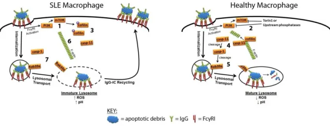
Herein we report that MRL/lpr MFs have impaired lysosomal maturation that prevents
the degradation of phagocytosed IgG-ICs (Fig. 1, 1). This defect is not unique to IgG-ICs as E.
coli opsonized with IgG fails to be degraded. Also, the defect is not ubiquitous to all internalized
cargo because TNP-ICs (2,4,6-trynitrophenol opsonized by IgG) are degraded. As a result, intact
IgG-ICs recycle to the cell membrane and accumulate on the cell surface 72 hours after being
phagocytosed (Fig. 1, 2). The prolonged residency of nuclear antigen in the phagolysosome
allows for the chronic activation of intracellular TLRs (Fig. 1, 3), and permeabilization of the
phagolysosomal membrane (Fig. 1, 4) results in dsDNA and IgG to gain access to the cytosol
and activate Aim2 (Fig. 1, 5) and TRIM21 (Fig. 1, 6). This heightens cell death and promotes
the secretion of IFNα in lupus-prone MRL/lpr MFs.
The impaired lysosomal maturation in MRL/lpr MFs is the result of active mTOR and its
mislocalization at the cell membrane (Fig. 2, 1) as treatment with Torin1 (mTORC1/C2
inhibitor) removes mTOR from the membrane and allows for lysosomal acidification and
degradation of IgG-ICs (Fig. 2, 2). Impairing mTORC1 with Rapamycin is not sufficient in
restoring lysosomal degradation suggesting a role for mTORC2. The activation of mTORC2
drives cofilin phosphorylation which prevents the depolymerization of actin following
9
cascade (Fig. 2, 4) that allows activated caspase-1 to cleave Rab39a (Fig. 2, 5); a necessary step
for lysosomal maturation. MRL/lpr MFs fail to efficiently depolymerize actin and recruit
caspase-11 on f-actin (Fig. 2, 6) and preventing caspase-1 mediated cleavage of Rab39 (Fig. 2,
10
Figure 1. Lupus-proneMFs fail to mature the lysosome and as a result activate
11
Figure 2. Signaling pathway regulating Rab39a mediated lysosomal maturation in
12
CHAPTER 2: Defects in lysosomal maturation facilitate the activation of innate sensors in
systemic lupus erythematosus
Defects in clearing apoptotic debris disrupt tissue and immunological homeostasis
leading to autoimmune and inflammatory diseases. Herein we report that macrophages from
lupus-prone MRL/lpr mice have impaired lysosomal maturation resulting in heightened ROS
production and attenuated lysosomal acidification. This diminishes their ability to degrade
apoptotic debris contained within IgG-immune complexes (IgG-ICs) and promotes recycling and
the accumulation of nuclear self-antigens at the membrane 72 hours after internalization.
Diminished degradation of IgG-ICs prolongs the intracellular residency of nucleic acids leading
to the activation of Toll-like receptors. It also promotes phagosomal membrane permeabilization
allowing dsDNA and IgG to leak into the cytosol and activate AIM2 and TRIM21. Collectively,
these underlying events promote the accumulation of nuclear antigens and activation innate
sensors that drives IFNα production and heightened cell death. These data identify a novel defect
in lysosomal maturation that provides a mechanism for the chronic activation of intracellular
innate sensors in systemic lupus erythematosus.
Introduction
The disposal of apoptotic debris is initiated by membrane changes that facilitate the
binding of IgM antibodies, acute phase proteins (CRP), and other serum opsonins to enhance
phagocytosis (132, 133). The disposal of apoptotic debris is crucial to immune homeostasis as
the accumulation of apoptotic debris (2-4) and the formation of immune complexes (ICs) (5)
13
clearance of apoptotic bodies in mice lacking scavenger receptors and complement proteins
induces spontaneous autoimmunity (15, 16). The idea that accumulated apoptotic bodies
contributes to SLE is further supported in human studies describing polymorphisms or decreased
expression of scavenger receptor, increased expression of FcγRs,or deficiencies in complement
(7-12). Despite these findings, it remains unclear whether macrophages harbor intrinsic defects
that contribute to impaired clearance (13, 14).
Apoptotic debris bound by IgG autoantibodies forms immune complexes (henceforth
referred to as IgG-ICs) that heighten autoantibody production by chronically stimulating
autoreactive B cell receptors (BCRs) and/or toll-like receptors (TLRs) upon delivery of nucleic
acids to the endosome (89, 92). In addition, the binding of IgG-ICs to FcγRs on myeloid cells
stimulates IFNα (134) and BAFF (135) secretion. In addition to stimulating surface receptors,
the activation of cytosolic sensors also impacts the pathology of SLE. Polymorphisms in the
cytosolic sensor to IgG (TRIM21) (119) and heightened expression of TRIM21 (120) and its
regulated genes (120, 121) have been identified in SLE patients, while two cytosolic sensors that
recognize dsDNA (p202 and AIM2) have been implicated in Type 1 IFN production in murine
lupus (101, 102). The involvement of other cytosolic sensors including, NLRP3/NLRP1 (105,
107) and STING (111, 112) have been more controversial. Despite the mounting evidence
implicating cell debris in the activation of innate sensors, a mechanism explaining how IgG-ICs
gain access to the cytosol and chronically activate intracellular receptors/sensors has never been
resolved.
Herein we show that lupus-prone MFs fail to fully mature lysosomes causing diminished
lysosomal acidification and the inability to degrade phagocytosed IgG-ICs. As a result, intact
14
antigens. The prolonged residency of intracellular IgG-ICs in the phagolysosome leads to
membrane permeabilization allowing dsDNA and IgG to leak into the cytosol and activate
cytosolic sensors AIM2 and TRIM21. Furthermore, accumulation of undegraded nucleic acids
inside the phagolysosome leads to the activation of TLR7 and TLR9. The combined activation
of these signaling pathways results in heightened cell death through inflammasome formation
and IFNα secretion.
Results
IgG-ICs accumulate in multiple murine models of autoimmunity
The accumulation of apoptotic debris has been identified in autoimmune diseases other
than SLE, including apoptotic beta-cells in diabetes (136), and apoptotic synoviocytes in
rheumatoid arthritis (137). Recent studies show that prior to disease, MFs from lupus-prone
mice (MRL/lpr and NZM2410) accumulate high levels of FcγR-bound IgG-ICs (Fig. 1A-B) (37).
Similarly, SLE patients experiencing active disease accumulate nuclear antigens on peripheral
blood mononuclear cells (37). Therefore, we wanted to assess whether accumulation of IgG-ICs
occurs in other autoimmune models by quantifying the levels of surface IgG and nuclear antigen
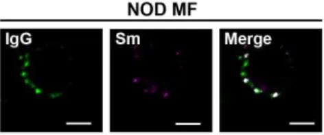
on MFs from murine models of diabetes (NOD) and rheumatoid arthritis (K/BxN). We found
that the levels of surface IgG on MFs from NOD and K/BxN mice were elevated (Fig. 1B), and
showed a punctate staining pattern similar to MRL/lpr MFs (Fig. S1). Surface Sm levels on
NOD MFs were slightly elevated, but absent on MFs from K/BxN mice. This suggests that in
other autoimmune diseases MFs accumulate IgG-ICs, but the antigen bound by IgG varies.
Since NOD, MRL/lpr and NZM2410 mice are genetically unrelated, these findings also suggest
15
membrane. Therefore understanding the mechanism underlying the accumulation of IgG-ICs on
the surface of MFs could elucidate a fundamental defect in autoimmunity.
Lupus-prone MFs phagocytose IgG-ICs but exhibit defective phagolysosome maturation
In SLE, the accumulation of apoptotic debris has been attributed to heightened cell death,
impaired clearance, decreased complement and increased IgG levels (2-4, 6, 17, 43, 138).
Whether lupus-prone MFs have intrinsic defects contributing to impaired clearance of apoptotic
debris is debated because there are no defined mechanisms underlying diminished clearance (13,
14). To identify the mechanism(s) underlying the accumulation of IgG-ICs, we formed ICs
using anti-nucleosome (PL2.3, IgG2a) bound to apoptotic blebs. This allows B6 and MRL/lpr
MFs to internalize physiologically relevant IgG-ICs and a means to compare phagocytosis,
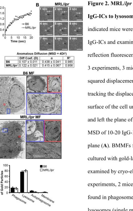
intracellular trafficking, and degradation in real time. Using TIRF microscopy, we found that B6
and MRL/lpr MFs had comparable diffusion coefficients prior to internalization, and they
internalized IgG-ICs at similar rates (Fig. 2A-B). Therefore, the accumulation of IgG-ICs on the
surface of MRL/lpr MFs is not the result of impaired phagocytosis.
Another possible explanation for accumulation of IgG-ICs on MRL/lpr MFs is improper
trafficking to lysosomes. However, cryo-electron microscopy showed that approximately 80%
of gold-labeled IgG-ICs reached lysosomal structures in B6 and MRL/lpr MFs within 2 hours of
phagocytosis (Fig. 2C-D). This indicates that intracellular trafficking is not impaired.
Impaired lysosomal degradation could promote membrane accumulation of IgG-ICs
despite their arrival at lysosomal structures. Lysosomes contain hydrolytic enzymes that degrade
cargo entering through multiple receptors including FcγRs (24). Activation of lysosomal
enzymes requires the termination of ROS and activation of the vacuolar H+-ATPase (V-ATPase)
16
acidotropic ratiometric dye during phagocytosis of IgG-ICs. In B6 MFs, real-time two-photon
microscopy identified vesicular fusion events resulting in large acidic structures (pH=4.0) (Fig.
3A-B). In MRL/lpr MFs, large acidic structures were rarely evident and vesicles failed to
sustain a pH below 5.5. To analyze larger numbers of MFs, we used ratiometric flow cytometry
to quantify the relative pH of the population. Within 30 minutes of exposure to IgG-ICs, B6
MFs reduced vesicular pH by 20% then de-acidified within 1 hour (Fig. 3C). Conversely,
MRL/lpr MFs showed an 8% drop in pH. Concurrent with the inability to fully acidify, MRL/lpr
MFs exhibited heightened and prolonged production of ROS (Fig. 3D). These results
demonstrate that MRL/lpr MFs are functionally impaired in lysosomal acidification, but the
impairment is not absolute as seen in lysosomal storage disorders. This is consistent with the
idea that antigen processing and MHC presentation remain at least partially intact in lupus-prone
mice (139, 140).
The impaired acidification and heightened ROS production suggests that MRL/lpr MFs
are not properly maturing the phagolysosome, thus preventing the degradation of IgG-ICs.
Maturation of the phagolysosome and autophagosome requires membrane stabilization, achieved
through the recruitment of lysosome-associated membrane proteins (LAMPs) and light chain 3
(LC3). To assess whether the phagolysosome fully matures, we quantified the levels of LAMP-1
and LC3A to distinguish autophagosomes (LC3A+, LAMP-1-) and autophagolysosomes (LC3A+,
LAMP-1+) from lysosomes (LC3A-, LAMP1+). Using confocal microscopy, we found that
MRL/lpr MFs showed a 2-fold reduction in the association of IgG-ICs with LC3A-, LAMP-1+
structures (Fig. 3E-F). Since IgG-ICs arrive at lysosomal structures (Fig. 2C-D), the reduced
association of LAMP-1 with vesicles containing IgG-ICs demonstrates that the impaired
17
Lupus-prone MFs recycle IgG-ICs to the cell membrane
Damaged membrane proteins traffic to the lysosome for degradation; however, impaired
degradation promotes their recycling back to the cell membrane (141, 142). We hypothesized
that a similar mechanism might promote recycling and the accumulation of FcγR-bound
IgG-ICs, since they also target lysosomal structures following activation (24). To test this, we
co-cultured MFs with fluorophore-conjugated IgG-ICs and monitored their localization over time.
Both B6 and MRL/lpr MFs bound similar levels of IgG-ICs, which were rapidly phagocytosed,
and evident within vesicular compartments at 24 hrs (Fig. 4A-B). By 72 hours, MFs from the
MRL/lpr mice recycled the IgG-ICs to the cell membrane, while B6 MFs retained them within
the cell. The IgG-ICs appeared to remain intact and bound by FcγRs as both the antibody and
apoptotic debris colocalized on the surface of the cell (Fig. S2) and the levels of surface FcγRI
and IgG on MRL/lpr MFs increased proportionately (37). Overall, this supports a model
wherein impaired maturation of the lysosome in MRL/lpr MFs diminishes degradation of
IgG-ICs inducing their recycling back to the membrane.
The levels of nuclear antigen on MFs from MRL/lpr mice lacking FcγRI (FcγRI
-/-MRL/lpr) are decreased 40% compared to cells from FcγRI+/+/MRL/lpr mice, and they remain
disease-free (37). This implicates the accumulation of IgG-ICs on FcγRI in SLE. Despite
FcγRI-/-MRL/lpr MFs binding 60% fewer IgG-ICs (Fig. 4A-B), they remain impaired in
lysosomal acidification and recycle internalized IgG-ICs. This demonstrates that FcγRI is not
the only receptor that recycles of ICs, and that loss of FcγRI decreases the amount of
IgG-ICs that are internalized.
To assess whether recycling is unique to ICs containing apoptotic debris, we bound
18
phagocytosed fewer E. coli-ICs (Fig 4C), 72 hours following phagocytosis the levels of LPS on
the cell surface increased 3-fold compared to those on B6 MFs (Fig. 4D). This indicates that E.
coli-ICs also fail to be degraded by MRL/lpr MFs. To assess whether all ICs were recycled in
MRL/lpr MFs, we cultured MFs with ICs formed by binding TNP20KLH to anti-TNP IgG2a
(TNP-ICs). Surprisingly, both MRL/lpr and B6 MFs phagocytosed and degraded the TNP-ICs
(Fig. 4E right). These data demonstrate that MRL/lpr MFs degrade some IgG-ICs and that
recycling is not unique to ICs containing apoptotic debris.
To assess whether impaired lysosomal acidification is sufficient to induce recycling of
IgG-ICs, we inhibited lysosome function in B6 MFs and assessed whether this induced IgG-ICs
to recycle. Concanamycin A prevents acidification and degradation of phagocytosed cargo by
specifically inhibiting the lysosomal V-ATPase. B6 MFs treated with concanamycin A recycled
IgG-ICs to levels similar as MRL/lpr (Fig. 4F). This indicates that diminished lysosomal
acidification is sufficient to promote recycling and accumulation of IgG-ICs.
Impaired lysosomal maturation permeabilizes the phagolysosomal membrane allowing
dsDNA and IgG to leak into the cytosol
Studies of microbial pathogens have shown that some intracellular bacteria prevent
phagosomal maturation resulting in bacterial antigens accessing the cytosol (143, 144). In a
similar manner, impaired lysosomal maturation in MRL/lpr MFs might allow antigens from
IgG-ICs to gain access to the cytosol and activate innate sensors. Therefore we selected two cytosolic
sensors (AIM2 and TRIM21), which recognize different components from IgG-ICs (dsDNA and
IgG), to determine whether the inability to mature the lysosome permeabilizes the
phagolysosome allowing antigens to leak into the cytosol. To assess whether nuclear antigens
19
fluorescent dsDNA. Immunoprecipitation of AIM2 from B6 and MRL/lpr MFs showed equal
levels of AIM2 protein (Fig. 5A). Despite equal amounts of protein, MRL/lpr MFs had a
2.5-fold increase in the amount of dsDNA bound to AIM2 compared to B6 (Fig. 5B). In contrast,
AIM2 from FcγRI-/-MRL/lpr MFs bound the same level of dsDNA as B6. This suggests that
although FcγRI-/-MRL/lpr MFs recycle IgG-ICs (Fig. 4A-B), they leak fewer antigens into the
cytosol. This might reflect decreased internalization of IgG-ICs, or that FcγR-specific signals
are necessary to permeabilize the phagolysosome.
AIM2 initiates inflammasome formation by recruiting pro-caspase-1 through the linker
molecule ASC. This cleaves and activates caspase-1 (102). To assess whether phagocytosis of
IgG-ICs by MFs induces inflammasome formation, we quantified activate caspase-1, and
enumerated cytosolic ASC foci (Fig. 5C-D). In the resting state, the number of MRL/lpr MFs
containing ASC foci was higher than B6, while in FcγRI-/-MRL/lpr mice they were comparable
to B6. However, 4 hours after co-culture with IgG-ICs, approximately 40% of the MRL/lpr MFs
exhibited ASC foci compared to 20% in B6 and FcγRI-/-MRL/lpr MFs (Fig. 5D). This was
consistent with diminished binding of dsDNA to AIM2 in B6 and FcγRI-/-MRL/lpr MFs (Fig.
5A-B). In MRL/lpr MFs, heightened formation of ASC foci coincided with a 4.5-fold increase
in activate caspase-1 compared to either B6 or FcγRI-/-MRL/lpr MFs (Fig. 5E). Similarly, ex
vivo splenic myeloid cells from MRL/lpr mice exhibited a 2-fold increase in active caspase-1
compared to B6 (Fig. 5F). Inflammasome formation was the consequence of lysosomal
dysfunction as B6 MFs treated with concanamycin A had high levels of ASC foci and caspase-1
activation (Fig. 5D-E). Hence, impaired lysosomal degradation of IgG-ICs allows nuclear
20
To corroborate the idea that diminished maturation of the lysosome promotes the
permeabilization of the phagolysosome allowing antigens to leak into the cytosol, we assessed
whether IgG from exogenous IgG-ICs activated TRIM21, a cytosolic sensor with high affinity
for IgG (113). We found that 4 hours after co-culture with IgG-ICs, MRL/lpr MFs showed a
2-fold increase in fluorophore tagged IgG bound to TRIM21 when compared to B6 (Fig. 6A-B).
The level of TRIM21-bound IgG in FcγRI-/-MRL/lpr MFs was not different from B6 indicating
that similar to nuclear antigen, diminished internalization of IgG-ICs reduces the amount of IgG
reaching the cytosol. Similarly, co-culture of MRL/lpr MFs with IgG-ICs increased the levels of
IgH/IgL bound by TRIM21 (Fig. 6A). We don’t believe that the heightened levels of IgG bound
to TRIM21 reflect the 8.8-fold increase in TRIM21 protein levels in MRL/lpr MFsbecause
FcγRI-/-MRL/lpr MFs also exhibited heightened levels of TRIM21 (Fig. 6A,C) and their levels of
IgG bound to TRIM21 were not different than B6 (Fig. 6A-B). Thus, although elevated TRIM21
may contribute to disease pathology, it alone is insufficient, unless diminished maturation of the
lysosome provides heightened levels of IgG ligand.
TRIM21 is an E3 ligase that possesses two unique functions. First, it inhibits type 1
interferon production by diminishing IRF protein levels through ubiquitination and proteasomal
degradation. Second, the binding of IgG to TRIM21 stabilizes IRF proteins and activates NF-κB
(114, 115). This heightens TLR activation and type 1 interferon production. To assess whether
TRIM21 was activated, we co-cultured B6 and MRL/lpr MFs with IgG-ICs and quantified
NF-κB activation by the nuclear translocation of p65. Resting MRL/lpr MFs exhibited slightly
elevated nuclear p65 levels; however, after co-culture with IgG-ICs, nuclear translocation of p65
was increased 2.5-fold (Fig. 6D-E). Loss of FcγRI in MRL/lpr MFs restored nuclear p65 levels
21
acidification because concanamycin A treated B6 MFs stimulated with IgG-ICs induced a
2.5-fold increase in nuclear p65 (Fig. 6D-E). It’s possible that NF-κB activation in response to
IgG-ICs was the result of the apoptotic debris binding to intracellular TLRs. To assess this
possibility, we co-cultured MRL/lpr MFs with apoptotic blebs lacking IgG. Like IgG-ICs,
apoptotic blebs recycled in MRL/lpr MF (Fig. S3) likely because they are opsonization by
C-reactive protein (CRP) and enter cells via FcγRI (145). Despite this, apoptotic blebs lacking IgG
did not translocate p65 to the nucleus indicating that IgG was responsible for NF-κB activation
(Fig. 6D-E). Thus, the inability to mature the phagolysosome allows IgG from ICs to leak into
the cytosol and activate TRIM21.
The activation of TRIM21 by IgG induces poly-ubiquitination and proteasomal
degradation (146). To assess the levels of TRIM21 activity in vivo, we quantified ubiquitinated
TRIM21 in ex vivo splenic myeloid cells from B6 and MRL/lpr mice. We found that splenic
myeloid cells from MRL/lpr mice had 85-fold more ubiquitinated TRIM21 compared to ex vivo
B6 myeloid cells (Fig. 6F-G). IgG was necessary for TRIM21 ubiquitination as comparable cells
from age-matched AID-/-MRL/lpr mice (lack IgG) had significantly less ubiquitinated TRIM21
(13.5-fold vs 85-fold) when compared to B6 myeloid cells. Acute activation of TRIM21
stabilizes IRF3, while chronic activation increases IRF7 levels by activating NF-κB (114, 115).
In ex vivo myeloid cells from MRL/lpr mice, we found that ubiquitinated TRIM21 was
coincident with nuclear translocation of p65 and heightened levels of IRF7 protein, but not with
heightened IRF3 (Fig. 6H). The finding that IRF7 protein levels are selectively increased
suggests that TRIM21 activation in myeloid cells from MRL/lpr mice is not an acute event; but
22
Impaired lysosomal maturation results in heightened intracellular TLR activation
Diminished lysosomal maturation allows IgG and nuclear antigens to leak into the
cytosol and activate innate sensors. However, a large fraction of IgG-ICs remains inside the
phagosome and is thus capable of activating TLRs. Coupled with heightened IRF7 levels from
activated TRIM21 (Fig. 6H), phagosomal TLR ligands could heighten IFNα secretion through
the activation of TLR7 and TLR9 (TLR7/9). To assess whether the prolonged residency of
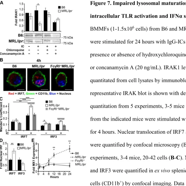
nuclear antigens within the phagolysosome activates intracellular TLRs, we quantified IRAK1
levels in MFs 24 hours after exposure to IgG-ICs. This time point was sufficient for B6 MFs to
degrade the IgG-ICs, but was prior to recycling in MRL/lpr MFs (Fig. 4A-B). We found that
MRL/lpr MFs exposed to IgG-ICs showed a 2.8-fold decrease in IRAK1 levels, consistent with
TLR activation (Fig. 7A). Chloroquine, an acidotropic molecule that binds double and single
stranded nucleotides and sterically hinders their binding to TLRs (147) restored IRAK1 levels
supporting that TLR7/9 are activated in MFs that fail to degrade IgG-ICs. Further, impairing
lysosomal acidification with concanamycin A prevents the degradation of IgG-ICs in B6 MFs
(Fig. 4F) and reduced IRAK1 to levels found in MRL/lpr MFs (Fig. 7A). Therefore, the
impaired lysosomal acidification in MRL/lpr MFs is sufficient to heighten TLR activation in the
presence of IgG-ICs.
Formation of the TLR-MyD88-IRAK1 complex downstream of TLR7/9 promotes
phosphorylation and nuclear translocation of IRF7 resulting in production of type 1 interferon
(148). To assess whether impaired degradation of IgG-ICs promotes nuclear translocation of
IRF7, we co-cultured MRL/lpr MFs with IgG-ICs and found they exhibited a 2-fold increase in
nuclear IRF7 levels (Fig. 7B-C) that was sustained for 24 hours. IgG-ICs did not localize all IRF
23
Reducing the lysosomal burden through loss of FcγRI also reduced the nuclear translocation of
IRF7 in MRL/lpr MFs to levels comparable to B6. These findings were not an artifact of
BMMFs because ex vivo myeloid cells from MRL/lpr mice also showed a 2-fold increase in
nuclear IRF7, while nuclear IRF3 was not elevated (Fig. 7D). Collectively, the data support a
model wherein impaired lysosomal maturation prolongs phagolysosomal residency of IgG-ICs
facilitating chronic intracellular TLR activation.
The inability to degrade IgG-ICs resulted in increased levels of IRF7 and heightened
intracellular TLR activation. Combined, these events could elevate IFNα secretion (148). To
assess this, we cultured MRL/lpr MFs with IgG-ICs (12 and 24 hours) and found they secreted
2- and 3-fold more IFNα (Fig. 7E). The production of IFNα was a consequence of FcγRI
mediated internalization because IFNα levels secreted by FcγRI-/-MRL/lpr MFs were comparable
to B6. This is consistent with a model wherein the accumulation of IgG-ICs in the
phagolysosome of MRL/lpr MFs drives heightened TLR activation and IFNα secretion as a
consequence of diminished lysosomal maturation.
Discussion
The mechanism underlying the accumulation of IgG-ICs in the periphery of SLE patients
has been highly debated. Observational studies in SLE patients have shown that MFs have
intrinsic defects in the phagocytosis of latex beads, apoptotic cells, IgG-ICs, bacteria, and yeast
(6, 8, 43, 44). Other studies have found that phagocytosis is intact, but the ability to degrade the
internalized cargo was impaired (43, 83, 84). We find that lupus-prone MRL/lpr MFs
phagocytose and traffic IgG-ICs to lysosomal structures, but that the lysosomal structures are
unable to mature and acidify. As a result, the IgG-ICs are not degraded and recycle back to the
24
antigens (37) and they express LAMP1/2 on the cell surface (86). This supports the idea that the
lysosomal compartment has been trafficked to the membrane and is consistent with our findings
showing that macrophages from lupus-prone mice accumulate high levels of nuclear antigens
(Fig. 1) as a consequence of recycling undegraded IgG-ICs from the lysosome (Fig. 4).
Interestingly IgG-ICs, apoptotic blebs, and E. coli-ICs recycled to the cell membrane while
TNP-ICs were degraded. This suggests that the size or content of the cargo impairs the lysosome.
Alternatively, the opsonin coating the incoming apoptotic debris could impact lysosomal
maturation. Combined this could explain the variability of previous studies aimed at identifying
phagocytic or lysosomal defects in SLE monocytes.
Spontaneous SLE has been linked to alterations in expression (98) and activation of
TLR7/9 (89, 92), dysregulation of the cytosolic sensors p202 and AIM2 (101, 102), and
polymorphisms (119) and heightened expression in TRIM21 (120). We now define that
defective degradation of FcγR-bound cargo in the lysosome is a critical upstream event that
overburdens phagolysosome. The prolonged intracellular residency of IgG-ICs promotes the
activation of TLRs and permeabilization of the phagolysosomal membrane allowing IgG and
nuclear antigen to access the cytosol and activate innate sensors. Other enzymes that are critical
in degrading nuclear antigens independent of lysosome function include RNase H2 (149), DNase
I (150, 151), and variants of DNase III (TREX1) (152). These have been implicated in SLE and
may operate in concert with impaired lysosomal maturation to promote autoimmunity.
Collectively, these findings describe the underlying events promoting the accumulation of
nuclear antigens and activation innate sensors that drive autoantibody, IFNα, and heightened
25
This study focused on defining how nuclear antigens accumulate on MFs, but since
nuclear antigen accumulates on DCs, B, and T cells (37) it is possible that other cells might
harbor defects in lysosomal maturation promoting other disease manifestations. For example,
diminished lysosomal maturation in pDCs could heighten secretion of IFNα (134). In contrast,
diminished lysosomal maturation in macrophages and neutrophils may heighten cell death (153).
Further, the presence of nuclear self-antigens on the cell surface could impact B cell tolerance.
For example, accumulation of nuclear antigens on the cell surface could provide a source of high
avidity antigen that renews BCR signaling and activates autoreactive B cells (154), facilitates
uptake of TLR ligands by BCR-mediated endocytosis (89), and positions autoreactive B cells to
further differentiate into memory cells if T-help is available (155). Thus, the same overarching
lysosomal defect may contribute to the activation of multiple cell types in SLE.
Overlapping autoimmune diseases are common in patients diagnosed with SLE including
diabetes (156), rheumatoid arthritis (157), and Sjögren’s syndrome (158). Recent GWAS studies
have identified common genetic polymorphisms including the major histocompatibility complex,
TNFAIP3, PTPN22 (159), STAT4 (160), and CD40 (161) that span multiple autoimmune
diseases, although their functional role in breaking tolerance is unknown. Our finding that
punctate IgG accumulates on MFs from multiple murine models of autoimmunity including SLE,
diabetes, and rheumatoid arthritis is interesting as it might reflect a defect common to multiple
autoimmune diseases. Further, the IgG did not colocalize with high levels of nuclear antigens,
suggesting that the antigens contained in the IgG-ICs might be disease-specific, and as a result,
activate the immune system in different ways. Therefore the accumulation of punctate IgG on
the surface of MFs from NOD and K/BxN mice raises the possibility that impaired lysosomal
26 Materials and Methods
Mice
C57BL/6(B6) and MRL/MpJ-Tnfrs6lpr/J (MRL/lpr; JAX mice Stock # 000485) colonies were maintained in an accredited animal facility at University of North Carolina at Chapel Hill
(UNC-CH). NZM2410 mice (162) were obtained from Dr. Gary Gilkeson,AID-/-MRL/lpr mice
(163) from Dr. Marilyn Diaz, NOD mice from Roland Tisch, and K/BxN mice (164) from
Christophe Benoist, and C57BL/6-Tg(UBC-GFP)30Scha/J (GFP-expressing) mice (165) from
Bill Goldman. We generated FcγRI-/-MRL/lpr mice by backcrossing FcγRI-/-C57BL/6 mice to
MRL/lpr mice for 10 generations.
Reagents
Antibodies specific for LAMP1 and CD11b were purchased from BD Biosciences, LC3A
from Cell Signaling, goat anti-rabbit IgG and rabbit anti-goat IgG from Molecular Probes;
AIM2, ASC, TRIM21, p65, IRAK1, IRF3, and IRF7 from Santa Cruz Biotechnologies, anti-IgG
from Jackson ImmunoResearch, and anti- LPS (E. coli J5) from Thermo Scientific.
Concanamycin A and chloroquine diphosphate salt were purchased from Sigma-Aldrich,
Immunogold conjugate EM streptavidin from BB International, and TNP20KLH from Biosearch
Technologies. Antibodies specific to Smith (Sm; 2.12.3), nucleosome (PL2-3) CD16/32 (2.4G2),
and TNP (Hy1.2) were purified from hybridoma culture supernatant using protein G-Sepharose
(GE Healthcare) then left unlabeled or conjugated with Alexa fluor according to the
manufacturer instructions (Molecular Probes). Fluorescent molecules LysoSensor,
dihydrorhodamine 123, and CellMask were purchased from Molecular Probes and FAM-FLICA
27
IRDye680- and IRDy800-conjugated antibodies (anti-rabbit, anti-mouse, anti-goat) were
purchased from LI-COR Biosciences.
Bone Marrow-derived MF (BMMF) cultures
Single-cell suspensions of bone marrow were prepared from the tibias and femurs of
C57BL/6 mice. Mononuclear cells were isolated using Lympholyte Separation Medium
(CEDARLANE Laboratories, Burlington, ON) plated in 60 mm petri dish with 6 mL
macrophage differentiation media (DMEM with 10% FBS, 10% L-cell supernatant, 1 mM
Sodium pyruvate, 50 μg/mL Gentamicin, 100 μg/mL Pen/Strep, 2mM L-Glutamine, 50 nM
β-ME and cultured overnight (37°C, 5% CO2). After the overnight culture, non-adherent cells were
plated into non-tissue culture treated 100 mm petri dishes (0.75-1 mL cells/petri dish) and 7 mL
fresh macrophage differentiation media was added to each dish. To promote macrophage
differentiation, cells were incubated for 6 days (37°C, 5% CO2). On day 4, culture medium was
replenished with an additional 5 mL of macrophage differentiation media. The resulting bone
marrow derived macrophages were removed from the dish by washing with ice cold PBS.
BMMF cultures were 98% CD11b+, I-Alo, and B7.2lo.
Formation of Immune Complexes
Apoptotic debris-containing immune complexes (IgG-IC): Single-cell suspensions of
thymocytes were prepared from 5-8 week mice, irradiated (600 rads) and cultured 16-18 hours
10 mL PBS (37°C, 5% CO2). Apoptotic thymocytes were centrifuged for 5 minutes (350 x g)
and the supernatant containing apoptotic debris was incubated with autoantibodies (2.12.3 or
PL2-3) on ice for 30 min (6.67 μg Ab/1 mL supernatant). Immune complexes were pelleted
28
with 10% FBS, 1 mM Sodium pyruvate, 50 μg/mL Gentamicin, 100 μg/mL Pen/Strep, 2mM
L-Glutamine, 50 nM β-ME).
TNP-containing immune complexes (TNP-ICs): To form TNP-ICs we incubated TNP20
-KLH with anti-TNP antibody (Hy1.2) on ice for 30 min (30 μg Ab/1 μg TNP--KLH). Immune
complexes were pelleted (160,000 x g) at 4°C for 45 min then resuspended in 200 μL R10 media
(as above).
Escherichia coli-containing immune complexes (E. coli-ICs): To form E. coli-ICs we
incubated GFP-expressing E. coli (166) with anti-LPS at room temperature for 2 hours (1.5
μg/6.25x106E. coli) in the presence of gentamycin (10 μg/1 mL). E. coli-ICs were cultured with
BMMFs at a MOI of 25.
Fluorescent Microscopy
All confocal microscopy was conducted using a Zeiss 710 confocal microscope with a 63
× 1.4 NA (oil) PLAN APO lens and Zeiss Zen software. All 2-photon microscopy was conducted
using an Olympus FlouView FV1000MPE multiphoton microscope with a 25 × 1.05 NA (water)
XLPlan N lens and Olympus FluoView software. Data was analyzed using ImageJ.
IgG-IC/apoptotic bleb localization: BMMFs were cultured in the presence of apoptotic
debris-containing IgG-Alexa488 ICs or GFP-expressing apoptotic debris for 2 hours in R10 media (as
above). After 2 hours the media was aspirated and cells were cultured in fresh R10 media.
CellMask and Hoechst 33342 were introduced to BMMFs 15 minutes prior to fixation at
indicated time points. Cells were fixed in room temperature with 2% paraformaldehyde and
transferred to 4°C for 15 min. Cells were resuspended in FluorSave and loaded onto coverslips
29
calculating the Mander’s coefficient of colocalization (ratio of colocalized pixels/total
fluorescent pixels).
2-photon microscopy/pH quantification: Two hours prior to imaging, BMMFs were
incubated (37°C, 5% CO2) on a glass bottom petri dish (MatTek Corp) in R10 media (rhodamine
free RPMI with 10% FBS and pen/strep [as above]). Immediately after adding 40 μL of IgG-ICs
and LysoSensor (2 mg/mL) cells were imaged for 1 hour. The dye was excited using two-photon
excitation (710 nm) and emissions at 420-460 nm and 495-540 nm were quantitated. The ratio of
the emission channels were used to determine the pH of the vesicles using a standard curve
generated by exciting LysoSensor with medium of varying pH, then quantifying the ratio of the
emission channels.
ASC localization/1 activation: BMMF were cultured with FAM-FLICA
caspase-1 for 20 minutes, fixed with 2% paraformaldehyde at the indicated time points, then incubated at
4°C for 15 min. Cells were blocked in 2.4G2 for 30 min at 4°C, stained with an anti-ASC
antibody and Hoechst 33342 (1 μg/ml) in permeabilization buffer (PBS with 0.05% Saponin and
0.5% BSA) for 30 min at 4°C. Cells were washed again, stained with goat anti-rabbit IgG-Alexa
647 in permeabilization buffer for 30 min at 4°C, washed, co-stained with anti-CD11b in FACS
media (2% FBS, 0.02% NaN3) for 30 min at 4°C, washed, resuspended in FluorSave and loaded
onto coverslips for microscopic imaging. The number of cells with cytosolic ASC foci were
counted and expressed as a percentage of the total cells. Total caspase-1 activation per cell was
quantified, background fluorescence subtracted, and fluorescence was normalized by cell area.
LAMP1/LC3A, IRF, and p65 localization: BMMFs, or splenic myeloid cells (CD11b+)
purified by positive selection were prepared as described for ASC localization/caspase-1
30
localization of IRF or p65 was quantified by calculating the Mander’s coefficient of
colocalization (ratio of colocalized pixels/total fluorescent pixels).
Flow Cytometry
All flow cytometry was conducted using an 18-color Becton Dickinson LSR II Flow
cytometer and data were acquired using Becton Dickinson FACSDiva 8.0.1 software.
Recycling flow: BMMFs were incubated (37°C, 5% CO2) with 40 μL Alexa488-labeled
IgG-ICs in R10 media (as above). To quantify surface bound ICs at 0 hours, phagocytic uptake
was impaired by culturing with IgG-ICs on ice for 2 hours. This was sufficient to allow the ICs
to bind to the surface of the cell but not be phagocytosed. For all other time points, cells were
incubated (37°C, 5% CO2) for 2 hours, then media was replaced to remove all unbound ICs. At
indicated time points, cells were blocked in 2.4G2 for 30 min on ice, washed and split into 2
samples. One sample was incubated with an anti-Alexa488 antibody (quenches Alexa488
fluorescence), while the other sample was left in FACS media (as above) for 30 min on ice. Both
samples were washed and fixed with 2% paraformaldehyde, and then incubated at 4°C for 15
min. Cells were resuspended in FACS media and the levels of surface IgG-IC were quantified by
flow cytometry. External IgG-ICs were calculated by subtracting the Alexa488 quenched sample
(internal IgG-ICs) from the unquenched sample (total IgG-ICs).
Ratiometric flow cytometry: BMMFs were incubated (37°C, 5% CO2) for 2 hours prior
to the addition of 40 μL of IgG-ICs in R10 media (as above). Concanamycin A (20 ng/mL) was
introduced to one sample from each cell type as a way to quantify an unacidified cell 2 hours
prior to addition of IgG-ICs and left on the cells throughout the experiment. IgG-ICs and
LysoSensor (2 mg/mL) were introduced for 30 min, aspirated, and replaced with fresh
31
flow cytometry. A UV laser (355 nm) was used to excite the dye and the MFI from the emission
channels (450/20 nm, 585/42 nm) was ratioed to quantify relative pH.
Ex vivo surface stain: Ex-vivo Splenic cells were fixed in room temperature 2%
paraformaldehyde and incubated for 15 minutes at 4°C. Cells were blocked in 2.4G2 for 30 min
at 4°C, washed, stained with anti-Sm (2.12.3) in FACS media (as above) for 30 min at 4°C,
washed, stained with anti-CD11b in FACS media for 30 min at 4°C, and washed. Cells were
resuspended in FACS media and the MFI of the surface Sm was determined by flow cytometry
and normalized to an isotype control.
Ex vivo caspase-1 activation: Ex-vivo splenic cells incubated with FAM-FLICA caspase-1
(5 μM) 20 minutes prior to fixation in FACs media (as above) at room temperature. Cells were
fixed in room temperature with 2% paraformaldehyde and transferred to 4°C for 15 min. Cells
were then blocked in 2.4G2 for 30 min at 4°C, washed, stained with anti-CD11b in FACS media
for 30 min at 4°C, and washed. Cells were resuspended in FACS media and caspase-1 activation
was determined by quantifying the MFI by flow cytometry.
Cryo-Electron Microscopy
BMMFs were incubated with 40 μL of gold-labeled IgG-ICs for 2 hours (37°C, 5% CO2).
Suspensions of macrophages were loaded into gold planchettes (model 16706897, well size 1.2
mm x 200 µm), which were placed in high-pressure-freeze (HPF) holders and torqued to make a
tight seal. Each sample was placed in the HPF chamber where the pressure was increased with
cyclohexane to about 2000 bar just milliseconds before a blast of liquid nitrogen cooled the
assembly at about 18,000 degrees/sec using a Leica EM PACT HPF. The pressure and cooling
curves were recorded and examined after each run to ensure consistency. Frozen samples were
32
2% osmium tetroxide in dry acetone cooled in liquid nitrogen. The vials were transferred cold to
a chamber at -90 °C in a Leica EM AFS freeze substitution device, where samples remained for
72 hours. The samples were warmed automatically using a program that increased the
temperature to -20 °C at 4 degrees/hour, held at -20 °C for 10 hours, then warmed at 4°/hour to
20°C. The warmed fixed samples were processed for transmission electron microscopy (TEM)
by washing with fresh acetone and replacing the acetone with propylene oxide, then embedding
in epon (EMS EMbed-812) and hardening at 60 °C. Thin sections, about 60-70 nm thick, were
cut with a diamond knife on a Leica Ultracut UTC and stained with uranyl acetate and lead
citrate. Stained thin sections were examined with an FEI Tecnai T12 G2 TEM at 80 kV using a
Gatan 794 digital camera and Gatan Digital Montage software to prepare up to 5x5 montages of
selected macrophages imaged at 6,000x to 30,000x.
Immunoprecipitation and Western Blot
Lysates were prepared by the addition of lysis buffer containing 1% CHAPS, 150 mM
NaCl, 10 mM Tris (pH 7.5), 2 mM sodium orthovanadate, 1 mM PMSF, 0.4 mM EDTA, 10 mM
NaF, and 1 μg/ml each of aprotinin, leupeptin, and α1-antitrypsin to cell pellets. Lysates were
held on ice for 10 min followed by the removal of particulate material by centrifugation at
12,000 × g for 10 min at 4°C.
Antibodies used in the immunoprecipitations were conjugated to cyanogen
bromide-activated Sepharose 4B according to manufacturer’s instruction (Amersham Pharmacia Biotech).
Approximately 2 μg of precipitating antibodies was incubated with 1.5×10⁶ cell equivalents of
cleared lysate for 1 hour at 4°C. Immunoprecipitates were washed twice with lysis buffer,
resuspended in reducing SDS-PAGE sample buffer, and then fractionated by 10% SDS-PAGE.