© 2019 by the Serbian Biological Society How to cite this article: Milić G, Gačić J, Mladenović-Marković A, Soldatović I, 655 Matić D, Popović S, Marković Ž, Damjanović S. Carotid intima-media thickness,
25-OH vitamin D, homocysteine and subclinical coronary artery atherosclerosis in patients with type 1 diabetes mellitus. Arch Biol Sci. 2019;71(4):655-63.
Carotid intima-media thickness, 25-OH vitamin D, homocysteine and subclinical
coronary artery atherosclerosis in patients with type 1 diabetes mellitus
Gordana Milić1,*, Jasna Gačić2, Ana Mladenović-Marković3, Ivan Soldatović4, Dragan Matić5, Srđan Popović1,
Željko Marković3 and Svetozar Damjanović1
1Clinic for Endocrinology, Diabetes and Metabolic Diseases, Clinical Center of Serbia, Belgrade, Serbia 2Clinical Hospital Center “Bežanijska kosa”, Belgrade, Serbia
3Center for Radiology and MR, Clinical Center of Serbia, Belgrade, Serbia
4Institute for Medical Statistics and Informatics, Faculty of Medicine, University of Belgrade, Serbia 5Clinic for Cardiology, Clinical Center of Serbia, Belgrade, Serbia
*Corresponding author: [email protected]
Received: July 6, 2019; Revised: August 12, 2019; Accepted: August 20, 2019; Published online: August 28, 2019
Abstract: Individuals with type 1 diabetes have an increased risk of premature atherosclerosis. The aim of this study was
to evaluate the possible predictive significance of elevated plasma total homocysteine (tHcy), lower serum 25-hydroxy vitamin D (25(OH)D) concentrations and increased carotid intima-media thickness (CIMT) for the development of coro-nary atherosclerosis in patients with type 1 diabetes mellitus (T1D) and no previous history of ischemic heart disease. The study included 73 patients previously diagnosed with T1D. The patients were divided into groups with and without non-obstructive moderate coronary artery stenosis. Coronary artery stenosis was examined using coronary multidetector computed tomographic angiography (MDCTA); CIMT was measured by B-mode ultrasound. The patients with moderate stenosis had significantly higher HbA1c (p<0.001), elevated tHcy (p<0.001), increased CIMTmax. (p<0.001) but lower 25(OH)D (p<0.001) in comparison to patients without detectable coronary atherosclerosis. Homocysteine (AUCHcy=0.955; p<0.001), vitamin D (AUCvit D=0.792; p<0.001) and CIMT max (AUCCIMT=0.743; p<0.001) (AUC or area under the curve) appear to be adequate markers for detecting stenosis of coronary arteries using receiver operating characteristic (ROC) curve analysis. Multivariate logistic regression analysis showed that serum homocysteine was the only significant predictor of moderate coronary artery stenosis. Our study implies that tHcy can be used as a reliable predictor of coronary artery atherosclerosis in patients with T1D. 25(OH)D and CIMT can also be used, but with lower diagnostic accuracy.
Keywords: type 1 diabetes mellitus; atherosclerosis; multidetector computed tomography coronary angiography; carotid
artery; coronary artery
INTRODUCTION
Preclinical coronary artery disease (CAD) is more often observed in patients with T1D, even at an early age. Furthermore, altered endothelial function is also noticed at an extremely early stage of T1D [1]. It is of interest to note that the extent of endothelial dysfunc-tion is in significant correladysfunc-tion with blood glucose concentration and is conversely related to the dura-tion of diabetes mellitus. The Pittsburgh Epidemiology of Diabetes Complications (EDC) study showed that adults with endothelial dysfunction symptoms were at a greater risk of developing CAD [2].
Previous studies analyzed endothelial function, arterial stiffness, CIMT, autonomic neuropathy and left ventricular (LV) function in T1D-associated hypergly-cemia with preclinical atherosclerosis [3-6]. After an 18-year follow-up period, the Oslo study confirmed correlation between HbA1c and the degree of athero-sclerosis examined by intravascular ultrasound. More precisely, a 1% increase of mean HbA1c was associated with a 6.4% increase in coronary vessel stenosis [7].
cardio-vascular risk factors [8]. In children, adolescents and adults with T1D, CIMT values were higher than in the control group without diabetes [1,9-11]. The Diabetes Control and Complications Trial/Epidemiology of Dia-betes Interventions and Complications (DCCT/EDIC) study showed that CIMT progression was more rapid in subjects with T1D within 6 years of follow-up [9].
Hyperhomocysteinemia is considered to be an independent risk factor for coronary atherosclerotic vascular disease [12]. Homocysteine is an amino acid, which is highly reactive. It is synthesized from another amino acid, methionine. It is worth pointing out that there are a number of enzymes that have a role in its metabolic pathway. Gene mutations involved in methionine metabolism, such as methylene tetrahy-drofolate reductase and methionine synthase reduc-tase and homocysteine (e.g., cystathionine β synthase (CBS)) underlie hyperhomocysteinemia [13]. Addi-tionally, serum folate and vitamin B12 deficiencies are among common nutritional causes of hyperho-mocysteinemia [14]. Since McCully [15] found the association between tHcy and atherosclerosis for the first time in 1969, there has been ample evidence confirming that elevated tHcy levels represent a car-diovascular risk factor in patients diagnosed with T1D. It is thought that the role of tHcy in endothelial dysfunction is mediated by mechanisms that include oxidative stress, nuclear factor κB (NF-κB) activation, inflammation and inhibition of endothelial nitric ox-ide synthase (eNOS) [16]. Cardiovascular disease has more frequently been associated with excessive levels of tHcy in patients diagnosed with T2D in comparison to a nondiabetic control group [17]. Thus far, several cross-sectional studies assessing the relationship be-tween tHcy and CAD in T1D patients have confirmed such a positive association [18-22].
Several studies have investigated vitamin D defi-ciency in relation to CAD [23-25]. According to Jain and Micinski [26], vitamin D contributes to oxida-tive stress inhibition and monocyte adhesion by me-diating the upregulation of glutamate cysteine ligase (GCL) and glutathione (GSH) in endothelial cells. Cell culture studies have showed that high glucose and/or acetoacetate are likely to trigger an increase in reactive oxygen species (ROS) and reduce cellu-lar cystathionine-γ-lyase (CSE) expression at mRNA and protein levels, thus leading to impaired cellular
H2S homeostasis [26]. Moreover, Li et al. [27] state that vitamin D is a negative regulator of the renin angiotensin aldosterone system (RAA system). An-other study reported that the prevalence of 25(OH)D deficiency was twice as high in the group of patients with T1D in comparison to a control group of healthy individuals [28,29]. The correlation between Vitamin D deficiency and risk of developing diabetes mellitus and other diabetes-related complications has been confirmed by other recent studies [29-34].
Studies in T1D have confirmed a positive correla-tion between tHcy and CIMT (EDIC DCCT study) [35] and a negative correlation between vitamin D concentrations and CIMT in the Young Finns Study [36]. Furthermore, an increase in plasma homocyst-eine concentration has been linked to a decrease in 25(OH)D levels in asymptomatic adults [37] and pa-tients with T2D [38].
The aim of the present study was to evaluate the possible predictive significance of hyperhomocyst-einemia, lower serum 25(OH)D concentrations and increased CIMT for the development of coronary atherosclerosis in asymptomatic patients with T1D.
MATERIALS AND METHODS Patient selection and data sampling
diabetic neuropathy. All patients were examined by an ophthalmologist, i.e. a retina specialist, in order to diagnose diabetic retinopathy.
Trained clinical staff obtained information on the demographic characteristics, medical history, family history and lifestyle via questionnaires and interviews. All subjects underwent detailed clinical examinations, including measuring blood pressure twice in a supine position and measuring their height and weight to cal-culate the body mass index (BMI). The BMI was calcu-lated by dividing the weight in kg with the height in me-ters squared. Laboratory analyses were conducted in the morning after at least 12 h of fasting. Lipids, cholesterol, HDL cholesterol and triglycerides were measured by a spectrophotometric method, while low density lipopro-tein (LDL) was calculated indirectly using the following formula: LDL=Cholesterol-(TG/2.2+HDL) [39]. C re-active protein (CRP) was quantified by latex-enhanced nephelometry using a Behring Nephelometer II ana-lyzer [40]. Glycated hemoglobin (HbA1c) was meas-ured (immune-inhibition method) as a parameter of long-term glucoregulation (3 months average amount of glucose); normal levels 4.0-6.0% [41]. The degree of albuminuria was assessed by the nephelometric method (Siemens Health Care Diagnostics Inc, Newark, USA). Serum homocysteine was measured using an Abbott Homocysteine assay on the Abbot AxSym analyzer, a fully automated fluorescence polarization immunoassay method from Abbott Diagnostics, USA. Vitamin D was measured by the chemiluminescent micro particle assay (CMIA) method (Siemens Health Care Diagnostics, USA) with a precision of 2.3%-3.9% within run and lin-earity>37-916 ng/mL 25(OH)D deficiency was defined for values <75nmol/l. Elevated serum homocysteine was defined for values >12 µmol/L. MDCT coronar-ography and ultrasound measurements of CIMT were performed in all patients on two separate days.
Multi-slice computer tomography (MSCT) protocol and coronary artery calcium score
All examinations were performed by a 64-slice CT Scanner (Light Speed GE, USA), in the CardIQX-press Proprogram (MIP, Angiographic View, Tree VR Vessel Analysis, VR Analysis, Cardiac Transpar-ency, Cardiac Reformat, EF and other tools). First, a non-contrast-enhanced prospective ECG-triggered CT was performed. This CT was used to calculate
the Agatston coronary artery calcium score (CACS) [42]. Next, MSCT-CA was performed after an intra-venous injection of a bolus (80-100 mL at 4-6 mL/s) of non-ionic iodinated contrast agent (iomeprol 400 mg/mL, Iomeron, Bracco, Italy), followed by a saline chaser (50 mL at 4-6 mL/s). If the heart rate was >65 bpm, additional intravenous beta-blockers (5-10 mg atenolol) were provided when tolerated.
Multidetector computer tomography (MDCT) data analysis
The overall CACS was recorded for each patient us-ing dedicated software (CardiIQXpress Pro program (MIP, Angiographic View, Tree VR Vessel Analysis, VR Analysis, Cardiac Transparency, Cardiac Refor-mat, EF and other tools )). The quantification of the overall CACS was based on the scoring algorithm of Agatston et al. [42], where coronary artery calcium was identified as a dense area located in the coronary artery and greater than 1 mm2, exceeding the
thresh-old of 130 Hounsfield units. All 16 coronary segments, as established in the American Heart Association classification, were taken into consideration. Subse-quently, the lesion was classified visually as obstructive (>50% luminal narrowing) or non-obstructive (≤50% luminal narrowing). In the present study, we classi-fied non-obstructive stenosis of coronary arteries as follows: minimal stenosis (<25% luminal narrowing) and moderate stenosis (25-50% luminal narrowing).
Assessment of carotid IMT and carotid ultrasonography
IMT measurements were obtained by B-mode ul-trasound (Siemens Accuson Antares device, USA). A single radiologist, who was blinded to the partici-pants, took all CIMT measurements and assessed the presence of plaque. This was performed using a linear array probe (10 MHz and 42 mm) with the patients in the supine position. Thereby, left and right common carotid arteries were examined. IMT was measured in the far wall of the common carotid artery, 1 cm proxi-mal to the carotid bulb, in the area free of plaques and at the optimum angle for measuring the CIMT of the proximal and distal walls.
con-firmed by electronic calipers. Left and right carotid bi-furcations as well as internal and common carotid ar-teries were examined for the presence of plaque. Sub-clinical atherosclerosis was defined as IMT≥0.9mm and/or the presence of ≥1cm carotid plaque.
Statistical analyses
Results are presented as counts (%), means±standard deviation (SD) or median (25th-75th percentile)
depend-ing on the data type and distribution. Groups were compared using parametric (t-test) and nonparametric (chi-square, Mann-Whitney U test) tests. Areas under the curve (AUC) and diagnostic accuracy
measure-ments were used (sensitivity, specificity, posi-tive and negaposi-tive predicposi-tive value, likelihood ratios) to assess significant markers of coro-nary artery atherosclerosis. Pearson’s cor-relation was used to assess the corcor-relations between the variables. Logistic regression was performed to evaluate the relationship between dependent variable and independ-ent variables. All p values less than 0.05 were considered significant. All data was analyzed using SPSS 20.0 (IBM Corp. Released 2011. IBM SPSS Statistics for Windows, Version 20.0. Armonk, NY: IBM Corp. USA).
RESULTS
The study included 73 patients with or with-out coronary artery stenosis. Out of the 73 patients, 45 patients had no signs of stenosis (61.64%) while 28 patients (38.36%) were diagnosed with moderate non-obstructive stenosis. The distribution of coronary artery stenosis (not patients) are shown in Sup-plementary Table S1. Coronary atherosclerosis was observed in 28 (38.36%) asymptomatic patients by coronary MDCTA. In most cases, moderate stenosis was detected in the right coronary artery (RCA).
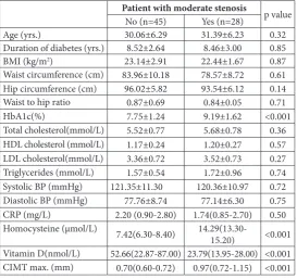
The clinical, anthropometric and biochemical characteristics of patients and CIMT are presented in Table 1. The average age and duration of diabetes was similar in both groups of patients with moderate stenosis and without coronary artery stenosis. HbA1c was significantly higher in patients with stenosis. Patients diagnosed with stenosis had higher serum homocysteine concentrations and lower serum vita-min D concentrations than the controls. We found a positive association between increased CIMT max and coronary stenosis.
CIMT measurements were obtained using diag-nostic accuracy measurements, i.e. by ultrasound, which is perceived as a highly specific test for detect-ing carotid artery stenosis in patients with type 1 dia-betes (Table 2).
Hyperhomocysteinemia, lower serum concentra-tion 25(OH)D and increased CIMT max appear to be sensitive and specific markers for detecting coronary
Table 1. Clinical, anthropometric and biochemical characteristics of patients and CIMT.
Patient with moderate stenosis p value
No (n=45) Yes (n=28)
Age (yrs.) 30.06±6.29 31.39±6.23 0.32
Duration of diabetes (yrs.) 8.52±2.64 8.46±3.00 0.85
BMI (kg/m2) 23.14±2.91 22.44±1.67 0.87
Waist circumference (cm) 83.96±10.18 78.57±8.72 0.61
Hip circumference (cm) 96.02±5.82 93.54±6.12 0.14
Waist to hip ratio 0.87±0.69 0.84±0.05 0.71
HbA1c(%) 7.75±1.24 9.19±1.62 <0.001
Total cholesterol(mmol/L) 5.52±0.77 5.68±0.78 0.36
HDL cholesterol (mmol/L) 1.17±0.24 1.20±0.27 0.57
LDL cholesterol(mmol/L) 3.36±0.72 3.52±0.73 0.27
Triglycerides (mmol/L) 1.57±0.54 1.72±0.96 0.74
Systolic BP (mmHg) 121.35±11.30 120.36±10.97 0.72
Diastolic BP (mmHg) 77.76±8.74 77.14±6.30 0.75
CRP (mg/L) 2.20 (0.90-2.80) 1.74(0.85-2.70) 0.50
Homocysteine (µmol/L) 7.42(6.30-8.40)
14.29(13.30-15.20) <0.001
Vitamin D(nmol/L) 52.66(22.87-87.00) 23.79(13.95-28.00) <0.001
CIMT max. (mm) 0.70(0.60-0.72) 0.97(0.72-1.15) <0.001
BMI – body mass index; BP – blood pressure;
CRP – C Reactive Protein, CIMT – carotid intima-media thickness;
Table 2. CIMT≥0.9mm as a screening test for detecting coronary artery stenosis.
Parameter (95% CI)
Sensitivity 0.714 (0.511-0.860)
Specificity 0.956 (0.836-0.992)
Positive predictive value 0.909 (0.693-0.984)
Negative predictive value 0.843 (0.708-0.925)
Likelihood ratio of a positive test 16.07 (4.064-63.555) Likelihood ratio of a negative test 0.299 (0.166-0.538)
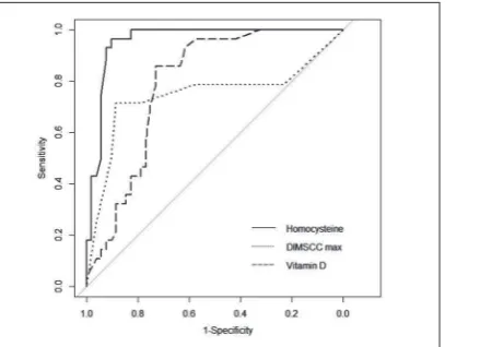
artery atherosclerosis using ROC analysis (AUCH-cy=0.955; (95%CI 0.990-1.000); p<0.001), vitamin D (AUCvit D=0,792; (95% CI 0.657-0.873); p<0.001) and CIMT max (AUCCIMT=0.743; (95%CI 0.643-0.910); p<0.001). Homocysteine proved to be the most reliable diagnostic marker of these three markers fol-lowed by the other two, respectively. The results of ROC analysis are presented in Fig. 1.
Pearson’s correlation analysis revealed a negative statistically significant correlation between serum homocysteine concentrations and serum vitamin D concentrations (r=-0.242; p=0.030), while there was a positive and statistically significant correlation between homocysteine and carotid IMT (r=0.456; p<0.001). However, the linear regression model re-vealed no significant relationship between homocyst-eine and serum vitamin D concentrations (b=-0.009 (95% CI -0.027 to 0.009); p=0.335) (Fig. 2). In addi-tion, no significant correlation was observed regarding CIMT and Vitamin D (r=0.091; p=0.422).
Multivariate logistic regression analysis of moder-ate coronary artery stenosis as a dependent variable is shown in Table 3. The analysis excluded CIMT be-cause this predictor was insignificant and the
confi-dence interval was extremely high when entered in multivariate analysis. The analysis showed that serum homocysteine concentration was the most reliable predictor of moderate coronary artery stenosis.
DISSCUSION
This study confirmed a positive correlation between increased HbA1c concentrations, increased plasma homocysteine concentrations, lower serum Vitamin D concentrations and elevated carotid IMT with coro-nary artery stenosis in asymptomatic patients with T1D. Coronary atherosclerosis was observed in one third of asymptomatic diabetic patients by coronary MDCTA. In most cases moderate RCA stenosis was detected. The diagnostic potential of noninvasive cor-onary MDCTA has been appreciated as it may help identify early atherosclerosis in asymptomatic diabetic patients [43]. In other words, not only can it be used to detect atherosclerosis in order to contribute to the primary prevention of CAD, but it can also be applied to atherosclerotic plaques in patients undergoing sta-tin treatment [44].
We confirmed that excessive levels of HbA1c and increased tHcy represent traditional risk factors for the development of coronary atherosclerosis in pa-tients with T1D. Our findings are in agreement with the results of previous studies, since we have been able to identify poor glycemic control as a predictor of CAD in these patients. This is in line with previ-ous research indicating a strong association between
Fig. 1. ROC analysis of investigated parameters. Fig. 2. Linear regression model analysis of relationship between homo-cysteine and serum vitamin D levels.
Table 3. Multivariate logistic regression analysis of moderate coro-nary artery stenosis as a dependent variable.
Variable p value OR (95% CI)
BMI 0.219 1.597 (0.757-3.370)
Homocysteine 0.008 4.427 (1.471-13.319)
Vitamin D 0.460 0.961 (0.865-1.067)
HbA1c variability and the number of patients diag-nosed with coronary artery stenosis [7]. Additionally, our study shows that coronary atherosclerosis is as-sociated with elevated tHcy, although this association has more frequently been observed in patients with type 2 diabetes [17]. A plethora of previous cross-sectional studies have reported a positive association between tHcy and diabetic microvascular complica-tions, whereas several studies have reported a cor-relation between hyperhomocysteinemia and CAD [18-22]. Nevertheless, the biological mechanism re-sponsible for vascular complications that occur as a consequence of the interaction between diabetes and elevated tHcy has not been fully elucidated. Hyperho-mocysteinemia associated with monocyte chemoat-tractant protein 1 (MCP-1) expression in the kidney via nuclear factor kappa-light-chain-enhancer of acti-vated B cells (NF-κB) activation may have contributed to renal injury in a hyperhomocysteinemic rat [21]. However, another study reported that the develop-ment of retinal vascular disease occurred due to the fact that tHcy superoxide from nicotinamide adenine dinucleotide phosphate (NADPH) oxidase led to im-paired endothelium-dependent nitrogen oxide (NO)-mediated dilation in the retinal arterioles [22].
The present study adds to the evidence that re-duced serum vitamin D levels may have a role in the development of coronary artery stenosis in patients with T1D. The findings of the Framingham Offspring study [23], the Health Professionals Follow-Up study [24] and a cross sectional study by Dobing [25] showed that low serum vitamin D was a risk factor for cardiovascular mortality in healthy subjects. The role of vitamin D in the pathophysiology of T1D has been explored in many studies including ones that in-vestigated its effects on immune-mediated destruction [30]. A study assessing the impact of specific vitamin D receptor polymorphisms on HLA class II histocom-patibility antigen DRB1 beta chain (HLADRB1) alleles did not identify it as a major risk factor for T1D [31]. The Finnish Birth Cohort Study reported that children who had been taking the recommended dose of 2000 IU/day of vitamin D were at a substantially lower risk of developing diabetes as opposed to those who had been receiving inadequate dosage [28]. Some studies have confirmed that severe vitamin D deficiency may lead to the development of diabetic neuropathy, retin-opathy and nephrretin-opathy in patients with T1D
[32-34]. An experimental study reported that vitamin D/ vitamin D receptor signaling in podocytes might help protect the kidney from diabetic injury [34]. Also, the association between vitamin D deficiency and age-re-lated macular degeneration should be considered [32]. Another experimental study examined the link be-tween vitamin D and the regulation of neurotrophins such as nerve growth factor (NGF) and neuronal Ca2+
homeostasis, along with its neuroprotective effect on peripheral nerves, respectively [33]. However, Sachs et al. [45] did not find evidence linking impaired vitamin D metabolism with increased risk of subclinical CVD in T1D. In addition, Serra-Planas et al. [29] evalu-ated the relationship between the concentrations of 25(OH)D and the presence of early atherosclerosis in asymptomatic T1D patients with no previous history of ischemic heart disease. Although T1D patients have lower concentrations and a 2-fold-higher prevalence of 25(OH)D deficiency than control individuals, they did not reveal an association between 25(OH)D con-centrations and subclinical CAD [29].
Moreover, we have also established a positive asso-ciation between elevated CIMT and both the number of cases diagnosed with coronary artery stenosis and elevated plasma homocysteine levels in T1D patients. The progression of CIMT caused by cardiovascular risk factors can predict further cardiovascular events [8]. In addition, the association between cardiovascu-lar risk and increased CIMT in patients with T1D has been observed in some studies [1,9-11]. In a study of young people with T1D who had been diagnosed with macrovascular disease or microalbuminuria, CIMT was increased by 25% (p<0.001) as compared to the control group of healthy individuals [11]. A significant correlation between CIMT and the percentage of the area affected by coronary vessel stenosis, which was measured by intravascular ultrasound, was reported in female patients [10]. Some studies on T1D have detected a positive correlation between CIMT and HbA1c [3] and CIMT and tHcy, respectively; [35] as well as a negative association between vitamin D concentrations and carotid IMT [38].
supplementation may help reduce homocysteine, thereby fostering the prevention of atherosclerotic vascular disease [37]. Homocysteine is metabolized by transsulfuration and remethylation (folate-dependent) pathways. The transsulfuration pathway represents a key route for homocysteine disposal and its conver-sion into cystathionine in the presence of the enzyme cystathionine-β-synthase (CBS) and cofactor vitamin B6. Hyperhomocysteinemia has been linked to CBS enzyme deficiency [13]. A significant increase in lower basal CBS mRNA levels was detected in mu-rine preosteoblasts after incubation with activated vitamin D. This suggests that CBS is a target gene of VDR and that vitamin D may modulate homocysteine metabolism while affecting its serum concentration [46]. However, one of the limitations of this study is that it is a cross-sectional study and that data could provide potential correlations between homocysteine levels and the development of atherosclerosis in pa-tients with T1D while further clinical investigations are still necessary.
CONCLUSION
Our study indicates that patients with T1D and hyper-homocysteinemia, lower 25(OH)D serum concentra-tions and increased CIMT have a higher risk of devel-oping coronary artery atherosclerosis. The presented data also suggest that tHcy can be used as a reliable predictor for the development of coronary artery ath-erosclerosis in asymptomatic patients with T1D.
Funding: This work received no funding.
Author contributions: Each author was included in all stages of manuscript preparation.
Conflict of interest disclosure: The authors do not have any conflict of interest.
REFERENCES
1. Krantz JS, Mack WJ, Hodis HN, Liu CR, Liu CH, Kaufman FR. Early onset of subclinical atherosclerosis in young per-sons with type 1 diabetes. J Pediatr. 2004;145(4):452-7. 2. Costacou T, Lopes-Virella MF, Zgibor JC, Virella G, Otvos J,
Walsh M, Orchard TJ. Markers of endothelial dysfunction in the prediction of coronary artery disease in type 1 diabetes. The Pittsburgh Epidemiology of Diabetes Complications Study. J Diabetes Complications. 2005;19(4):183-93.
3. Larsen JR, Brekke M, Bergengen L, Sandvik L, Arnesen H, Hanssen KF, Dahl-Jorgensen K. Mean HbA1c over 18 years predicts carotid intima media thickness in women with type 1 diabetes. Diabetologia. 2005;48(4):776-9.
4. Shishehbor MH, Hoogwerf BJ, Schoenhagen P, Marso SP, Sun JP, Li J, Klein AL, Thomas JD, Garcia MJ. Relation of hemoglobin A1c to left ventricular relaxation in patients with type 1 diabetes mellitus and without overt heart disease. Am J Cardiol. 2003;91(12):1514-7
5. Larsen JR, Sjøholm H, Berg TJ, Sandvik L, Brekke M, Hans-sen KF, Dahl-JørgenHans-sen K. Eighteen years of fair glycemic control preserves cardiac autonomic function in type 1 dia-betes. Diabetes Care. 2004;27(4):963-6.
6. Larsen J, Brekke M, Sandvik L, Arnesen H, Hanssen KF, Dahl-Jorgensen K. Silent coronary atheromatosis in type 1 diabetic patients and its relation to long-term glycemic control. Diabetes. 2002;51(8):2637-41.
7. Krolewski AS, Kosinski EJ, Warram JH, Leland OS, Busick EJ, Asmal AC, Rand LI, Christlieb AR, Bradley RF, Kahn CR. Magnitude and determinants of coronary artery disease in yuvenile onset, insulin-dependent diabetes mellitus. Am J Cardiol. 1987;59(8):750-5.
8. Sibal L, Agarwal SC, Home PD. Carotid intima-media thick-ness as a surrogate marker of cardiovascular disease in dia-betes. Diabetes METAB Synd Obes. 2011;4:23-34.
9. Polak JF, Backlund JY, Cleary PA, Harrington AP, O’Leary DH, Lachin JM, Nathan DM; DCCT/EDIC Research Group. Progression of carotid intima-media thickness during 12 years in the Diabetes Control and Complications Trial/Epi-demiology of Diabetes Interventions and Complications (DCCT/EDIC) Study. Diabetes. 2011;60(2):607-13. 10. Larsen JR1, Brekke M, Bergengen L, Sandvik L, Arnesen H,
Hanssen KF, Dahl-Jorgensen K. Mean HbA1c over 18 years predicts carotid intima-media thickness in women with type 1 diabetes. Diabetologia. 2005;48(4):776-9.
11. Sibal L, Aldibbiat A, Agarwal SC, Mitchell G, Oates C, Razvi S, Weaver JU, Shaw JA, Home PD. Circulating endothelial progenitor cells, endothelial function, carotid intima-media thickness dysfunction in people with type 1 diabetes without macrovascular disease or microalbuminuria. Diabetologia. 2009;52(8):1464-73.
12. Graham IM, Daly LE, Refsum HM, Robinson K, Bratt-ström LE, Ueland PM, Palma-Reis RJ, Boers GHJ, Sheahan RG, Israelsson B, Uiterwaal CS, Meleady R, McMaster D, Verhoef P, Witteman J, Rubba P, Bellet H, Wautrecht JC, de Valk HW, Sales Lúis AC, Parrot-Roulaud FM, Tan KS, Higgins I, Garcon D, Medrano MJ, Candito M, Evans AE, Andria G.Plasma homocysteine as a risk factor for vascular disease. The European Concerted Action Project. JAMA. 1997;277(22):1775-81.
13. Jhee KH, Kruger WD. The role of cystathionine β-synthase in homocysteine metabolism. Antioxid Redox Signal. 2005;7(5-6):813-22.
15. Boushey CJ, Beresford SA, Omenn GS, Motulsky AG. A quantitative assesment of plasma homocysteine as a risk factor for vascular disease. Probable benefits of increasing folic acid intakes. JAMA. 1995;274(13):1049-57.
16. Bagi Z, Ungvari Z, Szollár L, Koller A. Flow-induced con-striction in arterioles of hyperhomocysteinemic rats is due to impaired nitric oxide and enhanced thromboxane A(2) mediation. Arterioscler Thromb Vasc Biol. 2001;21(2):233-7. 17. Becker A, Kostense PJ, Bos G, Heine RJ, Dekker JM, Nijpels
G, Bouter LM, Stehouwer CD. Hyperhomocysteinaemia is associated with coronary events in type 2 diabetes. J Intern Med. 2003;253(3):293-300.
18. Buysschaert M, Jamart J, Dramais AS, Wallemacq P, Her-mans MP. Micro-and macrovascular complications and hyperhomocysteinemia in type 1 diabetic patients. Diabetes Metab. 2001;27(6):655-9.
19. Feng Y, Shan MQ, Bo L, Zhang XY, Hu J. Association of homocysteine with type 1 diabetes mellitus: a meta-analysis. Int J Clin Exp Med. 2015;8(8):12529-38.
20. Manna P, Gungor N, McVie R, Jain SK. Decreased cystathio-nine γ- lyase (CSE) activity in livers of type 1 diabetic rats and peripheral blood mononuclear cells (PBMC) of type 1 diabetic patients. J Biol Chem. 2014;289(17):11767-78. 21. Hwang SY, Woo CW, Au-Yeung KK, Siow YL, Zhu TY, O K.
Homocysteine stimulates monocyte chemoattractant pro-tein-1 expression in the kidney via nuclear factor-kappa B activation. Am J Physiol Renal. 2008;294(1):F236-44. 22. Omae T, Nagaoka T, Tanano I, Yoshida A. Homocysteine
inhibition of endothelium-dependent nitric oxide-mediated dilation of porcine retinal arterioles via enhanced superoxide production. Invest Ophthalmol Vis Sci. 2013;54(3):2288-95. 23. Wang TJ, Pencina MJ, Booth SL, Jacques PF, Ingelsson E,
Lanier K, Benjamin EJ, D’Agostino RB, Wolf M, Vasan RS. Vitamin D deficiency and risk of cardiovascular disease. Cir-culation. 2008;117(4):503-11.
24. Giovannucci E, Liu Y, Hollis BW, Rimm EB. 25 –hydroxyvi-tamin D and risk of myocardial infarction in men: a pro-spective study. Arch Intern Med. 2008;168(11):1174-80. 25. Dobnig H, Pilz S, Scharnagl H, Renner W, Seelhorst U,
Well-nitz B, Kinkeldei J, Boehm BO, Weihrauch G, Maerz W. Inde-pendent association of low serum 25-hydroxyvitamin D and 1,25-dihydroxyvitamin D levels with all-cause and cardio-vascular mortality. Arch Intern Med. 2008;168(12):1340-9. 26. Jain SK, Micinski D.Vitamin D up-regulates glutamate
cys-teine ligase and glutathione reductase and GSH formation and decreases ROS and MCP-1 and IL-8 secretion in high-glucose exposed U937 monocytes. Biochem Biophys Res. 2013;437(1):7-11.
27. Andres, V. Vitamin D puts the brakes on angiotensin II induced oxidative stress and vascular smooth muscle cells senescence. Atherosclerosis. 2014;236(2):444-7.
28. Hyppönen E, Läärä E, Reunanen A, Järvelin MR, Virtanen SM. Intake of vitamin D and risk of type 1 diabetes: a birth-cohort study. Lancet. 2001;358(9292):1500-3.
29. Serra-Planas E, Aguilera E, Granada ML, Soldevila B, Salinas I, Reverter JL, Pizarro E, Pellitero S, Alonso N, Mauricio D, Puig-Domingo M. High prevalence of vitamin D deficiency and lack of associations with subclinical atherosclerosis in
asymptomatic patients with type 1 diabetes mellitus from: a Mediterranean area. Acta Diabetologica. 2015;52(4):773-9. 30. Kamel MM, Fouad SA, Salaheldin O, Razek Ael-R, El-Fatah AI. Impact of vitamin D receptor gene polymorphism in pathogenesis of type 1 diabetes. Int J Clin Exp Med. 2014;7(12):5505-10.
31. Israni N, Goswami R, Kumar A, Rani R. Interaction of vita-min D receptor with HLADRB1 0301 in type 1 diabetes patients from North India. PloS One. 2009;4(12):e8023. 32. Singh A, Falk MK, Subhi Y, Sørensen TL. The association
between plasma 25-hydroxyvitamin D and subgroups age-related macular degeneration: a cross sectional study. PloS One. 2013;8(7):e70948.
33. Neveu I, Naveilhan P, Jehan F, Baudet C, Wion D, De Luca HF, Brachet P. 1,25-Dihydroxyvitamin D3 regulates the syn-thesis of nerve growth factor in primary cultures of glial cells. Brain Res Mol. 1994;24(1-4):70-6.
34. Wang Y, Deb DK, Zhang Z, Sun T, Liu W, Yoon D, Kong J, Chen Y, Chang A, Li YC. Vitamin D receptor signaling in podocytes protects against diabetic nephropathy. J Am Soc Nephrol. 2012;23(12):1977-86.
35. Basu A, Jenkins AJ, Stoner JA, Thorpe SR, Klein RL, Lopes-Virella MF, Garvey WT, Lyons TJ; DCCT/EDIC Research Group. Plasma homocysteine and carotid intima-media thickness in type 1 diabetes: A Prospective Study. Athero-sclerosis. 2014;236(1):188-95.
36. Juonala M, Voipio A, Pahkala K, Viikari JS, Mikkilä V, Kähönen M, Hutri-Kähönen N, Jula A, Burgner D, Sabin MA, Marniemi J, Loo BM, Laitinen T, Jokinen E, Taittonen L, Magnussen CG, Raitakari OT. Childhood 25(OH)Vita-minD levels and carotid intima-media thickness in adult-hood: the cardiovascular risk in young Finns study. J Clin Endocrinol Metab. 2015;100(4):1469-76.
37. Amer M, Qayyum R. The relathionship between 25-Hydroxyvitamin D and homocysteine in asymptomatic adults. J Clin Endocrinol Metab. 2014;99(2):633-8. 38. Mao X, Xing X, Xu R, Gong Q, He Y, Li S, Wang H, Liu C,
Ding X, Na R, Liu Z, Qu Y. Folic acid and vitamins D and B12 correlate with homocysteine in Chinese patients with type 2 diabetes mellitus, hypertension, or cardiovascular disease. Medicine (Baltimore). 2016;95(6):e2652.
39. Kind CRH, Pepys MB. The role of serum C-reactive pro-tein (CRP) measurement in clinical practice. Int Med. 1984;5:112-51.
40. Hafiane A, Genest J. High density lipoproteins: Measure-ment techniques and potential biomarkers of cardiovascular risk. BBA Clin. 2015;3:175-88.
41. Sherwani SI, Khan HA, Ekhzaimy A, Masood A, Sakharkar MK. Significance of HbA1c Test in Diagnosis and Prognosis of Diabetic Patients. Biomark Insights. 2016;11:95-104. 42. Agatston AS, Janowitz WR, Hildner FJ, Zusmer NR,
Viamonte M Jr, Detrano R. Quantification of coronary artery calcium using ultrafast computed tomography. J Am Coll Cardiol. 1990;15:827-32.
44. Zeb I, Li D, Nasir K, Malpeso J, Batool A, Flores F, Dail-ing C, Karlsberg RP, Budoff M. Effect of statin treatment on coronary plaque progression-a serial coronary angiography study. Atherosclerosis. 2013;231(2):198-204.
45. Sachs MC, Brunzell JD, Cleary PA, Hoofnagle AN, Lachin JM, Molitch ME, Steffes MW, Zinman B, de Boer IH; Diabetes Control and Complications Trial/Epidemiology of Diabetes Interventions and Complications Study Research Group. Cir-culating vitamin D metabolites and subclinical atherosclerosis in type 1 diabetes. Diabetes Care. 2013;36(8):2423-9. 46. Kriebitzsch C, Verlinden L, Eelen G, van Schoor NM, Swart
K, Lips P, Meyer MB, Pike JW, Boonen S, Carlberg C,
Vit-vitsky V, Bouillon R, Banerjee R, Verstuyf A. 1,25-Dihy-droxyvitamin D3 influences cellular homocysteine lev-els in murine preosteoblastic MC3T3-E1 cells by direct regulation of cysthationine βsynthase. J Bone Miner Res. 2011;26(12):2991-3000.
Supplementary Material