Available on-line at: www.oapub.org/edu
Copyright © The Author(s). All Rights Reserved. doi: 10.5281/zenodo.2556218 Volume 5 │ Issue 4 │ 2019
ELECTROCARDIOGRAPHIC CHARACTERISTICS OF
ATHLETES OF MOUNT CAMEROON ASCENT:
PREVENTION OF SUDDEN DEATH
Gassina Louis-Georges1, Mekoulou Ndongo Jerson1,
Guessogo Wiliam Richard1,2, Assomo Ndemba Peguy Brice1,
Tchoundjin Ernest1, Bika Lele Elysée Claude1,
Pepouomi Mama Nourdi1, Mandengue Samuel Honoré1, ,
Temfemo Abdou1,3,4i
1Exercise and Sport Physiology and Medicine Unit,
Faculty of Science, University of Douala, Douala, Cameroon
2National Institute of Youth and Sports,
Yaoundé, Cameroon
3Faculty of Medicine and Pharmaceutical Science,
University of Douala, Douala, Cameroon
4EA 3300 "Adaptations Physiologiques à l’Exercice
et Réadaptation à l’Effort", Faculté des Sciences du Sport, Université de Picardie Jules Verne, F-80025 Amiens, France
Abstract:
Mountain ultra-marathons participants are exposed to multiple internal and external stressors, from exercise and environment that can affect the cardiovascular response such as electrocardiographic profile (ECG). The aim of this study was to determine the electrocardiographic profile of athletes participating on the mount Cameroon race. Fifty-nine athletes and 50 non-athletes (31±7 vs 24±3 years) participated to the study during the 17th edition of Mount Cameroon ascent on February 18, 2012. ECG of
12-leads rest was measured in athletes and non-athletes. Resting heart rate (54±3 vs 71±9 bats/min) was lower (P<0.001) in athletes than non-athletes. P wave duration (108.5±1.8 vs 100.7±1.7 ms), PR interval (170.4±27.2 vs 155.8±22.3 ms), and RR interval (1072.3±188.2 vs 875.6±128.4 ms) were significantly higher (P<0.001) in athletes compared to non-athletes. However, on Rhythm and morphology abnormalities, athletes developed a sinus bradycardia (88.1 vs 8%), sinus arythmia (76.3 vs 22%) and left ventricular hypertrophy (54.2 vs 6%) higher than those of non-athletes (P<0.001). For the abnormalities of ECG conduction, atrioventricular block I (11.9 vs 6%),
incomplete Right Bundle Branch Block (5.1 vs 2 %), incomplete Left Branch Block (1.7 vs 0%), Left anterior fascicular block (1.7 vs 0%), T-wave inversion (V1-V6) (10.2 vs 8%), Short PR (3.4 vs 2%), Sus ST Segment shift (5.1 vs 4%) were similar in both groups. But, early repolarization (40.7 vs 16%) was significantly higher (P<0.001) in athletes than non-athletes. Mountain race athletes develop some cardiac ECG electric morpho-functional abnormalities.
Keywords: athletes, mountain race, electrocardiogram
1. Introduction
The Mount Cameroon ascent has taken on an international dimension and is rooted more in the Cameroonians socio-cultural realities. It is baptized «the race of Hope». It is an international ultra-endurance race very difficult because participants must perform a distance 42.000 m and rise to 4.095 m of altitude. This requires a good physical fitness, a steady and regular methodical training, a very strong psychological aspect and a good health. Participation in this race is subject to a set of medical examination.
In Cameroon, the National Federation of Athletics is responsible for the effectiveness of health status of participants. But, the essential measurements of health parameters are reduced only to the examination of the blood pressure, heart rate (HR) and to some cutaneous infections. The competitive sport, in its perpetual search of performance optimization by the training can train pathologies that can harm health following certain subsequent chronic cardiovascular adaptations (Corrado et al., 2003).
During the practice of an enduring sport, the cardiovascular risk of sudden cardiac death is transitorily increased what can reveal an ignored cardiopathy (Corrado et al., 2003, Mekoulou et al., 2017). Sudden cardiac death in athletes could due to underlying congenital or inherited cardiac abnormalities. Schmied and Borjesson (2014) showed the sudden cardiac death to be more prevalent in older athletes (> 35 years) due to atherosclerotic coronary heart disease. Thus, the place of the prevention seems to be obviousness in sport (Vetter, 2015). Early detection of cardiovascular pathologies is paramount in the practice of sport, because it makes it possible to limit the possible hazardous of sudden cardiac death by proposing a therapeutic attitude and/or a monitoring and by limiting the exposure to the maladjusted efforts (Pelliccia et al., 2005, Vetter & Dugan, 2013). However, the visit of non-counter-indications for the sportsmen is imperative and recommended by the European Society of Cardiology (Corrado et al.,
2005), American Heart Association (Maron et al., 2007) and many others learned
societies (Drezner et al., 2017).
Myerburg & Vetter 2007, Pellicia, 2006; 2007; Vetter 2014). However, the occurrence of many accidents attributed very often to a cardiac cause during sports competitions such as mountain climbs for example raises many questions.
To our knowledge, there is not enough scientific studies on Mount Cameroon ascent’s. Salah et al. (2012) assessed the impact of some anthropological and physiological (Blood pressure and heart rate) parameters on athlete’s performance of participants of the ascent of the mount Cameroon. There is no study on ECG characterization of participants of the ascent of the mount Cameroon. However, this race is a grueling footrace opportunity for the characterizing of participants ECG parameters in the context of high altitude competition. The aim of the present preliminary study was to characterize the athletes of Mount Cameroon race by their electrocardiographic parameters.
2. Materiel and Methods
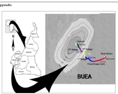
2.1 Presentation of the Mount Cameroon
The Mount is known as the Cameroon volcanic line (Tsafack et al., 2009). Commonly called the tank of the gods, it is considered as a live volcano localized at longitude 9° 170' East and Latitude 4° 203' North, part of with long part of the 1600 km chain of Cenozoic volcanic and sub-volcanic complexes extending from the Gulf of Guinea to the interior of the African continent (Tsafack et al., 2009). The ascent of Mount Cameroon is an annual sport event that takes place each month of February in the course of the main dry season in the South-West region of Cameroon hosts (The town of Buea). It is a very formidable endurance race, particularly difficult and physically demanding because participants must perform a distance 42.000 m uphill and downhill and rise to 4.095 m of altitude. The starting and finishing point is the Molyko stadium of Buea. The top of the mountain that marks the point of return is Fako, located 21 km from the starting point. Between the starting point and the summit is a prison site and three refuges sites which are the points of supply and control during the competition. From the starting point and successively, the prison site is located 6.6 km from the Molyko stadium, the first refuge site is located at 13.03 km or at6.43 km from the prison site, the second refuge site is at 16.73 km or at 3.7 km from the first refuge site, the third refuge site is at 19.53 km or at 2.8 km from the second refuge and the summit is at 21 km or at 1.47 km from the third refuge.
2.2 Participants
study was approved by the Ministry of Public Health and complied with the code of ethics of the Helsinki Declaration as amended in Seoul in October 2008.
2.3 Data collection
2.3.1 Anthropometric parameters
The size was measured using an iron rod graduated to the centimeter (Removable Mobile Height Chart - Seca 213, France) and, the weight was measured using a TANITA BC 532 scale impedance meter (Tanita Corporation, Tokyo, Japan). Using height and weight, we calculated the body mass index (BMI) according to the Quetelet formula.
BMI = Weight (kg) / [Height (m)]2.
2.3.2 Heart rate (HR)
It was recorded continuously during the rest period of 15 minutes in a quiet room without noise using a heart rate monitor (Polar RS800CX Electro Oy, Kempele, Finland) reliable, with minimal artifacts (Cassirame et al., 2007). Successive heart beats were analyzed using the software Polar Pro Trainer 5.1, and ectopic beats were visualized and manually replaced by interpolation of adjacent RR intervals. Smallest HR was considered as resting HR.
2.3.3 Electrocardiogram (ECG)
The resting ECG was performed using a 12-leads electrocardiograph CarTouch (Cardionics SA, Brussels) at a tape speed of 25 mm/s and an amplitude of 10 mm/Mv. Recordings were carried out according to the specifications of the American Heart Association (Fletcher et al., 2001, Kligfield et al., 2007). Pre-automatic interpreted ECGs by the device were printed on paper, reviewed and interpreted by two independent cardiologists specialized on athletes ECG interpretation. Electrocardiographic variables P wave, PR intervals, RR intervals between two heartbeats, corrected QTc of Bazett (1920), and QRS complex were retained for statistical analysis. For athletes, the ECG qualitative interpretation were in accordance with the specific Seattle interpretation criteria (Drezner et al., 2013) amended in 2014 (Sheikh et al., 2014) and the recent international recommendation of 2017 (Drezner et al., 2017). In another hand, Standard diagnostic criteria were used to define various ECG abnormalities in non-athletes (Surawicz & Knilans, 2008; Rautaharju et al., 2009).
2.3.4 Statistical analysis
3. Results
Table 1: Characteristics of athletes and controls
Characteristics Athletes
(n=59)
Non-athletes (n=50)
P-value
Age (Years) 31 ±7 24±3 <0.01
Weight (Kg) 66.1 ± 8.1 64.1 ± 10.5 0.3
Height (m) 1.72 ± 0.2 1.66 ± 0.1 0.8
BMI (Kg/m2) 22.5 ± 2.2 22.2 ± 3.2 0.3
Weekly training volume (hours/Week) 11 ± 3 --- ----
Subjects were males (59 athletes and 50 non-athletes). If the age was significantly (P<0.05) higher in athletes than non-athletes, means of weight, height and BMI of athletes and non-athletes were similar (P>0.05) (Table 1).
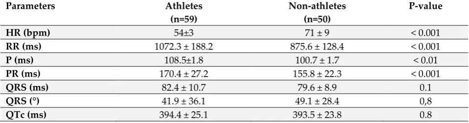
Table 2: Comparison of ECG parameter values between athletes and controls
Parameters Athletes
(n=59)
Non-athletes (n=50)
P-value
HR (bpm) 54±3 71 ± 9 < 0.001
RR (ms) 1072.3 ± 188.2 875.6 ± 128.4 < 0.001
P (ms) 108.5±1.8 100.7 ± 1.7 < 0.01
PR (ms) 170.4 ± 27.2 155.8 ± 22.3 < 0.001
QRS (ms) 82.4 ± 10.7 79.6 ± 8.9 0.1
QRS (°) 41.9 ± 36.1 49.1 ± 28.4 0,8
QTc (ms) 394.4 ± 25.1 393.5 ± 23.8 0.8
For Heart rate (HR) and the electrical parameters of ECG (Table 2), resting HR was significantly lower in athletes than in non-athletes (P<0.05). On the other hand RR, P, and PR were significantly higher in athletes than those of non-athletes (P<0.05). No significant difference was registered in QRS and QTc between athletes and non-athletes.
Table 3: Comparison of ECG abnormalities between athletes and controls
Abnormalities Athletes
(n=59)
Non-athletes ( n=50)
P-value
Rhythm and morphological abnormalities
Sinus bradycardia 52 (88.1 %) 4 (8%) < 0.001
Sinus arrhythmia 45(76.3) 11(22) < 0.001
Left ventricular hypertrophy (LVH) 32 (54.2%) 6 (12%) < 0.001
Conduction abnormalities
Atrioventricular block I (AVB I) 7 (11.9%) 3 (6%) 0.3
Incomplete Right Bundle Branch Block (IRBB) 3 (5.1%) 1 (2%) 0.6 Incomplete Left Branch Block (LBB) 1 (1.7%) 0 (0%) 0.8 Left anterior fascicular Block (LAFB) 1 (1.7%) 0 (0%) 0.8
T-wave Inversion (V1-V6) 6 (10.2%) 4 (8%) 0.7
Short PR (˂ 120ms) 2 (3.4%) 1 (2%) 0.6
Early repolarisation (V3 -V6) 24 (40.7) 8(16) < 0.001
Sinus bradycardia, and left ventricular hypertrophy (LVH) were more observed in athletes (P<0.05). Other cardiac morpho-functional abnormalities were observed in athletes and non-athletes without significant difference (P>0.05) (Table3).
4. Discussion
One of the main findings of this preliminary study was the presence of cardiac electrical morpho-functional changes in many athletes of the ascent of Mount Cameroon. However, the electrical and cardiac morpho-functional characterization of the ECG parameters of participants in the ascent of Mount Cameroon was consistent with that of the International Consensus Statement on Electrocardiographic Interpretive Criteria for Athletes (Drezner et al., 2017, Sharma et al., 2018).
Sinus bradycardia were observed both in athletes and non-athletes with the mean of resting HR (Table 2) significantly (P<0.05) lower in athletes than in non-athletes (Table 3). Resting sinus bradycardia, is commonly defined by a HR<60 beats/min, it is almost universal in athletes, depending on the type of sport and the level of training and competition (Balady et al., 1984). This abnormality is commonly reported in athletes and non-athletes even in adolescents (Sharma et al., 1999, Mekoulou et al., 2017). Sharma et al. (1999) reported 80% of sinus bradycardia in athletes compared to 19% in non-athletes in a study including 1000 athletes and 300 sedentary. In fact, bradycardia is generally observed in the practice of endurance sports (Sten et al., 2002). According to Brezner et al. (2013), HR is set by the balance between the sympathetic and parasympathetic nervous systems. In well-trained athletes, resting sinus bradycardia is a common finding due to increased vagal tone. For Sten et al. (2002), aerobic training may induce intrinsic adaptations in the sinus node with decreased automaticity resulting in a high prevalence of sinus bradycardia in endurance athletes. Thus, resting sinus bradycardia is particularly prevalent in endurance athletes due to increased vagal tone and possible structural atrial remodelling (Northcote et al., 1989; Noseworthy et al., 2011). In the present study no athletes presented a deep sinus bradycardia (HR<35 bpm).
athlete to an increased risk of sudden cardiac death if the auriculo-ventricular accessory pathway has the potential for fast ante-grade conduction. The presence of a short PR with or without delta wave is one of the resting ECG anomalies requiring a follow up of a cardiologist before the practice of enduring sport (Corrado et al., 2009; Drezner et al., 2013 ; Drezner et al., 2017).
The normal QRS should be less than or equal to 100 msec. Beyond that, there is an intraventricular conductive disorder. QRS duration predicts morality in patients with left ventricular dysfunction, hypertension (Liew, 2011) and cardiovascular mortality in the general population. Athletes or non-athletes had 82.4 ± 10.7 vs 79.6 ± 8.9, without significant difference between both groups (p = 0.1) in the present study.
The QTc interval calculated between the beginning of the QRS and the end of the T wave corresponded to the time required for depolarization and repolarization of the ventricles. QT interval has several sources of variability, including advanced age, gender, drugs, body mass index, autonomic changes, diabetes mellitus, dyslipidemia, smoking, heart failure, myocardial ischemia, hypertension, stroke, impaired renal function, liver cirrhosis, and electrolyte imbalances (Mozos et al., 2012). In the present study, average of QTc durations for non-athletes and even athletes were normal without difference between both groups. Applying Bazett's formula (Bazett, 1920), our study found no significant difference between mean QTc durations in athletes and non-athletes (table 2). The average value of QTc in non-athletes was superimposable to that of footballers in the study of Syransy et al. (2008) (403 ± 24 msec). Consequently, the lengthening of the QTc would not always be linked to sport (Carré & Cignon, 2001).
T-wave inversion has been observed in athletes without significant difference between non-athletes (table 3). T-wave inversions in the precordial derivations were observed in both athletes and non-athletes. T-wave inversion in precordial leads is a common finding and can appear to normal carry prognosis in the general population (Marcus et al., 2005; Mekoulou et al., 2017). Right precordial T-wave inversion in leads V1–V4 is a relatively rare finding in the middle-aged general population. A recent study of Pellicia et al. (2000) on ECG characterization in large athletic population has disproved the traditional idea that T-wave inversions are common and training-related ECG changes in the athletes. It had been reported a 2.7% prevalence of T-wave inversion in 1005 highly trained athletes and 2.3% in a large population of 32 652 young amateur athletes. In the same way Sharma et al. (1999) had noticed that the prevalence of T-wave inversion is similar among elite athletes and sedentary controls respectively 4.4% and 4.0%. The presence of a deep T-wave inversion (>2 mm in >2 adjacent) leads in an athlete is a non-specific but warning ECG sign of a potential cardiovascular disease with the risk of sudden cardiac death during sport. T wave inversion in inferior and/or lateral leads must raise the suspicion of ischemic heart disease, cardiomyopathy, aortic valve disease, systemic hypertension and LV non-compaction.
Left anterior fascicular Block (LAFB) have been more noticed in athletes than non-athletes despite non-significant (P>0.05) difference (Table 3).
Early repolarization is a physiological and benign ECG pattern in the general population of young people and athletes. In enduring trained athletes, early repolarization right precordial show typical features that may allow differentiation from some diseases. Early repolarization is commonly considered as an elevation of the QRS-ST junction (J-point) by ≥0.1mV affecting the inferior and/or lateral leads (Tikkanen et al., 2009). Early repolarization is more prevalent in athletes, young individuals, males and black ethnicity (Tikkanen et al., 2009). Early repolarization is related to parasympathetic activity enhancement and increased cardiovascular fitness. However, some authors (Haissaguerre et al., 2008; Tikkanen et al., 2009) argue that the presence of an early repolarization in infero-lateral derivations was associated with an increase of the risk of sudden cardiac death in the general population (Tikkanen et al., 2009). Atrioventricular block I illustrate a delay in auriculo-ventricular nodal conduction in athletes, due to increased vagal activity or intrinsic auriculo-ventricular node changes, and particularly resolves with the onset of exercise.
Incomplete RBBB is defined by a QRS duration <120 ms with a right bundle branch block pattern: terminal R wave in lead V1 (commonly characterized as an rSR’ pattern) and wide terminal S wave in leads I and V6 (Corrado et al., 2009; Drezner et al., 2017). On the physiological way, Kim et al. (2011) suggest that the mildly delayed in right ventricular conduction in athletes caused remodelling, which increased right ventricular cavity size and resultant increased conduction time, rather than an intrinsic delay within the His-Purkinje system Therefore, incomplete RBBB represents a phenotype of cardiac adaptation to exercise and in the absence of other features suggestive of disease does not require further evaluation.
On the physiological justification, it has been established that trained athletes commonly show intranodal conduction modifications such as (first-degree atrioventricular (AV) block and early repolarization, which result from physiological adaptation of the cardiac autonomic nervous system to enduring training conditioning, such as increased vagal tone and/or withdrawal of sympathetic activity at the rest (Holly et al., 1998, Corrado & McKenna, 2007). According to the international consensus for ECG interpretation in athletes (Drezner et al., 2017), this abnormality can be considered as chronic physiological adaptation to enduring sport, and have to be controlled before pre-participation to enduring competition.
5. Conclusion
physiological limits of these chronic adaptations and the risk of sudden cardiac death inherent in the practice of the sport.
Acknowledgment
The authors thank all the participants and the members of the Physiology and Medicine of Physical Activity and Sports (UPM-APS) of the University of Douala for their collaboration.
References
Balady GJ, Cadigan JB, Ryan TJ, 1984. Electrocardiogram of the athlete: an analysis of 289 professional football players. American Journal of Cardiology 53:1339-43. Bazett HC, 1920. An analysis of the time-relations of electrocardiograms. Heart 7:
353-370.
Brezner JA, Fischbach P, Froelicher V, Marek J, Pelliccia A, Prutkin JM, Schmied CM, Sharma S, Wilson MG, Ackerman MG, Anderson J, Ashley E, Asplund CA, Baggish AL, Börjesson M, Cannon BC, Corrado D, DiFiori JP, Harmon KG, Heidbuchel H, Owens DS, Paul S, Salerno JC, Stein R, Vetter VL, 2013. Normal electrocardiographic findings: recognizing physiological adaptations in athletes. British Journal of Sports Medicine 47:125-136.
Carre F, Cignon JC, 2001. Particularités électrocardiographiques de l’athlète : quelles limites? La revue du praticien. 51: 7-14.
Cassirame J, Tordi N, Mourot L, Rakobowchuk M, Regnard, 2007. Using of new beat to beat recorder system for traditional analysis of heart rate variability. Science and Sports 22:238-42.
Corrado D, Basso C, Pavei A, Michieli P, Schiavon M, Thiene G, 2006. Trends in sudden cardiovascular death in young competitive athletes after implementation of a pre-participation screening program. Journal American College of Cardiologie 296 (13):1593-1601.
Corrado D, Basso C, Rizzoli G, Schiavon M, Thiene G, 2003. Does sports activity enhance the risk of sudden death in adolescents and young adults? Journal American College of Cardiologie 42:1959-63.
Corrado D, Biff A, Basso C, et al., 2009. 12-lead ECG in the Athlete: physiological versus pathological abnormalities. British Journal of Sports Medicine 43:669-76.
Corrado D, McKenna WJ, 2007. Appropriate interpretation of the athlete’s electrocardiogram saves lives as well as money. European Heart Journal 28:1920-2.
Exercise Physiology and the Working Group of Myocardial and Pericardial Diseases of the European Society of Cardiology. European Heart Journal 26:516-24.
Drezner JA, Ackerman MJ, Anderson J, et al., 2013a. Electrocardiographic interpretation in Athletes: the 'Seattle criteria'. British Journal Sports Medicine 47:122-4.
Drezner JA, Fischbach P, Froelicher V, Marek J, Pelliccia A, Prutkin JM, et al., 2013b. Normal electrocardiographic findings: recognising physiological adaptations in athletes. British Journal of Sports Medicine 47:125-36.
Drezner JA, Sharma S, Baggish A, Papadakis M et al., 2017. International criteria for electrocardiographic interpretation in athletes. British Journal of Sports Medicine 1:1-28.
Fletcher GF, Balady GJ, Amsterdam EA, Chaitman B, Eckel R, Fleg J, et al., 2001. Exercise standards for testing and training a statement for healthcare professionals from the American Heart Association. Circulation 104:1694-740. Haissaguerre M, Derval N, Sacher F, Jesel L, Deisenhofer I, de Roy L, et al., 2008.
Sudden cardiac arrest associated with early repolarization. New England Journal Medicine 358:2016-23.
Heidbuchel H, Panhuyzen-Goedkoop N, Corrado D, et al., 2006. Recommendations for participation in leisure-time physical activity and competitive sports of patients with arrhythmias and potentially arhythmogenic conditions. Part 1: supraventricular arrhythmias and pacemakers. European Journal of Cardiovascular Preventive and Rehabilitation 13:475-84.
Holly RG, Shaffrath JD, Amsterdam EA, 1998. Electrocardiographic alterations associated with the hearts of athletes. Sports Medicine 25:139-48.
Kim JH, Noseworthy PA, McCarty D, et al., 2011. Significance of electrocardiographic right bundle branch block in trained Athletes. American Journal of Cardiology 107:1083-9.
Kligfield P, Gettes LS, Bailey JJ, Childers R, Deal EB, Hancock EW, et al., 2007. Recommendations for the standardization and interpretation of the electrocardiogram: part I: the electrocardiogram and its technology: a scientific statement from the American Heart Association Electrocardiography and Arrhythmias Committee, Council on Clinical Cardiology; the American College of Cardiology Foundation; and the Heart Rhythm Society. Circulation 115:1306-24.
Liew R, 2011. Electrocardiogram-based predictors of sudden cardiac death in patients with coronary artery disease, Clinical Cardiology 34 (8):466-473.
Marcus FI, 2005. Prevalence of T-wave inversion beyond V1 in young normal individuals and usefulness for the diagnosis of arrhythmogenic right ventricular cardiomyopathy/dysplasia. American Journal of Cardiology 95:1070-1.
American Heart Association council on nutrition, physical activity and metabolism. Endorsed by the American College of Cardiology Foundation. Circulation 115:1643-1655
Mekoulou NdongoJ, Assomo Ndemba PB, Temfemo A, Dzudie Tamdja A, Abanda
MH, Bika Lele CE, Tchoudjin E, Richard Guessogo W, Gassina LG, Mandengue SH, 2017. Pre and post exercise electrocardiogram pattern modifications in apparently healthy school adolescents in Cameroon. International Journal of Adolescent Medicine and Health. DOI: 10.1515/ijamh-2017-0071.
Mozos I, Serban C, Mihaescu R, 2012. The relation between arterial blood pressure variables and ventricular repolarization parameters. International Journal of Collaborative Research on Internal Medicine and Public Health 4(6):860-875. Myerburg RJ, Vetter VL, 2007. Electrocardiograms should be included in pre
participation screening of athletes. Circulation 116:2616-26.
Northcote R, Canning GP, Ballantyne D, 1989. Electrocardiographic findings in male veteran endurance athletes. British Heart Journal 61:155-60.
Noseworthy PA, Tikkanen JT, Porthan K, et al., 2011. The early repolarization pattern in the general population: clinical correlates and heritability. Journal of American College of Cardiology 57:2284-9.
Pelliccia A, Fagard R, Bjørnstad HH, Anastassakis A, Arbustini E, Assanelli D, et al., 2005. Recommendations for competitive sports participation in athletes with cardiovascular disease: a consensus document from the Study Group of Sports Cardiology of the Working Group of Cardiac Rehabilitation and Exercise Physiology and the Working Group of Myocardial and Pericardial Diseases of the European Society of Cardiology. European Heart Journal 26:1422-45.
Pelliccia A, Maron BJ, Culasso F, et al., 2000. Clinical significance of abnormal electrocardiographic patterns in trained athletes. Circulation 102:278-84.
Pellicia A, Di Paolo FM, Corrado D, Buccolieri C, Quattrini FM, Pisicchio C, et al., 2006. Evidence of efficacy of the Italian national pre-participation screening programme for identification of hypertrophic cardiomyopathy in competitive athletes. European Heart Journal 27:2196-2200.
Pellicia A, 2007. The pre-participation cardiovascular screening of competitive athletes: is it time to change the customary clinical practice? European Heart Journal, 287: 2703-2705.
Rautaharju PM, Surawicz B, Gettes LS, 2009. AHA/ACC/HRS Recommendations for the standardization and interpretation of the electrocardiogram. Circulation 119:241– 50.
Salah MA, Verla VS, Tonga C, 2012. Anthropometric and Hemodynamic Profiles of Athletes and Their Relevance to Performance in the Mount Cameroon Race of Hope. Asian Journal of Sports Medicine 3 (2): 99-104.
Stein R, Medeiros CM, Rosito GA, et al., 2002. Intrinsic sinus and atrioventricular node electrophysiological adaptations in endurance athletes. Journal of American College of Cardiology 39:1033-8.
Surawicz B, Knilans TK, 2008. Chou’s electrocardiography in clinical practice, 6th ed
Philadelphia: Saunders Elsevier.
Syransy AE, Ouattara S, Couloubaly I, 2008. Influence du sport sur la repolarisation ventriculaire chez les africains mélanodermes. Cardiologie Tropicale 33:129 Tikkanen JT, Anttonen O, Junttila MJ, Aro AL, Kerola T, Rissanen HA, et al., 2009.
Long-term outcome associated with early repolarization on electrocardiography. New England Journal of Medicine 361:2529-37.
Tsafack JPF, Wandji P, Bardintzeff JM, et al., 2009. The Mount Cameroon stratovolcano (Cameroon Volcanic Line, Central Africa): Petrology, geochemistry, isotope and age data. Geochemistry, Mineralogy and Petrology-Sofia 47:65-78.
Vetter VL, Dugan NP, 2013. A discussion of electrocardiographic screening and sudden cardiac death prevention: evidence and consensus. Current Opinion in Cardiology 28:139-51.
Vetter VL, 2015. Best practices for ECG screening in adolescents. Journal of Electrocardiology 48:316-23.
Vetter VL, 2014. Should Electrocardiographic screening of all infants, adolescents, and teenagers be performed? Circulation 130:688-97.
Appendix
Creative Commons licensing terms